Open Access

Gastrointestinal stromal tumors in the duodenum show increased contrast enhancement compared with those in the stomach on computed tomography

  • Authors:
    • Ryosuke Sato
    • Ryo Harada
    • Kenji Hashimoto
    • Tomoaki Tsutsui
    • Nao Hattori
    • Masafumi Inoue
    • Haruhiko Kobashi
    • Mami Morimoto
    • Maiko Tamura
    • Atsushi Hayashi
    • Masaya Iwamuro
  • View Affiliations

  • Published online on: August 4, 2022     https://doi.org/10.3892/mco.2022.2577
  • Article Number: 144
  • Copyright: © Sato et al. This is an open access article distributed under the terms of Creative Commons Attribution License.

Metrics: Total Views: 0 (Spandidos Publications: | PMC Statistics: )
Total PDF Downloads: 0 (Spandidos Publications: | PMC Statistics: )


Abstract

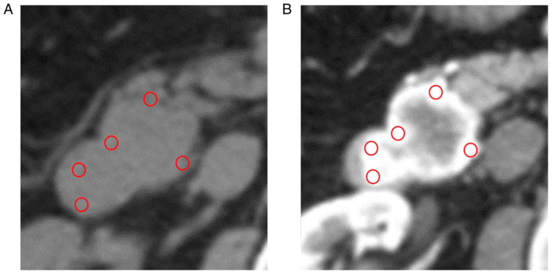
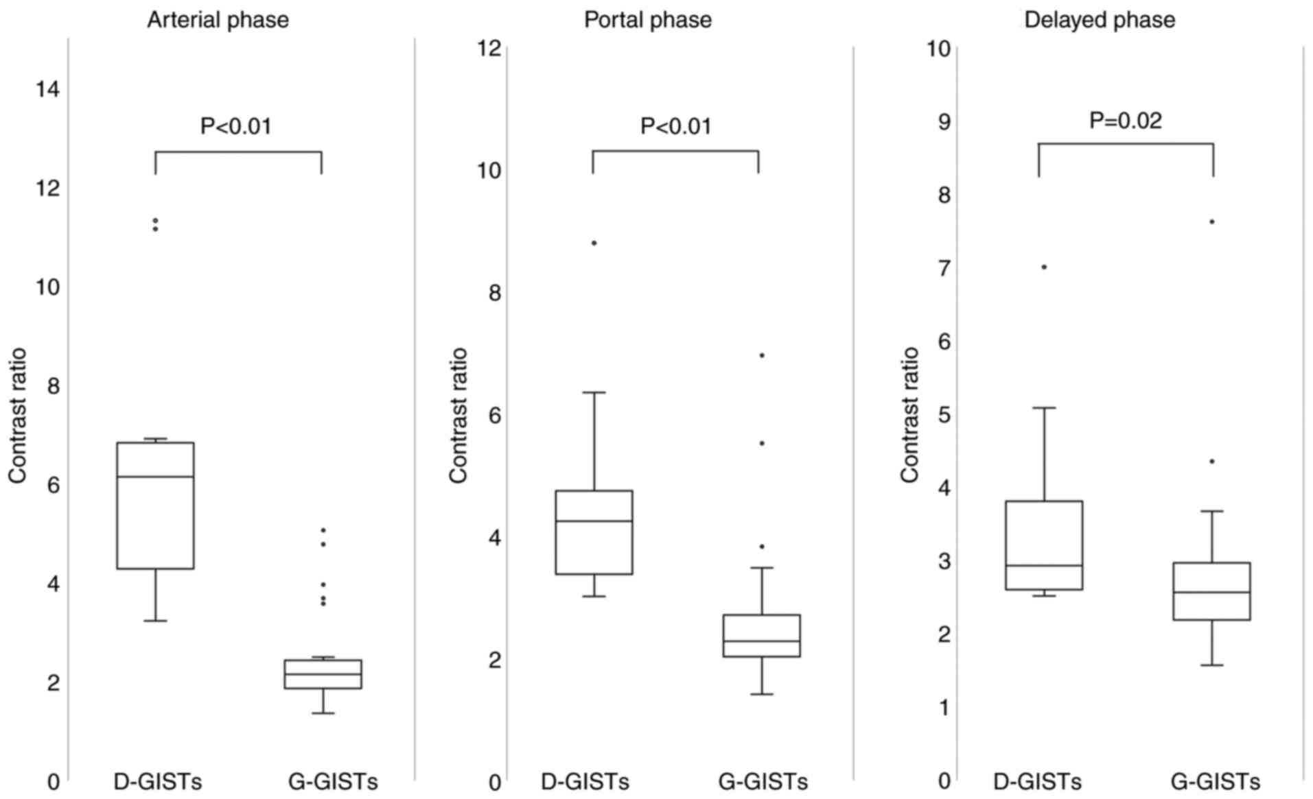
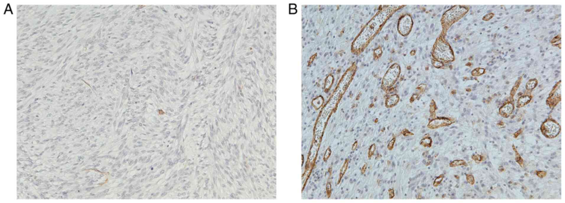
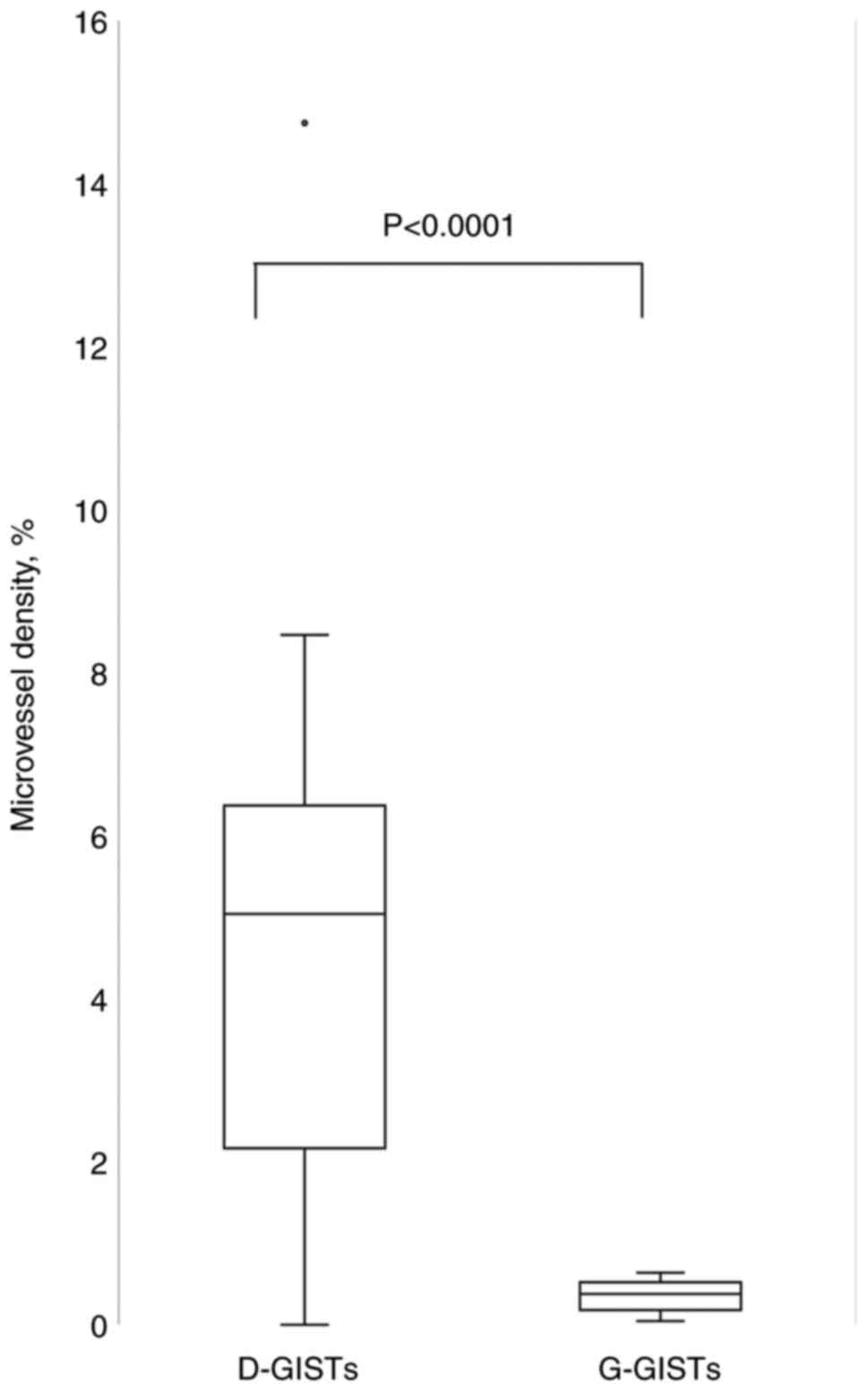
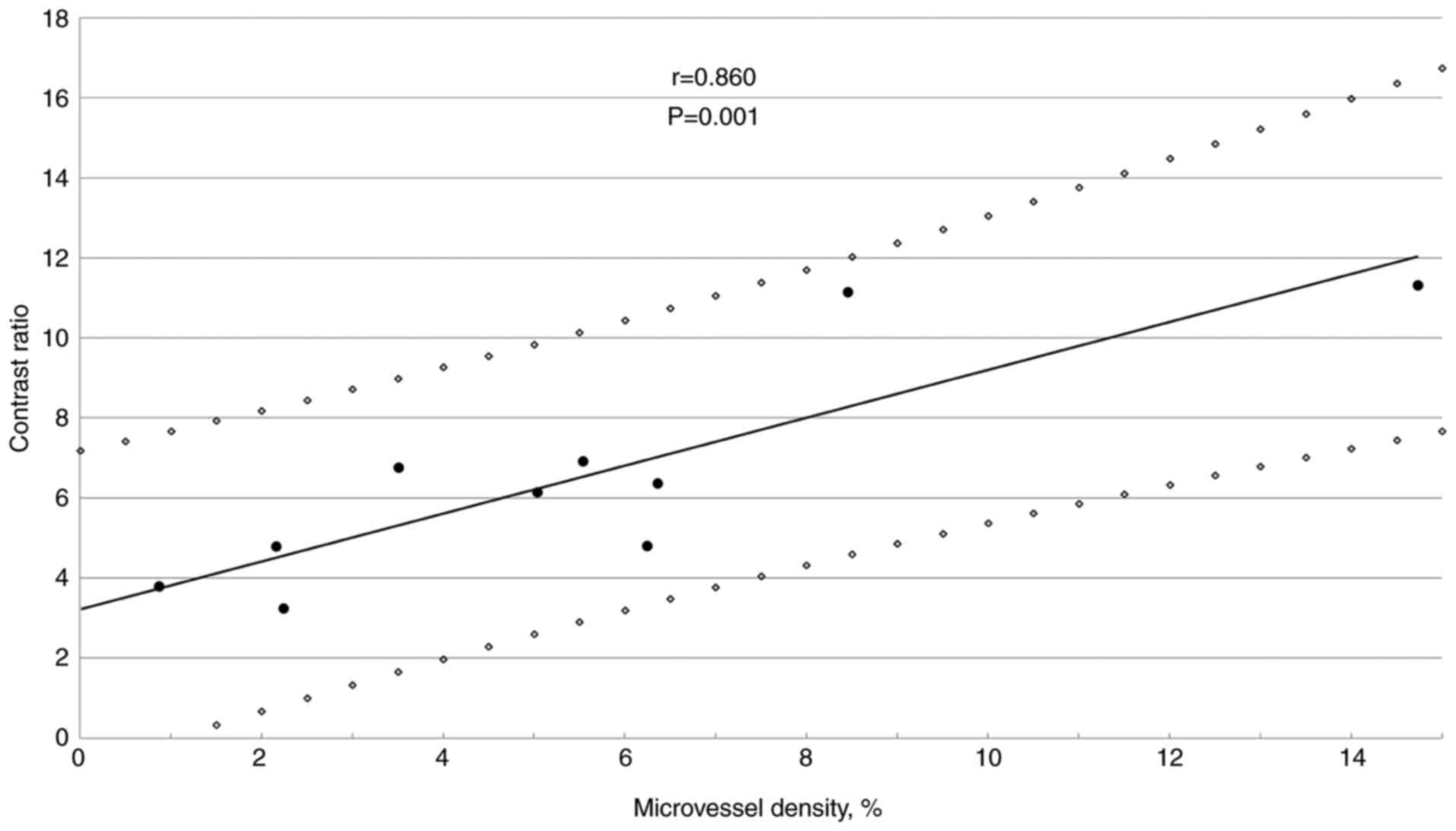
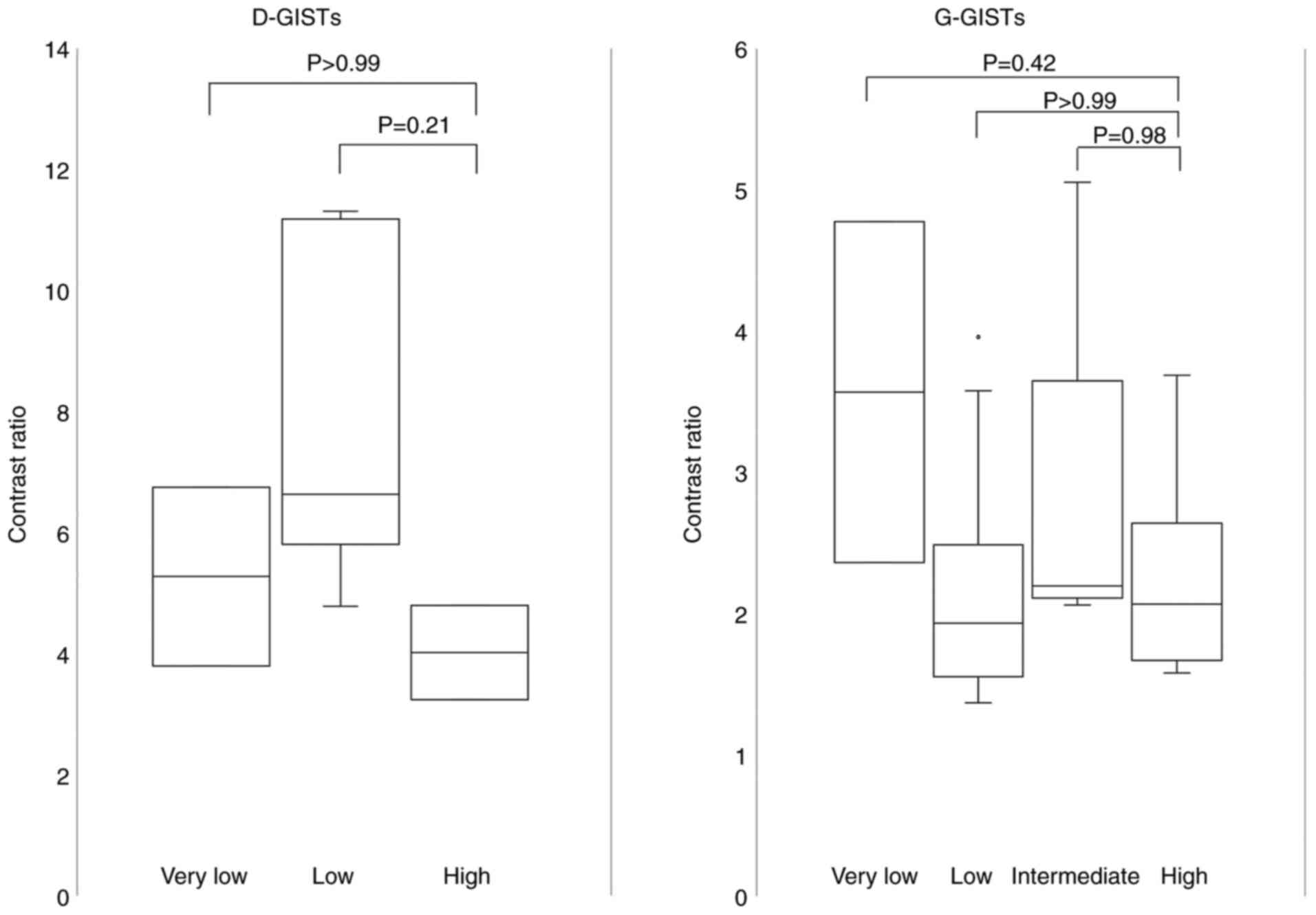
Duodenal gastrointestinal stromal tumors (D‑GISTs) are a rare and relatively small subset of GISTs whose imaging features are not well known. The present study aimed to evaluate the enhancement pattern of D‑GISTs compared with that of gastric GISTs (G‑GISTs) using dynamic computed tomography. This single‑center, retrospective, clinicopathological analysis was conducted on 10 patients with D‑GISTs who underwent surgery between June 2006 and October 2018. In the same period, 25 patients with G‑GISTs underwent surgery and were enrolled. The contrast ratio was defined as the ratio between Hounsfield units in contrast enhanced and unenhanced images in different phases, and these ratios were compared between the D‑GIST and G‑GIST groups. Furthermore, microvessel density, analyzed by immunohistochemical staining for CD31, was compared between the D‑GIST and G‑GIST groups. The contrast ratio of D‑GIST was significantly higher than that of G‑GIST in the arterial, portal and delayed phases (P<0.01, P<0.01 and P=0.02, respectively). The microvessel density of the D‑GISTs was significantly higher than that of the G‑GISTs (P<0.0001). D‑GISTs were more hypervascular than G‑GISTs on both imaging and pathological analyses.
View Figures
View References

Related Articles

Journal Cover

October-2022
Volume 17 Issue 4

Print ISSN: 2049-9450
Online ISSN:2049-9469

Sign up for eToc alerts

Recommend to Library

Copy and paste a formatted citation
x
Spandidos Publications style
Sato R, Harada R, Hashimoto K, Tsutsui T, Hattori N, Inoue M, Kobashi H, Morimoto M, Tamura M, Hayashi A, Hayashi A, et al: Gastrointestinal stromal tumors in the duodenum show increased contrast enhancement compared with those in the stomach on computed tomography. Mol Clin Oncol 17: 144, 2022
APA
Sato, R., Harada, R., Hashimoto, K., Tsutsui, T., Hattori, N., Inoue, M. ... Iwamuro, M. (2022). Gastrointestinal stromal tumors in the duodenum show increased contrast enhancement compared with those in the stomach on computed tomography. Molecular and Clinical Oncology, 17, 144. https://doi.org/10.3892/mco.2022.2577
MLA
Sato, R., Harada, R., Hashimoto, K., Tsutsui, T., Hattori, N., Inoue, M., Kobashi, H., Morimoto, M., Tamura, M., Hayashi, A., Iwamuro, M."Gastrointestinal stromal tumors in the duodenum show increased contrast enhancement compared with those in the stomach on computed tomography". Molecular and Clinical Oncology 17.4 (2022): 144.
Chicago
Sato, R., Harada, R., Hashimoto, K., Tsutsui, T., Hattori, N., Inoue, M., Kobashi, H., Morimoto, M., Tamura, M., Hayashi, A., Iwamuro, M."Gastrointestinal stromal tumors in the duodenum show increased contrast enhancement compared with those in the stomach on computed tomography". Molecular and Clinical Oncology 17, no. 4 (2022): 144. https://doi.org/10.3892/mco.2022.2577