Clinical and radiologic findings in angiomatous nasal polyps
- Authors:
- Published online on: May 29, 2025 https://doi.org/10.3892/mco.2025.2863
- Article Number: 68
-
Copyright: © Yeom et al. This is an open access article distributed under the terms of Creative Commons Attribution License.
Metrics:
Total
Views: 0 (Spandidos Publications: | PMC Statistics:
)
Total PDF Downloads: 0 (Spandidos Publications: | PMC Statistics:
)
Abstract
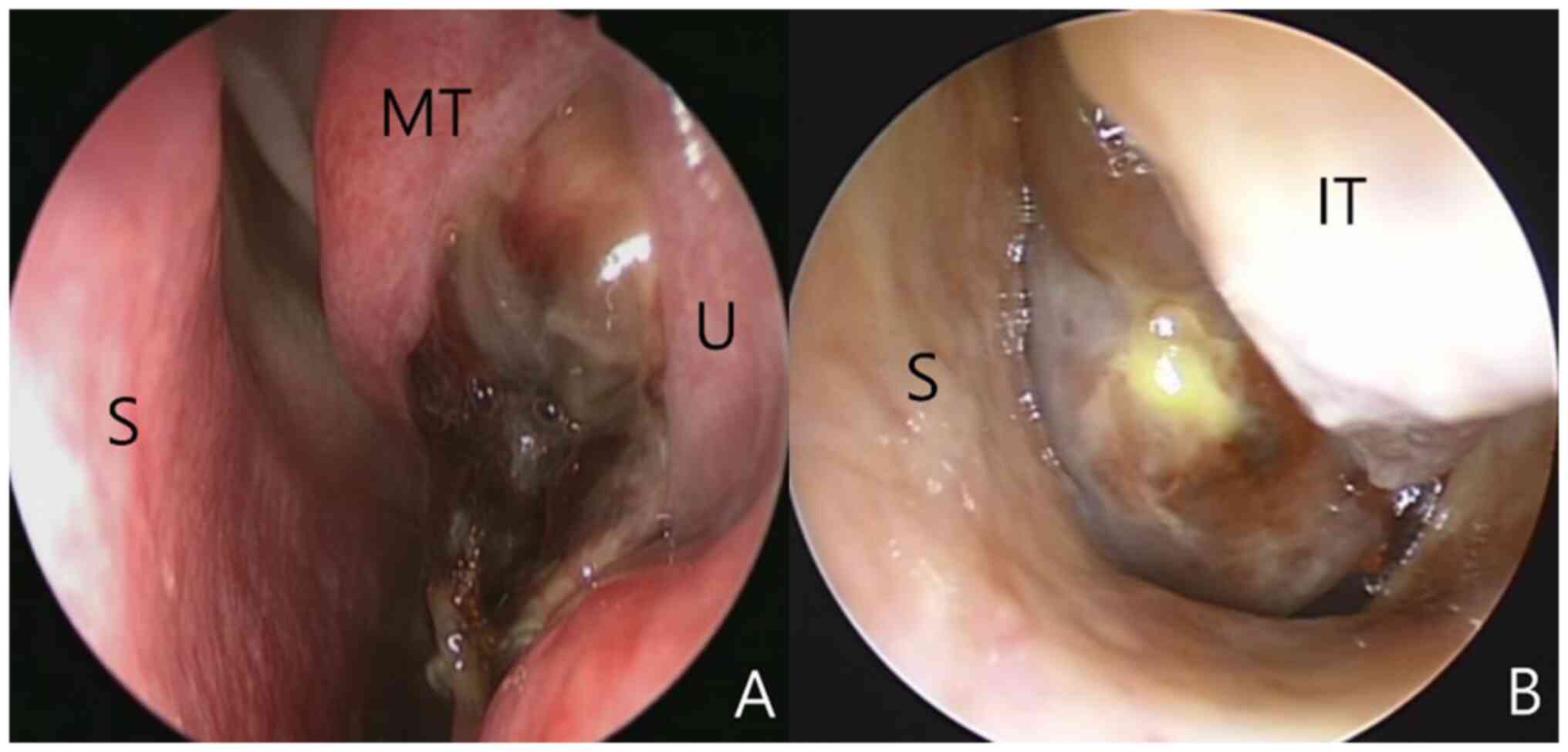
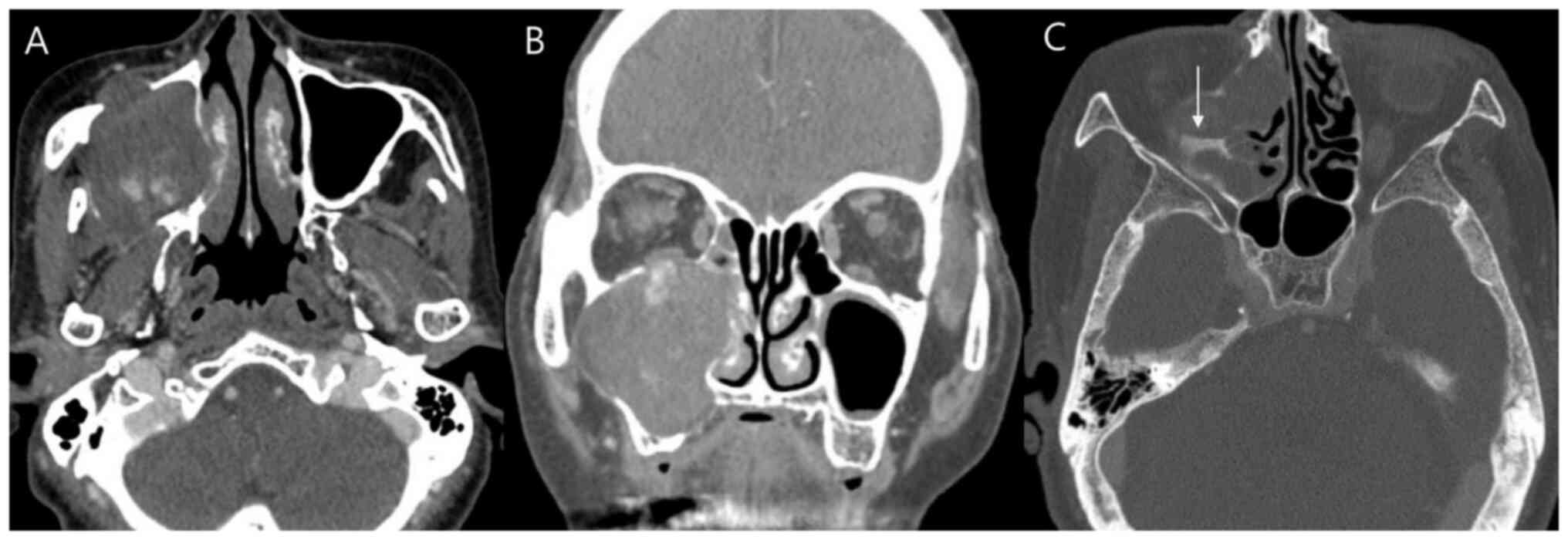
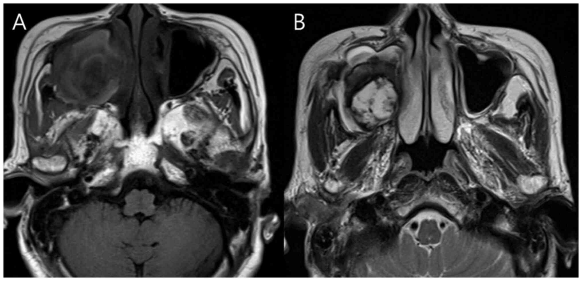
Angiomatous nasal polyps (ANPs), a rare subtype of inflammatory sinonasal polyps, pose a diagnostic challenge due to overlapping clinical and radiological features with other nasal tumors. This study aimed to evaluate the clinical and radiological characteristics of six pathologically confirmed ANPs. Six patients diagnosed with ANPs between December 2010 and May 2022 were retrospectively analyzed. All patients presented with nasal obstruction; the mean age was 42±28.4 years. The maxillary sinus was the most frequent origin site (66.7%). On computed tomography (CT) scans, all ANPs exhibited heterogeneous density; most (83.3%) demonstrated bony changes. Magnetic resonance imaging (MRI) was performed in four cases, all showing mild hyperintense signals on T1‑weighted images and heterogeneous hyperintensity on T2‑weighted images. A peripheral hypointense rim on T2‑weighted images was observed in three patients (75%). In conclusion, ANPs are challenging to distinguish from other nasal tumors based on clinical and radiological findings. While CT and MRI reveal certain typical characteristics, MRI provides more specific diagnostic clues.