Spandidos Publications Logo
  • About
    • About Spandidos
    • Aims and Scopes
    • Abstracting and Indexing
    • Editorial Policies
    • Reprints and Permissions
    • Job Opportunities
    • Terms and Conditions
    • Contact
  • Journals
    • All Journals
    • Oncology Letters
      • Oncology Letters
      • Information for Authors
      • Editorial Policies
      • Editorial Board
      • Aims and Scope
      • Abstracting and Indexing
      • Bibliographic Information
      • Archive
    • International Journal of Oncology
      • International Journal of Oncology
      • Information for Authors
      • Editorial Policies
      • Editorial Board
      • Aims and Scope
      • Abstracting and Indexing
      • Bibliographic Information
      • Archive
    • Molecular and Clinical Oncology
      • Molecular and Clinical Oncology
      • Information for Authors
      • Editorial Policies
      • Editorial Board
      • Aims and Scope
      • Abstracting and Indexing
      • Bibliographic Information
      • Archive
    • Experimental and Therapeutic Medicine
      • Experimental and Therapeutic Medicine
      • Information for Authors
      • Editorial Policies
      • Editorial Board
      • Aims and Scope
      • Abstracting and Indexing
      • Bibliographic Information
      • Archive
    • International Journal of Molecular Medicine
      • International Journal of Molecular Medicine
      • Information for Authors
      • Editorial Policies
      • Editorial Board
      • Aims and Scope
      • Abstracting and Indexing
      • Bibliographic Information
      • Archive
    • Biomedical Reports
      • Biomedical Reports
      • Information for Authors
      • Editorial Policies
      • Editorial Board
      • Aims and Scope
      • Abstracting and Indexing
      • Bibliographic Information
      • Archive
    • Oncology Reports
      • Oncology Reports
      • Information for Authors
      • Editorial Policies
      • Editorial Board
      • Aims and Scope
      • Abstracting and Indexing
      • Bibliographic Information
      • Archive
    • Molecular Medicine Reports
      • Molecular Medicine Reports
      • Information for Authors
      • Editorial Policies
      • Editorial Board
      • Aims and Scope
      • Abstracting and Indexing
      • Bibliographic Information
      • Archive
    • World Academy of Sciences Journal
      • World Academy of Sciences Journal
      • Information for Authors
      • Editorial Policies
      • Editorial Board
      • Aims and Scope
      • Abstracting and Indexing
      • Bibliographic Information
      • Archive
    • International Journal of Functional Nutrition
      • International Journal of Functional Nutrition
      • Information for Authors
      • Editorial Policies
      • Editorial Board
      • Aims and Scope
      • Abstracting and Indexing
      • Bibliographic Information
      • Archive
    • International Journal of Epigenetics
      • International Journal of Epigenetics
      • Information for Authors
      • Editorial Policies
      • Editorial Board
      • Aims and Scope
      • Abstracting and Indexing
      • Bibliographic Information
      • Archive
    • Medicine International
      • Medicine International
      • Information for Authors
      • Editorial Policies
      • Editorial Board
      • Aims and Scope
      • Abstracting and Indexing
      • Bibliographic Information
      • Archive
  • Articles
  • Information
    • Information for Authors
    • Information for Reviewers
    • Information for Librarians
    • Information for Advertisers
    • Conferences
  • Language Editing
Spandidos Publications Logo
  • About
    • About Spandidos
    • Aims and Scopes
    • Abstracting and Indexing
    • Editorial Policies
    • Reprints and Permissions
    • Job Opportunities
    • Terms and Conditions
    • Contact
  • Journals
    • All Journals
    • Biomedical Reports
      • Information for Authors
      • Editorial Policies
      • Editorial Board
      • Aims and Scope
      • Abstracting and Indexing
      • Bibliographic Information
      • Archive
    • Experimental and Therapeutic Medicine
      • Information for Authors
      • Editorial Policies
      • Editorial Board
      • Aims and Scope
      • Abstracting and Indexing
      • Bibliographic Information
      • Archive
    • International Journal of Epigenetics
      • Information for Authors
      • Editorial Policies
      • Editorial Board
      • Aims and Scope
      • Abstracting and Indexing
      • Bibliographic Information
      • Archive
    • International Journal of Functional Nutrition
      • Information for Authors
      • Editorial Policies
      • Editorial Board
      • Aims and Scope
      • Abstracting and Indexing
      • Bibliographic Information
      • Archive
    • International Journal of Molecular Medicine
      • Information for Authors
      • Editorial Policies
      • Editorial Board
      • Aims and Scope
      • Abstracting and Indexing
      • Bibliographic Information
      • Archive
    • International Journal of Oncology
      • Information for Authors
      • Editorial Policies
      • Editorial Board
      • Aims and Scope
      • Abstracting and Indexing
      • Bibliographic Information
      • Archive
    • Medicine International
      • Information for Authors
      • Editorial Policies
      • Editorial Board
      • Aims and Scope
      • Abstracting and Indexing
      • Bibliographic Information
      • Archive
    • Molecular and Clinical Oncology
      • Information for Authors
      • Editorial Policies
      • Editorial Board
      • Aims and Scope
      • Abstracting and Indexing
      • Bibliographic Information
      • Archive
    • Molecular Medicine Reports
      • Information for Authors
      • Editorial Policies
      • Editorial Board
      • Aims and Scope
      • Abstracting and Indexing
      • Bibliographic Information
      • Archive
    • Oncology Letters
      • Information for Authors
      • Editorial Policies
      • Editorial Board
      • Aims and Scope
      • Abstracting and Indexing
      • Bibliographic Information
      • Archive
    • Oncology Reports
      • Information for Authors
      • Editorial Policies
      • Editorial Board
      • Aims and Scope
      • Abstracting and Indexing
      • Bibliographic Information
      • Archive
    • World Academy of Sciences Journal
      • Information for Authors
      • Editorial Policies
      • Editorial Board
      • Aims and Scope
      • Abstracting and Indexing
      • Bibliographic Information
      • Archive
  • Articles
  • Information
    • For Authors
    • For Reviewers
    • For Librarians
    • For Advertisers
    • Conferences
  • Language Editing
Login Register Submit
  • This site uses cookies
  • You can change your cookie settings at any time by following the instructions in our Cookie Policy. To find out more, you may read our Privacy Policy.

    I agree
Search articles by DOI, keyword, author or affiliation
Search
Advanced Search
presentation
Molecular and Clinical Oncology
Join Editorial Board Propose a Special Issue
Print ISSN: 2049-9450 Online ISSN: 2049-9469
Journal Cover
October-2025 Volume 23 Issue 4

Full Size Image

Sign up for eToc alerts
Recommend to Library

Journals

International Journal of Molecular Medicine

International Journal of Molecular Medicine

International Journal of Molecular Medicine is an international journal devoted to molecular mechanisms of human disease.

International Journal of Oncology

International Journal of Oncology

International Journal of Oncology is an international journal devoted to oncology research and cancer treatment.

Molecular Medicine Reports

Molecular Medicine Reports

Covers molecular medicine topics such as pharmacology, pathology, genetics, neuroscience, infectious diseases, molecular cardiology, and molecular surgery.

Oncology Reports

Oncology Reports

Oncology Reports is an international journal devoted to fundamental and applied research in Oncology.

Experimental and Therapeutic Medicine

Experimental and Therapeutic Medicine

Experimental and Therapeutic Medicine is an international journal devoted to laboratory and clinical medicine.

Oncology Letters

Oncology Letters

Oncology Letters is an international journal devoted to Experimental and Clinical Oncology.

Biomedical Reports

Biomedical Reports

Explores a wide range of biological and medical fields, including pharmacology, genetics, microbiology, neuroscience, and molecular cardiology.

Molecular and Clinical Oncology

Molecular and Clinical Oncology

International journal addressing all aspects of oncology research, from tumorigenesis and oncogenes to chemotherapy and metastasis.

World Academy of Sciences Journal

World Academy of Sciences Journal

Multidisciplinary open-access journal spanning biochemistry, genetics, neuroscience, environmental health, and synthetic biology.

International Journal of Functional Nutrition

International Journal of Functional Nutrition

Open-access journal combining biochemistry, pharmacology, immunology, and genetics to advance health through functional nutrition.

International Journal of Epigenetics

International Journal of Epigenetics

Publishes open-access research on using epigenetics to advance understanding and treatment of human disease.

Medicine International

Medicine International

An International Open Access Journal Devoted to General Medicine.

Journal Cover
October-2025 Volume 23 Issue 4

Full Size Image

Sign up for eToc alerts
Recommend to Library

  • Article
  • Citations
    • Cite This Article
    • Download Citation
    • Create Citation Alert
    • Remove Citation Alert
    • Cited By
  • Similar Articles
    • Related Articles (in Spandidos Publications)
    • Similar Articles (Google Scholar)
    • Similar Articles (PubMed)
  • Download PDF
  • Download XML
  • View XML
Case Report

Lymph node metastasis in a patient with carcinoma in situ rectal cancer: A case report

  • Authors:
    • San-Ha Kim
    • Gi-Bong Chae
    • Seung-Koo Lee
    • Seung-Joo Nam
  • View Affiliations / Copyright

    Affiliations: Department of Internal Medicine, Kangwon National University Hospital, Chuncheon, Gangwon 24289, Republic of Korea, Department of Surgery, Kangwon National University School of Medicine, Chuncheon, Gangwon 24289, Republic of Korea, Department of Anatomical Pathology, Kangwon National University School of Medicine, Kangwon National University Hospital, Chuncheon, Gangwon 24289, Republic of Korea, Division of Gastroenterology and Hepatology, Department of Internal Medicine, Kangwon National University School of Medicine, Chuncheon, Gangwon 24289, Republic of Korea
  • Article Number: 91
    |
    Published online on: August 7, 2025
       https://doi.org/10.3892/mco.2025.2886
  • Expand metrics +
Metrics: Total Views: 0 (Spandidos Publications: | PMC Statistics: )
Metrics: Total PDF Downloads: 0 (Spandidos Publications: | PMC Statistics: )
Cited By (CrossRef): 0 citations Loading Articles...

This article is mentioned in:


Abstract

Colorectal cancer typically progresses from intramucosal carcinoma to invasive cancer, with lymph node metastasis considered possible only after submucosal invasion. Tumors confined to the mucosa are classified as carcinoma in situ (Tis), based on the American Joint Committee on Cancer, and are generally not thought to be capable of metastasis. The present study reports a rare case of lymph node metastasis in a 70‑year‑old female with Tis rectal cancer, who presented to Kangwon National University School of Medicine in December 2018. Although the tumor was confined to the mucosa without evidence of lymphovascular invasion, histopathological examination demonstrated metastasis to a regional lymph node. The patient underwent laparoscopic resection followed by concurrent chemoradiotherapy, and she has remained recurrence‑free for the past 6 years. This case challenges the conventional understanding of Tis‑stage colorectal cancer and highlights the need for the re‑evaluation of lymph node involvement in early‑stage cases. Further research is necessary to explore the mechanisms underlying lymph node metastasis in Tis rectal cancer.

Introduction

Colorectal cancer (CRC) is one of the most prevalent malignancies worldwide and remains a major cause of cancer-related morbidity and mortality. The disease typically follows a well-characterized adenoma-carcinoma sequence, progressing from benign adenomatous polyps to intramucosal carcinoma and ultimately to invasive cancer that penetrates the submucosa. Once the tumor invades beyond the mucosal layer, it gains access to lymphatic channels and blood vessels, which facilitates the spread to regional lymph nodes and distant organs.

According to the American Joint Committee on Cancer (AJCC) staging system, tumors confined to the mucosa, including the lamina propria and muscularis mucosae, are classified as carcinoma in situ (Tis) and designated as Stage 0(1). These lesions are considered to have an extremely low risk of metastasis, based on the long-standing belief that the colonic mucosa lacks lymphatic vessels. In contrast to other segments of the gastrointestinal tract - where basement membrane invasion often signifies the onset of metastatic potential- in colorectal cancer, only tumors that infiltrate the submucosa are regarded as biologically capable of metastasizing.

However, this traditional understanding has been increasingly challenged by emerging evidence. Several reports have described rare instances of local recurrence of distant metastasis arising from Tis-stage colorectal tumors, raising the possibility that lymphatic dissemination may occur even in the absence of submucosal invasion. Some of these atypical cases have involved poorly differentiated histology, extensive inflammation, or other high-risk features, but definitive lymph node metastasis in well-differentiated intramucosal carcinoma has remained undocumented.

Herein, we report a rare and unusual case of histologically confirmed lymph node metastasis in a 70-year-old female patient with Tis-stage rectal cancer, who initially presented to our hospital in December 2018. This case challenged the current staging paradigm and raises important questions regarding the metastatic potential of early-stage colorectal cancer. It underscores the need for reevaluation of staging criteria and highlights the importance of individualized clinical judgement even in lesions that are presumed to be biologically indolent.

Case report

A 70-year-old female presented to Kangwon National University School of Medicine in December 2018 for endoscopic removal of colon polyps identified during a screening colonoscopy. She had no prior history of colonoscopy and no significant medical history other than hypertension. Laboratory examination showed a white blood cell, hemoglobin, platelet count, blood urea nitrogen, creatinine, and carcinoembryonic antigen levels of 7,700/ul, 13.2 g/dl, 386,000/ul, 12.2 mg/dl, 0.8 mg/dl, and 4.2 ng/ml, 
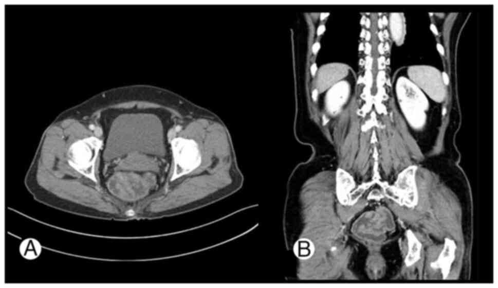
respectively. Abdominopelvic computed tomography (CT) revealed multiple polypoid lesions with conglomeration in the rectum with no other remarkable findings (Fig. 
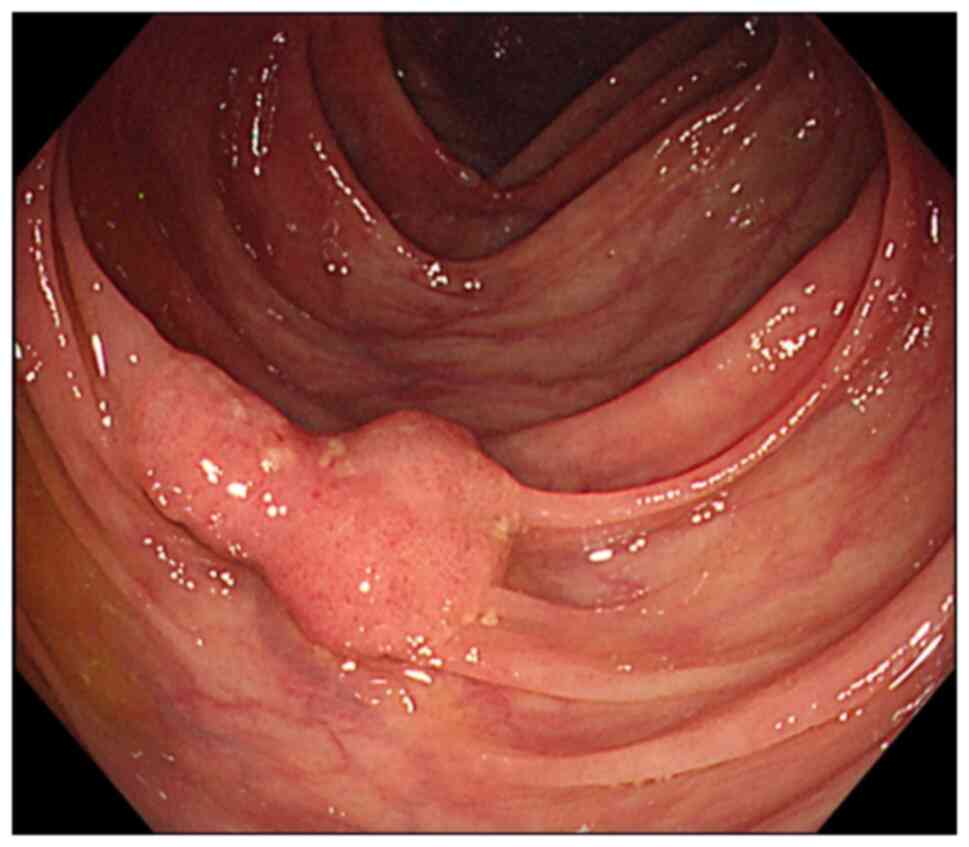
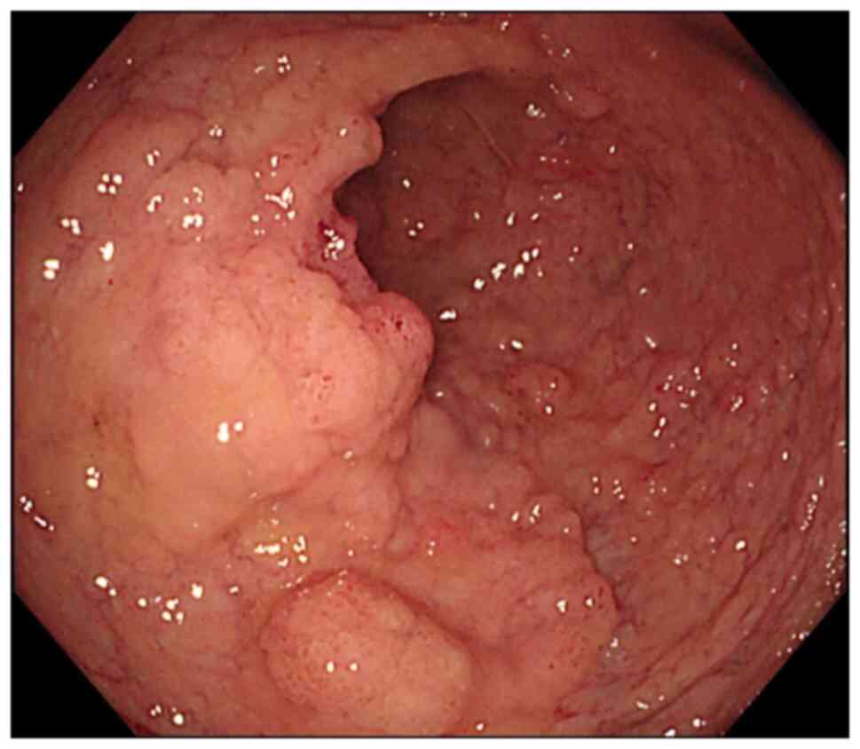
1). Colonoscopy identified two lesions, one in the ascending colon and another in the rectum (Figs. 2 and 3). In the ascending colon, 2 cm laterally spreading tumor (LST) was observed, and a biopsy revealed a tubulovillous adenoma with focal high-grade dysplasia. In the rectum, diffuse circumferential nodular mucosa with friability involving the whole rectum from the rectosigmoid junction to the anorectal junction was noted, and a biopsy of the most prominent nodular lesion revealed tubulovillous adenoma with low-grade dysplasia.

Abdominopelvic computed tomography
findings show numerous polypoid lesions with conglomeration in the
rectum. (A) A transverse (axial) CT image, and (B) a coronal view
image. No other remarkable findings were noted.

Figure 1

Abdominopelvic computed tomography findings show numerous polypoid lesions with conglomeration in the rectum. (A) A transverse (axial) CT image, and (B) a coronal view image. No other remarkable findings were noted.

Colonoscopy shows a 2 cm-sized
laterally spreading tumor in the ascending colon.

Figure 2

Colonoscopy shows a 2 cm-sized laterally spreading tumor in the ascending colon.

Colonoscopy shows diffuse nodular
mucosal lesions encircling the entire rectum.

Figure 3

Colonoscopy shows diffuse nodular mucosal lesions encircling the entire rectum.

For the ascending colonic LST lesions, endoscopic submucosal dissection was performed, which revealed a well-differentiated adenocarcinoma with a maximum submucosal invasion depth of 1 mm. Both the deep and lateral resection margins were clear, with no evidence of lymphovascular or perineural 
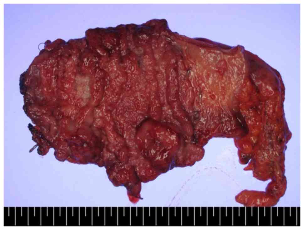
invasion. Due to extensive involvement of the entire rectal mucosa, the patient was referred for surgery for a large circumferential rectal lesion and underwent a laparoscopic ultra-low anterior resection (Fig. 4).

Gross surgical specimen of the rectal
lesion. The lower edge of the image includes a ruler for scale;
each black and white segment represents 5 mm.

Figure 4

Gross surgical specimen of the rectal lesion. The lower edge of the image includes a ruler for scale; each black and white segment represents 5 mm.

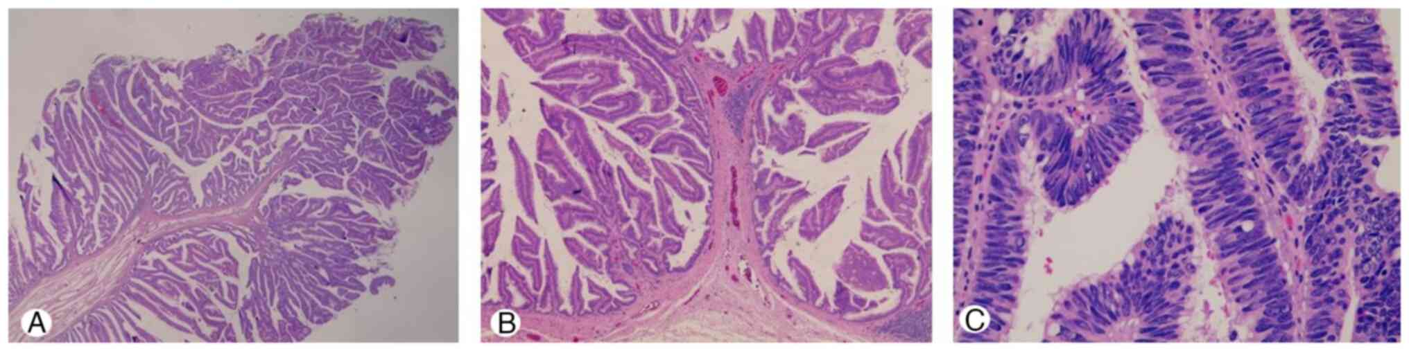
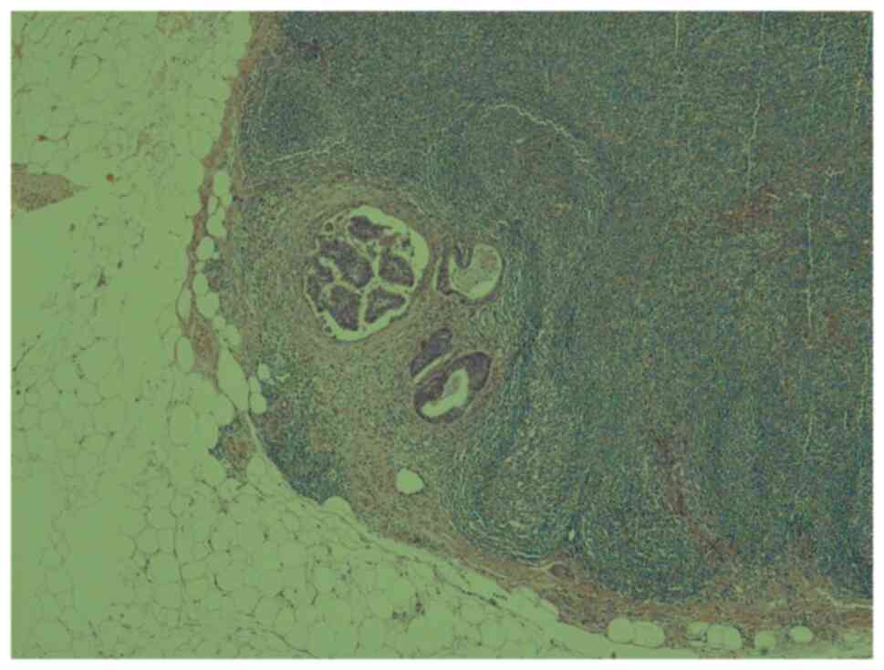
Histopathological examination of the resected specimen revealed a well-differentiated intramucosal adenocarcinoma (Tis) confined to the mucosa (Fig. 5). However, one regional lymph node adjacent to the rectum revealed metastasis (Fig. 6). Positron emission tomography-CT performed after surgery showed no evidence of distant metastasis. Although the current AJCC on Cancer TNM staging system (8th edition) for rectal cancer does not include a staging group for TisN1M0, the patient was classified as having stage IIIa rectal cancer following a multidisciplinary discussion. She underwent concurrent chemoradiotherapy for stage IIIa rectal cancer and has been followed up at an outpatient clinic for the past 6 years with no evidence of recurrence.

Microscopic examination of the
surgically resected rectum shows (A) highly-differentiated
villotubular proliferative glands (hematoxylin and eosin stain;
magnification, x15), (B) lesions with a characteristic frond-like
growth pattern and villous glandular dysplasia (hematoxylin and
eosin stain; magnification, x40), and (C) focal well-differentiated
adenocarcinoma lesion (hematoxylin and eosin stain; magnification,
x400).

Figure 5

Microscopic examination of the surgically resected rectum shows (A) highly-differentiated villotubular proliferative glands (hematoxylin and eosin stain; magnification, x15), (B) lesions with a characteristic frond-like growth pattern and villous glandular dysplasia (hematoxylin and eosin stain; magnification, x40), and (C) focal well-differentiated adenocarcinoma lesion (hematoxylin and eosin stain; magnification, x400).

Microscopic examination of one
regional lymph node shows metastatic tumor cells (hematoxylin and
eosin stain; magnification, x40).

Figure 6

Microscopic examination of one regional lymph node shows metastatic tumor cells (hematoxylin and eosin stain; magnification, x40).

Discussion

Tis refers to ‘carcinoma in situ,’ meaning that cancer cells are localized to their site of origin and have not invaded the surrounding tissue. In the gastrointestinal tract, Tis typically indicates a tumor confined to the epithelium without invasion of the lamina propria (i.e., intraepithelial tumor) but with penetration through the basement membrane, considered invasive cancer. However, in colorectal cancer, Tis refers to tumors that may invade up to the muscularis mucosa (i.e., intramucosal tumors) (1). It is generally believed that Tis-stage colorectal cancer confined to the mucosa has no potential for metastasis since the colonic mucosa lacks lymphatic vessels (2). Although some studies have reported cases of local recurrence or distant metastasis during follow-up of Tis-stage cancer (3-6), and there have been concerns regarding the potential for metastasis in poorly differentiated Tis colorectal cancer (7), no cases of lymph node metastasis have been confirmed in well-differentiated Tis tumors at the time of diagnosis. This study reports the first documented case of lymph node metastasis in a well-differentiated Tis tumor.

Recent research indicates that lymph node metastasis may be possible even in Tis-stage colorectal cancer. Previous study xplored the potential for cancer cell metastasis to lymph nodes in patients with Tis-stage colorectal cancer and confirmed the presence of lymphatic vessels in the lamina propria (8). Some studies have highlighted the challenges in detecting micrometastases in lymphatic vessels using hematoxylin and eosin staining in early-stage colorectal cancer, emphasizing the need for special staining techniques (9,10). By utilizing specific immunohistochemical staining methods, previous study accurately identified lymphatic vessels in the lamina propria of patients with early-stage colorectal cancer and assessed the potential for cancer cell metastasis. It has been proposed that a neoplastic or inflammatory microenvironment, which induces fibrotic or desmoplastic reactions, may facilitate focal lymphatic invasion, providing a pathway for metastasis in intramucosal colorectal cancer; however, the precise mechanism underlying lymph node metastasis in this case still remains unclear (3,6).

This case raises various important considerations for the diagnosis and treatment of Tis colorectal cancer. First, it suggests a novel metastatic pathway that differs from the current understanding and requires further research to elucidate the mechanisms underlying lymph node metastasis at this stage. Second, it underscores the need to consider the possibility of lymph node metastasis even at the Tis stage in clinical practice. It might be a prudent approach to include the evaluation of perirectal lymph nodes in the diagnosis and management of very early-stage colorectal cancer, despite the very low risk of lymph node metastasis.

Although traditionally considered non-metastatic, emerging evidence suggests that Tis-stage rectal cancers may, in rare cases, demonstrate lymphatic dissemination, necessitating a re-examination of current staging assumptions.

In addition to pathological findings, imaging studies also play a crucial role in the diagnosis and staging of colorectal cancer. However, conventional imaging modalities such as CT and PET-CT have limited sensitivity in detecting micrometastases in regional lymph nodes, particularly in Tis-stage tumors. In this case, no lymph node involvement was suspected preoperatively based on imaging studies, highlighting the limitations of current radiologic assessments.

Recent advancements in imaging, including radiomics and artificial intelligence, have opened new possibilities in preoperative lymph node evaluation. Radiomics-based approaches extract quantitative features-such as shape, texture, and enhancement patterns-from both intratumoral and peritumoral regions on CT, enabling the development of predictive models that outperform traditional size-based assessments (11). Furthermore, deep learning algorithms, such as convolutional neural networks, have shown promising results in identifying subtle imaging patterns associated with nodal metastasis, although challenges remain (12) regarding generalizability, dataset diversity, and clinical applicability. Meanwhile, PET-CT has demonstrated greater sensitivity than CT alone for detecting both nodal and distant metastases in colorectal cancer. In a recent study, PET-CT achieved a sensitivity of 66.7% for nodal metastasis, compared to 55.2% with CT, and 82.5% for distant metastasis (13). Nonetheless, the sensitivity of PET-CT for micrometastasis in early-stage tumors is still suboptimal. These findings suggest that although imaging technology continues to evolve, it remains imperfect in identifying lymph node involvement in Tis-stage cancer.

In conclusion, current colorectal cancer treatment protocols often overlook lymph node evaluation in Tis-stage colorectal cancer. However, this case highlights the need for a more comprehensive approach that considers the potential for lymph node metastasis, even in patients with Tis-stage cancer. Continued research integrating advanced imaging, pathological staining, and molecular analysis is warranted to better understand and detect early lymphatic dissemination.

Acknowledgements

Not applicable.

Funding

Funding: No funding was received.

Availability of data and materials

The data generated in the present study may be requested from the corresponding author.

Authors' contributions

SJN contributed to the conception of this study, participated in the clinical management of the patient and supervised the preparation of the manuscript. GBC performed the surgical treatment. SKL conducted the histopathological examination and confirmed the diagnosis. SHK conducted a comprehensive literature review, collected and organized the clinical data, analysed the case findings in relation to existing literature, and drafted the initial version of the manuscript. All authors discussed the results, revised the manuscript critically for important intellectual content, and approved the final manuscript for publication. SJN and SHK confirm the authenticity of all the raw data.

Ethics approval and consent to participate

Written informed consent was obtained from the patient for participation in the present study.

Patient consent for publication

Witten informed consent was obtained the patient for the publication of the present case report and any accompanying images.

Competing interests

The authors declare that they have no competing interests.

Authors' information

Professor Seung-Joo Nam ORCID: https://orcid.org/0000-0002-0349.

References

1 

Weiser MR: AJCC 8th edition: Colorectal cancer. Ann Surg Oncol. 25:1454–1455. 2018.PubMed/NCBI View Article : Google Scholar

2 

Fenoglio CM, Kaye GI and Lane N: Distribution of human colonic lymphatics in normal, hyperplastic, and adenomatous tissue. Its relationship to metastasis from small carcinomas in pedunculated adenomas, with two case reports. Gastroenterology. 64:51–66. 1973.PubMed/NCBI

3 

Lee HJ, Ye BD, Byeon JS, Kim J, Park YS, Hong YS, Yoon YS and Yang DH: Unusual local recurrence with distant metastasis after successful endoscopic submucosal dissection for colorectal mucosal cancer. Clin Endosc. 50:91–95. 2017.PubMed/NCBI View Article : Google Scholar

4 

Lee KH, Kim JS, Cheon KS, Song IS, Kang DY and Kim JY: TisN0M1 sigmoid colon cancer: A case report. Ann Coloproctol. 30:141–146. 2014.PubMed/NCBI View Article : Google Scholar

5 

Seo HJ, Kim YJ, Cho KB, Kim ES, Hwang IS, Baek SK and Park KS: Nodal metastasis after successful endoscopic submucosal dissection for colorectal mucosal cancer. Endoscopy. 43 (Suppl 2) UCTN:E374–E375. 2011.PubMed/NCBI View Article : Google Scholar

6 

Shia J and Klimstra DS: Intramucosal poorly differentiated colorectal carcinoma: Can it be managed conservatively? Am J Surg Pathol. 32:1586–8; author reply 1588-9. 2008.PubMed/NCBI View Article : Google Scholar

7 

Watson J, Matsui GY, Leaphart A, Wiegel J, Rainey FA and Lovell CR: Reductively debrominating strains of Propionigenium maris from burrows of bromophenol-producing marine infauna. Int J Syst Evol Microbiol 50 Pt. 3:1035–1042. 2000.PubMed/NCBI View Article : Google Scholar

8 

Rodrigo-Calvo MT, Saez de Gordoa K, Lopez-Prades S, Archilla I, Diaz A, Berrios M, Camps J, Musulen E and Cuatrecasas M: Tumour cell seeding to lymph nodes from in situ colorectal cancer. Cancers (Basel). 15(842)2023.PubMed/NCBI View Article : Google Scholar

9 

Fogt F, Zimmerman RL, Ross HM, Daly T and Gausas RE: Identification of lymphatic vessels in malignant, adenomatous and normal colonic mucosa using the novel immunostain D2-40. Oncol Rep. 11:47–50. 2004.PubMed/NCBI View Article : Google Scholar

10 

Kitagawa Y, Ikebe D, Hara T, Kato K, Komatsu T, Kondo F, Azemoto R, Komoda F, Tanaka T, Saito H, et al: Enhanced detection of lymphovascular invasion in small rectal neuroendocrine tumors using D2-40 and Elastica van Gieson immunohistochemical analysis. Cancer Med. 5:3121–3127. 2016.PubMed/NCBI View Article : Google Scholar

11 

Li A, Xiao Z and Zhou Z: RETRACTED: Preoperative Prediction of Lymph Node Metastasis in Colorectal Cancer Based on Intratumoral and Peritumoral CT Radiomics Nomogram. Cancer Treat Res Commun, 2025. https://doi.org/10.1016/j.ctarc.2025.100909.

12 

Keel B, Quyn A, Jayne D and Relton SD: State-of-the-art performance of deep learning methods for pre-operative radiologic staging of colorectal cancer lymph node metastasis: A scoping review. BMJ Open. 14(e086896)2014.PubMed/NCBI View Article : Google Scholar

13 

Engel R, Kudura K, Antwi K, Denhaerynck K, Steinemann D, Wullschleger S, Müller B, Bolli M and von Strauss Und Torney M: Diagnostic accuracy and treatment benefit of PET/CT in staging of colorectal cancer compared to conventional imaging. Surg Oncol. 57(102151)2024.PubMed/NCBI View Article : Google Scholar

Related Articles

  • Abstract
  • View
  • Download
  • Twitter
Copy and paste a formatted citation
Spandidos Publications style
Kim S, Chae G, Lee S and Nam S: Lymph node metastasis in a patient with carcinoma <em>in situ</em> rectal cancer: A case report. Mol Clin Oncol 23: 91, 2025.
APA
Kim, S., Chae, G., Lee, S., & Nam, S. (2025). Lymph node metastasis in a patient with carcinoma <em>in situ</em> rectal cancer: A case report. Molecular and Clinical Oncology, 23, 91. https://doi.org/10.3892/mco.2025.2886
MLA
Kim, S., Chae, G., Lee, S., Nam, S."Lymph node metastasis in a patient with carcinoma <em>in situ</em> rectal cancer: A case report". Molecular and Clinical Oncology 23.4 (2025): 91.
Chicago
Kim, S., Chae, G., Lee, S., Nam, S."Lymph node metastasis in a patient with carcinoma <em>in situ</em> rectal cancer: A case report". Molecular and Clinical Oncology 23, no. 4 (2025): 91. https://doi.org/10.3892/mco.2025.2886
Copy and paste a formatted citation
x
Spandidos Publications style
Kim S, Chae G, Lee S and Nam S: Lymph node metastasis in a patient with carcinoma <em>in situ</em> rectal cancer: A case report. Mol Clin Oncol 23: 91, 2025.
APA
Kim, S., Chae, G., Lee, S., & Nam, S. (2025). Lymph node metastasis in a patient with carcinoma <em>in situ</em> rectal cancer: A case report. Molecular and Clinical Oncology, 23, 91. https://doi.org/10.3892/mco.2025.2886
MLA
Kim, S., Chae, G., Lee, S., Nam, S."Lymph node metastasis in a patient with carcinoma <em>in situ</em> rectal cancer: A case report". Molecular and Clinical Oncology 23.4 (2025): 91.
Chicago
Kim, S., Chae, G., Lee, S., Nam, S."Lymph node metastasis in a patient with carcinoma <em>in situ</em> rectal cancer: A case report". Molecular and Clinical Oncology 23, no. 4 (2025): 91. https://doi.org/10.3892/mco.2025.2886
Follow us
  • Twitter
  • LinkedIn
  • Facebook
About
  • Spandidos Publications
  • Careers
  • Cookie Policy
  • Privacy Policy
How can we help?
  • Help
  • Live Chat
  • Contact
  • Email to our Support Team