Introduction
Low-grade serous ovarian cancer (LGSOC) is a rare,
distinct subtype of epithelial ovarian cancer, accounting for 2-5%
of all epithelial ovarian malignancies and 4-5% of serous tumors
globally (1-3).
LGSOC predominantly affects women at a slightly younger median age
than that associated with high-grade serous ovarian cancer, with
median ages at diagnosis being 54-56 years globally, compared with
~62 years in high-grade disease (3-6).
LGSOC is characterized by indolent clinical
behavior, slow progression and markedly prolonged survival times
compared with high-grade serous ovarian cancer, as well as minimal
responsiveness to conventional cytotoxic chemotherapy (1,5,6). The
hallmark histopathological features of LGSOC include
mild-to-moderate nuclear atypia and low mitotic activity (<12
mitoses per 10 high-power fields). These diagnostic criteria are
consistent with the 2020 World Health Organization (WHO)
Classification of Female Genital Tumors (7). Microscopically, LGSOC often shows
cuboidal or columnar cells with amphophilic cytoplasm and frequent
psammoma bodies, and is associated with borderline serous tumors,
supporting the hypothesis of a stepwise progression from benign or
borderline lesions (1,2).
Despite the slower progression and distinct tumor
biology compared with high-grade serous ovarian cancer, LGSOC often
presents at advanced stages, and optimal cytoreductive surgery
remains the cornerstone of initial treatment because the volume of
residual disease is the most meaningful predictor of survival
(1,2,5).
Platinum-based chemotherapy is the standard adjuvant therapy but
shows limited efficacy; therefore, hormonal maintenance therapy and
targeted agents are increasingly used (8). Epidemiological data from Indonesia
mirror global patterns, with the majority of patients with LGSOC
presenting in their mid-50s, similar to trends observed in
international cohorts (3-6),
and a high proportion of diagnoses at advanced International
Federation of Gynecology and Obstetrics (FIGO) stages (9). Survival outcomes are heavily
influenced by the stage at diagnosis and surgical success,
highlighting the universal need for improved early detection,
refined surgical strategies and expanded access to innovative
therapies (1,2,9).
Given the rarity of LGSOC, conducting robust
clinical research and accumulating sufficient patient data through
single-center studies presents substantial challenges. The limited
patient population and geographical dispersion necessitate
collaborative efforts to gain meaningful insights into the natural
history, molecular characteristics and optimal treatment approaches
of the disease. Therefore, multicenter studies involving multiple
institutions and research groups are key to overcoming the
limitations of small sample sizes inherent to rare types of cancer.
Moreover, this approach allows for comprehensive analyses of
clinical outcomes, identification of prognostic factors and the
development of novel therapies (4). This collaborative paradigm is
essential for advancing the understanding of LGSOC and ultimately
improving patient care for this challenging disease.
To date, a comprehensive LGSOC profile has not been
established in Indonesia. In low- and middle-income countries
(LMICs), challenges such as delayed referral from primary care
facilities, unequal access to advanced imaging, variability in
surgical expertise and the absence of robust national cancer
registries may substantially influence stage at diagnosis and
treatment outcomes. Consequently, real-world data from LMIC
settings are essential to understand how systemic healthcare
factors interact with tumor biology and surgical management in
shaping patient prognosis. To the best of our knowledge, the
present study represents the first multicenter analysis of LGSOC in
Indonesia and includes one of the largest cohorts reported in an
LMIC setting. The present study aimed to characterize the
clinicopathological features, surgical outcomes and recurrence
patterns in this cohort within a diverse national referral
network.
Materials and methods
Study design and patient
population
The present study encompassed nine tertiary cancer
centers [Dr. Soetomo General Academic Hospital and Universitas
Airlangga Hospital (Surabaya, Indonesia), Dr. Kariadi General
Hospital (Semarang, Indonesia), Dr. Cipto Mangunkusumo Hospital and
Gatot Soebroto Army Central Hospital (Jakarta, Indonesia), Ngoerah
General Hospital (Denpasar, Indonesia), Dr. Hasan Sadikin General
Hospital (Bandung, Indonesia), Ulin General Hospital (Banjarmasin,
Indonesia), Dr. Wahidin Sudirohusodo General Hospital (Makassar,
Indonesia)] located across seven provinces of Indonesia (East Java,
Central Java, West Java, Jakarta, Bali, South Kalimantan, and South
Sulawesi), representing major referral hubs that collectively
capture patients from diverse ethnic and geographic backgrounds.
This multicenter retrospective study used a total sampling approach
across nine referral hospitals in Indonesia. All patients diagnosed
with and undergoing surgery for LGSOC between January 2019 and
December 2024 were identified and included without age restriction.
The observed patient age range was 17-75 years. Inclusion criteria
comprised: I) Histopathologically confirmed LGSOC based on the 2020
WHO Classification of Female Genital Tumors, characterized by
low-grade nuclear atypia and low mitotic activity; and ii)
availability of clinical and surgical records. Patients with
incomplete data regarding the primary diagnosis, FIGO stage, or
surgical outcomes were excluded from the analysis (7). The presence of residual disease after
surgery was recorded as a binary variable: Optimal cytoreduction
(no visible residual disease) or suboptimal cytoreduction (any
visible residual disease). This binary classification was used
consistently throughout the study.
Data collection
Data were extracted from the patients' medical
records, and the following variables were collected: i) Demographic
data: Age at diagnosis, parity (nulliparous, primiparous,
multiparous) and BMI; ii) clinical characteristics: FIGO stage,
presence of comorbidities (hypertension and diabetes mellitus) and
presence of ascites at surgery; iii) treatment and pathological
data: Primary treatment modality (primary debulking surgery or
neoadjuvant chemotherapy), largest tumor diameter (cm), presence of
residual disease after surgery (optimal, no visible disease;
suboptimal, visible disease) and administration of adjuvant
chemotherapy; and iv) outcome data: Presence of disease recurrence
and patient condition at the last follow-up (alive without disease,
alive with disease or succumbed to disease).
Owing to heterogeneity in electronic medical record
systems across participating centers, certain variables (including
BMI, CA-125 levels and tumor size) were incompletely documented. No
data imputation methods were applied. For each statistical
comparison, analyses were conducted using available data; patients
with missing data for a given variable were excluded only from that
specific analysis. Molecular profiling data (such as KRAS, BRAF,
NRAS mutations and hormone receptor status) were not routinely
available in this retrospective dataset and were, therefore, not
analyzed.
Follow-up and outcome definition
Follow-up evaluations were conducted at ~6-month
intervals; however, specific schedules and imaging modalities were
not fully standardized across centers. Recurrence was defined as
clinical, radiological or biochemical evidence of disease after
completion of primary treatment. Radiological recurrence was based
on imaging findings (ultrasound, CT or MRI), whereas biochemical
recurrence was considered when CA-125 elevation was accompanied by
clinical suspicion or imaging confirmation. The timing of
recurrence was calculated from the completion of primary
treatment.
Statistical analysis
Descriptive statistics were used to summarize the
data collected. Continuous variables (age and tumor size) are
presented as a median value and range, and categorical variables
are presented as a frequency and percentage. Fisher's exact test
was used to evaluate associations between categorical variables
because several subgroup sample sizes were small. Kaplan-Meier (KM)
survival analysis with log-rank testing was performed to evaluate
differences in overall survival (OS) across subgroups. Cox
proportional hazards regression was conducted to estimate HRs,
including univariate analyses for all clinically relevant variables
and a restricted multivariate model incorporating a limited number
of key covariates selected a priori to minimize overfitting.
Analyses were performed in R (version 4.4.1; R Foundation for
Statistical Computing). P<0.05 was considered to indicate a
statistically significant difference.
Results
Patient demographic and
clinicopathological characteristics
A total of 160 patients met the inclusion criteria.
The demographic and clinical characteristics of the patients are
summarized in Table I. The median
age at diagnosis was 46 years (range, 17-75 years). The majority of
patients were multiparous (43.8%). Of all patients, half (50%)
presented with advanced-stage disease (stage III or IV). The median
tumor size was 14 cm (range, 2-40 cm) and 69 patients (43.1%) had
ascites at the time of surgery. BMI, tumor size and cancer
antigen-125 (CA-125) level data were incomplete in some cases owing
to differences in hospital medical record formats and inconsistent
documentation. Analyses involving these variables were thus
conducted using available case analysis, and denominators varied
accordingly across specific comparisons.
 | Table IBaseline demographic and clinical
characteristics. |
Table I
Baseline demographic and clinical
characteristics.
| Characteristic | Value |
|---|
| Median age, years
(min.-max.) | 45.5 (17-75) |
| Parity, n (%) | |
|
Nulliparous | 62 (38.8) |
|
Primiparous | 28 (17.5) |
|
Multiparous | 70 (43.8) |
| BMI, n (%) | |
|
Underweight | 17 (10.6) |
|
Normal | 53 (33.1) |
|
Overweight | 25 (15.6) |
|
Obese | 50 (31.3) |
|
Missing | 15 (9.4) |
| CA-125 level, n
(%) | |
|
Normal | 4 (2.5) |
|
Abnormal | 113 (70.6) |
|
Missing | 43 (26.9) |
| FIGO stage, n
(%) | |
|
I | 58 (36.3) |
|
II | 22 (13.7) |
|
III | 71 (44.4) |
|
IV | 9 (5.6) |
| Ascites, n (%) | |
|
Present | 69 (43.1) |
|
Absent | 91 (56.9) |
| Tumor size | |
|
Median, cm
(min.-max.) | 14 (2-40) |
|
Missing, n
(%) | 17 (10.6) |
| Metastasis, n
(%) | |
|
Omentum | |
|
Yes | 38 (23.8) |
|
No | 122 (76.3) |
|
Lymph
gland | |
|
Yes | 13 (8.1) |
|
No | 147 (91.9) |
| Comorbidity, n
(%) | |
|
Hypertension | |
|
Yes | 25 (15.6) |
|
No | 135 (84.4) |
|
Diabetes
mellitus | |
|
Yes | 9 (5.6) |
|
No | 151 (94.4) |
| Pelvic lymph node
dissection, n (%) | |
|
Yes | 38 (23.7) |
|
No | 122 (76.3) |
| Para-aortic lymph
node dissection, n (%) | |
|
Yes | 17 (10.6) |
|
No | 143 (89.4) |
Recurrence patterns and survival
outcomes
Of the patients with advanced stages of ovarian
cancer (III and IV), 72 (90%) underwent primary debulking surgery
and 8 (10%) underwent neoadjuvant chemotherapy (Table II). Patients who received
neoadjuvant chemotherapy were included in the overall surgical
outcome analysis based on residual disease status following
interval debulking surgery. Of all patients with advanced-stage
disease, 72.5% underwent optimal cytoreduction (no visible residual
disease) and 27.5% underwent suboptimal cytoreduction (with visible
residual disease). A detailed categorization (<1 cm vs. ≥1 cm)
was not possible because of documentation limitations. After
surgery, 119 patients (74.4%) received adjuvant chemotherapy.
Follow-up evaluations for recurrence were generally conducted at
~6-month intervals; however, the actual timing and methods varied
across centers. Disease recurrence was identified based on clinical
assessment supported by imaging modalities such as ultrasound, CT
or MRI when indicated by symptoms or rising CA-125 levels.
| Table IITreatment and patient outcomes. |
Table II
Treatment and patient outcomes.
| Characteristic | Value |
|---|
| Primary treatment,
n (%) | |
|
Primary
debulking surgery | 72 (90.0) |
|
FIGO
stage | |
|
III | 66 (91.7) |
|
IV | 6 (8.3) |
|
Neoadjuvant
chemotherapy | 8 (10.0) |
|
FIGO
stage | |
|
III | 5 (62.5) |
|
IV | 3 (37.5) |
| Residual disease, n
(%) | |
|
Yes | 22 (27.5) |
|
No (complete
debulking) | 58 (72.5) |
| Optimal
cytoreduction, n (%) | |
|
Stage
III | 51 (88.0) |
|
Stage
IV | 7 (12.0) |
| Adjuvant
chemotherapy, n (%) | |
|
Yes | 119 (74.4) |
|
Paclitaxel-carboplatin | 113 (70.6) |
|
Others | 6 (3.8) |
|
No | 41 (25.6) |
| Median follow-up
period, years (min.-max.) | 2 (1-6) |
| Recurrence, n
(%) | |
|
Yes | 16 (10.0) |
|
≥6
months | 10 (6.2) |
|
<6
months | 6 (3.8) |
|
No | 144 (90.0) |
| Current condition,
n (%) | |
|
Alive | 148 (92.5) |
|
Deceased | 12 (7.5) |
A total of 16 patients (10%) experienced disease
recurrence. Recurrence events included radiological and clinically
confirmed cases; isolated biochemical elevation without supportive
clinical or imaging findings was not classified as recurrence.
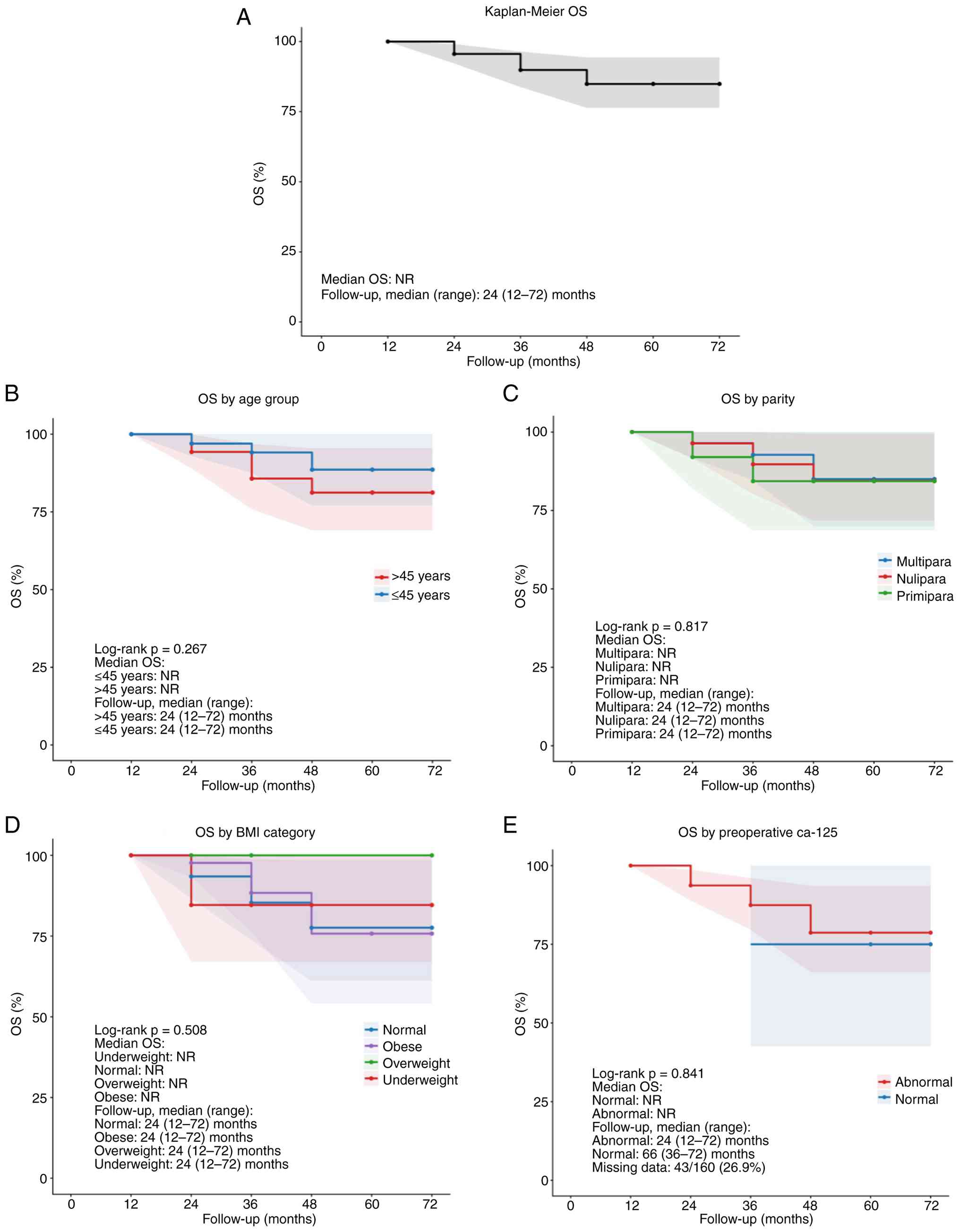
During the final assessment in June 2025, with a median follow-up
duration of 24 months, the estimated OS rates were 100% at 1 year,
95.6% (95% CI, 92.2-99.1) at 2 years, 89.9% (95% CI, 83.8-96.4) at
3 years and 84.9% (95% CI, 76.4-94.4) at 5 years. A total of 148
patients (92.5%) were alive and 12 patients (7.5%) had succumbed
(Table II). The median OS was not
reached during the follow-up period. Given the indolent biology of
LGSOC, this follow-up duration may underestimate long-term
recurrence and survival events. Analysis of prognostic factors
showed that the presence of a residual tumor after surgery was the
most substantial predictor of subsequent recurrence. Among the
patients, 31.8% (7/22) with residual disease experienced recurrence
compared with 6.5% (9/138) without residual disease (Table III).
| Table IIIAssociation of clinical factors with
outcomes. |
Table III
Association of clinical factors with
outcomes.
| Association
tested | Patient
distribution | Statistical
result | Effect
estimate | Key finding |
|---|
| Stage vs. residual
disease | Residual disease
present: 22 patients; no. residual disease: 138 patients | P<0.001 | - | Advanced stage was
significantly associated with suboptimal debulking |
| Residual disease
vs. recurrence | Recurrence in
residual disease group: 7/22 (31.8%); recurrence in no residual
disease group: 9/138 (6.5%) | P<0.001 | Absolute risk
difference: 25.3% (95% CI, 5.5-45.1%); relative risk: 4.88 (95% CI,
2.03-11.75) | Residual disease
was significantly associated with increased recurrence risk |
| Stage vs. tumor
size | Higher stage group
vs. lower stage group | P=0.128 | - | Higher stage was
not significantly associated with larger tumor size |
| Stage vs. current
condition | Worse condition
observed more frequently in advanced-stage patients | P=0.004 | - | Higher stage was
associated with worse clinical condition |
Association between residual disease
and recurrence risk
In this cohort, the overall recurrence rate was
10.0% (95% CI, 5.4-14.7). Patients with residual tumors after
surgery had a markedly higher recurrence rate [31.8% (95% CI,
12.4-51.3%)] when compared with those who achieved complete
debulking [6.5% (95% CI, 2.4-10.6%)]. The absolute risk difference
was 25.3% (95% CI, 5.5-45.1%), corresponding to a ~5-fold increase
in relative risk (4.88; 95% CI, 2.03-11.75). This association was
statistically significant, underscoring the prognostic importance
of achieving no residual disease at the time of primary surgery.
The key prognostic associations are summarized in Table III.
Prognostic factors associated with
surgical and survival outcomes
Analysis of prognostic factors showed that the
presence of a residual tumor after surgery was the most substantial
predictor of subsequent recurrence (P<0.001). Furthermore, the
cancer stage at diagnosis was a major factor in determining
surgical success. Patients with advanced-stage disease (stage
III/IV) were significantly more likely to undergo suboptimal
cytoreduction than those with early-stage disease (P<0.001;
Table III). Consistent with
these findings, the stage at diagnosis was significantly associated
with the disease status of patients at last follow-up (P=0.004;
Table III), with higher stages
being associated with worse survival outcomes. However, no
statistically significant association was found between cancer
stage and primary tumor size in this cohort (P=0.128). All reported
associations represent the results of unadjusted analyses.
Kaplan-Meier analysis of overall
survival according to baseline characteristics
KM curves for OS are shown in Fig. 1. The median OS was not reached
(Fig. 1A). No significant
differences in survival were observed across age groups (log-rank
P=0.267; Fig. 1B), parity
(log-rank P=0.817; Fig. 1C) or BMI
categories (log-rank P=0.508; Fig.
1D), with largely overlapping survival curves. Preoperative
CA-125 levels were also not associated with OS (log-rank P=0.841;
Fig. 1E), although interpretation
is limited by a high proportion of missing data (26.9%). These
findings suggest that baseline demographic and selected clinical
characteristics did not significantly influence OS in this
cohort.
Kaplan-Meier analysis of overall
survival according to clinicopathological and treatment
factors
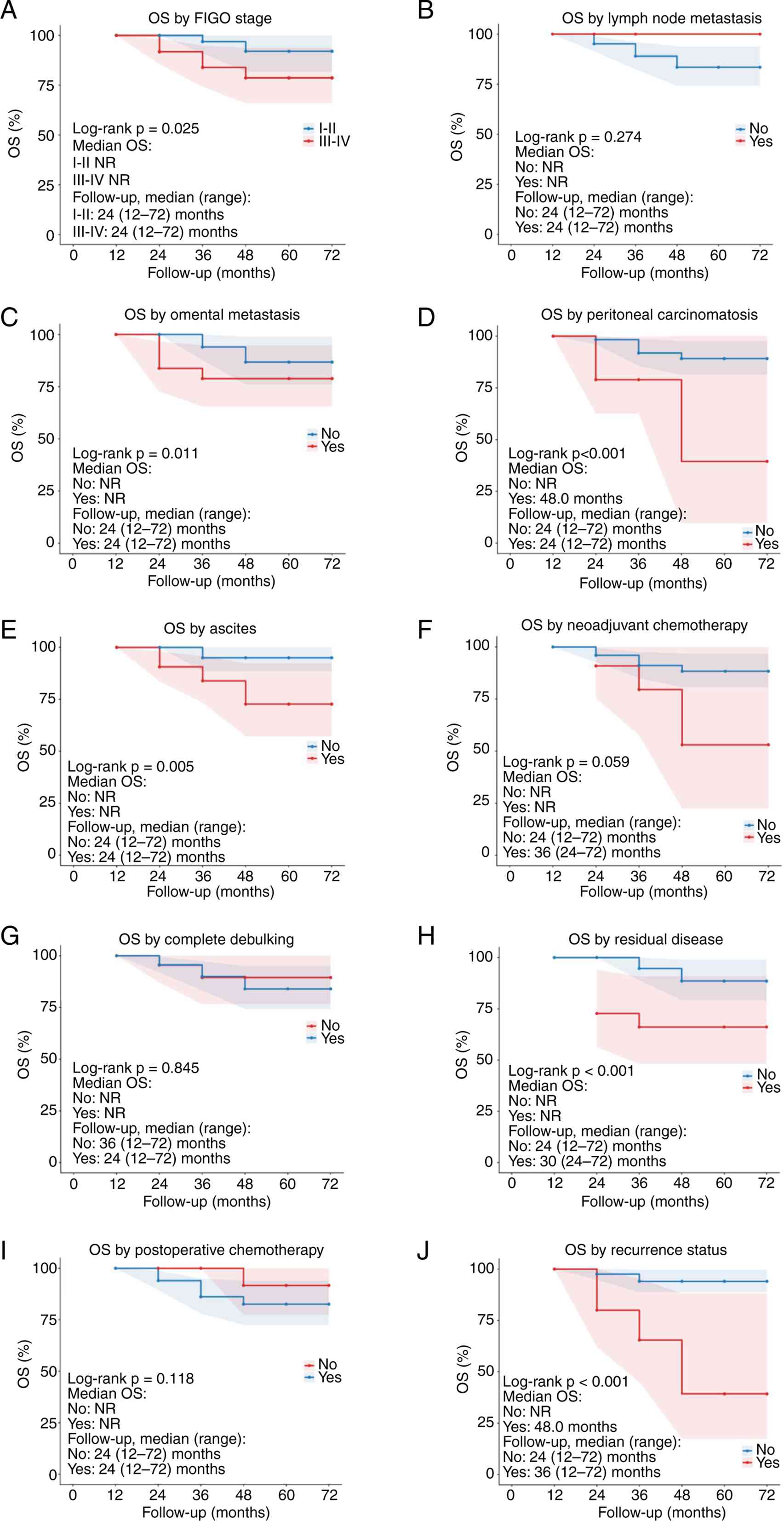
KM analyses of OS according to clinicopathological
and treatment-related factors are presented in Fig. 2. Patients with advanced-stage
disease (FIGO stages III-IV) demonstrated significantly worse OS
compared with those with early stage disease (stages I-II)
(log-rank P=0.025; Fig. 2A).
Similarly, the presence of omental metastasis (P=0.011), peritoneal
carcinomatosis (P<0.001), ascites (P=0.005), residual disease
after surgery (P<0.001) and recurrence (P<0.001) were all
significantly associated with worse survival outcomes (Fig. 2C-E, H and J).
By contrast, lymph node metastasis (P=0.274), administration of
neoadjuvant chemotherapy (P=0.059), completeness of debulking
(P=0.845) and postoperative chemotherapy (P=0.118) were not
significantly associated with OS (Fig.
2B, F, G and I),
although a trend toward worse survival was observed in patients
receiving neoadjuvant chemotherapy. Across the majority of
subgroups, median OS was not reached, except in patients with
peritoneal carcinomatosis and recurrence, where the median survival
was 48.0 months. Follow-up duration was broadly comparable between
groups.
Univariate and multivariate Cox
regression analyses of overall survival
According to univariate Cox regression analysis,
advanced FIGO stage, omental metastasis, peritoneal carcinomatosis,
ascites, residual disease and recurrence were significantly
associated with worse OS. Residual disease (HR, 7.93; 95% CI,
2.51-25.02; P<0.001) and recurrence (HR, 10.06; 95% CI,
3.17-31.92; P<0.001) demonstrated the strongest associations
with mortality (Table IV). In
multivariate analysis, only residual disease remained an
independent predictor of OS (adjusted HR, 5.59; 95% CI 1.44-21.66;
P=0.013), while FIGO stage was no longer statistically significant
(Table V).
| Table IVUnivariate Cox regression
analysis. |
Table IV
Univariate Cox regression
analysis.
| Variable | HR | 95% CI | P-value |
|---|
| FIGO stage
III-IV | 4.82 | 1.05-22.05 | 0.042 |
| Lymph node
metastasis | - | - | 0.998 |
| Omental
metastasis | 3.97 | 1.26-12.53 | 0.018 |
| Peritoneal
carcinomatosis | 6.42 | 2.00-20.59 | 0.002 |
| Ascites | 6.61 | 1.45-30.23 | 0.015 |
| Neoadjuvant
chemotherapy | 3.34 | 0.90-12.41 | 0.072 |
| Complete
debulking | 0.86 | 0.19-3.95 | 0.846 |
| Residual
disease | 7.93 | 2.51-25.02 | <0.001 |
| Postoperative
chemotherapy | 0.23 | 0.03-1.75 | 0.154 |
| Recurrence | 10.06 | 3.17-31.92 | <0.001 |
| Table VMultivariate Cox regression
analysis. |
Table V
Multivariate Cox regression
analysis.
| Variable | Adjusted HR | 95% CI | P-value |
|---|
| FIGO stages
III-IV | 2.05 | 0.34-12.27 | 0.433 |
| Residual
disease | 5.59 | 1.44-21.66 | 0.013 |
Discussion
The present multicenter study reports, to the best
of our knowledge, the first comprehensive overview of LGSOC in
Indonesia including one of the largest cohorts reported from an
LMIC. The findings confirm several established characteristics of
LGSOC, including its occurrence in a relatively younger population
in this setting and the substantial proportion of advanced-stage
presentation. In this cohort, residual disease after surgery and
advanced FIGO stage demonstrated the strongest unadjusted
associations with recurrence and survival outcomes.
Compared with international cohorts, the studied
Indonesian population demonstrated several distinct
characteristics. The median age at diagnosis (45.5 years) was
approximately a decade younger than the median of 54-56 years
reported in Western and East Asian studies (3-6),
suggesting potential demographic or referral differences within
Indonesia, such as earlier onset or differences in health-seeking
behavior. Despite the younger age, the proportion of patients
diagnosed at advanced stages (III-IV; 50%) was comparable to global
data, indicating that delays in presentation and diagnosis remain a
universal challenge (10,11). Obesity and elevated CA-125 levels
were common findings that paralleled those of other recent cohorts,
highlighting their relevance as risk and prognostic factors
(10,12). The overall rate of optimal
cytoreduction (72.5%) was slightly higher when compared with
several large single-center series (40-50%) (1,13),
which may reflect differences in stage composition and the surgical
focus of tertiary referral hospitals participating in the present
study. Collectively, these observations illustrate how patient
demographics, health-system factors and surgical centralization
shape outcomes in an LMIC context. This younger median age may
reflect Indonesia's population structure, which is demographically
younger than the populations of several Western countries, as well
as referral dynamics in which symptomatic younger patients are more
likely to reach tertiary centers. Biological variation cannot be
excluded; however, health-system factors likely have a substantial
effect on the observed differences between the findings of the
present study and those of previous reports.
Notably, optimal cytoreductive surgery was achieved
in 72.5% of cases, which is higher when compared with the rates
reported in several single-center series (40-50%) (1). Although the definition of optimal
cytoreduction required complete macroscopic clearance (no visible
residual disease), the relatively high R0 rate is most plausibly
explained by the inclusion of a substantial proportion of
early-stage cases (50%) rather than superior surgical
aggressiveness alone. Therefore, comparisons with cohorts composed
predominantly of advanced-stage disease should be interpreted
cautiously. Moreover, a direct comparison should consider
differences in patient composition; 50% of the present cohort
consisted of early-stage (I-II) disease, whereas several prior
studies included predominantly advanced-stage cases (III-IV)
(14,15). In addition, the definition of
optimal cytoreduction required complete macroscopic clearance (no
visible residual disease), compared with the <1 cm residual
threshold used in several international reports (16,17).
Although the present study adopted a stricter definition of optimal
cytoreduction, the relatively high rate of optimal surgery (72.5%)
is more plausibly explained by the inclusion of a large proportion
of early-stage cases (50%) rather than by the definitional
criterion itself. This emphasizes the importance of considering
stage distribution when comparing surgical outcomes across studies.
Schmeler and Gershenson (18)
reported optimal cytoreduction in 40-50% of LGSOC cases using the
<1 cm residual threshold, highlighting the uniqueness of the
present findings under a strict definition. This is important
because numerous studies have confirmed surgical completeness as
the principal prognostic factor for LGSOC, overshadowing the modest
impact of adjuvant chemotherapy owing to inherent chemoresistance
in the tumor (11,13). The observed adoption of adjuvant
chemotherapy (74.4%) fits with still-evolving standards; however,
recent meta-analyses suggest that chemotherapy has limited benefits
in LGSOC, with hormonal or targeted therapies emerging as promising
alternatives (8,13). The acceptance and utilization of
hormone maintenance therapy remained limited in the present cohort,
suggesting an area for improvement in clinical practice according
to updated global guidelines (1,13).
The most statistically significant predictors of
recurrence and survival in the present cohort were residual disease
after surgery and the FIGO stage at diagnosis, which mirrors the
conclusions of recent global and multicenter studies (1,11,13).
The present data showed that patients with advanced-stage disease
were more likely to exhibit suboptimal debulking and worse survival
than patients with early-stage disease, a relationship confirmed in
major registries and surveillance, epidemiology and end results
studies (11). These findings
reinforce the interpretation that, unlike high-grade serous ovarian
cancer, survival in LGSOC is more sensitive to the initial quality
of surgical cytoreduction than to systemic therapy (1,13).
These findings were further supported by
time-to-event analyses. KM curves demonstrated significantly worse
OS among patients with advanced FIGO stage, omental metastasis,
peritoneal carcinomatosis, ascites, residual disease and
recurrence. In univariate Cox regression, these variables were
significantly associated with an increased risk of mortality, with
residual disease (HR, 7.93) and recurrence (HR, 10.06)
demonstrating the strongest effects. In multivariate analysis, only
residual disease remained an independent predictor of OS (adjusted
HR, 5.59; 95% CI, 1.44-21.66; P=0.013), whereas FIGO stage lost
statistical significance after adjustment. This finding suggests
that the prognostic impact of disease stage may be partially
mediated through surgical outcomes, reinforcing the key importance
of complete cytoreduction in LGSOC (19).
A high CA-125 level at diagnosis, which was present
in 70.6% of cases, is also a recognized independent prognostic
factor of tumor burden and aggressiveness. However, its specific
impact on OS may be less than that of stage or surgical success
(11). Tumor size was not
significantly associated with tumor stage or outcome in the present
cohort, which agrees with recent reports highlighting the
prognostic importance of residual mass and disease stage (1,11).
Unlike some international studies that classify residual disease
according to size (<1 cm vs. ≥1 cm), the present dataset only
distinguished between ‘no visible disease’ and ‘any visible
residual disease’. This may partially explain the high rates of
optimal cytoreduction. Nevertheless, interpretation of CA-125
findings should be cautious owing to the incomplete documentation
in a subset of patients, which may introduce residual selection
bias.
Emerging literature from the past 5 years also
highlights the relevance of molecular targeted therapies,
particularly the use of MEK inhibitors for MAPK pathway mutations,
as superior or adjunctive options to chemotherapy in recurrent or
refractory settings (1,8,13).
The incorporation of precision medicine remains rare in LMIC,
including the present population, but warrants advocacy in tumor
board decision making.
In the present cohort, the recurrence rate was 10,
and 92.5% of patients were alive at the final follow-up point.
However, the median follow-up duration of 24 months is relatively
short for an indolent malignancy such as LGSOC, for which late
recurrences are well documented. Consequently, the reported
recurrence rate likely underestimates the true long-term recurrence
burden in this population, and extended follow-up is necessary for
more definitive survival assessment. Published international data
have reported 5-year OS rates of 59-89%, depending on the stage and
extent of cytoreduction (10,11,13).
These findings collectively confirm that long-term outcomes in
LGSOC are primarily driven by disease stage and surgical
completeness. Patients with early-stage disease universally show
good outcomes, highlighting the key importance of prompt diagnosis
and aggressive surgery.
The present findings concur with the latest global
evidence that achieving optimal cytoreductive surgery at the
earliest stage is paramount for favorable outcomes in patients with
LGSOC (20). Adjuvant
chemotherapy, although common, provides limited benefits; thus,
greater emphasis should be placed on potential hormone-based and
targeted regimens, as supported by emerging evidence. Continued
research, increased access to molecular diagnostics and adherence
to consensus guidelines are required to improve the prognosis of
patients with LGSOC, particularly in LMICs. Because follow-up
durations varied across centers, long-term survival analysis was
not feasible; nonetheless, the observed survival rate at the last
follow-up supports the generally favorable prognosis of LGSOC.
Beyond reflecting global patterns, the present
findings also highlight several features unique to the Indonesian
population. Patients in this cohort were diagnosed nearly a decade
earlier than international averages, and the study encompassed nine
tertiary hospitals across seven provinces, representing a
multiethnic population and variable access to oncology services.
Such diversity reflects the real-world complexity of cancer care in
Indonesia, an archipelagic nation where geographic distance and
unequal referral systems can influence the stage at diagnosis and
surgical outcomes.
Structural limitations within the healthcare system
likely contribute to these disparities. The distribution of
obstetricians and gynecologists remains uneven, with workforce
shortages and a tendency to concentrate in densely populated
regions, leaving a proportion of hospitals without adequate
specialist coverage (21).
Moreover, the presence of specialists alone does not ensure access
to high-quality oncological care, as resource constraints, limited
availability of advanced therapies and gaps in training may hinder
optimal management (22). These
challenges are further compounded by the scarcity and geographic
concentration of gynecological oncologists, with only 139
specialists nationwide and the majority located in Java, resulting
in substantial barriers to timely diagnosis and treatment,
particularly in remote regions (23).
Within the national health insurance system of
Indonesia, the tiered referral pathway may also contribute to
delays in diagnosis and treatment, creating a cascade of care that
disproportionately affects rare malignancies such as LGSOC, which
may remain underdiagnosed and present at more advanced stages.
Collectively, these findings underscore the importance of
strengthening specialist distribution, referral systems and
oncology infrastructure to improve equitable cancer care delivery
in LMIC settings.
The present study has several limitations. Ethnicity
and geographic distance to referral centers were not captured in
this dataset; however, given the multiethnic composition and wide
geographic distribution of Indonesia, these factors are expected to
contribute to variations in the stage at diagnosis and treatment
access. Formal analyses of OS and disease-free survival could not
be performed because of variability and incompleteness in follow-up
documentation across centers, a common limitation in multicenter
retrospective studies. First, as a retrospective analysis relying
on heterogeneous electronic medical records across multiple centers
in Indonesia, certain variables such as BMI, CA-125 levels and
tumor size were incompletely documented. In addition, the limited
number of recurrence (n=16) and mortality events (n=12) constrained
the statistical power of survival analyses and restricted the
complexity of multivariate modeling.
Despite these limitations, KM survival analysis with
log-rank testing and Cox proportional hazards regression were
performed to explore survival patterns and estimate effect sizes.
Given the small number of events, the multivariate model was
intentionally restricted to a limited number of clinically relevant
covariates to reduce the risk of overfitting; therefore, the
findings should be interpreted as exploratory rather than
definitive independent predictors.
Molecular profiling data, including KRAS, BRAF and
NRAS mutation status as well as hormone receptor expression, were
unavailable in this retrospective cohort. Given that 40-50% of
LGSOC tumors harbor MAPK pathway alterations, the absence of
molecular characterization limited the assessment of eligibility
for targeted therapies such as MEK inhibitors. Future prospective
registries incorporating molecular data will be essential to align
national management strategies with contemporary precision oncology
approaches.
Future research should prioritize improving the
quality and completeness of clinical documentation and establishing
a national ovarian cancer registry in Indonesia. These efforts
would enable robust survival analyses, support evaluation of
treatment patterns and facilitate the integration of molecular and
genomic data. In addition, such infrastructure would allow
meaningful collaboration with international datasets, enabling
comparative analyses of surgical outcomes, referral patterns and
treatment effectiveness across different healthcare settings.
From a clinical perspective, strengthening the
centralization of cytoreductive surgery in high-volume
gynecological oncology centers is essential to improve surgical
outcomes. The development of structured referral pathways from
secondary and district hospitals may reduce delays in treatment and
increase the likelihood of optimal cytoreduction. Expanding access
to hormonal and targeted therapies, including MEK inhibitors,
should also be prioritized to improve outcomes in patients with
LGSOC.
From a policy standpoint, these findings support
national strategies aimed at improving early detection and timely
referral. Public awareness initiatives targeting persistent
abdominal symptoms may contribute to earlier diagnosis and reduce
the proportion of patients presenting with advanced-stage disease.
Collectively, these efforts highlight the importance of coordinated
clinical, research and policy approaches to improve outcomes for
patients with LGSOC in resource-limited settings.
In conclusion, the present large multicenter
Indonesian cohort study revealed that the ability to achieve
complete surgical cytoreduction without residual disease was the
most potent predictor of favorable outcomes. The present findings
advocate for national health strategies focused on improving early
detection and ensuring that patients with LGSOC are managed in
specialized centers with the surgical expertise required to
maximize their chances of optimal surgical outcomes.
Acknowledgements
Not applicable.
Funding
Funding: No funding was received.
Availability of data and materials
The data generated in the present study may be
requested from the corresponding author.
Authors' contributions
BAT, KN, RAU, SR, FE, KYS, TMI, TWU, STW, HMARP and
ER contributed to conceptualization. KN, RAU and HAS confirm the
authenticity of all the raw data; BAT, KN and RAU contributed to
formal analysis; BAT, KN and RAU contributed to investigation. BAT,
KN, RAU and HAS contributed to methodology design. RAU contributed
to project administration. WS, SR, FE, KYS, TMI, VGEP, TWU, TDA,
STW, HMARP and ER contributed to data collection at each central
hospital. BAT and WS supervised the study. BAT and KN contributed
to validation. BAT, KN, RAU and HAS wrote the original draft. All
authors reviewed and edited the manuscript, and read and approved
the final manuscript.
Ethics approval and consent to
participate
The present study was approved by the Institutional
Review Board of Dr. Soetomo General Academic Hospital (approval no.
1095/KEPK/IX/2024; Surabaya, Indonesia). All data used in the
present study are available for independent analysis for the
purposes of additional data analysis or to ensure reproducibility
of the present study in other centers. Informed consent for
participation was waived in accordance with the approval granted by
the institutional ethics committee.
Patient consent for publication
Not applicable.
Competing interests
The authors declare that they have no competing
interests.
Use of artificial intelligence tools
During the preparation of this work, artificial
intelligence tools were used to improve the readability and
language of the manuscript and to enhance the literature search,
and subsequently, the authors revised and edited the content
produced by the artificial intelligence tools as necessary, taking
full responsibility for the ultimate content of the present
manuscript.
References
|
1
|
Alhusaini H, Badran A, Al Juhani A,
Alshamsan B, Alsagaih Y, Alqayidi AA, Sheikh A, Elhassan T,
Maghfoor I, Elshentenawy A and Elshenawy MA: Outcome and prognostic
factors of low-grade serous ovarian cancer: An observational
retrospective study. Mol Clin Oncol. 21(47)2024.PubMed/NCBI View Article : Google Scholar
|
|
2
|
Giannecchini GV, da Silva JL, de Oliveira
Bretas G, dos Santos ALS, Baltar LFR and de Melo AC: Exploring
novel approaches in the systemic therapy of low-grade serous
carcinoma of the ovary: A literature review. Front Med (Lausanne).
11(1366603)2024.PubMed/NCBI View Article : Google Scholar
|
|
3
|
Zwimpfer TA, Tal O, Geissler F, Coelho R,
Rimmer N, Jacob F and Heinzelmann-Schwarz V: Low grade serous
ovarian cancer - A rare disease with increasing therapeutic
options. Cancer Treat Rev. 112(102497)2023.PubMed/NCBI View Article : Google Scholar
|
|
4
|
Arora T, Mullangi S, Vadakekut ES and
Lekkala MR: Epithelial Ovarian Cancer. In: StatPearls. StatPearls
Publishing, Treasure Island, FL, 2024.
|
|
5
|
Babaier A, Mal H, Alselwi W and Ghatage P:
Low-grade serous carcinoma of the ovary: The current status.
Diagnostics (Basel). 12(458)2022.PubMed/NCBI View Article : Google Scholar
|
|
6
|
Nakayama K, Razia S, Ishibashi T and Kyo
S: Current concept of low-grade serous ovarian carcinoma. Transl
Cancer Res. 13:6–10. 2024.PubMed/NCBI View Article : Google Scholar
|
|
7
|
Höhn AK, Brambs CE, Hiller GGR, May D,
Schmoeckel E and Horn LC: 2020 WHO classification of female genital
tumors. Geburtshilfe Frauenheilkd. 81:1145–1153. 2021.PubMed/NCBI View Article : Google Scholar
|
|
8
|
Cobb L and Gershenson D: Novel
therapeutics in low-grade serous ovarian cancer. Int J Gynecol
Cancer. 33:377–384. 2023.PubMed/NCBI View Article : Google Scholar
|
|
9
|
Rustamadji P, Wiyarta E, Nuryanto KH,
Anggraeni TD, Kusuma F, Purwoto G, Winarto H, Heliyanti T, Tjahjadi
H, Hayati A, et al: A decade of ovarian cancer in indonesia:
Epidemiology and survival analysis from 2010 to 2020. J Clin Med.
14(1692)2025.PubMed/NCBI View Article : Google Scholar
|
|
10
|
Zuhdy M, Abdelkhalek M, Gaballa K, Hamdy M
and Refky B: Prognostic factors and clinical hallmarks of low grade
serous ovarian carcinoma: A single center study. Eur J Gynaecol
Oncol. 44:98–104. 2023.
|
|
11
|
Bao M, Jia Q, Tang H, Dong Z, Shan W, Chen
Y, Zhang S, Wei W, Qin Z, Wang H, et al: Study on prognostic
factors of low-grade serous ovarian cancer and establishment of
nomogram prognostic model. Cancer in Females. 1:1–9. 2024.
|
|
12
|
Giro A, Herrmann T, Bauer A, Pinard C,
Godiveau M, Passildas J, Abrial C and Durando X: Predictive and
prognostic factors in epithelial ovarian cancer: A review. Indian J
Gynecol Oncol. 23(45)2025.
|
|
13
|
Wang Q, Cao SH, Li YY, Zhang JB, Yang XH
and Zhang B: Advances in precision therapy of low-grade serous
ovarian cancer: A review. Medicine (Baltimore).
103(E34306)2024.PubMed/NCBI View Article : Google Scholar
|
|
14
|
Barlin JN, Long KC, Tanner EJ, Gardner GJ,
Leitao MM Jr, Levine DA, Sonoda Y, Abu-Rustum NR, Barakat RR and
Chi DS: Optimal (≤1 cm) but visible residual disease: Is extensive
debulking warranted? Gynecol Oncol. 130:284–288. 2013.PubMed/NCBI View Article : Google Scholar
|
|
15
|
Suidan RS, Ramirez PT, Sarasohn DM,
Teitcher JB, Mironov S, Iyer RB, Zhou Q, Iasonos A, Paul H, Hosaka
M, et al: A multicenter prospective trial evaluating the ability of
preoperative computed tomography scan and serum CA-125 to predict
suboptimal cytoreduction at primary debulking surgery for advanced
ovarian, fallopian tube, and peritoneal cancer. Gynecol Oncol.
134:455–461. 2014.PubMed/NCBI View Article : Google Scholar
|
|
16
|
Chang SJ and Bristow RE: Evolution of
surgical treatment paradigms for advanced-stage ovarian cancer:
Redefining ‘optimal’ residual disease. Gynecol Oncol. 125:483–492.
2012.PubMed/NCBI View Article : Google Scholar
|
|
17
|
Shah MM, Estes JM and Alvarez RD: No
Residual Disease: The New Definition for Optimal Cytoreduction in
Ovarian Cancer? Clin Ovarian Other Gynecol Cancer [Internet].
2012;5:45–47. 2012.
|
|
18
|
Schmeler KM and Gershenson DM: Low-grade
serous ovarian cancer: A unique disease. Curr Oncol Rep.
10:519–523. 2008.PubMed/NCBI View Article : Google Scholar
|
|
19
|
Wohrer H, Koual M, Bentivegna E, Benoit L,
Metairie M, Bolze PA, Kerbage Y, Raimond E, Akladios C, Carcopino
X, et al: Prognostic impact of cytoreductive surgery conducted with
primary intent, versus cytoreductive surgery after neoadjuvant
chemotherapy, in the management of patients with advanced
epithelial ovarian cancers: A multicentre, propensity score-matched
study from the FRANCOGYN group. BJOG. 130:1511–1520.
2023.PubMed/NCBI View Article : Google Scholar
|
|
20
|
Di Lorenzo P, Conteduca V, Scarpi E,
Adorni M, Multinu F, Garbi A, Betella I, Grassi T, Bianchi T, Di
Martino G, et al: Advanced low grade serous ovarian cancer: A
retrospective analysis of surgical and chemotherapeutic management
in two high volume oncological centers. Front Oncol.
12(970918)2022.PubMed/NCBI View Article : Google Scholar
|
|
21
|
Tjokroprawiro BA, Sulistya HA, Muharram
FR, Ulhaq RA, Izza A, Prasetyo B, Novitasari K, Wiweko B, Habibie
PH, Lukmana AAI and Asari MMH: Geospatial access to emergency
obstetric surgery in indonesia: Is travel time for access too long?
Ann Glob Heal. 90(82)2024.PubMed/NCBI View Article : Google Scholar
|
|
22
|
Tjokroprawiro BA, Novitasari K, Ulhaq RA,
Sulistya HA and Martini S: Investigation of the trends and
associated factors of ovarian cancer in Indonesia: A systematic
analysis of the Global Burden of Disease study 1990-2021. PLoS One.
20(e0313418)2025.PubMed/NCBI View Article : Google Scholar
|
|
23
|
Tjokroprawiro BA, Sulistya HA, Rahma AA,
Novitasari K, Ulhaq RA, Yuliati I, Utami TW and Anggraeni TD:
Geographic barriers to gynaecological cancer care in Indonesia: A
geospatial and infrastructure analysis. BMJ Open.
16(e112112)2026.PubMed/NCBI View Article : Google Scholar
|