1. Introduction
Head and neck malignant tumors refer to all
malignant tumors extending from the base of the skull to above the
clavicle and anterior to the cervical spine, including malignant
tumors in the soft tissues of the head and face, ears, nose,
pharynx, larynx, oral cavity, salivary glands, soft tissues of the
neck and thyroid gland. They generally exclude intracranial,
cervical and intraocular tumors. The incidence rate of these tumors
accounts for ~5% of all systemic malignant tumors, posing a serious
threat to human health (1). Early
diagnosis and precision treatment of cancer play a crucial role in
improving patients' prognosis and quality of life. For decades,
researchers have collected vast amounts of clinical data in various
forms to tackle cancer. However, it is difficult for individual
clinicians to handle such a massive dataset. With the development
of artificial intelligence (AI), this problem is gradually being
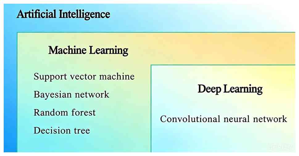
solved. As an important subfield of AI, machine learning (ML),
which can autonomously learn, analyze, and process big data, and
deep learning (DL), as an important branch of ML, have become
popular tools for medical researchers. Various learning techniques,
including Convolutional Neural Network (CNN), Support Vector
Machine, Bayesian Network, Random Forest (RF) and Decision Tree
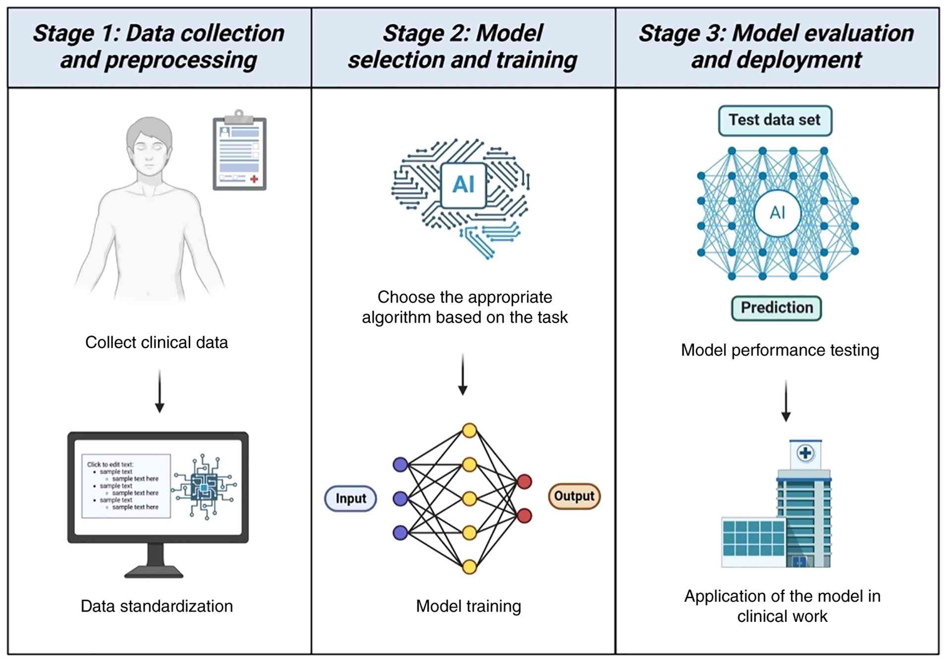
(DT), have been widely applied in cancer research to develop
predictive models for effective and accurate decision-making
(Figs. 1 and 2) (2,3).
With the rapid development of AI technology,
clinical research is entering a new stage of intelligence and
data-driven at an unprecedented speed. In the current era of
increasingly complex medical practices and diverse disease spectrum
changes, the deep integration of traditional statistical methods,
medical experience and emerging technologies is gradually building
a research paradigm centered on precision medicine. A group of
scholars at the forefront of interdisciplinary research are
exploring how AI can be efficiently applied to disease diagnosis,
prediction and individualized treatment pathways, providing
scientific support for achieving precision medicine.
AI-based medical imaging has emerged as a powerful
tool. By integrating multi-omics data such as radiomics, electronic
medical records and pathology, it constructs risk stratification
models that can identify high-risk populations and assist
clinicians in early screening. Leveraging its advantages of being
non-invasive, cost-effective and rapid, it significantly optimizes
the cancer diagnosis and monitoring processes (4-6).
AI in medical radiotherapy can automate manual tasks, enhance
safety measures, optimize adaptive treatment, and improve processes
such as image segmentation, generating synthetic CT scans,
automatic planning, and motion tracking, thereby enhancing the
efficiency and quality of the treatment process (7,8). AI
can extract imaging features that were previously unresolvable from
medical imaging and categorize them into clinical ‘phenotypes’.
Positron emission tomography-computed tomography and single photon
emission computed tomography have been widely used in the detection
and staging of cancer lesions, aiding in improved prediction of
prognosis and detection of adverse events (9). Introducing AI into the pathological
images of tissue samples can reveal structural features in
histological images that are unobservable to the human eye,
efficiently diagnose disease types, and generate corresponding
biomarker profiles (10). Current
research on AI-assisted clinical diagnosis and treatment not only
focuses on enhancing the performance of the model itself but also
strives to develop interpretability to ensure the credibility of
the decision-making process.
As individuals' emphasis on health increases,
clinicians' work becomes increasingly busy. The use of AI to assist
doctors in diagnosis and treatment can not only improve clinical
efficiency but also reduce errors. AI has shown great potential in
the medical field. In the present review, focus is addressed on the
application of AI in early warning, early diagnosis and precision
treatment of head and neck tumors.
2. Diagnosis
Nasopharyngeal carcinoma (NPC) and malignant tumors
of the pharynx often lack specific manifestations in the early
clinical stages. Early diagnosis of patients relies on the
detection of lesions through endoscopy. Therefore, it is
particularly important for endoscopists to observe the
morphological characteristics of the mass through endoscopy and
make preliminary judgments. However, it is challenging for the
human eye to distinguish between mucosal inflammation, cysts and
early-stage tumors under endoscopy. Junior doctors need extensive
training to develop the ability to identify the microvascular
characteristics of tumors. For endoscopists with limited training
and experience, the diagnostic accuracy and consistency for early
NPC and malignant throat tumors are not ideal. Therefore,
developing a computer-aided diagnostic system to assist doctors in
diagnosis is of great significance (Table I) (11-14).
 | Table IApplication of artificial
intelligence in otolaryngology. |
Table I
Application of artificial
intelligence in otolaryngology.
| First author/s,
year | Model task | Data source | Algorithm | Model
performance | (Refs.) |
|---|
| Li et al,
2025 | Early
classification and detection of vocal cord leukoplakia | 378 optical
coherence tomography images from 12 patients | Random Forest | Accuracy=92.59%,
Recall=93.25% | (15) |
| Sampieri et
al, 2024 | Real-time
delineation of laryngeal cancer boundaries | 3,933 images of
laryngeal cancer from 557 patients | Deep Learning | Dice Similarity
Coefficient=0.83, Accuracy=0.97 | (16) |
| Tie et al,
2024 | Early diagnosis of
vocal cord cancer | 7,057 laryngoscope
images from 426 patients | Deep Learning | Accuracy=0.879,
sensitivity=0.882, specificity=0.875, AUC=0.936 | (17) |
| Tamashiro et
al, 2020 | Pharyngeal cancer
detection | 5,403 laryngoscope
images of 202 cases of superficial pharyngeal cancer and 45 cases
of advanced pharyngeal cancer | Deep Learning | AUC=0.8, NBI
sensitivity=85.6%, WLI sensitivity=70.1% | (18) |
| Li et al,
2018 | Detection of
nasopharyngeal malignant tumors | 28,966
nasopharyngeal endoscopy images of 7951 patients | Deep Learning | Accuracy=88.7%,
sensitivity=91.3%, specificity=83.1%, AUC=0.938 | (21) |
| Xu et al,
2022 | Identify NPC and
non-cancerous diseases | 4,783
nasopharyngeal endoscopy images from 671 patients | Deep Learning | S-DCNN (Siamese
deep CNN): Accuracy=95.7%, sensitivity=97.0%, specificity=94.3%,
AUC=0.985 | (22) |
| He et al,
2023 | Real-time detection
of NPC during nasopharyngeal endoscopy | 2,429 video frames
of nasopharyngeal endoscopy from 690 patients | Deep Learning | Precision=0.825,
Recall=0.743, Mean Average Precision=0.814 | (23) |
| Xu et al,
2024 | Accurate diagnosis
of NPC | 12,087
nasopharyngeal endoscopic images and 309 videos from 1108
patients | Deep Learning | Fujian image
dataset: Accuracy=0.956, Sensitivity=0.948, Specificity=0.963,
AUC=0.981 | (24) |
| | | | | Fujian video
dataset: Accuracy=0.906, Sensitivity=0.847, Specificity=0.934,
AUC=0.953 | |
| | | | | Jiangxi image
dataset: accuracy=0.880, sensitivity=0.931, specificity=0.827,
AUC=0.937 | |
| He et al,
2025 | Real-time
AI-assisted detection and segmentation | There are a total
of 8,816 frames of nasal endoscopy videos from 707 patients | Deep Learning | NBI:
Accuracy=95.8%, Accuracy=93.1%, Recall=96.0%, Specificity=97.2%,
AUC=0.998 and 0.977 | (25) |
| | | | | WLI:
Accuracy=95.0%, Accuracy=93.5%, Recall=97.2%, Specificity=93.5%,
AUC=0.977 and 0.970 | |
| Peng et al,
2021 | Assist in the
diagnosis and management of thyroid nodules | 22,354 ultrasound
images from 111,114 patients | Deep Learning | Accuracy=0.877,
AUROC=0.922, NPV=0.97, PPV=0.95 | (26) |
| Wang et al,
2024 | Pathological
diagnosis of thyroid cells | 11,254 full-slide
images from 4037 patients | Deep Learning | Accuracy=0.707,
Average AUROC=0.936 | (28) |
| Li et al,
2023 | Real-time detection
of throat cancer | 31,543 NBI and WLI
images from 2,382 patients | Deep Learning | Multimodal:
Accuracy=0.956, Sensitivity=0.948, Specificity=0.964, PPV=0.965,
NPV=0.946 | (29) |
| | | | | Single-modality
WLI: Accuracy=0.948, Sensitivity=0.885, Specificity=0.988,
PPV=0.978, NPV=0.932 | |
| | | | | Single-modality
NBI: Accuracy=0.935, Sensitivity=0.978, Specificity=0.798,
PPV=0.94, NPV=0.917 | |
| Kwon et al,
2025 | Diagnosing
early-stage glottic cancer using laryngeal imaging and voice
analysis | Pusan National
University Hospital dataset | DT, CNN | The accuracy of the
laryngeal image model is 87.88%; the accuracy of the speech DT is
89.06%; and the accuracy of the combined learning of the two is
95.31% | (30) |
| Kim et al,
2024 | Diagnosing
laryngeal diseases based on voice | From January 2015
to December 2022, the voices of male patients who underwent voice
assessments and had voice changes within three weeks | SVM, gradient
boosting tree, artificial neural network, CNN | SVM:
Accuracy=0.6828, Precision=0.6451 Recall=0.6905 | (32) |
| | | | | Gradient Boosting
Tree: Accuracy=0.7152, Precision=0.6790, Recall=0.6769 | |
| | | | | Artificial neural
network: Accuracy=0.6407, Precision=0.6577, Recall=0.5192 | |
| | | | | CNN:
Accuracy=0.7530, Precision=0.7515, Recall=0.7264 | |
The classical ML model shows unique value in the
task of small amount of data. Li et al (15) constructed a ML model for early
staging and detection of laryngeal cancer using only 378 optical
coherence tomography images of 12 patients. The intraoperative
sensitivity of 84.21% was obtained, which can assist in diagnosing
whether the vocal cord leukoplakia further progressed to vocal cord
cancer, and clarify the direction for subsequent treatment
(15). ML models such as DT and RF
rely on artificial precise cutting features (such as shape and
texture), which endows the model with high transparency and
interpretability, and is helpful for clinical understanding of
model logic. However, it also brings inherent limitations. Manually
defining features requires a lot of time, has high requirements for
operators, and it is difficult to exhaust lesion features by manual
definition.
With the ability of automatically extracting
multi-level features, DL has achieved excellent performance in
large sample endoscopic image analysis and has become the preferred
technology for AI models. Sampieri et al (16) trained a DL model based on 3,933
laryngeal cancer images of 557 patients to demarcate the laryngeal
cancer boundary under laryngoscope, and its accuracy is equivalent
to that of human physicians. Tie et al (17) developed a vocal cord leukoplakia
diagnosis model using 7,057 images of 426 patients, which
significantly improved the area under curve (AUC) and accuracy of
laryngologists. The DL model can automatically learn image features
and avoid manual features, so it can surpass the traditional
learning model in tasks with clear standards and sufficient data.
Narrow band imaging (NBI) further amplifies this advantage, which
enhances the display of mucosal blood vessels and fine structures
through specific narrow-band spectra, providing high-quality input
for DL. Tamashiro et al (18) developed a CNN diagnosis system
based on NBI to detect all cancer lesions in 1,912 validation
images in the detection of pharyngeal malignant tumors. The
sensitivity of NBI images is as high as 85.6%, markedly higher than
70.1% of white light imaging (WLI) images, which also confirms the
advantages of NBI in cancer screening (18).
In the field of endoscopic NPC recognition, several
studies have developed an AI model based on static nasal endoscopic
images, which has proved its feasibility and recognition
performance (19-22).
However, these models lack the ability of real-time dynamic
detection, which is not conducive to rapid diagnosis and guidance
of biopsy. In recent years, researchers began to focus on the
real-time detection of NPC in nasal endoscopic video. He et
al (23) developed an NPC
detection model based on DL using the ‘you only look once’ network.
The model shows excellent performance and stability in the test.
The accuracy, recall, average precision mean and F1 score of the
internal test set are 0.977, 0.943, 0.977 and 0.960, respectively,
and those of the external test set are 0.825, 0.743, 0.814 and
0.780(23). Xu et al
(24) developed a DL model for NPC
diagnosis using nasal endoscopic imaging omics, which integrates a
large number of static images and videos. The model shows high
accuracy, sensitivity and specificity in the real environment,
which is superior to the existing models that mainly rely on
biopsy, MRI and CT images. The AUC in the two hospital datasets is
0.981 and 0.937, respectively (24). The NPC SDNet multimodal real-time
nasal endoscopy new DL neural network model built by He et
al (25) allows feature
extraction, segmentation and lesion feature description
simultaneously in the process of image processing. The diagnostic
accuracy of NPC SDNet is 94.0%, and it processes 1,000 frames per
min, which is better than the accuracy of clinicians of different
professional levels (68.9-88.2%), proving that it can help
clinicians improve the diagnostic accuracy of NPC (25).
In other application scenarios of head and neck
tumors such as imaging and pathology, the advantages of the DL
model are also reflected. The DL model thynet developed by Peng
et al (26) was used to
identify benign and malignant nodules by thyroid ultrasound, with
an area under the receiver operating characteristic curve of 0.922,
which was significantly higher than that of 0.839 for radiologists.
After assisting decision-making, the unnecessary puncture
proportion was reduced, and the missed diagnosis rate was slightly
reduced. Its advantage was that deep features such as echo texture
and boundary morphology were stably extracted from massive pixels,
avoiding the fatigue and bias of human eye perception (26). Fine needle aspiration for thyroid
nodules with high suspicion of malignancy is essential for
appropriate therapeutic intervention, but there are still a small
number of samples that produce uncertain cytological results
(27). The thyroid patch-oriented
WSI ensemble recognition (thyropowe) AI-assisted model developed by
Wang et al (28) using
whole slide images can improve the specificity of primary
cytopathologists in diagnosing thyroid cancer from 0.887 to 0.993,
and the accuracy from 0.877 to 0.948. This system is helpful for
rapid and accurate cellular diagnosis of thyroid nodules, thus
enhancing the diagnostic ability of cytopathologists, and is a
potential solution to alleviate the problem of insufficient
cytopathologists (28). These
studies once again show the outstanding advantages of DL in image
global feature learning.
However, the black box characteristic of DL makes
its decision-making process difficult to disassemble, and the model
performance is highly dependent on large-scale and high-quality
annotation data, which is prone to over fitting problems. When the
imaging conditions change, the generalization ability of the model
may be significantly attenuated.
By fusing the complementary information of different
modes, multimodal learning effectively overcomes the upper
performance limit of single-mode model. The laryngopharyngeal AI
diagnostic system developed by Li et al (29) integrates laryngoscope white light
and NBI images at the same time. The accuracy rate of the
multimodal model reaches 0.94, which is better than using either
mode alone, and is equivalent to that of expert laryngologists
(29). Kwon et al (30) further introduced acoustic mode,
fused laryngeal image and voice DT results, and the classification
accuracy increased from 87.88 and 89.06% of single mode to 95.31%,
reflecting the performance breakthrough brought by multiple modal
information complementation. Nowadays, an increasing number of
otolaryngologists have tried to combine voice analysis and
endoscopic technology with AI (31,32).
The mechanism advantage of multimodal fusion is that it enhances
the robustness and generalization ability of the model, and
different modes provide complementary evidence, so that the system
can understand complex lesions from multiple dimensions. However,
this strategy also brings new challenges. Multimodal input requires
higher standardization of data acquisition, equipment compatibility
and consistency of inspection process, which increases the
complexity of the system. When introducing voice and other modes
that are easily disturbed by age, sex, vocalization habits and
environmental noise, the problem of feature stability and cross
mechanism generalization must be solved to ensure the reliable
application of the model.
3. Treatment and prognosis
In clinical treatment, AI models are applied in the
formulation of personalized treatment strategies for various
cancers. For instance, by integrating data from imaging,
histopathology, clinical information and other sources, AI can
predict a patient's treatment response, thereby guiding the
implementation of systemic treatment or translational therapy,
improving efficacy and saving resources.
The treatment for Squamous Cell Carcinoma of the
Head and Neck (HNSCC) primarily involves surgery, supplemented by
radiotherapy and chemotherapy. The comprehensive treatment of
malignant tumors requires the rational and planned application of
existing methods based on the patient's physical condition, tumor
pathological type, extent of invasion and tendency, in order to
significantly improve the cure rate (33). Postoperative chemoradiotherapy is
the standard treatment for cancer with positive resection margins
or extracapsular extension, but the benefits of chemotherapy for
patients with other intermediate-risk characteristics remain
unclear. To predict patients with intermediate-risk HNSCC who may
benefit from chemoradiotherapy, Howard et al (34) trained three ML survival models:
DeepSurv, neural multitask logistic regression and random survival
forests. From the National Cancer Database of the United States,
33,527 patients who underwent radical surgery and adjuvant
chemoradiotherapy or radiotherapy alone were retrospectively
selected. The median follow-up time was 43.2 months, and there was
no significant difference in the prognostic accuracy among the
three models. Treatment was administered according to the
recommendations of the ML model, and the survival rate was
significantly improved in all models. The hazard ratio was 0.79 for
DeepSurv, 0.83 for neural multitask logistic regression, and 0.90
for random survival forest model. Among the three models, no
survival benefit of chemotherapy was observed in patients
recommended to receive radiotherapy alone, indicating that these
models identified a subgroup of patients who only needed
radiotherapy alone (34). Based on
clinical data such as MRI, tumor TN staging, histological subtypes
and plasma EBV DNA levels, Zhang et al (35) developed a DL model to assess
distant metastasis-free survival in locally advanced NPC and
explored the value of additional chemotherapy for different risk
groups on the basis of concurrent chemoradiotherapy. By combining
clinical variables with prognostic significance for distant
metastasis-free survival with coefficients weighted through
logistic regression analysis, the researchers established a
prediction model for distant metastasis-free survival. Patients
were divided into high-risk and low-risk groups based on the median
risk score. Subgroup survival analysis was conducted on patients
who received concurrent chemoradiotherapy alone and those who
received additional chemotherapy plus concurrent chemoradiotherapy
in different risk groups. The distant metastasis-free survival
period of patients in the high-risk group was significantly shorter
than that of patients in the low-risk group. In the low-risk group,
patients who received concurrent chemoradiotherapy alone had a
longer distant metastasis-free survival period than those who
received additional chemotherapy plus concurrent chemoradiotherapy.
However, in the high-risk group, there was no significant
difference between patients who received concurrent
chemoradiotherapy alone and those who received additional
chemotherapy plus concurrent chemoradiotherapy (35).
Radiotherapy is the primary treatment method for
non-metastatic NPC. Among radiation therapy techniques,
intensity-modulated radiotherapy (IMRT) is the most widely used,
offering improved 5-year local regional control and overall
survival with low toxic side effects. However, its efficacy in
locally advanced NPC is unsatisfactory (36-38).
Hu et al (39) utilized ML
algorithms to construct a prognostic model for patients with stage
III-IV advanced NPC after their initial IMRT. A total of 427
patients were included, with an average follow-up period of 7.16
years. Three algorithms, namely logistic regression, DT and RF,
were employed. Among them, RF demonstrated the best predictive
performance, identifying six most significant predictors as
Epstein-Barr virus DNA, aspartate aminotransferase, body mass
index, age, blood glucose level and alanine aminotransferase
(39). Radiation-induced temporal
lobe injury is a severe sequela of radiotherapy in patients with
NPC, having a significant adverse impact on their quality of life,
physical and emotional functioning, and even survival. Although the
use of IMRT in NPC has reduced the incidence of
radiotherapy-related complications, some patients still report such
events (40-42).
Wen et al (43)
retrospectively analyzed data from 8,194 patients with NPC who
received IMRT and developed a predictive model incorporating
dosimetric and clinical variables. D0.5cc (the dose delivered to
0.5 cm3 of the temporal lobe) was the most predictive
dose-related feature, with the highest AUC of 0.799. D0.5cc65.06 Gy
was the tolerable dose for the temporal lobe and reducing D0.5cc
could reduce the risk of temporal lobe damage, especially in
elderly patients with advanced disease.
The application of AI in precision oncology enables
the analysis of tumor genomic data, aiding in the design of
innovative clinical trials and the development of personalized
combination therapy strategies tailored to individual biomarker
characteristics (44,45). Circular RNA (circRNA) is involved
in various biological processes and plays a crucial role in disease
diagnosis, treatment and prognosis. Wang et al (46) proposed a computational model based
on collaborative learning using circRNA multi-view functional
annotation to predict potential circRNA disease associations, which
demonstrates favorable performance in predicting candidate
disease-related circRNAs. Miao et al (47) established a tumor drug sensitivity
prediction model to assist doctors in designing personalized tumor
treatment plans, including targeted drugs and non-specific
chemotherapy drugs. The model was tested on 87 molecular targeted
drugs and non-specific chemotherapy drugs, and the results showed
that the method could effectively predict tumor drug sensitivity,
with an average sensitivity of 0.98 and specificity of
0.97(47).
Immunogenic cell death (ICD) is a type of regulatory
cell death capable of activating adaptive immune responses, capable
of reshaping the tumor immune microenvironment (TIME) by emitting
danger signals or damage-associated molecular patterns. Wang et
al (48) established and
validated a prognostic model for HNSCC related to ICD, identifying
two subtypes associated with ICD through consensus clustering. The
high ICD subtype is associated with favorable clinical outcomes,
abundant immune cell infiltration, and high activity of immune
response signals. This stratification has important clinical
implications for predicting the prognosis of patients with HNSCC
and for immunotherapy (48). Kim
et al (49) developed a
high-throughput immunogenic cell death inducer screening system
utilizing real-time image analysis based on AI. This system screens
for ICD inducers by identifying the typical morphology of dead
cells that undergo ICD. In a blinded test, it efficiently
identified three ICD inducers, which were then verified through
analyses of cell death type, DAMP release, and immune activation.
This system significantly reduces the time and resources required
for screening and can also detect subtle morphological differences
that are difficult to detect manually (49). Patients with cancer with different
prognoses often exhibit differences in their TIME and response to
immune checkpoint inhibitors (ICIs). Cao et al (50) constructed and validated a
prognostic combined cell death index model consisting of 16 genes
by combining ICD and necroptosis characteristics. This model can
predict the response to ICIs in esophageal squamous cell carcinoma
(ESCC). They found that HOOK1 can induce necroptosis in ESCC cells
and inhibit their proliferation and migration, while CUL4A exhibits
the opposite effect. Immunoprecipitation experiments and data from
ESCC patients support the view that HOOK1 and CUL4A may act as
tumor suppressors and oncogenes, respectively (50).
The application of AI in the medical field has taken
healthcare a significant step towards individualization and
precision. AI assists doctors in formulating chemotherapy and
radiotherapy plans, enabling patients with cancer to achieve
maximum therapeutic effects with minimal side effects. AI helps in
the search for potential biomarkers and targets, making
immunotherapy more accessible to a wider range of patients with
cancer. AI-enhanced healthcare will become the mainstream direction
of future medical care.
4. Explainable AI (XAI)
Although AI is highly useful in cancer diagnosis and
prognostic prediction, it lacks the necessary accountability,
transparency and reliability in decision-making for cancer
diagnosis and prognosis, rendering it difficult to be applied
clinically. Therefore, it is necessary to develop intelligent
models that can explain their predictions, so that clinicians can
use them reliably.
Kausar (51)
developed a model based on ML and XAI to predict the survival of
patients with HNSCC using microRNA (miRNA) sequences and clinical
datasets. ML models were developed using RF, Cox Regression, Lasso
Regression, Elastic Regression and Gradient Boosting to predict the
survival risk of patients with HNSCC. Dysregulated miRNAs can lead
to cancer metastasis and can be used to predict patient survival.
Based on this, an XGBoost model based on XAI was developed to
explain the prediction results by displaying specific miRNAs
involved in the survival of patients with HNSCC. Experiments showed
that the important miRNAs identified by the interpretable model for
predicting the survival of patients with HNSCC were consistent with
the prognostic-related miRNAs reported in biological studies,
demonstrating the reliability of the ML model for predicting the
survival of patients with HNSCC, which can be used by clinicians
for treatment decisions (51).
Researchers in the field of medical image analysis
are increasingly using XAI to explain the results of their
algorithms. Dörrich et al (52) developed an interpretable CNN for
evaluating histopathology of head and neck cancer. Initially, a CNN
model was trained to classify tumor and non-tumor tissues. Then,
XAI techniques were applied to explore the important features that
contributed to the CNN model's decision-making. The interpretable
AI method demonstrated that the CNN model's decisions relied on
features consistent with the opinions of pathology experts. This
confirmed that the CNN model could predict head and neck cancer
with high accuracy and had promising prospects in assisting
pathologists in evaluating cancer slides (52).
Multiple research reports have confirmed that the
introduction of XAI helps to enhance the transparency and
reliability of models, bridging the gap between algorithm output
and clinical trust. However, the explanation is only an
approximation of the model's decision-making process and is not
completely faithful to the original model, thus inevitably reducing
its accuracy on the primary task. Another issue is how to evaluate
whether XAI technology can provide favorable explanations (53).
5. Current restrictions
The use of AI has brought numerous conveniences to
the medical industry. However, the adoption of AI solutions and
their implementation in healthcare are still in their infancy.
Although numerous models have shown favorable generalization in
clinical testing, it is still difficult to effectively implement
them in clinical work. This situation is partly due to the fact
that the impact of AI in healthcare is mostly preclinical research,
which takes place in artificial simulation environments, making it
difficult to infer the actual effectiveness of models in real-world
clinical settings (54,55). On the other hand, patients tend to
trust treatment decisions led by doctors (56).
ML and DL models require large datasets to
accurately classify or predict different tasks. Due to the
non-interchangeability of healthcare systems across regions,
obtaining sufficient data often requires collaboration among
multiple large hospitals, which creates a barrier for building
models with more complex and precise algorithms. Overfitting is
also a problem that must be addressed in model development. The
model may learn the relationship between patient variables and
unrelated outcomes, such as specific pixels or textures. Therefore,
the algorithm may work well in the training dataset, but it may
provide inaccurate results when predicting future outcomes. In
addition, the incidence rate of some diseases varies significantly
across different age groups and sexes. The incidence rate of HNSCC
is higher in male than in female patients. In diagnostic and
treatment decision-making experiments related to HNSCC, male
patients usually account for a higher proportion. The bias caused
by the imbalance in sex ratio cannot be effectively evaluated, and
even traditional AI algorithms may amplify this potential bias,
resulting in inconsistent accuracy of model results across
different populations (57,58).
What's worse, the reliability of AI is often broadly
described based on its working principle, which cannot directly
prove the accuracy of its output results. In clinical application
scenarios, the input data is complex and multidimensional, making
it more difficult to interpret the visual output results of AI.
There is no way of knowing whether the model has captured effective
information fragments, or whether the importance weights assigned
by the model to different data are reasonable. For example, the
authors once built a multimodal diagnosis model for early vocal
cord cancer, using voice and laryngoscope images as input
information. In experiments, the accuracy of disease diagnosis
using single-modality data was not consistent, and it was not
possible to determine the weight allocation of the two-modality
data when the multimodal model performed diagnostic tasks.
Researchers analyze the decision-making behavior of AI by probing
the input and output or approximating its decision logic, but this
cannot fully capture the behavior of the underlying model, and the
performance of model interpretation is rarely tested. Currently,
there is no unified evaluation metric in the field of interpretable
AI. This means that the interpreted model decisions may be correct
or incorrect, and similarly, the interpretation may also be correct
or incorrect (59).
Ethics has been a topic that has plagued AI since
its inception. Although existing guidelines for the implementation
and use of AI ethics draw on principles such as autonomy,
non-maleficence, beneficence and justice from medical ethics, and
emphasize transparency, interpretability, accountability and
fairness, they do not provide a clear definition of AI ethics
(60). At the current stage, the
public has various concerns about the application of AI in the
medical field. How to clearly define the responsible parties in
case patients adopt AI decision-making schemes and adverse medical
outcomes occur? Is there any unauthorized commercial use or leakage
of patients' clinical data? All these issues require us to
continuously improve relevant laws and regulations and enhance
regulatory technology, so that in the future, precision medicine
can strike a balance between new AI technologies and humanistic
care, efficiency improvement and risk control (61).
6. Prospect
In recent years, the great potential of AI has
greatly increased the related research in otolaryngology head and
neck surgery. In the clinical diagnosis and treatment of
Otolaryngology Head and neck surgery, imaging examination has
irreplaceable core value. The application of AI technology in this
field is increasingly in-depth. From the early auxiliary
recognition of static laryngoscope and nasal endoscope images, it
is gradually expanded to the real-time analysis of dynamic
laryngoscope and video nasal endoscope, and then to a multimodal
joint analysis framework that integrates images, clinical texts and
physiological signals. With the continuous innovation of algorithm
architecture and training strategy, the diagnosis efficiency of AI
system has been significantly improved, the missed detection rate
has been effectively controlled, and its adaptability to complex
clinical scenarios has been continuously enhanced. This technology
iteration process not only effectively optimized the existing
workflow but also showed broad prospects for clinical
transformation.
At the molecular biology level, AI can efficiently
screen tumor related markers, predict key inhibitory targets, and
evaluate the prognosis and recurrence risk of patients based on
multi-omics data, while providing intelligent screening and
verification path for the development of new antitumor drugs. In
the clinical treatment, the AI assisted decision system can
optimize the radiotherapy and chemotherapy scheme of Otolaryngology
Head and neck, accurately regulate the drug dose, significantly
reduce the toxic and side effects while improving the curative
effect, especially help to avoid unnecessary damage to brain tissue
caused by head irradiation, and improve the quality of life of
patients. The application of AI runs through the whole chain from
molecular typing, efficacy prediction to scheme optimization and
risk control, showing the strong trend of continuous iteration and
multi-field integration of AI in the diagnosis and treatment of
Otolaryngology Head and neck tumors. Its clinical application has
broad prospects and is expected.
Medical AI still faces significant challenges from
construction to clinical transformation, among which the-black
box-problem is particularly critical. The black box makes the
algorithm decision-making process lack the necessary transparency,
and it is difficult for users to understand its reasoning basis and
internal logic. Current research frontiers and future directions
mainly include developing more sophisticated explanation methods to
mitigate the negative correlation between model performance and
interpretability. Research also encompasses establishing unified
interpretability evaluation metrics and enhancing the inherent
interpretability of complex models such as deep learning, with the
goal of achieving interpretability and traceability at each
execution step. By increasing the transparency of technology, we
can enhance people's trust in AI.
To ensure the safe and reliable operation of AI
systems, the focus should be on rigorous and thorough verification
procedures. The only effective way to prove the decisions made by
AI systems is through thorough, careful and meticulous safety and
verification work. Instead of requiring complex AI systems to
provide partial explanations, thorough and rigorous verification of
these systems should be advocated for, among as numerous different
groups as possible, to maximize the guarantee that marginalized
groups are not disproportionately affected by any specific
system.
In terms of ethical challenges, academic disciplines
and governments have made concerted efforts to develop ethical
guidelines for the implementation and use of AI, clarifying the
future development path of AI. In 2021, UNESCO issued the
‘Recommendation on the Ethics of AI’, which is the world's first
normative framework specifically for AI ethics. A total of 193
member states have officially adopted this ethical framework. China
also introduced the ‘Ethical Norms for New-Generation AI’ in the
same year, aiming to integrate ethics into the entire life cycle of
AI and provide ethical guidance for individuals, legal entities,
and other relevant institutions engaged in AI-related activities.
The IEEE Global Initiative provides a platform for
multi-stakeholders and the public from related scientific and
engineering fields to exchange ideas, reach broad consensus on
pressing ethical and social issues, as well as candidate
recommendations for the development and implementation of these
technologies, and explore how to establish ethical and socially
compliant applications for intelligent and autonomous systems and
technologies, aligning them with established values and ethical
principles, while prioritizing human welfare in specific cultural
contexts (https://standards.ieee.org/wp-content/uploads/import/documents/other/ead_v2.pdf).
7. Conclusion
In the present review, the roles of multiple ML and
DL models in the diagnosis, treatment and prognosis of head and
neck malignancies were specifically described. AI models have been
applied in various fields such as clinical decision-making,
precision treatment, molecular mechanisms and targeted drug
development, indicating that the application of AI in medicine is
rapidly advancing. The integration of AI technology into healthcare
heralds a new era of medical innovation. AI can help improve
clinical efficiency, reduce healthcare costs, and advance clinical
research. Automation technology can achieve or surpass the
performance of expert physicians in certain diagnostic or research
tasks in a shorter time. Healthcare systems and professionals can
recognize and harness the potential of AI technology, ensuring that
responsibility and ethics are combined to benefit patients.
Acknowledgements
Not applicable.
Funding
Funding: The present study work was supported by the Ningbo Top
Medical and Health Research Program (grant no. 2023030514), the
2024 Ningbo Public Welfare Science and Technology Plan Key Project
(grant no. 2024S032), the key project of the Ningbo Education
Science Planning in 2025 (grant no. 2025YZD001), the 2024 Teaching
and Research Project of Ningbo University (grant no. JYXM2024122),
the 2022 postgraduate course construction project of Medical School
of Ningbo University, and Ninghai Science and Technology Project
(grant no. 05), the Natural Science Foundation of Zhejiang
Provincial (grant no. LQ21H130001), the Ningbo Health Branding
Subject Fund (grant no. PPXK2018-02), the Ningbo Clinical Research
Center for Otolaryngology Head and Neck Disease (grant no.
2022L005), the Ningbo ‘Technology Innovation 2025’ Major Special
Project (grant no. 2020Z097), the Ningbo Natural Science Foundation
(grant no. 2023J213), the 2022 postgraduate course construction
project of Medical School of Ningbo University, and Key Project of
Huili Foundation (grant no. 2022ZD003), and Ningbo Top Medical and
Health Research Program (grant no. 2023030514).
Availability of data and materials
Not applicable.
Authors' contributions
SYQ wrote the major part of the manuscript. DY and
HZ revised and reviewed the manuscript. All authors read and
approved the final version of the manuscript. Data authentication
is not applicable.
Ethics approval and consent to
participate
Not applicable.
Patient consent for publication
Not applicable.
Competing interests
The authors declare that they have no competing
interests.
References
|
1
|
Global Burden of Disease 2019 Cancer
Collaboration. Kocarnik JM, Compton K, Dean FE, Fu W, Gaw BL,
Harvey JD, Henrikson HJ, Lu D, Pennini A, et al: Cancer incidence,
mortality, years of life lost, years lived with disability, and
disability-adjusted life years for 29 cancer groups from 2010 to
2019: A systematic analysis for the global burden of disease study
2019. JAMA Oncol. 8:420–444. 2022.PubMed/NCBI View Article : Google Scholar
|
|
2
|
Swanson K, Wu E, Zhang A, Alizadeh AA and
Zou J: From patterns to patients: Advances in clinical machine
learning for cancer diagnosis, prognosis, and treatment. Cell.
186:1772–1791. 2023.PubMed/NCBI View Article : Google Scholar
|
|
3
|
Varlamova EV, Butakova MA, Semyonova VV,
Soldatov SA, Poltavskiy AV, Kit OI and Soldatov AV: Machine
learning meets cancer. Cancers (Basel). 16(1100)2024.PubMed/NCBI View Article : Google Scholar
|
|
4
|
Potočnik J, Foley S and Thomas E: Current
and potential applications of artificial intelligence in medical
imaging practice: A narrative review. J Med Imaging Radiat Sci.
54:376–385. 2023.PubMed/NCBI View Article : Google Scholar
|
|
5
|
Gore JC: Artificial intelligence in
medical imaging. Magn Reson Imaging. 68:A1–A4. 2020.PubMed/NCBI View Article : Google Scholar
|
|
6
|
Chen G, Zhang J, Zhuo D, Pan Y and Pang C:
Identification of pulmonary nodules via CT images with hierarchical
fully convolutional networks. Med Biol Eng Comput. 57:1567–1580.
2019.PubMed/NCBI View Article : Google Scholar
|
|
7
|
Zadnorouzi M and Abtahi SMM: Artificial
intelligence (AI) applications in improvement of IMRT and VMAT
radiotherapy treatment planning processes: A systematic review.
Radiography (Lond). 30:1530–1535. 2024.PubMed/NCBI View Article : Google Scholar
|
|
8
|
Minaee S, Boykov Y, Porikli F, Plaza A,
Kehtarnavaz N and Terzopoulos D: Image segmentation using deep
learning: A survey. IEEE Trans Pattern Anal Mach Intell.
44:3523–3542. 2022.PubMed/NCBI View Article : Google Scholar
|
|
9
|
McGale JP, Chen DL, Trebeschi S, Farwell
MD, Wu AM, Cutler CS, Schwartz LH and Dercle L: Artificial
intelligence in immunotherapy PET/SPECT imaging. Eur Radiol.
34:5829–5841. 2024.PubMed/NCBI View Article : Google Scholar
|
|
10
|
Wen Z, Wang S, Yang DM, Xie Y, Chen M,
Bishop J and Xiao G: Deep learning in digital pathology for
personalized treatment plans of cancer patients. Semin Diagn
Pathol. 40:109–119. 2023.PubMed/NCBI View Article : Google Scholar
|
|
11
|
Yang Q, Liu Z, Sun H, Jiao F, Zhang B and
Chen J: A narrative review: Narrow-band imaging endoscopic
classifications. Quant Imaging Med Surg. 13:1138–1163.
2023.PubMed/NCBI View Article : Google Scholar
|
|
12
|
McCoul ED, Ganeshan V and Nguyen T: Nasal
endoscopy: What have we been missing? Int Forum Allergy Rhinol.
14:1277–1281. 2024.PubMed/NCBI View Article : Google Scholar
|
|
13
|
Chabrillac E, Dupret-Bories A, Vairel B,
Woisard V, De Bonnecaze G and Vergez S: Narrow-band imaging in
oncologic otorhinolaryngology: State of the art. Eur Ann
Otorhinolaryngol Head Neck Dis. 138:451–458. 2021.PubMed/NCBI View Article : Google Scholar
|
|
14
|
Fu ZY, Li DP, Shen CL, Wang JP, Han YX,
Chen SW, Ding Z, Zhang L, Liang BY, Yin SY, et al: Narrow-band
imaging in head and neck carcinomas: A systematic review and
meta-analysis. Laryngoscope. 135:34–44. 2025.PubMed/NCBI View Article : Google Scholar
|
|
15
|
Li X, Sun S, Zhang Y, Liu C, Yan Y, Song
W, Chen X, Hong W and Yue S: Accurate and timely intraoperative
classification of vocal cord leukoplakia by machine learning
assisted handheld OCT. Biomed Opt Express. 16:4190–4202.
2025.PubMed/NCBI View Article : Google Scholar
|
|
16
|
Sampieri C, Azam MA, Ioppi A, Baldini C,
Moccia S, Kim D, Tirrito A, Paderno A, Piazza C, Mattos LS and
Peretti G: Real-time laryngeal cancer boundaries delineation on
white light and narrow-band imaging laryngoscopy with deep
learning. Laryngoscope. 134:2826–2834. 2024.PubMed/NCBI View Article : Google Scholar
|
|
17
|
Tie CW, Li DY, Zhu JQ, Wang ML, Wang JH,
Chen BH, Li Y, Zhang S, Liu L, Guo L, et al: Multi-instance
learning for vocal fold leukoplakia diagnosis using white light and
narrow-band imaging: A multicenter study. Laryngoscope.
134:4321–4328. 2024.PubMed/NCBI View Article : Google Scholar
|
|
18
|
Tamashiro A, Yoshio T, Ishiyama A,
Tsuchida T, Hijikata K, Yoshimizu S, Horiuchi Y, Hirasawa T, Seto
A, Sasaki T, et al: Artificial intelligence-based detection of
pharyngeal cancer using convolutional neural networks. Dig Endosc.
32:1057–1065. 2020.PubMed/NCBI View Article : Google Scholar
|
|
19
|
Abd Ghani MK, Mohammed MA, Arunkumar N,
Mostafa SA, Ibrahim DA, Abdullah MK, Jaber MM, Abdulhay E,
Ramirez-Gonzalez G and Burhanuddin MA: RETRACTED ARTICLE:
Decision-level fusion scheme for nasopharyngeal carcinoma
identification using machine learning techniques. Neural Comput
Appl. 32:625–638. 2020.
|
|
20
|
Mohammed MA, Abd Ghani MK, Arunkumar N,
Hamed RI, Mostafa SA, Abdullah MK and Burhanuddin MA: RETRACTED
ARTICLE: Decision support system for nasopharyngeal carcinoma
discrimination from endoscopic images using artificial neural
network. J Supercomput. 76:1086–1104. 2018.
|
|
21
|
Li C, Jing B, Ke L, Li B, Xia W, He C,
Qian C, Zhao C, Mai H, Chen M, et al: Development and validation of
an endoscopic images-based deep learning model for detection with
nasopharyngeal malignancies. Cancer Commun (Lond).
38(59)2018.PubMed/NCBI View Article : Google Scholar
|
|
22
|
Xu J, Wang J, Bian X, Zhu JQ, Tie CW, Liu
X, Zhou Z, Ni XG and Qian D: Deep learning for nasopharyngeal
carcinoma identification using both white light and narrow-band
imaging endoscopy. Laryngoscope. 132:999–1007. 2022.PubMed/NCBI View Article : Google Scholar
|
|
23
|
He Z, Zhang K, Zhao N, Wang Y, Hou W, Meng
Q, Li C, Chen J and Li J: Deep learning for real-time detection of
nasopharyngeal carcinoma during nasopharyngeal endoscopy. iScience.
26(107463)2023.PubMed/NCBI View Article : Google Scholar
|
|
24
|
Xu Y, Wang J, Li C, Su Y, Peng H, Guo L,
Lin S, Li J and Wu D: Advancing precise diagnosis of nasopharyngeal
carcinoma through endoscopy-based radiomics analysis. iScience.
27(110590)2024.PubMed/NCBI View Article : Google Scholar
|
|
25
|
He R, Jie P, Hou W, Long Y, Zhou G, Wu S,
Liu W, Lei W, Wen W and Wen Y: Real-time artificial
intelligence-assisted detection and segmentation of nasopharyngeal
carcinoma using multimodal endoscopic data: A multi-center,
prospective study. EClinicalMedicine. 81(103120)2025.PubMed/NCBI View Article : Google Scholar
|
|
26
|
Peng S, Liu Y, Lv W, Liu L, Zhou Q, Yang
H, Ren J, Liu G, Wang X, Zhang X, et al: Deep learning-based
artificial intelligence model to assist thyroid nodule diagnosis
and management: a multicentre diagnostic study. Lancet Digit
Health. 3:e250–e259. 2021.PubMed/NCBI View Article : Google Scholar
|
|
27
|
Kim TH and Krane JF: The evolution of
‘atypia’ in thyroid fine-needle aspiration specimens. Diagn
Cytopathol. 50:146–153. 2022.PubMed/NCBI View
Article : Google Scholar
|
|
28
|
Wang J, Zheng N, Wan H, Yao Q, Jia S,
Zhang X, Fu S, Ruan J, He G, Chen X, et al: Deep learning models
for thyroid nodules diagnosis of fine-needle aspiration biopsy: A
retrospective, prospective, multicentre study in China. Lancet
Digit Health. 6:e458–e469. 2024.PubMed/NCBI View Article : Google Scholar
|
|
29
|
Li Y, Gu W, Yue H, Lei G, Guo W, Wen Y,
Tang H, Luo X, Tu W, Ye J, et al: Real-time detection of
laryngopharyngeal cancer using an artificial intelligence-assisted
system with multimodal data. J Transl Med. 21(698)2023.PubMed/NCBI View Article : Google Scholar
|
|
30
|
Kwon I, Wang SG, Shin SC, Cheon YI, Lee
BJ, Lee JC, Lim DW, Jo C, Cho Y and Shin BJ: Diagnosis of early
glottic cancer using laryngeal image and voice based on ensemble
learning of convolutional neural network classifiers. J Voice.
39:245–257. 2025.PubMed/NCBI View Article : Google Scholar
|
|
31
|
Song Q and Li X: Application and
development of voice analysis and endoscopic technology combined
with artificial intelligence in the diagnosis and treatment of
throat disease. Lin Chuang Er Bi Yan Hou Tou Jing Wai Ke Za Zhi.
36:647–650. 2022.PubMed/NCBI View Article : Google Scholar : (In Chinese).
|
|
32
|
Kim HB, Song J, Park S and Lee YO:
Classification of laryngeal diseases including laryngeal cancer,
benign mucosal disease, and vocal cord paralysis by artificial
intelligence using voice analysis. Sci Rep. 14(9297)2024.PubMed/NCBI View Article : Google Scholar
|
|
33
|
Johnson DE, Burtness B, Leemans CR, Lui
VWY, Bauman JE and Grandis JR: Head and neck squamous cell
carcinoma. Nat Rev Dis Primers. 6(92)2020.
|
|
34
|
Howard FM, Kochanny S, Koshy M, Spiotto M
and Pearson AT: Machine learning-guided adjuvant treatment of head
and neck cancer. JAMA Netw Open. 3(e2025881)2020.PubMed/NCBI View Article : Google Scholar
|
|
35
|
Zhang L, Wu X, Liu J, Zhang B, Mo X, Chen
Q, Fang J, Wang F, Li M, Chen Z, et al: MRI-based deep-learning
model for distant metastasis-free survival in locoregionally
advanced nasopharyngeal carcinoma. J Magn Reson Imaging.
53:167–178. 2021.PubMed/NCBI View Article : Google Scholar
|
|
36
|
Ng WT, Soong YL, Ahn YC, AlHussain H, Choi
HCW, Corry J, Grégoire V, Harrington KJ, Hu CS, Jensen K, et al:
International recommendations on reirradiation by intensity
modulated radiation therapy for locally recurrent nasopharyngeal
carcinoma. Int J Radiat Oncol Biol Phys. 110:682–695.
2021.PubMed/NCBI View Article : Google Scholar
|
|
37
|
Chen S, Yang D, Liao X, Lu Y, Yu B, Xu M,
Bin Y, Zhou P, Yang Z, Liu K, et al: Failure patterns of recurrence
and metastasis after intensity-modulated radiotherapy in patients
with nasopharyngeal carcinoma: Results of a multicentric clinical
study. Front Oncol. 11(693199)2022.PubMed/NCBI View Article : Google Scholar
|
|
38
|
Kong FF, Ni MS, Zhai RP, Ying HM and Hu
CS: Local control and failure patterns after intensity modulated
radiotherapy with reduced target volume delineation after induction
chemotherapy for patients with T4 nasopharyngeal carcinoma. Transl
Oncol. 16(101324)2022.PubMed/NCBI View Article : Google Scholar
|
|
39
|
Hu D, Wang Y, Ji G and Liu Y: Using
machine learning algorithms to predict the prognosis of advanced
nasopharyngeal carcinoma after intensity-modulated radiotherapy.
Curr Probl Cancer. 48(101040)2024.PubMed/NCBI View Article : Google Scholar
|
|
40
|
Bin X, Zhu C, Tang Y, Li R, Ding Q, Xia W,
Tang Y, Tang X, Yao D and Tang A: Nomogram based on clinical and
radiomics data for predicting radiation-induced temporal lobe
injury in patients with non-metastatic stage T4 nasopharyngeal
carcinoma. Clin Oncol (R Coll Radiol). 34:e482–e492.
2022.PubMed/NCBI View Article : Google Scholar
|
|
41
|
Dong J, Ng WT, Wong CHL, Li JS, Bollen H,
Chow JCH, Eisbruch A, Lee AWM, Lee VHF, Ng SP, et al: Dosimetric
parameters predict radiation-induced temporal lobe necrosis in
nasopharyngeal carcinoma patients: A systematic review and
meta-analysis. Radiother Oncol. 195(110258)2024.PubMed/NCBI View Article : Google Scholar
|
|
42
|
Guan Y, Yu BB, Liu S, Luo HY and Huang ST:
Research trends of radiation induced temporal lobe injury in
patients with nasopharyngeal carcinoma from 2000 to 2022: A
bibliometric analysis. Radiat Oncol. 18(151)2023.PubMed/NCBI View Article : Google Scholar
|
|
43
|
Wen DW, Lin L, Mao YP, Chen CY, Chen FP,
Wu CF, Huang XD, Li ZX, Xu SS, Kou J, et al: Normal tissue
complication probability (NTCP) models for predicting temporal lobe
injury after intensity-modulated radiotherapy in nasopharyngeal
carcinoma: A large registry-based retrospective study from China.
Radiother Oncol. 157:99–105. 2021.PubMed/NCBI View Article : Google Scholar
|
|
44
|
Fountzilas E, Tsimberidou AM, Vo HH and
Kurzrock R: Clinical trial design in the era of precision medicine.
Genome Med. 14(101)2022.PubMed/NCBI View Article : Google Scholar
|
|
45
|
Prelaj A, Miskovic V, Zanitti M, Trovo F,
Genova C, Viscardi G, Rebuzzi SE, Mazzeo L, Provenzano L, Kosta S,
et al: Artificial intelligence for predictive biomarker discovery
in immuno-oncology: A systematic review. Ann Oncol. 35:29–65.
2024.PubMed/NCBI View Article : Google Scholar
|
|
46
|
Wang Y, Liu X, Shen Y, Song X, Wang T,
Shang X and Peng J: Collaborative deep learning improves
disease-related circRNA prediction based on multi-source functional
information. Brief Bioinform. 24(bbad069)2023.PubMed/NCBI View Article : Google Scholar
|
|
47
|
Miao R, Chen HH, Dang Q, Xia LY, Yang ZY,
He MF, Hao ZF and Liang Y: Beyond the limitation of targeted
therapy: Improve the application of targeted drugs combining
genomic data with machine learning. Pharmacol Res.
159(104932)2020.PubMed/NCBI View Article : Google Scholar
|
|
48
|
Wang X, Wu S, Liu F, Ke D, Wang X, Pan D,
Xu W, Zhou L and He W: An immunogenic cell death-related
classification predicts prognosis and response to immunotherapy in
head and neck squamous cell carcinoma. Front Immunol.
12(781466)2021.PubMed/NCBI View Article : Google Scholar
|
|
49
|
Kim E, Jang D, Kim M, Heo J, Shin J, Chung
JY, Choi M, Yang WH, Lee H and Cha JH: High-throughput screening
system for immunogenic cell death inducers using artificial
intelligence-based real-time image analysis. Comput Biol Med.
196(110727)2025.PubMed/NCBI View Article : Google Scholar
|
|
50
|
Cao K, Zhu J, Lu M, Zhang J, Yang Y, Ling
X, Zhang L, Qi C, Wei S, Zhang Y and Ma J: Analysis of multiple
programmed cell death-related prognostic genes and functional
validations of necroptosis-associated genes in oesophageal squamous
cell carcinoma. EBioMedicine. 99(104920)2024.PubMed/NCBI View Article : Google Scholar
|
|
51
|
Kausar N: Machine learning and explainable
artificial intelligence reveals the MicroRNAs associated with
survival of head and neck squamous cell carcinoma patients. Comput
Biol Chem. 118(108503)2025.PubMed/NCBI View Article : Google Scholar
|
|
52
|
Dörrich M, Hecht M, Fietkau R, Hartmann A,
Iro H, Gostian AO, Eckstein M and Kist AM: Explainable
convolutional neural networks for assessing head and neck cancer
histopathology. Diagn Pathol. 18(121)2023.PubMed/NCBI View Article : Google Scholar
|
|
53
|
van der Velden BHM, Kuijf HJ, Gilhuijs KGA
and Viergever MA: Explainable artificial intelligence (XAI) in deep
learning-based medical image analysis. Med Image Anal.
79(102470)2022.PubMed/NCBI View Article : Google Scholar
|
|
54
|
Waring J, Lindvall C and Umeton R:
Automated machine learning: Review of the state-of-the-art and
opportunities for healthcare. Artif Intell Med.
104(101822)2020.PubMed/NCBI View Article : Google Scholar
|
|
55
|
Upadhyay U, Gradisek A, Iqbal U, Dhar E,
Li YC and Syed-Abdul S: Call for the responsible artificial
intelligence in the healthcare. BMJ Health Care Inform.
30(e100920)2023.PubMed/NCBI View Article : Google Scholar
|
|
56
|
Busch F, Hoffmann L, Xu L, Zhang LJ, Hu B,
García-Juárez I, Toapanta-Yanchapaxi LN, Gorelik N, Gorelik V,
Rodriguez-Granillo GA, et al: Multinational attitudes toward AI in
health care and diagnostics among hospital patients. JAMA Netw
Open. 8(e2514452)2025.PubMed/NCBI View Article : Google Scholar
|
|
57
|
Aung YYM, Wong DCS and Ting DSW: The
promise of artificial intelligence: A review of the opportunities
and challenges of artificial intelligence in healthcare. Br Med
Bull. 139:4–15. 2021.PubMed/NCBI View Article : Google Scholar
|
|
58
|
Chen RJ, Wang JJ, Williamson DFK, Chen TY,
Lipkova J, Lu MY, Sahai S and Mahmood F: Algorithmic fairness in
artificial intelligence for medicine and healthcare. Nat Biomed
Eng. 7:719–742. 2023.PubMed/NCBI View Article : Google Scholar
|
|
59
|
Ghassemi M, Oakden-Rayner L and Beam AL:
The false hope of current approaches to explainable artificial
intelligence in health care. Lancet Digit Health. 3:e745–e750.
2021.PubMed/NCBI View Article : Google Scholar
|
|
60
|
Weidener L and Fischer M: Teaching AI
ethics in medical education: A scoping review of current literature
and practices. Perspect Med Educ. 12:399–410. 2023.PubMed/NCBI View Article : Google Scholar
|
|
61
|
Elendu C, Amaechi DC, Elendu TC, Jingwa
KA, Okoye OK, John Okah M, Ladele JA, Farah AH and Alimi HA:
Ethical implications of AI and robotics in healthcare: A review.
Medicine (Baltimore). 102(e36671)2023.PubMed/NCBI View Article : Google Scholar
|