Introduction
Gastric cancer is a notable contributor to
cancer-related morbidity and mortality worldwide, ranking fifth in
terms of incidence and fourth in terms of mortality among all
malignancies (1) Patients with
gastric cancer in China represent over one-half of the worldwide
total. Recent national data from 2022 reveal that gastric cancer
has the third highest incidence and mortality rates among all
cancer types in the country (2).
Despite advancements in diagnosis and treatment, the overall
survival (OS) rate remains low, with a 5-year survival rate
<50%, dropping to <30% for advanced stages of the disease.
Although trastuzumab has improved survival in human epidermal
growth factor receptor-2 (HER-2)-positive disease and established
anti-HER-2 therapy as a standard of care (3,4),
overall outcomes remain poor. HER-2-positive tumors comprise 12-20%
of gastric cancer cases (5) and
represent a distinct therapeutic entity defined by the efficacy of
anti-HER-2 agents. Although HER-2 upregulation portends inferior
survival and heightened metastatic potential relative to
HER-2-negative disease, existing evidence largely derives from
dichotomous comparisons between these subgroups (6-9).
Although research on the specific subtype of disease with low HER-2
expression in gastric cancer has increased (10,11),
few studies have further differentiated this subgroup of
HER-2-positive gastric cancer, leading to a limited understanding
of whether variations in clinical characteristics and prognosis
exist among patients with HER-2-positive gastric cancer with
differing levels of HER-2 protein expression. Clinical data
comparing immunohistochemistry (IHC) 2+ and 3+ HER-2-positive
tumors, scored according to the Rüschoff/Hofmann grading system
(12,13), remain limited. The present study
stratified HER-2-positive gastric cancer by protein expression to
evaluate its impact on clinicopathological features and
survival.
Patients and methods
Patient enrollment and data
collection
The present retrospective study examines clinical
data from patients with HER-2-positive gastric cancer who underwent
a radical gastrectomy at the Zhangzhou Affiliated Hospital of
Fujian Medical University (Zhangzhou, China) between March 2017 and
March 2023. All patients were of Asian ethnicity and originated
from Zhangzhou, Fujian, China. Inclusion criteria necessitated
patients to meet the following conditions: Preoperative clinical
and imaging assessments indicating stage I-III gastric cancer
according to the 8th edition of the American Joint Committee on
Cancer Tumor-Node-Metastasis (TNM) system (14); successful radical resection (D2
lymphadenectomy and negative resection margins); postoperative
pathological confirmation of gastric adenocarcinoma (International
Classification of Diseases for Oncology no. C16 M8140/3); HER-2
status determined as IHC 3+ or 2+, with positive fluorescence in
situ hybridization (FISH) in postoperative pathological
specimens; and availability of comprehensive medical records.
Exclusion criteria encompassed distant organ metastasis, concurrent
malignancies, residual gastric cancer, incomplete medical
documentation, or perioperative mortality attributed to surgical
complications such as bleeding, septic shock or multiple organ
failure. The study was approved by the Institutional Ethics Review
Board of Zhangzhou Affiliated Hospital of Fujian Medical University
(approval no. 2025LWB426).
Treatment strategy
All patients were treated according to clinical TNM
staging. cT1-3N0-1M0 cases proceeded directly to radical
gastrectomy, whereas cT3-4N1-3M0 cases received 2-4 cycles of
neoadjuvant chemotherapy (fluoropyrimidine- or taxane-based plus
platinum: FLOT, FOLFOX, XELOX/SOX or taxane-cisplatin) before
surgery. Doses were as follows: Docetaxel, 50 mg/m2;
oxaliplatin, 85-130 mg/m2; 5-FU, 2,400-2,600
mg/m2; capecitabine, 1,000 mg/m2 BID; S-1, 40
mg/m2 BID; cisplatin, 70-75 mg/m2; all on day
1, every 2-3 weeks. Adjuvant fluoropyrimidine-based chemotherapy
was given to patients with pII (pT1-2N3M0) or pIII (pT3-4aN1-3M0)
disease. Perioperative trastuzumab was not routinely used, in line
with National Comprehensive Cancer Network, European Society for
Medical Oncology, Chinese Society of Clinical Oncology and Japanese
Gastric Cancer Association guidelines (15-18).
Detection of HER-2 expression
The expression of HER-2 protein in gastric cancer
tissues was assessed using the Elivision two-step
immunohistochemical method. The HER-2/neu rabbit monoclonal
antibody (4B5; catalog number 790-2991; prediluted) from Roche
Diagnostics (Shanghai) Co., Ltd., was applied on a Roche
BenchmarkXT automated IHC platform. HER-2 IHC scoring was
independently performed by two experienced gastrointestinal
pathologists who were blinded to clinical outcomes. Discrepant or
equivocal cases were resolved by consensus review using a
multi-headed microscope. Immunostaining of HER-2 was categorized as
0, 1+, 2+ or 3+ utilizing the Rüschoff/Hofmann grading system
(12,13). Tumor tissues with a score of 2+
underwent fluorescence in situ hybridization (FISH) analysis
to confirm that they were indeed HER-2+. Scores of 0
were designated as HER-2-negative; 1+ and FISH-non-amplified 2+
cases were classified as low expression; and 3+ and FISH-amplified
2+ cases were considered HER-2-positive. FISH was performed on 4-µm
FFPE sections using the Abbott PathVysion HER-2 DNA Probe Kit.
Specimens were fixed in 10% neutral buffered formalin at room
temperature for 24-48 h before processing and embedding.
Deparaffinized sections were post-fixed in 10% neutral buffered
formalin for 10 min at room temperature, then processed per the
manufacturer's protocol (pretreatment at 80˚C for 30 min, protease
digestion at 37˚C for 10 min, overnight hybridization at 37˚C with
HER-2/neu and CEP17 probes). Tumor cells were identified on the
corresponding H&E sections. At least 20 consecutive nuclei were
scored, and the HER-2/CEP17 ratio was calculated. A HER-2/CEP17
ratio ≥2 indicated a HER-2-positive result, while a ratio <1.8
indicated a HER-2-negative result. Samples with ratios between 1.8
and 2 necessitated the evaluation of an additional 20-40 tumor
cells, with a final ratio <1.8 classified as HER-2-negative and
≥2 as HER-2-positive. For cases between 1.8 and 2, a further 80-160
nuclei were counted, and the assessment was independently repeated
by at least two observers to reach a consensus.
Statistical analysis
The associations between HER-2 protein expression
and clinical characteristics were assessed through χ2
testing. DFS and OS were analyzed using Kaplan-Meier curves with
log-rank testing for between-group comparisons, and univariate and
multivariate Cox regression analyses were performed to identify the
associations between clinicopathological features and survival
outcomes. All statistical analyses were conducted using SPSS 18.0
software (SPSS, Inc.), with P<0.05 used to indicate a
statistically significant difference.
Results
Characteristics of the overall
population
A total of 99 patients with HER-2-positive gastric
cancer were included in the present study (Table I). The age of participants ranged
from 32 to 86 years, with a median age of 68 years. The majority of
individuals (58.6%) were aged ≥65 years, and the cohort was
predominantly male (75.8%). The cohort primarily consisted of
patients with intestinal-type gastric cancer (79.8%),
well-differentiated tumors (57.6%), T3-4 infiltration depth
(73.7%), stage III disease (59.6%), tumor size <5 cm (60.6%),
lymph node metastasis (60.6%), LVI (76.8%), and PNI (75.8%). All
patients exhibited proficient mismatch repair (pMMR), and 20
patients (20.2%) underwent neoadjuvant therapy. As of the last
follow-up in February 2025, recurrence was observed in 47 patients
(47.5%), and 33 patients (33.3%) passed away.
 | Table IClinical characteristics of patients
with gastric cancer. |
Table I
Clinical characteristics of patients
with gastric cancer.
| Characteristics | n (%) |
|---|
| Total | 99 (100.0) |
| Sex | |
|
Male | 75 (75.8) |
|
Female | 24 (24.2) |
| Age, years | |
|
<65 | 41 (41.4) |
|
≥65 | 58 (58.6) |
|
Differentiation | |
|
Poor | 42 (42.4) |
|
Well | 57 (57.6) |
| Lymphovascular
invasion | |
|
No | 23 (23.2) |
|
Yes | 76 (76.8) |
| TNM stage | |
|
I-II | 40 (40.4) |
|
III | 59 (59.6) |
| Tumor invasion | |
|
T1-2 | 26 (26.3) |
|
T3-4 | 73 (73.7) |
| HER-2
expression | |
|
IHC 2+ | 38 (38.4) |
|
IHC 3+ | 61 (61.6) |
| Lauren type | |
|
Intestinal | 79 (79.8) |
|
Non-intestinal | 20 (20.2) |
| Tumor size, cm | |
|
<5 | 60 (60.6) |
|
≥5 | 39 (39.4) |
| Perineural
invasion | |
|
No | 24 (24.2) |
|
Yes | 75 (75.8) |
| Lymph node
metastasis | |
|
Negative | 39 (39.4) |
|
Positive | 60 (60.6) |
| Neoadjuvant
therapy | |
|
No | 79 (79.8) |
|
Yes | 20 (20.2) |
| Follow-up
outcome | |
|
Recurrence | 47 (47.5) |
|
Death | 33 (33.3) |
Univariate and multivariate analysis
of the overall population
Univariate and multivariate analyses were conducted
to determine the prognostic risk factors for HER-2-positive gastric
cancer. The univariate Cox regression analysis showed that HER-2
expression (P=0.002), tumor size (P=0.003), depth of tumor invasion
(P=0.002), presence of lymph node metastasis (P<0.001), TNM
stage (P<0.001), PNI (P=0.001) and LVI (P=0.007) were associated
with poor DFS. Furthermore, the multivariable Cox regression
analysis revealed that HER-2 expression (P=0.039), lymph node
metastasis (P=0.030) and PNI (P=0.040) were independent prognostic
factors impacting DFS among patients with gastric cancer (Table II). Notably, the univariate Cox
regression analysis showed that tumor size (P=0.014), tumor
invasion (P=0.010), TNM stage (P=0.002), lymph node metastasis
(P=0.009), and PNI (P=0.018) were associated with decreased OS.
HER-2 expression (P=0.074) did not exhibit a significant
association with OS in patients with gastric cancer. The
multivariable Cox regression analysis revealed no independent
prognostic factors for OS in these patients (Table III).
| Table IIUnivariate and multivariate Cox
regression analysis of disease-free survival. |
Table II
Univariate and multivariate Cox
regression analysis of disease-free survival.
| | Univariate
analysis | Multivariate
analysis |
|---|
| Variables | HR (95% CI) | P-value | HR (95% CI) | P-value |
|---|
| Sex
(male/female) | 1.081 | 0.516-2.006 | 0.960 | | | |
| Age (<65/≥65
years) | 1.030 | 0.577-1.838 | 0.921 | | | |
| Differentiation
(well/poor) | 0.778 | 0.437-1.384 | 0.393 | | | |
| TNM stage
(I-II/III) | 4.392 | 2.048-9.421 | <0.001 | 0.759 | 0.219-2.629 | 0.664 |
| Lauren type
(intestinal/non-intestinal) | 1.281 | 0.637-2.578 | 0.487 | | | |
| Tumor size
(<5/≥5 cm) | 2.372 | 1.332-4.228 | 0.003 | 1.718 | 0.934-3.159 | 0.082 |
| Tumor invasion
(T1-2/T3-4) | 5.277 | 1.889-14.74 | 0.002 | 1.510 | 0.416-5.477 | 0.531 |
| Lymph node
metastasis (no/yes) | 4.842 | 2.166-10.82 | <0.001 | 3.796 | 1.140-12.63 | 0.030 |
| Perineural invasion
(no/yes) | 9.978 | 2.417-41.18 | 0.001 | 7.592 | 1.101-52.36 | 0.040 |
| Lymphovascular
invasion (no/yes) | 4.113 | 1.474-11.47 | 0.007 | 0.337 | 0.081-1.397 | 0.134 |
| HER-2 expression
(IHC 2+/IHC 3+) | 3.045 | 1.504-6.167 | 0.002 | 2.167 | 1.038-4.520 | 0.039 |
| Table IIIUnivariate and multivariate analysis
of overall survival. |
Table III
Univariate and multivariate analysis
of overall survival.
| | Univariate
analysis | Multivariate
analysis |
|---|
| Variables | HR (95% CI) | P-value | HR (95% CI) | P-value |
|---|
| Sex
(male/female) | 0.948 | 0.623-1.441 | 0.801 | | | |
| Age (<65/≥65
years) | 0.829 | 0.581-1.184 | 0.303 | | | |
| Differentiation
(well/poor) | 1.645 | 0.911-1.180 | 0.154 | | | |
| TNM stage
(I-II/III) | 4.354 | 1.680-11.28 | 0.002 | 1.699 | 0.329-8.767 | 0.526 |
| Lauren type
(intestinal/non-intestinal) | 1.827 | 0.848-3.937 | 0.124 | | | |
| Tumor size
(<5/≥5 cm) | 2.364 | 1.188-4.703 | 0.014 | 1.657 | 0.810-3.390 | 0.167 |
| Tumor invasion
(T1-2/T3-4) | 6.611 | 1.581-27.64 | 0.010 | 2.443 | 0.393-15.19 | 0.338 |
| Lymph node
metastasis (no/yes) | 3.239 | 1.337-7.849 | 0.009 | 1.340 | 0.337-5.332 | 0.678 |
| Perineural invasion
(no/yes) | 5.630 | 1.347-23.53 | 0.018 | 1.525 | 0.241-9.646 | 0.654 |
| Lymphovascular
invasion (no/yes) | 2.422 | 0.850-6.896 | 0.098 | | | |
| HER-2expression
(IHC 2+/IHC 3+) | 2.015 | 0.934-4.349 | 0.074 | | | |
Different characteristics of gastric
cancer with different HER-2 protein expression levels
HER-2 positivity was identified in 99 patients: 61
(61.6%) were IHC 3+ and 38 (38.4%) were IHC 2+ with FISH
confirmation. These 38 FISH-positive cases represent 22.6% (38/168)
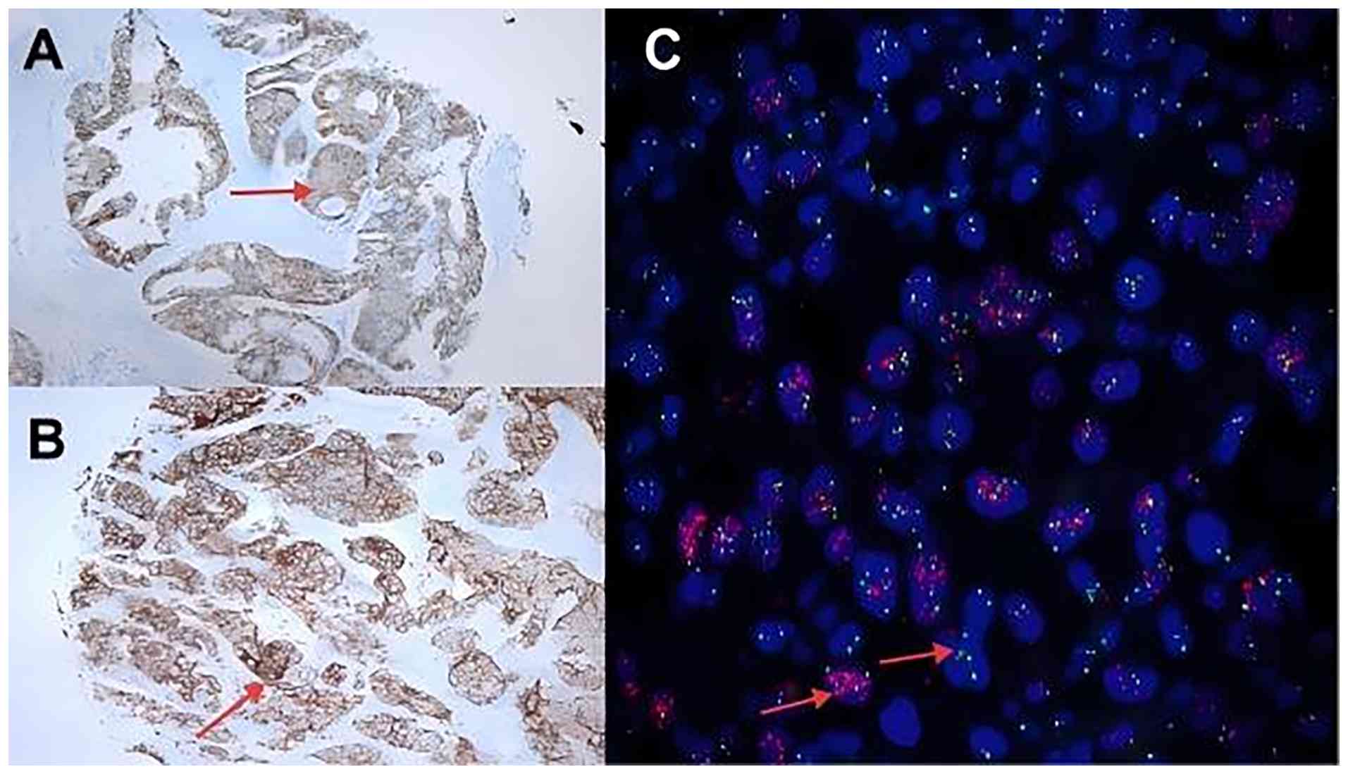
of the initial IHC 2+ screening cohort (Fig. 1). Based on an analysis of
clinicopathological characteristics according to HER-2 expression,
it was found that PNI and LVI were more frequent in HER-2 IHC 3+
patients (51/61 and 51/61, respectively) than in IHC 2+ patients
(24/38 and 25/38, respectively), with P=0.021 and P=0.041 for the
respective comparisons. However, there were no statistically
significant differences in terms of sex, age, TNM stage, Lauren
classification, tumor size, tumor invasion or lymph node metastasis
between the two groups (Table
IV).
| Table IVCharacteristics of gastric cancer
with different HER-2 protein expression. |
Table IV
Characteristics of gastric cancer
with different HER-2 protein expression.
|
Characteristics | HER-2 IHC 2+ | HER-2 IHC 3+ | P-value |
|---|
| Sex, n (%) | | | 0.919 |
|
Male | 29 | 46 | |
|
Female | 9 | 15 | |
| Age, n (%) | | | 0.912 |
|
<65
years | 16 | 25 | |
|
≥65
years | 22 | 36 | |
| Differentiation, n
(%) | | | 0.713 |
|
Poor | 17 | 25 | |
|
Well | 21 | 36 | |
| TNM stage, n
(%) | | | |
|
I-II | 19 | 21 | 0.125 |
|
III | 19 | 40 | |
| Lauren type, n
(%) | | | 0.232 |
|
Intestinal | 28 | 51 | |
|
Non-intestinal | 10 | 10 | |
| Tumor size, n
(%) | | | 0.093 |
|
<5
cm | 27 | 33 | |
|
≥5 cm | 11 | 28 | |
| Tumor invasion, n
(%) | | | 0.059 |
|
T1-2 | 14 | 10 | |
|
T3-4 | 24 | 51 | |
| Lymph node
metastasis, n (%) | | | 0.088 |
|
No | 19 | 20 | |
|
Yes | 19 | 41 | |
| Perineural
invasion, n (%) | | | 0.021 |
|
No | 14 | 10 | |
|
Yes | 24 | 51 | |
| Lymphovascular
invasion, n (%) | | | 0.041 |
|
No | 13 | 10 | |
|
Yes | 25 | 51 | |
| Neoadjuvant
therapy, n (%) | | | |
|
No | 33 | 46 | 0.168 |
|
Yes | 5 | 15 | |
Survival outcome
The study involved 99 patients, with a median DFS
time of 29 months [95% CI, 21-37) and a median OS time of 39 months
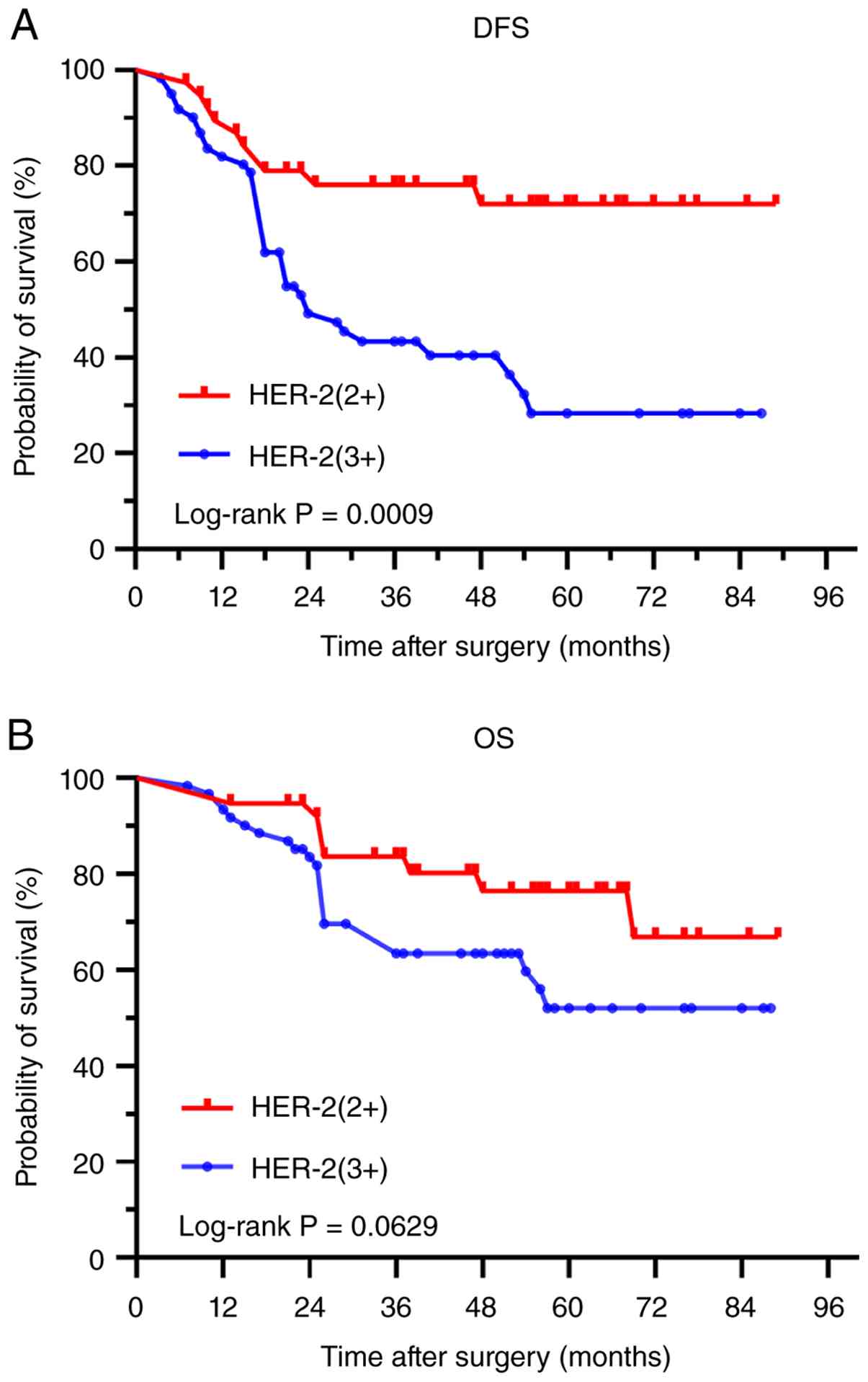
(95% CI, 36-50). Kaplan-Meier survival analysis revealed a
significantly shorter median DFS time in patients with HER-2 IHC 3+
compared with those in patients with HER-2 IHC 2+ (24 months vs.
not reached, P=0.002). The disease-free survival rates at 1, 3 and
5 years post-operation were notably lower in patients with HER-2
IHC 3+ gastric cancer compared with those in patients with HER-2
IHC 2+ gastric cancer (81.96, 43.38 and 28.34% vs. 89.47, 76.02 and
72.02%; P=0.0009). However, the OS rates at 1, 3, and 5 years
post-surgery exhibited no differences between patients with HER-2
IHC 3+ gastric cancer and those with HER-2 IHC 2+ gastric cancer
(93.84, 63.47 and 52.01% vs. 94.74, 83.59 and 76.43%; P=0.0629)
(Fig. 2).
Discussion
The HER-2 gene, located on the long arm of
chromosome 17, encodes the ERBB-2 receptor protein situated on the
cell membrane (19). This protein
belongs to the epidermal growth factor receptor family and
regulates various cellular functions, such as differentiation,
apoptosis, adhesion, migration and growth, through the tyrosine
kinase pathway upon activation (20). Overexpression or amplification of
HER-2/ERBB-2 has been observed in breast cancer (21), colorectal cancer (22), lung cancer (23), tumors of the reproductive system
(24), and gastric cancer
(25).
Upregulation/amplification of HER-2 in breast cancer
is associated with an unfavorable prognosis. Similarly, the HER-2
gene is an important prognostic factor for gastric cancer. Aznab
et al (6) observed a marked
difference in survival and mortality rates between patients with
HER-2-positive and HER-2-negative gastric cancer. The analysis of
HER-2 status and prognostic indicators in 97 patients with gastric
cancer revealed a strong association between HER-2 status and the
stage of the disease. Zhang et al (7) reported that patients with HER-2
upregulation had higher recurrence rates and shorter survival
times. Moreover, HER-2 upregulation was identified as an
independent prognostic factor following surgery for T3 gastric
adenocarcinoma. Kurokawa et al (8) conducted a large-scale multicenter
study to evaluate the prognostic implications of HER-2 expression
in gastric cancer. The research demonstrated that patients with
HER-2 positivity had markedly poorer survival outcomes compared
with HER-2-negative individuals. Cheng et al (26) pooled 30 studies (n=9,945) and
showed HER-2 upregulation was associated with worse OS in both
resectable (HR, 1.56; 95% CI, 1.32-1.85) and advanced (HR, 1.70;
95% CI, 1.23-2.35) gastric cancer, supporting HER-2 positivity as
an unfavorable prognostic biomarker. However, Kung et al
(9) reported no marked differences
in OS or DFS between patients with HER-2-negative and
HER-2-positive gastric cancer after surgical resection (n=334),
suggesting that HER-2 is not an independent prognostic factor.
Notably, HER-2-positive tumors were associated with a higher risk
of distant lymphatic recurrence. The present study identified HER-2
expression, tumor size, tumor invasion, lymph node metastasis, TNM
stage, PNI and LVI as factors associated with reduced DFS in
patients with HER-2-positive gastric cancer. Importantly, HER-2
expression, lymph node metastasis and PNI were identified as
independent prognostic factors influencing DFS in patients with
gastric cancer, which is consistent with the findings of Zhang
et al (27). Additionally,
the present study indicated that tumor size, tumor invasion, lymph
node metastasis, PNI and LVI were associated with reduced OS time;
however, no factors, including HER-2 protein expression, were
independent prognostic factors in HER-2-positive gastric cancer.
This result is in line with previous research by Fisher et
al (28). Although individual
studies report inconsistent prognostic implications of HER-2 in
gastric cancer, most investigations and meta-analyses support HER-2
positivity as a marker of poor survival (7-9,26,27).
Endogenous HER-2 upregulation drives an aggressive phenotype,
largely through preferential formation of HER2/HER3 heterodimers
that constitutively activate the PI3K/Akt and MAPK pathways,
thereby promoting proliferation, angiogenesis,
epithelial-mesenchymal transition and chemoresistance (29).
HER-2 status in clinical practice is primarily
assessed by IHC and FISH. Although FISH is considered the gold
standard owing to its high sensitivity, specificity and
reproducibility, its widespread use is limited by technical
complexity, specialized equipment requirements and high cost. IHC
is a commonly used method to evaluate HER-2 protein expression due
to its cost-effectiveness, rapid detection, and broad applicability
in gastric and breast cancer (30,31).
In the present study, the IHC method was employed for the initial
screening of HER-2-positive gastric cancer. For cases with
ambiguous IHC interpretation results, the FISH method was
subsequently utilized for confirmation, which not only reduced
testing costs but also enhanced the accuracy of the results. For
the first time, HER-2-positive gastric cancer was stratified by
protein expression level to evaluate its association with
clinicopathological features and survival. These findings provide
evidence to guide prognostic assessment and targeted therapy in
this patient population.
In the present study, HER-2 protein expression was
not significantly associated with sex, age, tumor differentiation,
TNM stage, Lauren classification, tumor size, depth of invasion or
lymph node status. However, HER-2-positive tumors, particularly IHC
3+ cases, were more frequently characterized by perineural and
lymphovascular invasion. Although the current TNM classification
does not include LVI and PNI, these factors are widely recognized
by researchers as being linked to the aggressive biological
behavior of tumors (32-34).
LVI refers to the invasion, destruction or presence of tumor cells
within the lumen of small blood vessels and lymphatic vessels in
and around the tumor area under microscopic examination (35). A previous study found that LVI is a
prognostic factor for gastric cancer. Positive LVI in patients with
gastric cancer was linked to malignancy, deeper invasion, lymph
node metastasis and a worse prognosis (36). Wu et al (37) showed that LVI correlates with
deeper invasion, greater nodal burden and lower overall survival
rate. PNI, an independent dissemination pathway, is a
well-established predictor of early recurrence and poor prognosis
in gastric cancer. Patients with gastric cancer positive for PNI
have higher rates of postoperative local recurrence and lower OS
rates (38). Sun et al
(39) reported that, in patients
with locally advanced gastric cancer receiving neoadjuvant therapy
followed by surgery, PNI and lymph node metastasis were independent
prognostic factors. These findings underscore that both LVI and PNI
are key determinants of survival in gastric cancer. Patients with
both perineural and lymphovascular invasion in gastric cancer face
an increased risk of lymph node metastasis and a poorer prognosis
(34). Studies show that nerves,
together with blood vessels and lymphatic vessels, collectively
constitute the external microenvironment for tumor growth; their
interaction with tumor cells promotes tumorigenesis and progression
(40,41). A study also showed that VEGF
expression is upregulated in gastric cancer tissues. By promoting
angiogenesis and generating structurally immature vessels with
defective basement membranes, VEGF facilitates tumor cell
intravasation and hematogenous dissemination (42). PNI is currently considered a
special mode of tumor metastasis; it involves complex interactions
between tumor cells, stromal cells, surrounding immune cells and
nerve fibers (40,43). Current understanding of the
biological mechanisms underlying LET/PNI in gastric cancer is
limited, and the interaction with the HER-2 gene is even more
poorly understood. This gap highlights the imperative for future,
in-depth research to elucidate these clinically important
mechanisms.
Patients with HER-2 IHC 3+ gastric cancer may
exhibit unfavorable biological characteristics associated with
increased recurrence and risk of metastasis. The present study
found that HER-2 IHC 3+ patients had significantly shorter median
DFS times compared with HER-2 2+ patients. Additionally, the DFS
rates at 1, 3 and 5 years post-surgery were notably lower for
patients with HER-2 3+ gastric cancer compared with patients with
HER-2 2+ gastric cancer, indicating a heightened vulnerability to
postoperative recurrence and metastasis. Although 1-, 3- and 5-year
OS rates did not differ significantly between patients with HER-2
IHC 3+ and 2+ gastric cancer, survival trends suggested a potential
disadvantage for HER-2 IHC 3+ cases, a pattern that may become more
pronounced with extended follow-up.
In conclusion, patients with HER-2 IHC 3+ gastric
cancer demonstrate elevated rates of post-surgical recurrence and
metastasis, resulting in a reduced OS time. It is advisable to
maintain vigilant monitoring and implement intensified
postoperative surveillance protocols for this patient group to
detect signs of tumor recurrence and metastasis promptly.
Furthermore, the potential efficacy of anti-HER-2 therapy in
enhancing survival outcomes among these individuals warrants
additional investigation.
Limitations include the single-center, retrospective
design and selection of only surgically resectable cases, limiting
generalizability. Potential bias from HER-2 heterogeneity across
Lauren subtypes and the absence of dynamic HER-2 monitoring may
further affect interpretation. Larger, multi-center prospective
studies with serial HER-2 assessments are needed.
In conclusion, HER-2 is a significant negative
prognostic factor associated with poorer DFS, although data on OS
remain limited. HER-2 IHC 3+ gastric cancer exhibits a higher
prevalence of PNI and LVI, as well as an elevated risk of
postoperative recurrence and metastasis.
Acknowledgements
Not applicable.
Funding
Funding: The present study was supported by the Startup Fund for
Scientific Research, Fujian Medical University (grant no.
2022QH1276).
Availability of data and materials
The data generated in the present study may be
requested from the corresponding author.
Authors' contributions
HS contributed substantially to study conception,
design, data acquisition, analysis, and interpretation, and wrote
the main manuscript. SL conceived the research idea and critically
revised the manuscript. HS and HH confirm the authenticity of all
the raw data. XX contributed to data acquisition through active
participation in both data analysis and interpretation, and
co-wrote the first draft. HH prepared the figures and tables,
involving data selection and organization for visual presentation,
and contributed to manuscript revision. HH was also actively
engaged in the interpretation of pathological data, playing a
crucial role in elucidating the pathological findings and their
correlation with the clinical data. All authors commented on
previous versions of the manuscript. All authors read and approved
the final version of the manuscript.
Ethics approval and consent to
participate
All procedures involving human participants were
performed according to the ethical standards of the institutional
and national research committee and with the 1964 Helsinki
Declaration and its later amendments or comparable ethical
standards. The study was approved by the Institutional Research
Ethics Committee of Zhangzhou Hospital Affiliated with Fujian
Medical University (approval no. 2025LWB426). As a retrospective
study, the requirement for informed consent was waived by the
ethics committee.
Patient consent for publication
Not applicable.
Competing interests
The authors declare that they have no competing
interests.
References
|
1
|
Sung H, Ferlay J, Siegel RL, Laversanne M,
Soerjomataram I, Jemal A and Bray F: Global cancer statistics 2020:
GLOBOCAN estimates of incidence and mortality worldwide for 36
cancers in 185 countries. CA Cancer J Clin. 71:209–249.
2021.PubMed/NCBI View Article : Google Scholar
|
|
2
|
Xia C, Dong X, Li H, Cao M, Sun D, He S,
Yang F, Yan X, Zhang S, Li N and Chen W: Cancer statistics in China
and United States, 2022: Profiles, trends, and determinants. Chin
Med J (Engl). 135:584–590. 2022.PubMed/NCBI View Article : Google Scholar
|
|
3
|
Bang YJ, Van Cutsem E, Feyereislova A,
Chung HC, Shen L, Sawaki A, Lordick F, Ohtsu A, Omuro Y, Satoh T,
et al: Trastuzumab in combination with chemotherapy versus
chemotherapy alone for treatment of HER2-positive advanced gastric
or gastro-oesophageal junction cancer (ToGA): A phase 3,
open-label, randomised controlled trial. Lancet. 376:687–697.
2010.PubMed/NCBI View Article : Google Scholar
|
|
4
|
Boku N: HER2-positive gastric cancer.
Gastric Cancer. 17:1–12. 2014.PubMed/NCBI View Article : Google Scholar
|
|
5
|
Meric-Bernstam F, Johnson AM, Dumbrava E,
Raghav K, Balaji K, Bhatt M, Murthy RK, Rodon J and Piha-Paul SA:
Advances in HER2-targeted therapy: Novel agents and opportunities
beyond breast and gastric cancer. Clin Cancer Res. 25:2033–2041.
2019.PubMed/NCBI View Article : Google Scholar
|
|
6
|
Aznab M, Maleksabet D, Khazaei S, Khazaei
M and Rezaei M: The role of human epidermal growth factor receptor
(HER2/neu) in the prognosis of patients with gastric cancer. Asian
Pac J Cancer Prev. 20:1989–1994. 2019.PubMed/NCBI View Article : Google Scholar
|
|
7
|
Zhang Z, Wang B, Huang G, Wang R, Song J,
Lin S, Wang W, Jiang Y, Huang Q and Wang L: Overexpression of HER-2
protein is a high-risk factor for patients with surgically-resected
stage T3 gastric adenocarcinoma. Clin Lab. 63:115–125.
2017.PubMed/NCBI View Article : Google Scholar
|
|
8
|
Kurokawa Y, Matsuura N, Kimura Y, Adachi
S, Fujita J, Imamura H, Kobayashi K, Yokoyama Y, Shaker MN,
Takiguchi S, et al: Multicenter large-scale study of prognostic
impact of HER2 expression in patients with resectable gastric
cancer. Gastric Cancer. 18:691–697. 2015.PubMed/NCBI View Article : Google Scholar
|
|
9
|
Kung CY, Huang KH, Lin SC, Hung YP, Chen
MH, Chao Y, Lo SS, Wu CW, Li AFY, Fang WL and Lan YT:
Clinicopathologic features and prognosis of HER2-positive gastric
cancer. Int Surg. 108:68–77. 2024.
|
|
10
|
Narita Y, Mizuno T, Ishizuka Y, Sakakida
T, Masuishi T, Taniguchi H, Kadowaki S, Honda K, Ando M, Tajika M,
et al: Clinicopathological and prognostic significance of HER2-low
expression in advanced gastric cancer: A retrospective
observational study. Oncologist. 30(oyae328)2025.PubMed/NCBI View Article : Google Scholar
|
|
11
|
Gokon Y, Nakashima Y, Ohki Y, Ogino T,
Hatoyama K, Shimizu K, Kashiwadate T, Katsura K, Abe T and Sato K:
Prognostic significance of low HER2 expression in gastric cancer: A
retrospective, single-center analysis. BMC Cancer.
24(1081)2024.PubMed/NCBI View Article : Google Scholar
|
|
12
|
Hofmann M, Stoss O, Shi D, Büttner R, van
de Vijver M, Kim W, Ochiai A, Rüschoff J and Henkel T: Assessment
of a HER2 scoring system for gastric cancer: Results from a
validation study. Histopathology. 52:797–805. 2008.PubMed/NCBI View Article : Google Scholar
|
|
13
|
Rüschoff J, Dietel M, Baretton G, Arbogast
S, Walch A, Monges G, Chenard MP, Penault-Llorca F, Nagelmeier I,
Schlake W, et al: HER2 diagnostics in gastric cancer-guideline
validation and development of standardized immunohistochemical
testing. Virchows Arch. 457:299–307. 2010.PubMed/NCBI View Article : Google Scholar
|
|
14
|
Amin MB, Edge SB, Greene FL, Byrd DR,
Brookland RK, Washington MK, Gershenwald JE, Compton CC, Hess KR,
Sullivan DC, et al: AJCC cancer staging manual. 8th ed. New York:
Springer; 2017. 1032 p. ISBN 978-3-319-40617-6.
|
|
15
|
Ajani JA, D'Amico TA, Almhanna K, Bentrem
DJ, Chao J, Das P, Denlinger CS, Fanta P, Farjah F, Fuchs CS, et
al: Gastric cancer, version 3.2016, NCCN clinical practice
guidelines in oncology. J Natl Compr Canc Netw. 14:1286–1312.
2017.
|
|
16
|
Smyth EC, Verheij M, Allum W, Cunningham
D, Cervantes A and Arnold D: ESMO Guidelines Committee. Gastric
cancer: ESMO clinical practice guidelines for diagnosis, treatment
and follow-up. Ann Oncol. 27(suppl 5):v38–v49. 2016.PubMed/NCBI View Article : Google Scholar
|
|
17
|
Chinese Society of Clinical Oncology
(CSCO) Guidelines Working Committee. Guidelines of Chinese Society
of Clinical Oncology (CSCO): Primary Gastric Cancer. 2017.V1.
Beijing: People's Medical Publishing House; 2017. ISBN
978-7-117-24380-3. https://www.pmphmall.com/gdsdetail/609686-284313.
|
|
18
|
Japanese Gastric Cancer Association.
Japanese gastric cancer treatment guidelines 2014 (ver. 4). Gastric
Cancer. 20:1–19. 2017.PubMed/NCBI View Article : Google Scholar
|
|
19
|
Wang T, Amemiya Y, Henry P, Seth A, Hanna
W and Hsieh ET: Multiplex ligation-dependent probe amplification
can clarify HER2 status in gastric cancers with ‘Polysomy 17’. J
Cancer. 6:403–408. 2015.PubMed/NCBI View Article : Google Scholar
|
|
20
|
Sekaran A, Kandagaddala RS, Darisetty S,
Lakhtakia S, Ayyagari S, Rao GV, Rebala P, Reddy DB and Reddy DN:
HER2 expression in gastric cancer in Indian population-an
immunohistochemistry and fluorescence in situ hybridization study.
Indian J Gastroenterol. 31:106–110. 2012.PubMed/NCBI View Article : Google Scholar
|
|
21
|
Dempsey N, Sandoval A and Mahtani R:
Metastatic HER2-positive breast cancer: Is there an optimal
sequence of therapy? Curr Treat Options Oncol. 24:1120–1137.
2023.PubMed/NCBI View Article : Google Scholar
|
|
22
|
Benli Y, Arikan H and Akbulut-Caliskan O:
HER2-targeted therapy in colorectal cancer: A comprehensive review.
Clin Transl Oncol. 27:3607–3624. 2025.PubMed/NCBI View Article : Google Scholar
|
|
23
|
Odintsov I, Makarem M, Nishino M, Bachert
SE, Zhang T, LoPiccolo J, Paweletz CP, Gokhale PC, Ivanova E,
Saldanha A, et al: Prevalence and therapeutic targeting of
high-level ERBB2 amplification in NSCLC. J Thorac Oncol.
19:732–748. 2024.PubMed/NCBI View Article : Google Scholar
|
|
24
|
Edjtemaei R, Nili F, Jahanzad I, Ameli F
and Ghasemi D: HER-2 overexpression in female genital tract clear
cell carcinomas: Evaluation of different scoring guidelines,
clinicopathological features and prognostic impact. Ann Diagn
Pathol. 66(152184)2023.PubMed/NCBI View Article : Google Scholar
|
|
25
|
Matsusaka S, Nashimoto A, Nishikawa K,
Miki A, Miwa H, Yamaguchi K, Yoshikawa T, Ochiai A, Morita S, Sano
T, et al: Clinicopathological factors associated with HER2 status
in gastric cancer: Results from a prospective multicenter
observational cohort study in a Japanese population (JFMC44-1101).
Gastric Cancer. 19:839–851. 2016.PubMed/NCBI View Article : Google Scholar
|
|
26
|
Cheng J, Cai M, Wang G and Tao K: HER2
becomes a novel survival biomarker for gastric cancer patients: A
pooled analysis. Ther Adv Med Oncol. 16(12716649)2024.PubMed/NCBI View Article : Google Scholar
|
|
27
|
Zhang X, Fu M, Li Z, Shen F, Chen H, Wang
Z and Zheng J: The expression and prognostic predictive value of
PD-L2 in patients undergoing radical gastric cancer surgery. Future
Sci OA. 11(2526993)2025.PubMed/NCBI View Article : Google Scholar
|
|
28
|
Fisher SB, Fisher KE, Squires MR III,
Patel SH, Kooby DA, El-Rayes BF, Cardona K, Russell MC, Staley CA
III, Farris AB III and Maithel SK: HER2 in resected gastric cancer:
Is there prognostic value? J Surg Oncol. 109:61–66. 2014.PubMed/NCBI View Article : Google Scholar
|
|
29
|
Gravalos C and Jimeno A: HER2 in gastric
cancer: A new prognostic factor and a novel therapeutic target. Ann
Oncol. 19:1523–1529. 2008.PubMed/NCBI View Article : Google Scholar
|
|
30
|
Sakaguchi M, Maebayashi T, Aizawa T and
Ishibashi N: Whole-brain radiation therapy for breast cancer
patients with dural metastasis without concomitant brain metastasis
and leptomeningeal metastasis. Anticancer Res. 38:6405–6411.
2018.PubMed/NCBI View Article : Google Scholar
|
|
31
|
The Society of Breast Cancer China
Anti-Cancer Association; Breast Oncology Group of the Oncology
Branch of the Chinese Medical Association. Guidelines and
Specifications for the Diagnosis and Treatment of Breast Cancer in
China (2024 Edition). Chinese J Cancer. 33:1092–1187. 2023.
|
|
32
|
Kang JH, Son IT, Kim BC, Park JH, Kim JY
and Kim JW: Recurrence-free survival outcomes based on novel
classification combining lymphovascular invasion, perineural
invasion, and T4 status in stage II-III colon cancer. Cancer Manag
Res. 14:2031–2040. 2022.PubMed/NCBI View Article : Google Scholar
|
|
33
|
Sun Q, Liu T, Liu P, Luo J, Zhang N, Lu K,
Ju H, Zhu Y, Wu W, Zhang L, et al: Perineural and lymphovascular
invasion predicts for poor prognosis in locally advanced rectal
cancer after neoadjuvant chemoradiotherapy and surgery. J Cancer.
10:2243–2249. 2019.PubMed/NCBI View Article : Google Scholar
|
|
34
|
Zhang F, Chen H, Luo D, Xiong Z, Li X, Yin
S, Jin L, Chen S, Peng J and Lian L: Lymphovascular or perineural
invasion is associated with lymph node metastasis and survival
outcomes in patients with gastric cancer. Cancer Med. 12:9401–9408.
2023.PubMed/NCBI View Article : Google Scholar
|
|
35
|
Li P, He H, Zhu C, Ling YH, Hu WM, Zhang
XK, Luo RZ, Yun JP, Xie D, Li YF and Cai MY: The prognostic
significance of lymphovascular invasion in patients with resectable
gastric cancer: A large retrospective study from Southern China.
BMC Cancer. 15(370)2015.PubMed/NCBI View Article : Google Scholar
|
|
36
|
Fujikawa H, Koumori K, Watanabe H, Kano K,
Shimoda Y, Aoyama T, Yamada T, Hiroshi T, Yamamoto N, Cho H, et al:
The clinical significance of lymphovascular invasion in gastric
cancer. In Vivo. 34:1533–1539. 2020.PubMed/NCBI View Article : Google Scholar
|
|
37
|
Wu L, Liang Y, Zhang C, Wang X, Ding X,
Huang C and Liang H: Prognostic significance of lymphovascular
infiltration in overall survival of gastric cancer patients after
surgery with curative intent. Chin J Cancer Res. 31:785–796.
2019.PubMed/NCBI View Article : Google Scholar
|
|
38
|
Zhao B, Lv W, Mei D, Luo R, Bao S, Huang B
and Lin J: Perineural invasion as a predictive factor for survival
outcome in gastric cancer patients: A systematic review and
meta-analysis. J Clin Pathol. 73:544–551. 2020.PubMed/NCBI View Article : Google Scholar
|
|
39
|
Sun Y, Yang L, Wang C, Zhao D, Cai J, Li
W, Zhang W, Huang J and Zhou A: Prognostic factors associated with
locally advanced gastric cancer patients treated with neoadjuvant
chemotherapy followed by surgical resection. Oncotarget.
8:75186–75194. 2017.PubMed/NCBI View Article : Google Scholar
|
|
40
|
Lv C, Zhou T, Chen W, Yin XD, Yao JH and
Zhang YF: Preliminary study correlating CX3CL1/CX3CR1 expression
with gastric carcinoma and gastric carcinoma perineural invasion.
World J Gastroenterol. 20:4428–4432. 2014.PubMed/NCBI View Article : Google Scholar
|
|
41
|
Gao P, Zhou G, Zhang Q, Su ZX, Zhang TG,
Xiang L, Wang Y, Zhang SL and Mu K: Lymphangiogenesis in gastric
carcinoma correlates with prognosis. J Pathol. 218:192–200.
2009.PubMed/NCBI View Article : Google Scholar
|
|
42
|
Tao H, Qin L, Lin Y and Wang R: Expression
of vascular endothelial growth factor and its prognostic
significance in gastric carcinoma. World J Gastroenterol.
2:128–130. 1996.
|
|
43
|
Amit M, Na'ara S and Gil Z: Mechanisms of
cancer dissemination along nerves. Nat Rev Cancer. 16:399–408.
2016.PubMed/NCBI View Article : Google Scholar
|