1. Introduction
Diffuse large B-cell lymphoma (DLBCL), the most
common aggressive subtype of non-Hodgkin lymphoma (NHL), accounts
for ~30-40% of all NHL cases and is characterized by marked
aggressiveness, biological heterogeneity and potential curability.
Although 60-70% of newly diagnosed patients are cured with the
R-CHOP regimen, up to 30-40% develop primary refractory disease or
relapse (1). Outcomes in
refractory disease remain extremely poor, although newer
strategies, such as the FDA-approved Pola-R-CHP regimen,
immunotherapy and targeted agents, have appreciably improved
survival. Because the benefit of these strategies depends heavily
on early and accurate risk stratification, reliable prognostic
models that can inform individualized treatment decisions (TD)
represent a core clinical need.
Positron emission tomography-computed tomography
(PET/CT) is a radionuclide tracer imaging technique. Its principle
is to track the metabolic activity of specific biological
substrates using tracers labeled with positron-emitting
radionuclides: Multiple rings of detectors capture the photons
emitted after positron annihilation, and computer processing then
generates images that combine anatomical information on target
organs or diseased tissue with the corresponding physiological
parameters, thereby supporting diagnosis (2). By integrating metabolic functional
imaging with anatomical localization, this technique has
substantially reshaped the diagnostic and therapeutic paradigm of
DLBCL. The tracer most widely used in clinical practice is the
glucose analog 18F-fluorodeoxyglucose
(18F-FDG). After intravenous injection,
18F-FDG enters cells through glucose transporters and is
phosphorylated intracellularly; because it can be neither
metabolized further nor transported back across the cell membrane,
it remains trapped within the cell for several hours. Under
conditions of stable glucose metabolism, the distribution of
18F-FDG therefore reflects glucose utilization
throughout the body. The core advantage of 18F-FDG
PET/CT lies in its ability to trace the glycolytic activity of
tumor cells, allowing sensitive detection of metabolically active
lesions that conventional examinations may miss and thereby
improving the accuracy of staging (3). PET/CT also provides objective
evidence for response assessment, prognostic stratification, and
individualized treatment adjustment through quantitative measures,
such as metabolic tumor volume (MTV), total lesion glycolysis
(TLG), and the maximum distance between lesions (Dmax), and through
dynamic parameters that capture metabolic change before and after
treatment, such as the change in maximum standardized uptake value
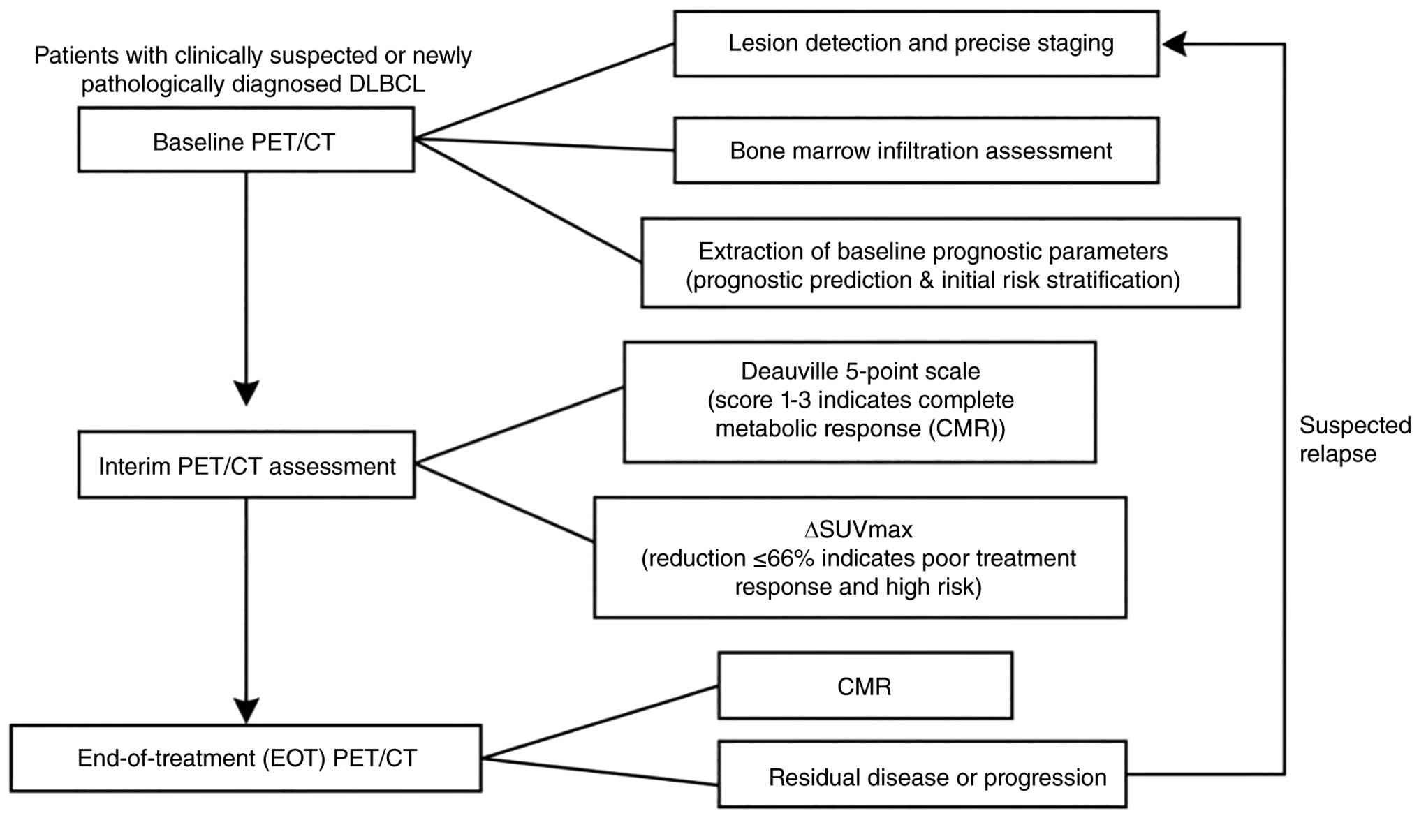
(ΔSUVmax) and the Deauville score. PET/CT has thus become
indispensable for the diagnosis, staging, interim monitoring, and
prognostic prediction of DLBCL. The present study systematically
reviews its role across the comprehensive management of the disease
(Fig. 1).
2. Lesion extent and staging
PET/CT-assisted staging
PET/CT has become a pivotal component of the
clinical management of DLBCL. Its particular value lies in the
ability to identify, through metabolic imaging, extranodal lesions
that conventional modalities such as contrast-enhanced CT and
ultrasound readily miss. PET/CT can clearly delineate sites that
appear negative or indeterminate on conventional examination,
particularly extranodal or deep-seated disease such as microscopic
splenic involvement and retroperitoneal or peritoneal lesions
(4), and it provides precise
localization for biopsy, thereby increasing the diagnostic yield of
pathological sampling (5). Whereas
conventional CT defines disease extent solely on the basis of
lesion size, PET/CT distinguishes tumor from normal tissue on the
basis of metabolic activity and thereby reclassifies the disease
stage in ~30% of patients, most often by upstaging (6). In patients who relapse or progress
after treatment, PET/CT also permits re-evaluation of disease
extent and informs the adjustment of subsequent treatment
strategies.
Detection of bone marrow involvement
(BMI) by PET/CT
Accurate detection of BMI is important for TDs and
prognostic assessment, both at initial staging and during response
evaluation in DLBCL. No universally accepted gold standard for
detecting BMI in lymphoma currently exists. Standard iliac crest
bone marrow biopsy (BMB) has well-recognized limitations in
identifying marrow involvement in DLBCL, and ancillary techniques
such as flow cytometry are therefore often added in clinical
practice to improve sensitivity. In recent years, PET/CT, as a
non-invasive functional imaging modality, has shown substantial
value for the assessment of BMI in DLBCL.
On PET/CT, bone marrow uptake in patients with DLBCL
is broadly classified as diffuse homogeneous uptake (dPET; higher
than liver uptake) or focal uptake (fPET) (7). Focal uptake can substitute directly
for BMB in the diagnosis of BMI, whereas diffuse uptake, although
it does not entirely exclude BMI, generally has no prognostic
impact and may be regarded as PET-negative (7). Notably, marrow involvement in DLBCL
more often produces a focal pattern than diffusely increased uptake
(8). Diffusely increased uptake
may arise from reactive marrow hyperplasia, inflammation, or
post-chemotherapy change, and may also reflect diffuse lymphomatous
infiltration; its diagnostic specificity is nevertheless lower than
that of focal uptake (9). Although
markedly increased diffuse FDG uptake (with an SUV exceeding that
of the liver) often suggests a high likelihood of marrow
involvement, its clinical significance remains uncertain.
Several studies have compared PET/CT and BMB for the
detection of BMI in DLBCL. PET/CT has greater sensitivity for focal
marrow lesions (10); in one
series (8), the sensitivity of
PET/CT was 94%, compared with only 24% for BMB. In patients with a
negative BMB but focal marrow uptake on PET/CT, the involvement
indicated by PET/CT carries independent prognostic value and helps
identify disease missed by biopsy (11). PET/CT also has a high negative
predictive value (NPV) at post-treatment restaging, up to 100% in
one study (12), suggesting that
patients with a negative PET/CT result may not require repeat BMB
to confirm complete remission.
Other studies have found that PET/CT can still miss
marrow involvement detected by BMB, particularly when infiltration
is diffuse rather than focal or when an indolent lymphoma component
is present (13,14). Histological examination therefore
remains important for staging and TDs in patients with early-stage
disease. Moreover, visual assessment and quantitative metabolic
parameters do not consistently outperform BMB for prognostication,
and BMB has been reported to remain an independent predictor of
progression-free survival (PFS) and overall survival (OS) in some
patients (14).
In summary, PET/CT has high sensitivity and a high
NPV for the evaluation of BMI in DLBCL, and it is a valuable
complement to BMB, particularly for the detection of focal lesions
and the assessment of treatment response. Because false-negative
results occur and findings are inconsistent across studies,
however, PET/CT cannot fully replace BMB, especially in newly
diagnosed high-risk patients and in those with clinically suspected
diffuse infiltration, in whom the combined use of both methods is
more prudent. The respective indications for the two modalities can
be defined more precisely. When BMB is unavailable or the patient
declines an invasive procedure, focal uptake on PET/CT provides a
reliable basis for diagnosing BMI; at post-treatment restaging, the
very high NPV of a negative PET/CT result can obviate unnecessary
repeat biopsy; and when a comprehensive assessment of total tumor
burden at both nodal and extranodal sites is required, PET/CT
provides markedly more information than BMB. Conversely, BMB
remains irreplaceable in several settings: In newly diagnosed DLBCL
in which PET/CT shows only diffuse uptake without definitive
evidence of tumor infiltration, BMB with histology and flow
cytometry is still required for confirmation; when clinical
suspicion of marrow involvement is high but PET/CT is negative (for
example, an indolent lymphoma component or minimal diffuse
infiltration), BMB is a necessary complement; and when TDs depend
closely on marrow pathology [for example, whether to add central
nervous system (CNS) prophylaxis or to modify chemotherapy
intensity], BMB remains the gold standard. For patients with
high-risk disease at baseline staging [for example, an IPI score
≥3, markedly elevated lactate dehydrogenase (LDH), or multiple
extranodal lesions], concurrent PET/CT and BMB are recommended,
because the two modalities are complementary and their combined use
minimizes the rate of missed diagnosis: Focal uptake on PET/CT can
replace BMB for diagnosis, whereas BMB should still be performed
when only diffuse uptake is observed or when PET/CT is negative but
clinical suspicion remains high.
PET/CT-guided target volume
delineation for radiotherapy
The contribution of PET/CT to lesion detection
extends beyond staging. In radiotherapy, PET/CT allows the extent
of tumor infiltration to be delineated more precisely, reduces the
dose delivered to normal tissue, and improves both the safety and
the efficacy of treatment. In DLBCL, PET/CT has substantially
improved the accuracy of target volume delineation: By displaying
tumor metabolic activity, it distinguishes active disease from
post-treatment fibrosis or necrosis more clearly than conventional
CT and therefore defines the extent of infiltration more
accurately. Radiotherapy guided by this functional metabolic
information achieves a more conformal dose distribution,
concentrating high doses within the true tumor volume while sparing
the surrounding normal organs as far as possible. In mediastinal
disease, for example, PET/CT can effectively separate tumor from
the adjacent pericardium and lung, markedly reducing the dose
delivered to these critical structures and lowering the risk of
late complications such as radiation pneumonitis and myocardial
injury (15). Radiotherapy plans
based on PET/CT have been shown to maintain, and possibly to
improve, local tumor control while enhancing treatment safety,
ultimately offering patients improved outcomes and quality of
life.
3. Prognostic prediction and response
assessment by PET/CT
Early identification of patients with DLBCL who
respond poorly to R-CHOP is essential if more effective alternative
treatments are to be explored. The International Prognostic Index
(IPI), the traditional and most widely used prognostic tool in
DLBCL, stratifies patients according to five clinical variables:
Age, performance status, LDH level, Ann Arbor stage and the number
of extranodal sites involved, and provides clinicians with an
initial basis for risk assessment. The IPI nevertheless has
important limitations in the era of precision medicine: As a static
clinical score, it captures neither the intrinsic biological
heterogeneity of the tumor nor its metabolic burden, and it does
not permit dynamic monitoring of response. By revealing tumor
functional metabolism non-invasively and dynamically, PET/CT
enables precise management throughout the treatment course, from
baseline risk assessment through interim response monitoring to
end-of-treatment (EOT) evaluation and has become a key tool for
predicting survival outcomes in DLBCL.
Baseline prognostic parameters on
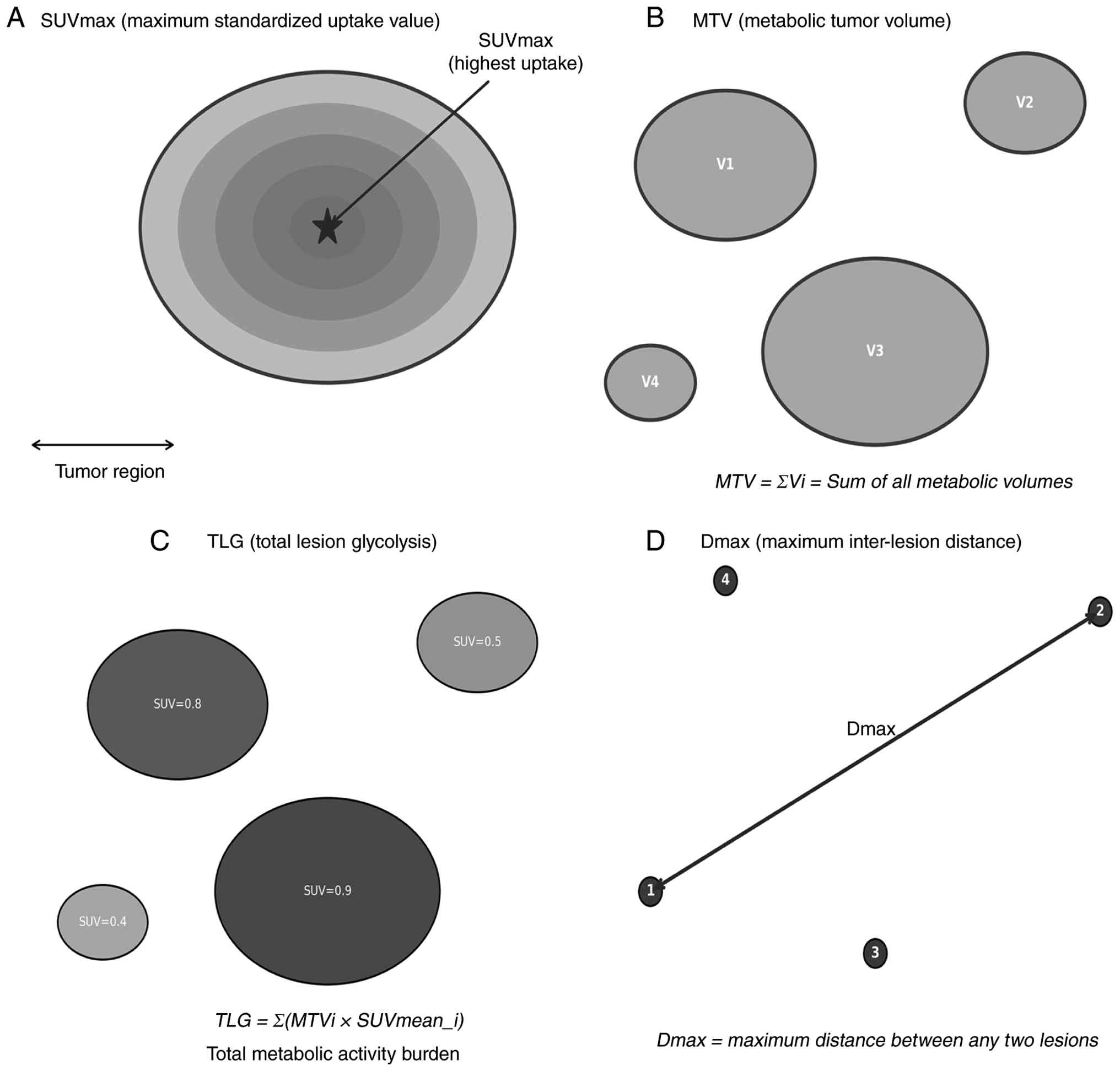
PET/CT. SUVmax
The standardized uptake value (SUV) is a
semi-quantitative measure of FDG uptake, reported as SUVmax,
SUVmean, or SUVpeak, that reflects tumor metabolic activity by
relating the radioactivity concentration within the tumor to the
injected dose. The prognostic value of baseline SUV in DLBCL is
complex. Some studies support a predictive role on grounds of
biological plausibility, in that a high SUV may indicate a more
aggressive tumor and have reported an association between high
baseline SUVmax and both PFS and OS (16). More detailed evidence (17), however, indicates that the
predictive power of SUV alone is limited and unstable, mainly
because SUVmax represents the metabolic activity of only the single
most active point within the tumor and captures neither total tumor
burden nor spatial heterogeneity. In current prognostic models, SUV
therefore serves primarily as a basic measurement, whereas
parameters that characterize the disease more comprehensively, such
as MTV and TLG, have proved to be more robust and independent
prognostic indicators. In short, although baseline SUV is
biologically meaningful, it is generally insufficient to predict
long-term outcome reliably on its own; it must be incorporated into
composite models that also include tumor burden, clinical indices
such as the IPI, and molecular features if risk stratification is
to be precise.
At the level of biological mechanism,
18F-FDG uptake is clearly linked to tumor cell
proliferative activity. Ki-67, the most widely used marker of
cellular proliferation, indicates the proportion of cells in the
G1, S, G2 and M phases of the cell cycle. Several studies have
demonstrated a significant positive correlation between SUVmax and
the Ki-67 index in DLBCL lesions (18,19).
Papajík et al (20)
analyzed 149 patients with newly diagnosed NHL and found that Ki-67
correlated positively with SUVmax, with the DLBCL subtype showing
the highest level of FDG uptake. Mechanistically, rapidly
proliferating tumor cells require large amounts of glucose to meet
the demands of glycolytic energy production and biosynthesis, which
upregulates glucose transporter 1 and hexokinase 2 and thereby
drives high FDG uptake. The strength of the correlation between
SUVmax and Ki-67 nevertheless varies among studies, because SUVmax
reflects glucose metabolic flux whereas Ki-67 measures the
proliferative fraction at a single time point; the two do not
capture identical biological dimensions, which helps explain why
SUVmax alone has limited prognostic value. Studies directly
examining the relationship between SUVmax and other proliferation
markers, such as proliferating cell nuclear antigen (PCNA) and
argyrophilic nucleolar organizer regions, remain scarce in DLBCL,
and the available evidence remains strongest for Ki-67.
MTV and TLG
MTV is the total volume of disease delineated at a
given standardized uptake value threshold, whereas TLG is the sum,
across all lesions, of the product of MTV and SUVmean. Baseline MTV
and TLG are established independent risk factors for PFS and OS
(21-23).
MTV improves the risk stratification provided by the
IPI and by the National Comprehensive Cancer Network IPI (24-26),
particularly in identifying patients at genuinely high risk.
Although the traditional clinical indices are widely used, they
discriminate poorly within a heterogeneous patient population. By
quantifying the total metabolic tumor burden, MTV identifies truly
high-risk patients more accurately, patients who often have poor
outcomes even when the international prognostic indices place them
in a low- or intermediate-risk category.
Combining MTV with other clinical or imaging
features is an effective way to refine risk stratification further.
One study integrated MTV with a measure of spatial tumor
dissemination and thereby improved risk stratification in DLBCL
(27): By combining MTV with the
maximum distance between the two farthest lesions (Dmax), the
investigators separated patients into three prognostic groups, and
the high-risk group, with both high MTV and high Dmax, had a
significantly lower 4-year PFS rate. Combining the two measures
therefore identifies genuinely high-risk individuals within
conventional stage groupings more precisely and optimizes risk
stratification. MTV can also be combined with molecular subtyping.
Within the cell-of-origin (COO) classification, the germinal center
B-cell-like (GCB) and non-GCB subtypes carry inherently different
prognoses. As early as 2016, Cottereau et al (1) showed, in 81 patients with newly
diagnosed DLBCL treated with R-CHOP or a CHOP-like regimen, that
patients with a high MTV had significantly lower 5-year PFS and OS
than those with a low MTV; more importantly, high MTV still
distinguished patients with a poor prognosis from those with a
favorable prognosis within the molecularly low-risk group. These
findings indicate that integrating molecular and PET/CT imaging
features at diagnosis can produce a more refined, individualized
prognostic model.
Risk stratification models have not remained
confined to static baseline parameters. In a pioneering study,
Eertink et al (28)
extended the approach to dynamic assessment during treatment,
combining baseline PET radiomics features (including age, MTV, and
the distance between the largest lesion and the farthest lesion,
Dmax-bulk) with a quantitative measure of interim metabolic
response (ΔSUVmax as a continuous variable) to construct a dynamic
prediction model. This model significantly improved risk
stratification in DLBCL, increasing the accuracy of risk
reclassification by 22% relative to conventional models and
providing a sound basis for decisions on early treatment
intensification with, for example, chimeric antigen receptor (CAR)
T cells or bispecific antibodies.
In summary, the use of PET/CT in DLBCL has evolved
from simple response assessment to prognostic prediction and
precise risk stratification. Baseline MTV, a powerful quantitative
index, together with the IPI, molecular subtyping, and even early
dynamic metabolic response, now forms part of a multidimensional,
modern system of risk stratification.
Dmax. Cottereau et al (1) were the first to propose that
dissemination features, the maximum distance between the two
farthest lesions (Dmax-patient) and the distance between the
largest lesion and the farthest lesion (Dmax-bulk), are prognostic
factors in DLBCL (27), and these
measures have since been widely adopted. Dmax and the spatial
dissemination index (SDmax) are independent predictors of PFS and
OS (29) and reflect the spatial
spread of disease. SDmax and MTV characterize the disease in
complementary ways: The former describes the pattern of
dissemination, the latter the tumor burden. Notably, SDmax carries
significant value for baseline risk stratification in DLBCL
independently of MTV. Cottereau et al (30) also found that a revised prognostic
index based on Dmax and Eastern Cooperative Oncology Group
performance status (ECOG PS), the Dmax-revised prognostic index,
identified an ultra-high-risk population, with an area under the
curve significantly greater than that of the IPI. Dmax is thus a
quantitative measure of lymphoma dissemination that is
significantly associated with prognosis in DLBCL and that
stratifies patient risk effectively (Fig. 2).
Interim PET/CT assessment
Interim PET/CT is a critical tool for assessing the
response to first-line therapy in DLBCL. To minimize false-positive
and false-negative results, international lymphoma organizations
and investigators worldwide have continued to refine the
interpretation criteria, successively proposing visual methods such
as the International Harmonization Project criteria and the
Deauville five-point scale, as well as semi-quantitative measures
such as the ΔSUVmax. Current guidelines nevertheless remain
cautious about the routine clinical use of interim PET/CT and state
explicitly that treatment should not be changed on the basis of
interim PET/CT findings alone unless there is clear evidence of
disease progression.
Timing of interim PET/CT. Response assessment
during treatment for DLBCL is categorized by timing: Early PET
after two cycles of chemotherapy (I-PET2), interim PET after four
cycles (I-PET4), and EOT PET after completion of all therapy.
I-PET2 mainly reflects the initial chemosensitivity of rapidly
proliferating cells and helps identify, at an early stage, patients
with a favorable or a poor response, including those with no
response. I-PET4 better captures the regrowth of lymphoma cells,
reflected in a high proliferation index at that time point, and is
therefore used mainly to identify patients likely to progress or
relapse. In DLBCL, the first interim PET/CT is recommended after
two rather than four cycles of treatment. When the I-PET2 result is
negative, subsequent PET/CT scans add little to the monitoring of
relapse (31), whereas a positive
I-PET2 result warrants repeat imaging.
In a study of 1,692 patients with DLBCL that
examined the timing of interim PET, Eertink et al (32) found that, during R-CHOP therapy,
I-PET2 and I-PET4 predicted a favorable response with comparable
accuracy. The optimal time point and criteria should therefore be
chosen according to the clinical question: The best time to assess
response is after two cycles; patients with a good response on
I-PET2 are suitable candidates for de-escalation trials; and those
with an unfavorable response, as defined by the ΔSUVmax criterion
at I-PET4, may be enrolled in randomized trials of novel
therapies.
Yuan et al (33) explored an even earlier time point
and showed that interim PET/CT after a single cycle of chemotherapy
is feasible in DLBCL and has predictive value similar to that of
I-PET2. The Deauville score and MTV measured after one cycle could
not, however, predict the final treatment response or one-year
disease-free status.
Interpretation criteria for interim
PET/CT
Deauville score. The Deauville scoring
system, a key tool for response assessment, has been examined
extensively. A negative interim PET/CT result [Deauville score 1-3,
indicating complete metabolic response (CMR)] is generally
associated with a favorable prognosis (33,34);
its core value lies in distinguishing treatment-sensitive from
potentially resistant patients according to the degree of metabolic
response. In one study of patients with low-risk DLBCL, interim
PET/CT after four cycles of R-CHOP identified favorable responders
(Deauville score 1-3) in whom the number of chemotherapy cycles
could be reduced from six to four without compromising the 2-year
PFS rate; this approach also significantly reduced the risk of
adverse events such as neutropenia, febrile neutropenia and
infection (35).
Yuan et al (33) further reported that a Deauville
score of 4 on interim PET/CT has limited prognostic value in DLBCL,
whereas a score of 5 carries considerably greater prognostic
significance. The Deauville system is thus not only an effective
prognostic tool but also an important quantitative basis for
individualizing treatment and optimizing therapeutic strategy.
ΔSUVmax. The ΔSUVmax is a core quantitative
measure for interim PET/CT and reflects the relative change in
tumor metabolic activity between baseline and interim imaging. It
is calculated as ΔSUVmax=[(baseline SUVmax-interim SUVmax)/baseline
SUVmax] x100%. A reduction of ≤66% at interim assessment identifies
patients at very high risk (36-38).
By quantifying dynamic changes in metabolic activity, ΔSUVmax
overcomes the qualitative limitations of the Deauville score and
provides a key tool for the precise adjustment of treatment in
DLBCL. As an independent predictor, it has proved superior to
visual assessment in several studies (39,40).
Combining visual assessment using the Deauville
score with ΔSUVmax to classify response on a 0-3 scale further
improves predictive accuracy: Patients with a score of 0 have a
significantly improved prognosis than all other groups, with
improved positive predictive value (PPV) and specificity (41). Another study assessed the
prognostic value of several PET parameters, including the Deauville
score, at baseline and at interim assessment, and found that
baseline MTV and ΔSUVmax were independent predictors of PFS and OS
and that the EOT Deauville score was also significantly associated
with PFS (42). Notably, the
combination of an interim Deauville score of 4-5 with a ΔSUVmax
reduction of ≤70% identified a subgroup with a particularly poor
prognosis.
Interim PET/CT across molecular subtypes.
DLBCL is molecularly heterogeneous, and the greatest differences in
biological behavior and treatment response lie between the GCB and
non-GCB subtypes. Sun et al (43) found that interim PET/CT interpreted
with the Deauville score independently predicted PFS in patients
with GCB DLBCL but had no predictive value for OS. In patients with
non-GCB disease, by contrast, the I-PET-negative group had
significantly improved PFS and OS than the I-PET-positive group.
Receiver operating characteristic analysis showed that interim PET
based on ΔSUVmax predicted PFS and OS effectively in the non-GCB
subtype but not in the GCB subtype; after stratification by the
optimal ΔSUVmax threshold, this measure predicted both PFS and OS
in patients with non-GCB disease.
These findings highlight the subtype-dependent
prognostic value of interim PET and underline the need to take the
pathological subtype of DLBCL into account in clinical practice. In
patients with GCB disease, an interim Deauville score of 3 may
indicate a relatively favorable prognosis, so that the original
regimen can reasonably be continued. In patients with non-GCB
disease, failure to achieve a ΔSUVmax reduction of ≥66% strongly
suggests that the original regimen is insufficiently effective and
that treatment should be modified early, for example, by switching
to a novel therapy such as Pola-R-CHP or CAR T-cell therapy.
Nevertheless, when making TDs based on interim
PET/CT, it is critical to recognize that molecular subtyping does
not fully capture DLBCL's biological aggressiveness. High-grade
B-cell lymphomas with MYC and BCL2 and/or BCL6 rearrangements
(‘double-hit’ or ‘triple-hit’ lymphomas), as defined by the WHO
classification, constitute a distinct histological entity with an
aggressive course and poor response to R-CHOP, irrespective of COO.
These patients often require intensified regimens or early trial
referral, and their management should not be guided by interim PET
results alone, since chemosensitivity on imaging does not guarantee
durable remission. Thus, the integration of interim PET/CT into
therapeutic decision-making must be accompanied by comprehensive
histopathological and genetic evaluation, including fluorescence
in situ hybridization for MYC, BCL2 and BCL6 rearrangements,
to ensure that the most aggressive variants are identified and
treated accordingly.
Interim PET/CT is a pivotal tool for assessing the
response to treatment in DLBCL. With respect to timing, I-PET2 and
I-PET4 have comparable predictive value: A negative I-PET2 result
indicates a favorable prognosis, whereas a positive result warrants
re-evaluation. With respect to interpretation, the Deauville score
is widely applied, whereas quantitative measures such as ΔSUVmax (a
reduction of ≤66% indicating high risk) may predict outcome more
precisely and may outperform visual assessment. The molecular
subtype of DLBCL also influences the value of assessment, interim
PET/CT being of greater prognostic utility in non-GCB disease.
Clinical application should therefore integrate the timing of
assessment, the interpretation criteria used, the molecular
subtype, and the histological characteristics in order to guide
individualized TDs.
EOT PET/CT assessment
A negative EOT PET/CT result (Deauville score 1-3)
is closely associated with significantly prolonged PFS and OS
(44,45), whereas a positive result (Deauville
score 4-5) indicates a high risk of relapse. CMR at the end of
treatment is an independent predictor of PFS and OS and a reliable
prognostic biomarker in DLBCL, and its predictive value remains
stable in long-term survival analyses. Han et al (34) showed that PET/CT parameters
measured after a single cycle of chemotherapy were unrelated to
long-term survival, whereas a negative EOT PET/CT result was the
key predictor of long-term outcome; early chemotherapy-induced
tumor regression therefore has considerably less prognostic value
than EOT assessment.
In summary, PET/CT provides a dynamic monitoring
network across the entire course of DLBCL management, with routine
imaging recommended at baseline, interim, and end of treatment. At
baseline, MTV, TLG and Dmax reflect tumor burden and dissemination,
offering prognostic value beyond the IPI and guiding initial
treatment intensity; however, no universally validated cut-offs
exist for these parameters, reported thresholds vary widely with
segmentation methods, populations and regimens, so they should be
used for risk stratification rather than binary classification. At
interim, the Deauville scale and ΔSUVmax enable early
identification of high-risk patients; a ΔSUVmax reduction of ≤66%
is the most validated threshold for predicting poor outcomes,
whereas the Deauville score lacks a standardized semi-quantitative
cut-off, and its PPV varies with timing and context. At end of
treatment, a negative PET/CT strongly predicts long-term survival,
while a positive result indicates high relapse risk and warrants
further investigation.
For clinical decision-making: Elevated baseline MTV,
TLG and Dmax in patients with high-risk IPI further stratify
prognosis within this already high-risk group, suggesting closer
monitoring or risk-adapted strategies. At interim, ΔSUVmax
reduction >66% with Deauville 1-3 supports continuing current
therapy, whereas ΔSUVmax ≤66% or Deauville 4-5, without clear
progression, should not routinely modify first-line therapy; these
findings should prompt multidisciplinary discussion to exclude
false-positives (for example, inflammation and infection), and
patients should continue planned therapy until EOT reassessment. At
end of treatment, negative scans allow surveillance, while positive
scans require biopsy confirmation before salvage therapy due to
false-positive rates. Only the ΔSUVmax ≤66% threshold is
consistently validated across cohorts; MTV, TLG and Dmax lack
internationally accepted cut-offs, with reported values varying
considerably by segmentation methods, populations and protocols.
All PET/CT findings must therefore be interpreted cautiously,
integrated with clinical and pathological data, and applied in
local context rather than mechanistically, with further
standardization and prospective validation needed.
4. PET/CT-based radiomics models
PET/CT-based radiomics has shown considerable value
in the diagnosis and treatment of DLBCL in recent years. Radiomics
permits the high-throughput extraction and quantification, from
standard PET/CT images, of features that are imperceptible to the
human eye, and thereby reveals tumor heterogeneity more
comprehensively.
Construction of prognostic models
Several studies have confirmed that radiomics
features extracted from baseline PET/CT have independent prognostic
value. In one study, semi-automatic segmentation was used to
extract 490 radiomics features, and a model incorporating MTV,
SUVmax and Dmax substantially outperformed the conventional IPI
(46). Combining radiomics
features with clinical factors such as the IPI refined risk
stratification further and allowed more precise identification of
high-risk patients (47,48).
Various strategies for feature selection and fusion
have been explored in order to optimize radiomics models. Jiang
et al (49) used a
cross-combination approach to generate 49 candidate
feature-selection pipelines and derived radiomics features
(R-features) significantly associated with PFS and OS. Another
study compared several methods of feature reduction and found that,
irrespective of the approach to lesion selection or dimensionality
reduction, patient-level conventional PET features such as MTV,
together with measures of disease dissemination, consistently
showed the best predictive value (50).
Although different PET segmentation methods (for
example, a fixed SUV threshold or a percentage threshold) alter the
calculated values of individual radiomics features, the
discriminative performance of models built with different
segmentation methods does not differ substantially (51), which supports the robustness of the
radiomics approach.
Models for subtyping and
identification of transformation
Beyond prognostication, PET/CT radiomics has shown
considerable promise in supporting the molecular subtyping of
DLBCL, particularly the distinction between the GCB subtype and the
non-GCB subtype [chiefly the activated B-cell-like (ABC) subtype].
This distinction is clinically important, because the GCB and ABC
subtypes differ in pathogenesis and prognosis and often require
different treatment strategies. Work in this field is moving from
conventional metabolic parameters such as SUVmax toward more
complex radiomics features.
In a retrospective study of lymphomas presenting
with bulky mediastinal masses, classical Hodgkin lymphoma, primary
mediastinal B-cell lymphoma and gray zone lymphoma, Abenavoli et
al (52) found significant
differences among these subtypes in FDG PET texture features and
metabolic heterogeneity, providing proof of concept for the use of
PET/CT radiomics to differentiate lymphoma subtypes.
Jiang et al (53) developed and validated a baseline
PET/CT radiomics model for the non-invasive identification of
histological transformation of indolent follicular lymphoma into
aggressive DLBCL, demonstrating that radiomics features can capture
the profound changes in tumor biology that accompany
transformation. Clinically, such a model offers a valuable
non-invasive alternative for patients who, for various reasons,
cannot undergo repeat tumor tissue sampling for genetic sequencing.
PET/CT is therefore likely to extend beyond staging and response
assessment and to become embedded in the decision-making loop of
precision medicine, with non-invasive analysis of tumor biology
providing key evidence to guide individualized treatment.
In summary, PET/CT radiomics improves the accuracy
of prognostic stratification in DLBCL and extends the role of
imaging to molecular subtyping and the identification of
histological transformation, reflecting a shift from conventional
metabolic parameters toward multidimensional, high-throughput
features. Standardization of image acquisition and segmentation,
the development of automated analysis workflows, and large-scale
prospective studies are still needed before radiomics can move from
a research tool to routine clinical use in the individualized
management of DLBCL.
5. Applications in special clinical
scenarios
PET/CT in CAR T-cell therapy
In the setting of CAR T-cell therapy, patients with
DLBCL typically undergo PET/CT at the time of the TD, at CAR T-cell
infusion (TT), and 1 month (M1) and 3 months (M3) after treatment.
Metabolic parameters derived from these examinations have
considerable value for predicting both survival and toxicity. Cohen
et al (54) found, on
univariate analysis, that a TD SUVmax >17.1 and a TT SUVmax
>12.1 were significantly associated with shorter OS; on
multivariate analysis, three factors were significantly associated
with shorter OS: a TD SUVmax >17.1, an LDH level >450 U/l,
and an ECOG performance status >1. At early response assessment
(M1), patients with a Deauville score >3 had a significantly
shorter median OS, and a ΔSUVmax of ≤66% relative to the TD
baseline also indicated a poor prognosis. With respect to toxicity,
the pre-infusion SUVmax correlates strongly with the grade of
cytokine release syndrome (CRS) (Spearman r=0.806, P<0.001) and
the pre-infusion TLG moderately so (r=0.534, P<0.001),
indicating that metabolic parameters acquired before infusion can
give advance warning of toxicity (55). Taken together, these findings
suggest that pre-treatment SUVmax helps select patients likely to
benefit from CAR T-cell therapy, that the Deauville score and
ΔSUVmax at 1 month allow early identification of patients at high
risk of treatment failure, and that pre-infusion SUVmax provides an
early warning for toxicity management.
PET/CT in autologous hematopoietic
stem cell transplantation (auto-HSCT)
Pre-transplant PET/CT effectively predicts survival
in patients with DLBCL undergoing auto-HSCT (56). Sauter et al (57) found that patients with a
pre-transplant Deauville score of 1-3 had significantly higher
3-year PFS (77% vs. 49%) and OS (86% vs. 54%) than those with a
score of 4. Qiao et al (58) showed that, although the magnitude
of the change in SUVmax (using a threshold of 66%) predicted
neither PFS nor OS, a higher pre-transplant SUVmax (>6) was
significantly associated with shorter PFS. Pre-transplant PET/CT
therefore not only predicts survival in DLBCL but is also
clinically useful for identifying patients at high risk of
transplant failure and for selecting those who require intensified
intervention.
6. Emerging alternative technologies
18F-FDG PET/CT is the cornerstone of
staging and response assessment in DLBCL, but it has intrinsic
limitations in distinguishing tumor from inflammation or infection
and in characterizing tumor heterogeneity. These gaps have driven
the development of novel tracers and of PET/MR, which aim to
compensate for the shortcomings of 18F-FDG PET/CT and to
provide richer molecular imaging information for precision medicine
(Table I).
 | Table IEmerging PET tracers and advanced
imaging technologies in diffuse large B-cell lymphoma. |
Table I
Emerging PET tracers and advanced
imaging technologies in diffuse large B-cell lymphoma.
| Category |
Tracer/Technology | Key Features |
|---|
| Cell proliferation
tracers | 18F-FLT
(Fluorothymidine) | • Higher tumor
specificity |
| | | • Correlates with
Ki-67 |
| | | • Improved PPV for
early response |
| Antigen-targeted
tracers |
68Ga/89Zr-CD20,
Ga-PentixaFor | • Assess CD20
expression |
| | | • Predict rituximab
efficacy |
| | | • CXCR4-targeted
imaging |
| Amino acid
tracers | 11C-MET
(Methionine) | • High
tumor-to-background |
| | | • Unaffected by
inflammation |
| | | • Short half-life
(20 min) |
| PET/MR
technology | Integrated
PET/MR | • Reduced radiation
exposure |
| | | • Superior soft
tissue contrast |
| | | • DWI for early
response |
Novel PET tracers
Among the tracers currently available,
18F-FDG is the conventional metabolic agent.
Proliferation tracers include
3'-deoxy-3'-18F-fluorothymidine (18F-FLT),
which is phosphorylated by thymidine kinase 1 (TK-1) and thereby
retained within the cell; TK-1 activity peaks during the DNA
synthesis phase. Uptake of 18F-FLT correlates well with
classic markers of tumor proliferation such as the Ki-67 index,
PCNA expression, and the S-phase fraction, while remaining low in
inflammatory lesions. 18F-FLT can therefore distinguish
tumor tissue from inflammation more specifically than
18F-FDG and has considerable potential for early
response assessment and treatment monitoring. Because FLT may have
greater tumor specificity than FDG (59,60),
FLT PET offers a particular advantage for the early assessment of
response to chemotherapy, chemoimmunotherapy, radiotherapy, or
combinations of these. Minamimoto et al (61) demonstrated that, in DLBCL, the PPV
of interim FLT PET/CT was significantly higher than that of interim
FDG PET/CT with both qualitative and semi-quantitative
interpretation; even with simple visual assessment, the PPV of FLT
PET/CT remained superior to that of FDG PET/CT.
Tracers of amino acid metabolism, such as
¹¹C-methionine (¹¹C-MET), offer another perspective, since tumor
cells require abundant amino acids for protein synthesis. ¹¹C-MET
provides high tumor-to-background contrast and is unaffected by
inflammation, and it is significantly superior to
18F-FDG for imaging cerebral lymphoma and for
delineating tumor margins precisely. Its clinical application is
limited, however, by the very short physical half-life of ¹¹C (20
min), which requires on-site cyclotron production.
Antigen- and receptor-targeted
tracers
Tracers directed against antigens on the surface of
DLBCL cells, such as 68Ga- or 89Zr-labeled
anti-CD20 antibodies (for example, 89Zr-DFO-rituximab),
allow CD20 expression to be assessed in vivo across all
lesions, providing an intuitive means of predicting the efficacy of
rituximab and of conducting pharmacokinetic studies. Radiolabeled
antibodies against CD22 can similarly be used to assess suitability
for the corresponding targeted therapies. Particularly promising is
68Ga-PentixaFor, a PET molecular probe directed at C-X-C
chemokine receptor 4 (CXCR4). CXCR4 is highly expressed in a range
of tumors, including multiple myeloma, lymphoma and
aldosterone-producing adenoma, and in inflammatory disease, and
68Ga-PentixaFor binds it specifically, permitting highly
sensitive imaging of these conditions. This agent may assist not
only in prognostic stratification and the identification of
high-risk patients but also in selecting patients suitable for
CXCR4-antagonist therapy.
68Ga-labeled prostate-specific membrane
antigen (PSMA) ligands warrant a note of caution: although
developed for prostate cancer, PSMA is also expressed in the
neovascular endothelium of tumors, so that some highly vascularized
extranodal DLBCL lesions may show non-specific uptake that must be
recognized during image interpretation.
PET/MR
Integrated PET/MR has emerged as a more advanced
molecular imaging technology than PET/CT and offers several
advantages in DLBCL. First, it substantially reduces radiation
exposure, which is particularly important for young patients who
require repeated follow-up imaging. Second, MR is more sensitive
for the detection of extranodal disease, especially involvement of
the CNS, liver, bone, and bone marrow; combined with
diffusion-weighted imaging, it can reveal treatment-induced changes
in cellularity at an earlier stage and thus provide both functional
and metabolic evidence for response assessment. Although PET/CT
remains the standard for staging, the multiparametric capability of
PET/MR gives it considerable potential for precise staging,
response monitoring, and prognostic assessment.
In summary, 18F-FLT, ¹¹C-MET, the various
antigen-targeted tracers, and PET/MR are powerful complements to
18F-FDG PET/CT and hold considerable promise for
specific clinical challenges in DLBCL. Future work will need to
overcome the limitations of existing tracers and to move these
novel molecular probes from clinical studies into routine practice,
ultimately enabling the personalized and precise management of
DLBCL.
7. Limitations
Several limitations of PET/CT in clinical practice
deserve attention. First, its sensitivity for CNS lesions is
relatively low, chiefly because the high physiological FDG uptake
of normal brain tissue masks lymphomatous lesions; such cases
require cerebrospinal fluid examination or MRI to improve
diagnostic accuracy. Second, physiological FDG uptake in other
normal structures, such as the gastrointestinal tract and bone
marrow, can interfere with image interpretation and produce
false-positive results. Third, in the assessment of BMI, PET/CT has
limited ability to detect heterogeneous or indolent disease, for
which BMB remains necessary for definitive diagnosis.
More fundamental problems are shared across the
field. i) There is no uniform standard for image segmentation,
which directly affects the comparability of parameters and the
generalizability of prognostic models. ii) Parameter measurement is
not standardized across centers: Differences in scanners,
reconstruction algorithms and patient preparation limit the
cross-center comparability of semi-quantitative measures such as
SUV and hinder the pooling and dissemination of results from
multicenter studies. iii) Visual assessment is subjective:
interpretation of the Deauville score depends on the reader's
experience, and inter-observer variability is substantial,
particularly at the boundary between scores 3 and 4;
semi-quantitative parameters compensate only in part, since their
thresholds and measurement procedures are also not standardized.
These limitations point to an urgent need for internationally
accepted, standardized guidelines covering PET/CT acquisition,
reconstruction, segmentation and interpretation, in order to
improve the reliability and reproducibility of the technique in the
precision management of DLBCL.
8. Summary and outlook
PET/CT has become a core tool in the comprehensive
management of DLBCL, with value spanning diagnosis, response
assessment and prognostic prediction. At diagnosis and staging, its
high metabolic sensitivity refines clinical staging and
significantly improves the detection of BMI. At interim assessment,
dynamic monitoring using visual interpretation and
semi-quantitative parameters effectively identifies high-risk
patients. For prognosis, a negative PET/CT result at the end of
treatment is a strong predictor of long-term survival, whereas
baseline MTV, TLG and Dmax are established independent prognostic
indicators, MTV in particular being combinable with clinical and
imaging measures to optimize risk stratification. In special
settings such as CAR T-cell therapy and auto-HSCT, PET/CT provides
key evidence for patient selection, early warning of toxicity and
survival prediction.
Several priorities must be addressed if PET/CT is to
be applied more precisely and more widely in DLBCL. First,
internationally unified standards for image acquisition,
reconstruction, segmentation and quantitative measurement should be
established, including definition of the optimal thresholding
algorithm for MTV and improved quality-control procedures for
Deauville scoring and standardized SUV measurement, so as to
enhance the reproducibility of clinical data and the comparability
of multicenter studies. Secondly, because most
machine-learning-based prognostic models are currently derived from
retrospective studies, multicenter prospective cohort studies with
large samples are required to validate rigorously the predictive
value of radiomics features combined with clinical parameters, to
establish automated and standardized analytical workflows, and to
translate these models into clinical decision-making. Third, the
particular advantages of novel tracers such as 18F-FLT,
¹¹C-MET, and 68Ga-PentixaFor for discriminating tumor
from inflammation and for assessing proliferative activity and
receptor expression should be exploited by overcoming current
limitations in production technology, cost and evidence base, by
conducting multicenter clinical trials, by establishing
standardized protocols for administration and interpretation, and
by exploring theranostic strategies that pair these tracers with
targeted agents.
In summary, through the coordinated advancement of
standardization, prospective validation, translation of novel
tracers and multimodal imaging, PET/CT and its derived imaging
biomarkers are expected to form a more powerful, precise, and
generalizable system for prognostic prediction and therapeutic
decision-making, ultimately supporting the continued development of
individualized precision medicine for DLBCL.
Acknowledgements
Not applicable.
Funding
Funding: No funding was received.
Availability of data and materials
Not applicable.
Authors' contributions
HJ, HS, DZ and LB made substantial contributions to
the conception and design of the study, drafted the article and
revised it critically for important intellectual content, and read
and approved the final version of the manuscript. Data
authentication is not applicable.
Ethics approval and consent to
participate
Not applicable.
Patient consent for publication
Not applicable.
Competing interests
The authors declare that they have no competing
interests.
References
|
1
|
Cottereau AS, Lanic H, Mareschal S,
Meignan M, Vera P, Tilly H, Jardin F and Becker S: Molecular
profile and FDG-PET/CT total metabolic tumor volume improve risk
classification at diagnosis for patients with diffuse Large B-cell
lymphoma. Clin Cancer Res. 22:3801–3809. 2016.PubMed/NCBI View Article : Google Scholar
|
|
2
|
Basu S, Kwee TC, Surti S, Akin EA, Yoo D
and Alavi A: Fundamentals of PET and PET/CT imaging. Ann N Y Acad
Sci. 1228:1–18. 2011.PubMed/NCBI View Article : Google Scholar
|
|
3
|
Jaini S and Dadachova E: FDG for therapy
of metabolically active tumors. Semin Nucl Med. 42:185–189.
2012.PubMed/NCBI View Article : Google Scholar
|
|
4
|
Adams HJ, de Klerk JM, Fijnheer R, Dubois
SV, Nievelstein RA and Kwee TC: CT-based versus FDG-PET/CT-based
NCCN international prognostic index risk stratification in DLBCL. J
Natl Compr Canc Netw. 13:171–176. 2015.PubMed/NCBI View Article : Google Scholar
|
|
5
|
Ferrari S, Weber A, Marra P, Tebaldi P,
Pavoni C, Barbui AM, Gritti G, Dulcetta L, Carbone FS, Muglia R, et
al: Enhancing the diagnostic accuracy of core needle biopsy in
patients with lymphoproliferative disorders by an optimized
protocol. Radiol Med. 130:844–853. 2025.PubMed/NCBI View Article : Google Scholar
|
|
6
|
Gallamini A and Borra A: Role of PET in
lymphoma. Curr Treat Options Oncol. 15:248–261. 2014.PubMed/NCBI View Article : Google Scholar
|
|
7
|
Chen Y, Zhou M, Liu J and Huang G:
Prognostic value of bone marrow FDG uptake pattern of PET/CT in
newly diagnosed diffuse large B-cell lymphoma. J Cancer.
9:1231–1238. 2018.PubMed/NCBI View Article : Google Scholar
|
|
8
|
Berthet L, Cochet A, Kanoun S,
Berriolo-Riedinger A, Humbert O, Toubeau M, Dygai-Cochet I, Legouge
C, Casasnovas O and Brunotte F: In newly diagnosed diffuse large
B-cell lymphoma, determination of bone marrow involvement with
18F-FDG PET/CT provides better diagnostic performance and
prognostic stratification than does biopsy. J Nucl Med.
54:1244–1250. 2013.PubMed/NCBI View Article : Google Scholar
|
|
9
|
Salaun PY, Gastinne T, Bodet-Milin C,
Campion L, Cambefort P, Moreau A, Le Gouill S, Berthou C, Moreau P
and Kraeber-Bodéré F: Analysis of 18F-FDG PET diffuse bone marrow
uptake and splenic uptake in staging of Hodgkin's lymphoma: A
reflection of disease infiltration or just inflammation? Eur J Nucl
Med Mol Imaging. 36:1813–1821. 2009.PubMed/NCBI View Article : Google Scholar
|
|
10
|
Khan AB, Barrington SF, Mikhaeel NG, Hunt
AA, Cameron L, Morris T and Carr R: PET-CT staging of DLBCL
accurately identifies and provides new insight into the clinical
significance of bone marrow involvement. Blood. 122:61–67.
2013.PubMed/NCBI View Article : Google Scholar
|
|
11
|
Aide N, Talbot M, Fruchart C, Damaj G and
Lasnon C: Diagnostic and prognostic value of baseline FDG PET/CT
skeletal textural features in diffuse large B cell lymphoma. Eur J
Nucl Med Mol Imaging. 45:699–711. 2018.PubMed/NCBI View Article : Google Scholar
|
|
12
|
Jackson AE, Smeltzer JP, Habermann TM,
Jones JM, Burnette B, Ristow K, Wiseman GA, Macon WR, Nowakowski GS
and Witzig TE: The utility of restaging bone marrow biopsy in
PET-negative patients with diffuse large B-cell lymphoma and
baseline bone marrow involvement. Am J Hematol. 89:865–867.
2014.PubMed/NCBI View Article : Google Scholar
|
|
13
|
Alzahrani M, El-Galaly TC, Hutchings M,
Hansen JW, Loft A, Johnsen HE, Iyer V, Wilson D, Sehn LH, Savage
KJ, et al: The value of routine bone marrow biopsy in patients with
diffuse large B-cell lymphoma staged with PET/CT: A Danish-Canadian
study. Ann Oncol. 27:1095–1099. 2016.PubMed/NCBI View Article : Google Scholar
|
|
14
|
Adams HJ, Kwee TC, Fijnheer R, Dubois SV,
Nievelstein RA and de Klerk JM: Bone marrow
18F-fluoro-2-deoxy-D-glucose positron emission tomography/computed
tomography cannot replace bone marrow biopsy in diffuse large
B-cell lymphoma. Am J Hematol. 89:726–731. 2014.PubMed/NCBI View Article : Google Scholar
|
|
15
|
Paumier A, Bakkour M, Ghalibafian M,
Beaudre A, Blanchard P, Martinetti F and Girinsky T: Involved-node
radiotherapy combined with deep-inspiration breath-hold technique
in patients with Hodgkin lymphoma. Cancer Radiother. 16:85–90.
2012.PubMed/NCBI View Article : Google Scholar : (In French).
|
|
16
|
Choi JH, Lim I, Byun BH, Kim BI, Choi CW,
Kang HJ, Shin DY and Lim SM: The role of 18F-FDG PET/CT in patients
with diffuse large B-cell lymphoma after radioimmunotherapy using
131I-rituximab as consolidation therapy. PLoS One.
17(e0273839)2022.PubMed/NCBI View Article : Google Scholar
|
|
17
|
Xie M, Wu K, Liu Y, Jiang Q and Xie Y:
Predictive value of F-18 FDG PET/CT quantization parameters in
diffuse large B cell lymphoma: A meta-analysis with 702
participants. Med Oncol. 32(446)2015.PubMed/NCBI View Article : Google Scholar
|
|
18
|
Zhao T, Yuan L, Li JL, Zhao M, Lin YM,
Xing J and Bao LL: Prognostic value of baseline 18F-FDG PET/CT
combined with clinicopathological characteristics in diffuse large
B-Cell lymphoma. Zhongguo Shi Yan Xue Ye Xue Za Zhi. 33:365–372.
2025.PubMed/NCBI View Article : Google Scholar : (In Chinese).
|
|
19
|
Wu X, Pertovaara H, Korkola P, Vornanen M,
Eskola H and Kellokumpu-Lehtinen PL: Glucose metabolism correlated
with cellular proliferation in diffuse large B-cell lymphoma. Leuk
Lymphoma. 53:400–405. 2012.PubMed/NCBI View Article : Google Scholar
|
|
20
|
Papajík T, Mysliveček M, Sedová Z,
Buriánková E, Procházka V, Koranda P, Raida L, Kubová Z, Palová M,
Kučerová L, et al: Standardised uptake value of 18F-FDG on staging
PET/CT in newly diagnosed patients with different subtypes of
non-Hodgkin's lymphoma. Eur J Haematol. 86:32–37. 2011.PubMed/NCBI View Article : Google Scholar
|
|
21
|
Alderuccio JP, Reis IM, Hamadani M,
Nachiappan M, Leslom S, Kahl BS, Ai WZ, Radford J, Solh M, Ardeshna
KM, et al: PET/CT biomarkers enable risk stratification of patients
with Relapsed/Refractory diffuse large B-cell lymphoma enrolled in
the LOTIS-2 clinical trial. Clin Cancer Res. 30:139–149.
2024.PubMed/NCBI View Article : Google Scholar
|
|
22
|
Song MK, Yang DH, Lee GW, Lim SN, Shin S,
Pak KJ, Kwon SY, Shim HK, Choi BH, Kim IS, et al: High total
metabolic tumor volume in PET/CT predicts worse prognosis in
diffuse large B cell lymphoma patients with bone marrow involvement
in rituximab era. Leuk Res. 42:1–6. 2016.PubMed/NCBI View Article : Google Scholar
|
|
23
|
Park HL, Han EJ, O JH, Choi BO, Park G,
Jung SE, Yahng SA, Eom KS and Cho SG: On Behalf Of Catholic
University Lymphoma Group: Early interim chemotherapy response
evaluation by F-18 FDG PET/CT in diffuse large B cell lymphoma.
Diagnostics (Basel). 10(1002)2020.PubMed/NCBI View Article : Google Scholar
|
|
24
|
Zhang H, Xu Z, Zhou W, Chen J, Wei Y, Wu
H, Wei X and Feng R: Metabolic tumor volume from baseline [18 F]FDG
PET/CT at diagnosis improves the IPI stratification in patients
with diffuse large B-cell lymphoma. Ann Hematol. 103:5891–5902.
2024.PubMed/NCBI View Article : Google Scholar
|
|
25
|
Zhao P, Yu T and Pan Z: Prognostic value
of the baseline 18F-FDG PET/CT metabolic tumour volume (MTV) and
further stratification in low-intermediate (L-I) and
high-intermediate (H-I) risk NCCNIPI subgroup by MTV in DLBCL MTV
predict prognosis in DLBCL. Ann Nucl Med. 35:24–30. 2021.PubMed/NCBI View Article : Google Scholar
|
|
26
|
Shagera QA, Cheon GJ, Koh Y, Yoo MY, Kang
KW, Lee DS, Kim EE, Yoon SS and Chung JK: Prognostic value of
metabolic tumour volume on baseline 18F-FDG PET/CT in addition to
NCCN-IPI in patients with diffuse large B-cell lymphoma: Further
stratification of the group with a high-risk NCCN-IPI. Eur J Nucl
Med Mol Imaging. 46:1417–1427. 2019.PubMed/NCBI View Article : Google Scholar
|
|
27
|
Cottereau AS, Nioche C, Dirand AS, Clerc
J, Morschhauser F, Casasnovas O, Meignan M and Buvat I: 18F-FDG PET
dissemination features in diffuse large B-Cell lymphoma are
predictive of outcome. J Nucl Med. 61:40–45. 2020.PubMed/NCBI View Article : Google Scholar
|
|
28
|
Eertink JJ, Heymans MW, Wiegers SE, Bes
AL, Duhrsen U, Huttmann A, Kurch L, Barrington SF, Mikhaeel GN,
Lugtenburg PJ, et al: Risk prediction in diffuse large B-cell
lymphoma improves when combining baseline PET features with interim
PET response. Haematologica. 110:2413–2421. 2025.PubMed/NCBI View Article : Google Scholar
|
|
29
|
Wang F, Cui S, Lu L, Shao X, Yan F, Liu Y,
He B, Wang J, Cao Y, Yue Y, et al: Dissemination feature based on
PET/CT is a risk factor for diffuse large B cell lymphoma patients
outcome. BMC Cancer. 23(1165)2023.PubMed/NCBI View Article : Google Scholar
|
|
30
|
Cottereau AS, Meignan M, Nioche C,
Capobianco N, Clerc J, Chartier L, Vercellino L, Casasnovas O,
Thieblemont C and Buvat I: Risk stratification in diffuse large
B-cell lymphoma using lesion dissemination and metabolic tumor
burden calculated from baseline PET/CT†. Ann Oncol. 32:404–411.
2021.PubMed/NCBI View Article : Google Scholar
|
|
31
|
Zhang X, Fan W, Xia ZJ, Hu YY, Lin XP,
Zhang YR, Li ZM, Liang PY and Li YH: Use of subsequent PET/CT in
diffuse large B-cell lymphoma patients in complete remission
following primary therapy. Chin J Cancer. 34:70–78. 2015.PubMed/NCBI View Article : Google Scholar
|
|
32
|
Eertink JJ, Burggraaff CN, Heymans MW,
Dührsen U, Hüttmann A, Schmitz C, Müller S, Lugtenburg PJ,
Barrington SF, Mikhaeel NG, et al: Optimal timing and criteria of
interim PET in DLBCL: A comparative study of 1692 patients. Blood
Adv. 5:2375–2384. 2021.PubMed/NCBI View Article : Google Scholar
|
|
33
|
Yuan L, Kreissl MC, Su L, Wu Z, Hacker M,
Liu J, Zhang X, Bo Y, Zhang H, Li X and Li S: Prognostic analysis
of interim 18F-FDG PET/CT in patients with diffuse large B cell
lymphoma after one cycle versus two cycles of chemotherapy. Eur J
Nucl Med Mol Imaging. 46:478–488. 2019.PubMed/NCBI View Article : Google Scholar
|
|
34
|
Han EJ, Park HL, Yahng SA, Min GJ, Choi
BO, Park G, O JH and Cho SG: Catholic University Lymphoma Group.
Prognostic value of very early interim FDG PET/CT after single
cycle of chemotherapy for 10-Year survival in diffuse Large B-Cell
lymphoma. Cancers (Basel). 17(926)2025.PubMed/NCBI View Article : Google Scholar
|
|
35
|
Shi Q, He Y, Yi HM, Mu RJ, Jiang XF, Fu D,
Dong L, Qin W, Xu PP, Cheng S, et al: Positron emission
tomography-adapted therapy in low-risk diffuse large B-cell
lymphoma: Results of a randomized, phase III, non-inferiority
trial. Cancer Commun (Lond). 43:896–908. 2023.PubMed/NCBI View Article : Google Scholar
|
|
36
|
Casasnovas RO, Meignan M,
Berriolo-Riedinger A, Bardet S, Julian A, Thieblemont C, Vera P,
Bologna S, Brière J, Jais JP, et al: SUVmax reduction improves
early prognosis value of interim positron emission tomography scans
in diffuse large B-cell lymphoma. Blood. 118:37–43. 2011.PubMed/NCBI View Article : Google Scholar
|
|
37
|
Rekowski J, Hüttmann A, Schmitz C, Müller
SP, Kurch L, Kotzerke J, Franzius C, Weckesser M, Bengel FM,
Freesmeyer M, et al: Interim PET evaluation in diffuse Large B-cell
lymphoma using published recommendations: Comparison of the
deauville 5-Point scale and the ΔSUVmax method. J Nucl Med.
62:37–42. 2021.PubMed/NCBI View Article : Google Scholar
|
|
38
|
Zucca E, Cascione L, Ruberto T,
Facchinelli D, Schär S, Hayoz S, Dirnhofer S, Giovanella L,
Bargetzi M, Mamot C and Ceriani L: Prognostic models integrating
quantitative parameters from baseline and interim positron emission
computed tomography in patients with diffuse large B-cell lymphoma:
Post-hoc analysis from the SAKK38/07 clinical trial. Hematol Oncol.
38:715–725. 2020.PubMed/NCBI View Article : Google Scholar
|
|
39
|
Li X, Xie X, Zhang L, Li X, Li L, Wang X,
Fu X, Sun Z, Zhang X, Li Z and Wu J: Research on the midterm
efficacy and prognosis of patients with diffuse large B-cell
lymphoma by different evaluation methods in interim PET/CT. Eur J
Radiol. 133(109301)2020.PubMed/NCBI View Article : Google Scholar
|
|
40
|
Itti E, Meignan M, Berriolo-Riedinger A,
Biggi A, Cashen AF, Véra P, Tilly H, Siegel BA, Gallamini A,
Casasnovas RO and Haioun C: An international confirmatory study of
the prognostic value of early PET/CT in diffuse large B-cell
lymphoma: Comparison between Deauville criteria and ΔSUVmax. Eur J
Nucl Med Mol Imaging. 40:1312–1320. 2013.PubMed/NCBI View Article : Google Scholar
|
|
41
|
Zhao W, Wu X, Huang S, Wang H and Fu H:
Evaluation of therapeutic effect and prognostic value of 18F-FDG
PET/CT in different treatment nodes of DLBCL patients. EJNMMI Res.
14(20)2024.PubMed/NCBI View Article : Google Scholar
|
|
42
|
Michaud L, Bantilan K, Mauguen A,
Moskowitz CH, Zelenetz AD and Schöder H: Prognostic value of
18F-FDG PET/CT in diffuse large B-Cell lymphoma treated with a
Risk-Adapted immunochemotherapy regimen. J Nucl Med. 64:536–541.
2023.PubMed/NCBI View Article : Google Scholar
|
|
43
|
Sun N, Qiao W, Wang T, Xing Y and Zhao J:
Prognostic value of interim PET/CT in GCB and non-GCB DLBCL:
Comparison of the Deauville five-point scale and the ΔSUVmax
method. BMC Cancer. 24(1583)2024.PubMed/NCBI View Article : Google Scholar
|
|
44
|
Kostakoglu L, Martelli M, Sehn LH, Belada
D, Carella AM, Chua N, Gonzalez-Barca E, Hong X, Pinto A, Shi Y, et
al: End-of-treatment PET/CT predicts PFS and OS in DLBCL after
first-line treatment: Results from GOYA. Blood Adv. 5:1283–1290.
2021.PubMed/NCBI View Article : Google Scholar
|
|
45
|
Lee JW, Oh D, Eom KY, Kim JH, Kim WC,
Chung MJ and Lee JH: The prognostic value of PET/CT evaluation with
Deauville score on the recurrence and survival in diffuse large
B-cell lymphoma: A multi-institutional study of KROG 17-02. Clin
Exp Metastasis. 37:125–131. 2020.PubMed/NCBI View Article : Google Scholar
|
|
46
|
Eertink JJ, van de Brug T, Wiegers SE,
Zwezerijnen GJC, Pfaehler EAG, Lugtenburg PJ, van der Holt B, de
Vet HCW, Hoekstra OS, Boellaard R and Zijlstra JM: 18F-FDG PET
baseline radiomics features improve the prediction of treatment
outcome in diffuse large B-cell lymphoma. Eur J Nucl Med Mol
Imaging. 49:932–942. 2022.PubMed/NCBI View Article : Google Scholar
|
|
47
|
Dondolin R, Garrou F, Almasri M, Terzi Di
Bergamo L, Cosentino C, Bruscaggin A, Salehi M, Talotta D, Bruna R,
Rivolta GM, et al: Integration of [18F]FDG-PET radiomics with
liquid biopsy improves outcome prediction in newly diagnosed
diffuse large B-cell lymphoma. Leukemia. 39:2207–2214.
2025.PubMed/NCBI View Article : Google Scholar
|
|
48
|
Zhang X, Chen L, Jiang H, He X, Feng L, Ni
M, Ma M, Wang J, Zhang T, Wu S, et al: A novel analytic approach
for outcome prediction in diffuse large B-cell lymphoma by [18F]FDG
PET/CT. Eur J Nucl Med Mol Imaging. 49:1298–1310. 2022.PubMed/NCBI View Article : Google Scholar
|
|
49
|
Jiang C, Li A, Teng Y, Huang X, Ding C,
Chen J, Xu J and Zhou Z: Optimal PET-based radiomic signature
construction based on the cross-combination method for predicting
the survival of patients with diffuse large B-cell lymphoma. Eur J
Nucl Med Mol Imaging. 49:2902–2916. 2022.PubMed/NCBI View Article : Google Scholar
|
|
50
|
Eertink JJ, Zwezerijnen GJC, Cysouw MCF,
Wiegers SE, Pfaehler EAG, Lugtenburg PJ, van der Holt B, Hoekstra
OS, de Vet HCW, Zijlstra JM and Boellaard R: Comparing lesion and
feature selections to predict progression in newly diagnosed DLBCL
patients with FDG PET/CT radiomics features. Eur J Nucl Med Mol
Imaging. 49:4642–4651. 2022.PubMed/NCBI View Article : Google Scholar
|
|
51
|
Eertink JJ, Pfaehler EAG, Wiegers SE, van
T, Brug D, Lugtenburg PJ, Hoekstra OS, Zijlstra JM, de Vet HCW and
Boellaard R: Quantitative radiomics features in diffuse large
B-Cell lymphoma: Does segmentation method matter? J Nucl Med.
63:389–395. 2022.PubMed/NCBI View Article : Google Scholar
|
|
52
|
Abenavoli EM, Barbetti M, Linguanti F,
Mungai F, Nassi L, Puccini B, Romano I, Sordi B, Santi R, Passeri
A, et al: Characterization of Mediastinal bulky lymphomas with
FDG-PET-Based radiomics and machine learning techniques. Cancers
(Basel). 15(1931)2023.PubMed/NCBI View Article : Google Scholar
|
|
53
|
Jiang C, Qian C, Jiang Q, Zhou H, Jiang Z,
Teng Y, Xu B, Li X, Ding C and Tian R: Virtual biopsy for
non-invasive identification of follicular lymphoma histologic
transformation using radiomics-based imaging biomarker from PET/CT.
BMC Med. 23(49)2025.PubMed/NCBI View Article : Google Scholar
|
|
54
|
Cohen D, Luttwak E, Beyar-Katz O, Hazut
Krauthammer S, Bar-On Y, Amit O, Gold R, Perry C, Avivi I, Ram R
and Even-Sapir E: [18F]FDG PET-CT in patients with DLBCL treated
with CAR-T cell therapy: A practical approach of reporting pre- and
post-treatment studies. Eur J Nucl Med Mol Imaging. 49:953–962.
2022.PubMed/NCBI View Article : Google Scholar
|
|
55
|
Gui J, Li M, Xu J, Zhang X, Mei H and Lan
X: [18F]FDG PET/CT for prognosis and toxicity prediction of diffuse
large B-cell lymphoma patients with chimeric antigen receptor
T-cell therapy. Eur J Nucl Med Mol Imaging. 51:2308–2319.
2024.PubMed/NCBI View Article : Google Scholar
|
|
56
|
Winter A, Rybicki L, Shah SN, Jagadeesh D,
Gerds AT, Hamilton BK, Liu H, Dean R, Sobecks R, Pohlman B, et al:
Prognostic value of pre-transplant PET/CT in patients with diffuse
large B-cell lymphoma undergoing autologous stem cell
transplantation. Leuk Lymphoma. 59:1195–1201. 2018.PubMed/NCBI View Article : Google Scholar
|
|
57
|
Sauter CS, Matasar MJ, Meikle J, Schoder
H, Ulaner GA, Migliacci JC, Hilden P, Devlin SM, Zelenetz AD and
Moskowitz CH: Prognostic value of FDG-PET prior to autologous stem
cell transplantation for relapsed and refractory diffuse large
B-cell lymphoma. Blood. 125:2579–2581. 2015.PubMed/NCBI View Article : Google Scholar
|
|
58
|
Qiao W, Zhao J, Xing Y, Wang C and Wang T:
Predictive value of [18F]fluoro-2-deoxy-D-glucose
positron emission tomography for clinical outcome in patients with
relapsed/refractory diffuse large B-cell lymphoma prior to and
after autologous stem cell transplant. Leuk Lymphoma. 55:276–282.
2014.PubMed/NCBI View Article : Google Scholar
|
|
59
|
Wang R, Xu B, Liu C, Guan Z, Zhang J, Li
F, Sun L and Zhu H: Prognostic value of interim fluorodeoxyglucose
and fluorothymidine PET/CT in diffuse large B-cell lymphoma. Br J
Radiol. 91(20180240)2018.PubMed/NCBI View Article : Google Scholar
|
|
60
|
Schelhaas S, Heinzmann K, Bollineni VR,
Kramer GM, Liu Y, Waterton JC, Aboagye EO, Shields AF, Soloviev D
and Jacobs AH: Preclinical Applications of
3'-Deoxy-3'-[18F]Fluorothymidine in Oncology -A Systematic review.
Theranostics. 7:40–50. 2017.PubMed/NCBI View Article : Google Scholar
|
|
61
|
Minamimoto R, Fayad L, Advani R, Vose J,
Macapinlac H, Meza J, Hankins J, Mottaghy F, Juweid M and Quon A:
Diffuse large B-cell lymphoma: Prospective multicenter comparison
of early interim FLT PET/CT versus FDG PET/CT with IHP, EORTC,
deauville, and PERCIST criteria for early therapeutic monitoring.
Radiology. 280:220–229. 2016.PubMed/NCBI View Article : Google Scholar
|