Introduction
Cutaneous melanoma is an aggressive malignancy
arising from melanocytes and represents one of the most common
cancers in the United States, ranking in the top five malignancies
in both men and women (1). Its
incidence has risen faster than any other cancer worldwide among
white populations, particularly those with fair skin and excessive
sun exposure (2). The development
of cutaneous melanoma results from a complex interplay between
genetic susceptibility, phenotypic characteristics, and
environmental exposures. Approximately 90% of melanoma cases are
sporadic and arise from the accumulation of somatic mutations,
largely driven by ultraviolet (UV) radiation exposure and
age-related genetic alterations (3,4).
Familial melanoma accounts for 7-15% of cases, whereas highly
penetrant germline mutations are identified in only approximately
2% of patients (5,6). Cutaneous melanoma typically presents
as a new or evolving pigmented lesion, often characterized by
changes in size, shape, or colour and occasionally accompanied by
pruritus, bleeding, or ulceration. Diagnosis is established by
histopathological examination of a biopsy specimen. Disease staging
is based on key prognostic factors, including Breslow thickness,
ulceration, lymph node involvement, and the presence of distant
metastases. Prognosis is strongly stage-dependent, with excellent
outcomes in localized disease following surgical excision and
significantly poorer survival in advanced stages due to the high
metastatic potential of melanoma (7). Over the past decade, the therapeutic
landscape of advanced melanoma has been revolutionized by the
introduction of immune checkpoint inhibitors, including anti-PD-1
agents (pembrolizumab and nivolumab), anti-CTLA-4 therapy
(ipilimumab), and anti-LAG-3 therapy (relatlimab), as well as BRAF-
and MEK-targeted therapies, resulting in significant improvements
in patient survival (8).
Nevertheless, metastatic melanoma remains a highly aggressive and
clinically heterogeneous disease characterized by a marked
propensity for distant spread. It most frequently involves regional
lymph nodes, lungs, liver, and brain (9). Although melanoma can metastasize to
virtually any organ, urinary bladder involvement is exceedingly
rare in clinical practice despite being more frequently reported in
autopsy series (10). Here, we
report a case of cutaneous melanoma metastasizing to the urinary
tract during adjuvant treatment with the anti-PD-1 monoclonal
antibody pembrolizumab.
Case report
A 79-year-old woman, a former smoker with no family
history of malignancy, was diagnosed in July 2023 with a
superficial spreading melanoma of the back. Histopathological
examination revealed a Clark level IV lesion with a Breslow
thickness of 4.3 mm (pT4b), a mitotic rate of 9/mm², and evidence
of lymphovascular invasion. Surgical margins were narrow, and a
wider excision with sentinel lymph node biopsy was performed. The
definitive pathological stage was pT4b N2a. Tumour PD-L1 analysis
demonstrated negative expression. BRAF mutational status was
assessed on representative formalin-fixed, paraffin-embedded tumour
tissue by real-time polymerase chain reaction using TaqMan probes
and the BRAF Mutation Detection Kit (EntroGen, Tarzana, CA, USA),
after selection of tumour-enriched areas on haematoxylin and
eosin-stained sections. The assay investigated clinically relevant
BRAF hotspot mutations, including codon 600 alterations, according
to the manufacturer's protocol. No BRAF mutation was detected, and
the tumour was classified as BRAF wild-type. No next-generation
sequencing or conventional sequencing was performed. Initial
staging assessment with computed tomography (CT) scan and
18F-fluoro-2-deoxy-D-glucose positron emission tomography [(18F)FDG
PET] showed no evidence of metastatic disease. The patient
subsequently initiated adjuvant immunotherapy with pembrolizumab at
a fixed dose of 200 mg every 21 days, scheduled for one year. In
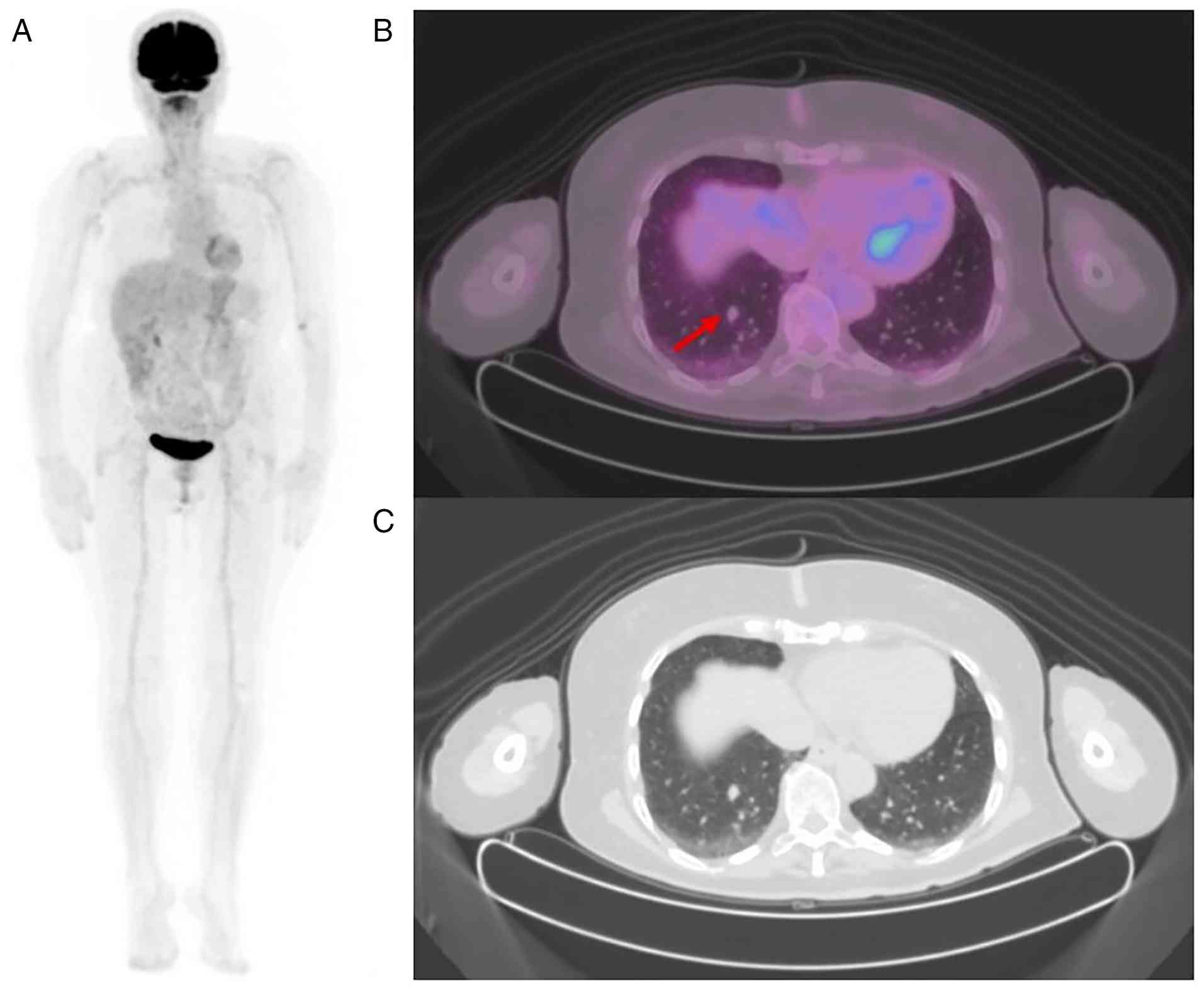
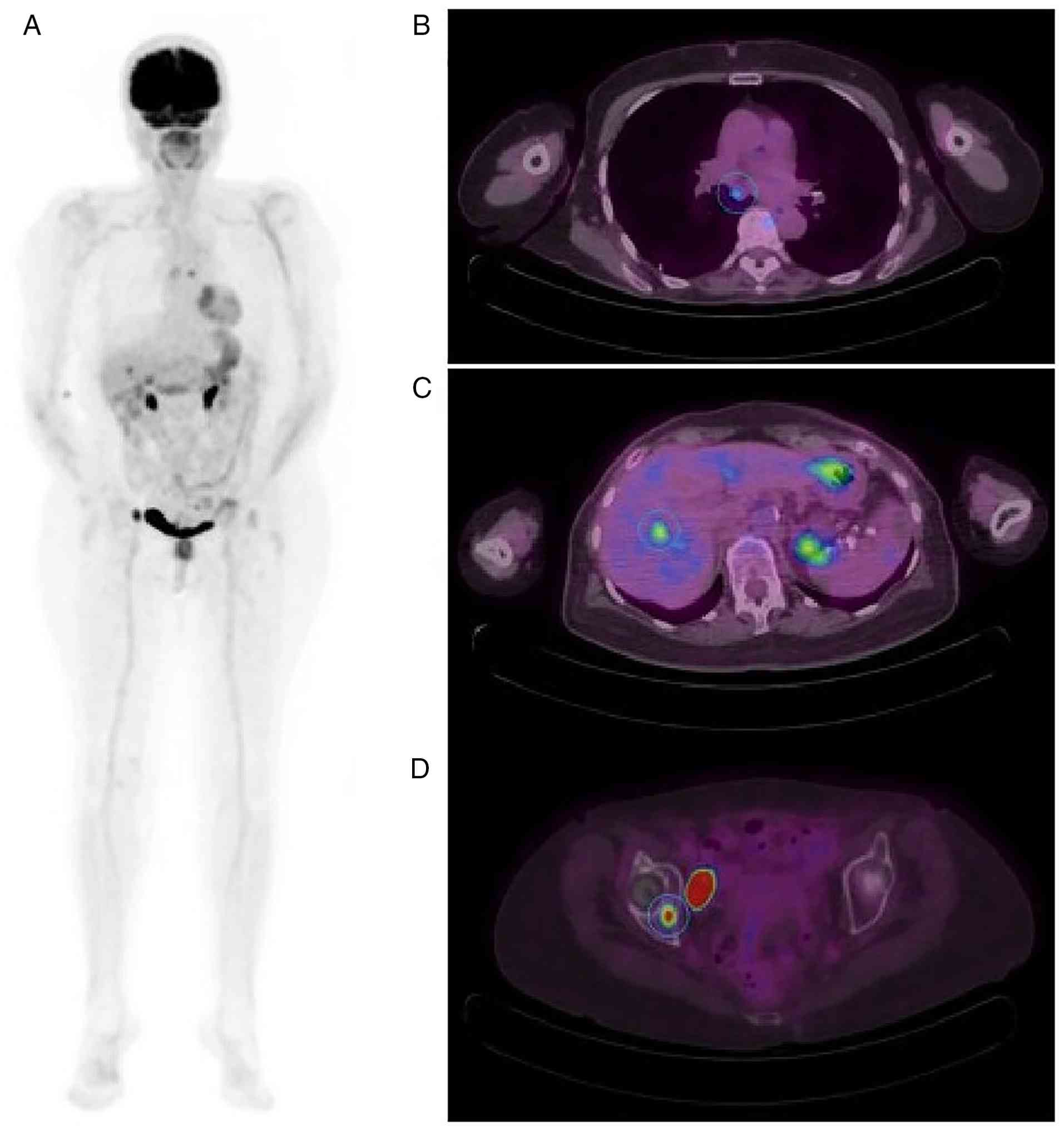
April 2024, restaging with CT and FDG-PET imaging revealed a newly
developed right lower-lobe pulmonary nodule measuring approximately
1 cm, with a maximum standardized uptake value (SUVmax) of 2
(Fig. 1). Given the absence of
additional metastatic sites, after multidisciplinary discussion,
the patient underwent surgical excision of the lung nodule, and
pathological evaluation confirmed the metastatic nature of the
lesion. Pembrolizumab adjuvant therapy was continued until August
2024, when the patient presented with recurrent haematuria. Prompt
urological evaluation led to the identification, on cystoscopic
exam, of an 8 mm bladder lesion. The lesion was highly suspicious
for malignancy; therefore, transurethral resection bladder tumour
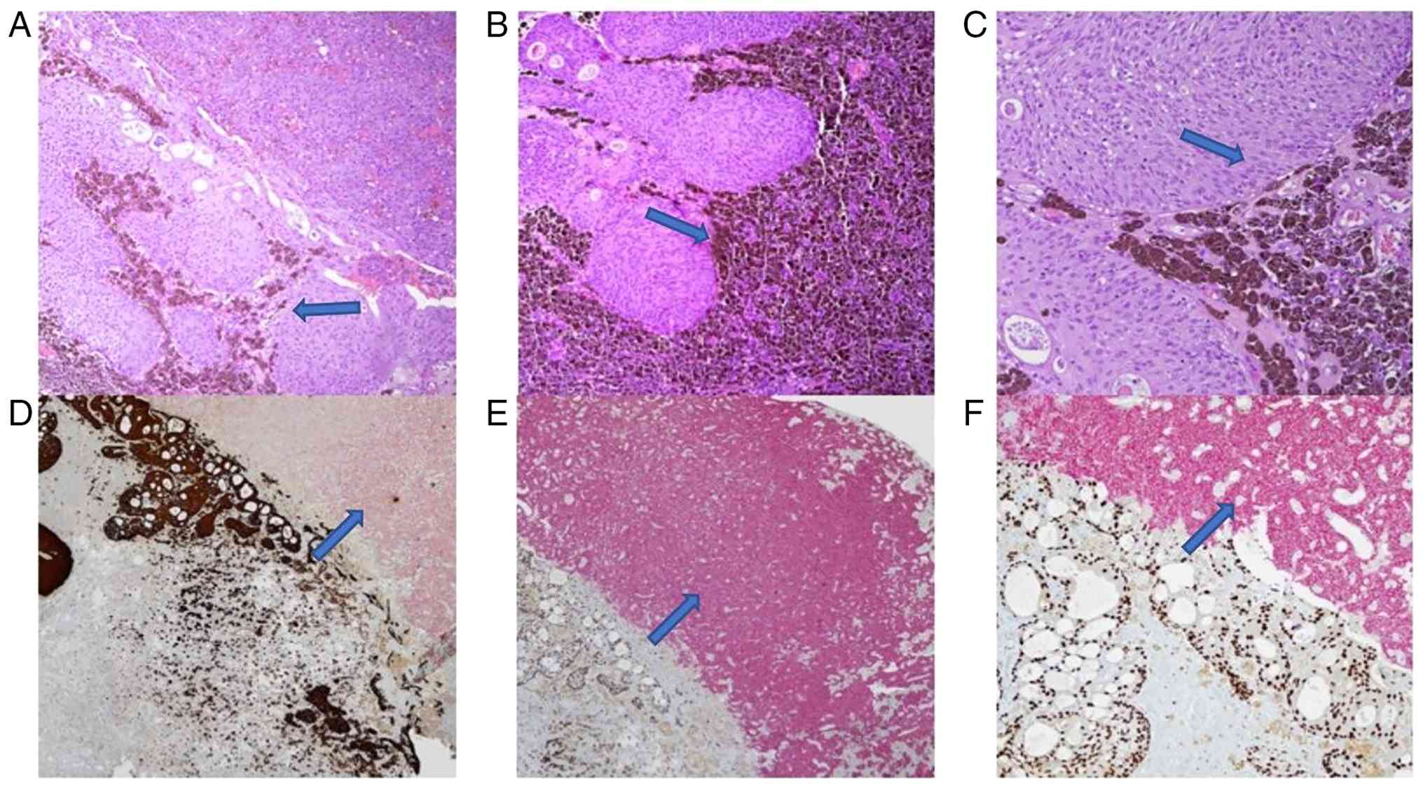
(TURBT) was performed. An accurate histopathological analysis
demonstrated the presence of metastatic cells from melanoma
involving the bladder wall, with a molecular profile characterized
by PD-L1 negativity and BRAF wild-type status (Fig. 2). Histological and
immunohistochemical analyses were performed on formalin-fixed,
paraffin-embedded tissue obtained from the transurethral resection
of the bladder lesion. Four-micrometre-thick sections were prepared
for haematoxylin and eosin staining and immunohistochemistry. The
immunohistochemical panel included dual staining for
pan-cytokeratin/SOX-10 and GATA3/HMB45. The following ready-to-use
primary antibodies were used according to the manufacturers'
protocols: anti-pan cytokeratin AE1/AE3/PCK26, mouse monoclonal
antibody cocktail, Roche Diagnostics/Ventana, REF 760-2595; SOX-10,
clone SP267, rabbit monoclonal primary antibody, Cell Marque/Roche
Diagnostics, Roche no. 07560389001; GATA3, clone L50-823, mouse
monoclonal primary antibody, Cell Marque/Roche Diagnostics, Roche
no. 07107749001; and CONFIRM anti-Melanosome HMB45, clone HMB45,
mouse monoclonal primary antibody, Roche Diagnostics/Ventana, REF
790-4366. The antibodies were used as prediluted automated-platform
reagents. Slides were evaluated by light microscopy using a Zeiss
Axio Imager 2 microscope equipped with a digital camera system,
Carl Zeiss Microscopy GmbH, Jena, Germany, and representative
photomicrographs were acquired at x10 and x20 magnification. The
patient continued immunotherapy until December 2024. Restaging
imaging performed in January 2025 revealed systemic disease
progression with involvement of the lymph nodes, liver, and bone
(Fig. 3). Consequently, in
February 2025, the patient initiated first-line systemic therapy
for metastatic disease with a combination of nivolumab and
ipilimumab, completing three of the four planned cycles due to the
onset of transient thyrotoxicosis, which resolved spontaneously
after discontinuation of ipilimumab. The patient achieved an
initial partial response to treatment followed by stable disease at
subsequent imaging assessments. The patient is currently receiving
nivolumab with no additional immune-related adverse events. As of
March 2026, her disease remains clinically and radiologically
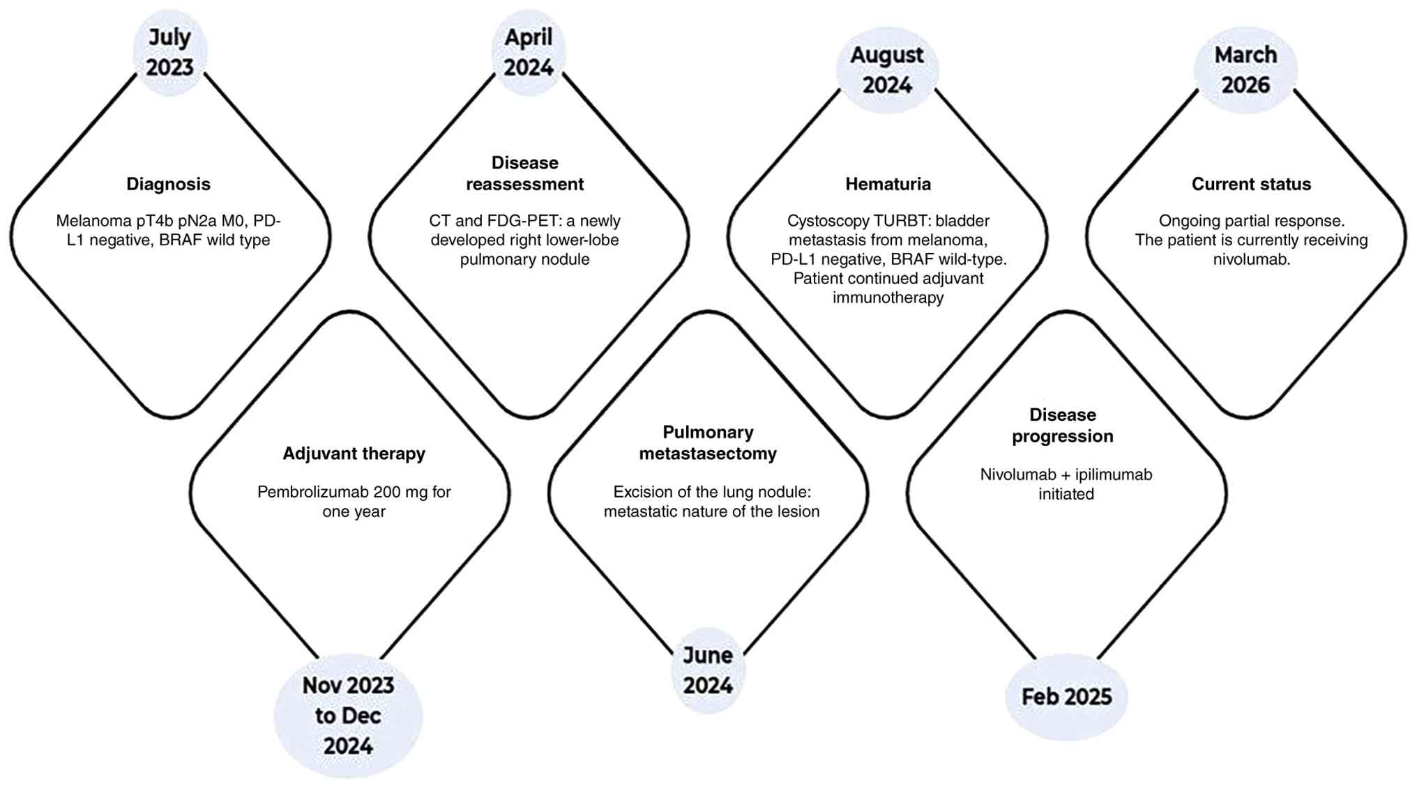
stable. A summary of the patient's clinical course and treatment
timeline is reported in Fig.
4.
Discussion
Although nearly all cancers have the potential to
metastasize to the bladder, secondary bladder neoplasms account for
only 2-3% of all malignant bladder tumours, most commonly
originating from adjacent organs such as the colon, prostate,
rectum, or cervix (10). In rare
cases, the primary sites include the stomach (4.3%), skin (3.9%),
lung (2.8%), and breast (2.5%) (11-13).
The most common histological type of secondary bladder neoplasm is
adenocarcinoma, whereas malignant melanoma is considerably rarer
(10). Primary melanoma of the
bladder is exceedingly rare, representing just 0.2% of all
melanomas. Melanoma metastases to the urinary bladder are rare in
clinical practice, with approximately thirty cases reported in the
literature over the past few decades (10). A summary of available literature
(10,14-28)
is reported in Table I. However,
autopsy studies have shown bladder involvement in 18-37% of
patients with disseminated melanoma, highlighting a higher
incidence of bladder metastases in this setting (23). These metastases are commonly
associated with synchronous lesions in other organs, suggesting an
unfavourable prognosis (23,29).
The mechanisms underlying bladder metastasis from melanoma remain
incompletely understood. Hematogenous dissemination is considered
the most likely pathway, owing to the highly vascular nature of
melanoma and the rich vascular supply of the bladder. This
hypothesis is supported by the frequent coexistence of bladder
metastases with synchronous distant metastatic sites, including
lung, liver, bone, and brain involvement (30). Nevertheless, the discrepancy
between the relatively high prevalence observed in autopsy studies
and the rarity of clinically detected cases suggests that
additional biological factors may influence metastatic colonization
and tumour outgrowth within the bladder microenvironment (31). Local stromal composition,
immune-cell infiltration, endothelial adhesion molecules, and
organ-specific chemokine signalling may contribute to the
establishment of clinically evident bladder metastases only in
selected patients (32).
Furthermore, the frequent asymptomatic presentation likely
contributes to underdiagnosis during the early phases of
dissemination. Symptomatic presentation is uncommon, occurring in
only about 15% of cases, with gross haematuria being the most
frequent manifestation (19),
though urinary retention, dysuria, increased frequency, flank pain,
or suprapubic pain may also occur (33). In the majority of cases, patients
are asymptomatic, with metastases discovered incidentally on
systemic follow-up imaging, including abdominal CT scans (20,23).
Radiologically, melanomas, whether primary or metastatic, are
hyperintense on T1-weighted magnetic resonance imaging (MRI) in
24-47% of cases, owing to the paramagnetic properties of melanin
and its copper, manganese, and zinc content, which shorten T1
relaxation times (34,35). Haemorrhage, present in up to 50% of
cases, can further enhance T1 signal, although poorly
differentiated lesions may lack this feature (35). Recognition of this pattern is
essential, and a T1-hyperintense bladder mass in a patient with
known melanoma should prompt high suspicion (25). Histologically, pigmented bladder
lesions provide an important diagnostic clue and should be
confirmed immunohistochemically, with positive staining for S-100,
HMB45, MART-1/Melan-A, tyrosinase, and MITF (29). Treatment of metastatic melanoma to
the bladder should be individualized, taking into account the
patient's overall condition, extent of metastatic disease, and
symptom burden (36). Therapeutic
options include TURBT, partial or radical cystectomy, radiotherapy,
and systemic therapies. TURBT serves both diagnostic and palliative
roles, relieving haematuria and urinary symptoms, although it
rarely achieves complete tumour control. Radical cystectomy may be
considered in selected patients with isolated lesions and good
performance status; however, even in such cases, survival seldom
exceeds three years (36,37). The management of bladder metastases
from melanoma should follow a multidisciplinary approach,
integrating surgical, systemic, and supportive treatments. The
therapeutic landscape of advanced melanoma has dramatically evolved
with the introduction of immune checkpoint inhibitors and targeted
therapies. In patients harbouring the BRAF V600E mutation, targeted
therapies with BRAF and MEK inhibitors, as well as immune
checkpoint inhibitors (anti-CTLA-4, anti-PD-1, anti-LAG3), have
demonstrated superior overall and progression-free survival
compared with conventional chemotherapy or interleukin-2 therapy
(38). Immunotherapy has
transformed the therapeutic landscape, achieving 5-year overall
survival rates of up to 49.5% (23). However, treatment resistance and
immune-related adverse events continue to pose significant clinical
challenges. In our patient, metastatic dissemination occurred
despite adequate local control of the primary melanoma and adjuvant
treatment with pembrolizumab. Several high-risk pathological
features of the primary lesion, including increased Breslow
thickness, lymphovascular invasion, nodal involvement, and elevated
mitotic activity, likely contributed to the aggressive biological
behaviour of the disease. In contrast to most cases reported in the
literature, we performed molecular profiling of the primary
melanoma and metastatic sites, to study the evolution of PD-L1
expression and genomic alterations during tumour progression over
time. Interestingly, molecular profiling remained substantially
consistent across the primary tumour and metastatic sites, with
persistent PD-L1 negativity and absence of BRAF alterations,
supporting a model of metastatic spread driven by pre-existing
resistant clones rather than the acquisition of new driver
alterations during progression. This may reflect immune-selective
pressure under PD-1 blockade leading to outgrowth of pre-existing
resistant clones rather than true genomic evolution of the tumour
(39). Another clinically relevant
aspect of this case is the development of oligoprogression during
adjuvant pembrolizumab therapy. Oligoprogression is increasingly
recognized as a distinct evolutionary pattern during immune
checkpoint inhibition, in which a limited number of metastatic
sites acquire resistance while the remaining disease burden remains
controlled by systemic therapy. This phenomenon may reflect spatial
and temporal heterogeneity in tumour evolution, with resistant
subclones emerging under immune-selective pressure (40). In melanoma, acquired resistance to
anti-PD-1 therapy has been associated with several mechanisms,
including defects in interferon-γ signalling, impaired antigen
presentation through alterations in β2-microglobulin or HLA
expression, T-cell exhaustion, compensatory upregulation of
alternative inhibitory checkpoints, and development of an
immunosuppressive tumour microenvironment enriched in regulatory T
cells and myeloid-derived suppressor cells. The persistent PD-L1
negativity observed in both the primary tumour and metastatic
lesions in our patient may reflect a poorly inflamed tumour
microenvironment, potentially consistent with an immune-desert or
non-inflamed phenotype associated with reduced sensitivity to PD-1
blockade (41,42). In our patient, both the isolated
pulmonary metastasis and the subsequent solitary bladder lesion
were amenable to local treatment while systemic pembrolizumab was
maintained. This strategy was supported by the hypothesis that most
tumour sites could still remain partially controlled by ongoing
immune checkpoint inhibition, whereas locally resistant clones
could be eradicated through surgery. Emerging evidence in melanoma
and other solid tumours suggests that local ablative approaches,
including surgery or radiotherapy, may prolong the duration of
benefit from immunotherapy in selected patients with
oligoprogression, delaying the need for systemic treatment
escalation and preserving quality of life (43). Historically, surgery was primarily
palliative-performed to relieve symptoms such as anaemia or urinary
obstruction-rather than to extend survival. However, in the era of
modern immunotherapy, surgical resection of oligometastatic lesions
has been associated with longer overall survival in selected
patients (44). Despite these
advances, the prognosis of metastatic melanoma remains poor, with a
median survival of approximately 12 months and a 5-year survival
rate ranging between 5 and 19% (37,45).
An additional noteworthy aspect of this case is the durable disease
control achieved with nivolumab plus ipilimumab after progression
during adjuvant pembrolizumab therapy. Combined CTLA-4 and PD-1
blockade may partially overcome resistance to prior anti-PD-1
monotherapy by enhancing T-cell priming, broadening the repertoire
of antitumor T-cell responses, and restoring immune activation
within resistant tumour sites (46,47).
This observation further supports the biological rationale for
sequential and combinatorial immunotherapeutic strategies in
metastatic melanoma, even after progression during prior anti-PD-1
treatment. In conclusion, metastatic bladder melanoma carries a
particularly poor prognosis and the highest mortality risk among
sites of melanoma dissemination. Even with surgical resection and
systemic therapy, no patient has been reported to survive beyond
three years following bladder metastasis resection (35). Since the advent of ICIs, management
has remained primarily palliative, focusing on symptom control and
quality-of-life improvement. Nevertheless, this case highlights how
modern immunotherapeutic strategies, combined with local treatment
approaches for oligoprogressive disease, may contribute to
prolonged disease control even in patients with atypical metastatic
sites. Early recognition of unusual metastatic localizations,
careful multidisciplinary evaluation, and integration of local and
systemic therapies are essential to optimize clinical outcomes in
selected patients with metastatic melanoma.
 | Table ICase reports of melanoma metastasis
to the bladder. |
Table I
Case reports of melanoma metastasis
to the bladder.
| Author | Year | Age | Gender | Presentation | Timing
(Syn/Met) | Other metastatic
sites | Treatment | OS | Molecular
profile | Primary tumour | (Refs.) |
|---|
| Walsh et
al | 1966 | 68 | Male | Haematuria | Met | NR | TURBT | NR | NR | Cutaneous
melanoma | (14) |
| deKernion et
al | 1975 | 56 | Male | Haematuria | Met | Small bowel and
liver | Immunotherapy with
BCG | 11 months | NR | Cutaneous
melanoma | (15) |
| Arapantoni-Dadioti
et al | 1995 | 28 | Female | Pain and burning
during micturition | Met | Skin, lung,
brain | Patient refused any
further treatment | NR | NR | Melanoma | (16) |
| Ergen et
al | 1995 | 65 | Male | Right flank pain
and haematuria | Met | Gastroduodenal
mucosa | NR | NR | NR | Cutaneous
melanoma | (17) |
| Demirkesen et
al | 2000 | 45 | Female | Urinary frequency,
macroscopic haematuria and right inguinal swelling | Met | Skin, lymph nodes,
and meninges | Resection,
chemo-immunotherapy protocol that consisted of cisplatin, DTIC,
fotomustin and interferon α, followed by immunotherapy protocol of
interferon α and interleukin-2 | 13 months | NR | Melanoma | (18) |
| Ozan Efesoy et
al | 2011 | 60 | Female | Gross haematuria
and weight loss | Met | Brain, lymph nodes,
and lung | Transurethral
resection | 7 months | NR | Cutaneous
melanoma | (19) |
| Eric S. Wisenbaugh
et al | 2012 | 81 | Male | Microscopic
haematuria Urinary frequency and hesitancy | Met | None | TURBT | NR | NR | Cutaneous
melanoma | (20) |
| Eric S. Wisenbaugh
et al | 2012 | 84 | Male | Incidental
finding | Met | Small bowel | TURBT | 4 months | NR | Cutaneous
melanoma | (20) |
| Eric S. Wisenbaugh
et al | 2012 | 85 | Male | Gross
haematuria | Met | Abdominal lymph
nodes, stomach, lung | TURBT | 4 months | NR | Cutaneous
melanoma | (20) |
| Eric S. Wisenbaugh
et al | 2012 | 62 | Female | Incidental on
restaging CT | Met | NR | TURBT | 5 months | NR | Uveal melanoma | (20) |
| Arvind Rishi et
al | 2014 | 61 | Female | Gross
haematuria | Met | Subcutaneous
tissue, brain, lungs | TURBT | 2 months | BRAF V600E
mutation | Cutaneous
melanoma | (21) |
| Rahoui Moez et
al | 2020 | 31 | Male | Haematuria and low
urinary tract signs | Met | Lymph nodes | Monopolar loop
electrocautery followed by chemotherapy | NR | NR | Cutaneous
melanoma | (22) |
| Raj A. Kumar et
al | 2021 | 78 | Female | Haematuria | Met | Lung and
meninges |
Ipilimumab/nivolumab combination
immunotherapy | NR | BRAF V600E
mutation | Cutaneous
melanoma | (23) |
| Jesús
Machuca-Aguado et al | 2022 | 78 | Male | Haematuria | Met | Lung, liver, lymph
nodes and bone | 2015 haemostatic
radiotherapy and 2017 TURBT | 36 months | NR | Uveal melanoma | (24) |
| Jesús
Machuca-Aguado et al | 2022 | 56 | Male | Incidental
finding | Met | NR | NR | NR | NR | Uveal melanoma | (24) |
| Duarte Vieira e
Brito et al | 2022 | 73 | Male | Haematuria | Met | Suspected pulmonary
and peritoneal metastasis | TURBT | NR | NR | Uveal melanoma | (9) |
| Maria Clara
Fernandes et al | 2022 | 47 | Female | Lower urinary tract
symptoms and haematuria | Met | Left inguinal
nodes, spleen, lungs, brain and endometrium | TURBT | NR | NR | Cutaneous
melanoma | (25) |
| Takashi Ueno et
al | 2024 | 86 | Male | Incidental
finding | Met | Liver | TURBT followed by
best supportive care | NR | BRAF wild-type | Uveal melanoma | (10) |
| Takashi Ueno et
al | 2024 | 57 | Male | Right abdominal
discomfort | Met | None | TURBT | 22 years | NR | Cutaneous
melanoma | (10) |
| Takashi Ueno et
al | 2024 | 79 | Female | Haematuria | Met | Lung, lymph nodes,
adrenal glands, suspected peritoneal nodule | TURBT followed by
pembrolizumab | NR | NR | Cutaneous
melanoma | (10) |
| Abdelmounim
Boughaleb et al | 2024 | 75 | Female | Haematuria | Met | Breast, liver,
lymph nodes, bone, and multifocal subcutaneous and muscular
lesions | TURBT followed by
continuation of immunotherapy with nivolumab and ipilimumab | NR | NR | Cutaneous
melanoma | (26) |
| Tu Anh Do et
al | 2024 | 53 | Female | Incidental
finding | Met | Lymph nodes | Chemotherapy with
dacarbazine | 10 months | BRAF wild-type | Cutaneous
melanoma | (27) |
| Marwin Anandrati
et al | 2024 | 71 | Female | Haematuria | Met | None | Surgery followed by
immunotherapy | NR | NR | Cutaneous
melanoma | (28) |
| Mariapia Marafioti
et al | 2026 | 79 | Female | Haematuria | Met | Lung, lymph nodes,
liver, bone | Adjuvant
immunotherapy with pembrolizumab for one year; first-line systemic
therapy with a combination of nivolumab and ipilimumab | Patient alive | Negative PD-L1
expression, BRAF wild-type | Cutaneous
melanoma | |
Acknowledgements
Not applicable.
Funding
Funding: No funding was received.
Availability of data and materials
The data generated in the present study are included
in the figures and/or tables of this article.
Authors' contributions
Conceptualization was conducted by MMa, MMu, ML and
MS. Formal analysis and data interpretation were performed by MMa,
MMu, GC, MIP and MV. Data acquisition and clinical management were
carried out by MMa, MMu, LP, RL, GC, MV, ML and MS. Literature
analysis, writing of the original draft, and reviewing and editing
of the manuscript was performed by MMa and MMu. Supervision was
provided by MIP, ML and MS. Project administration was conducted by
MMa and MMu. All authors have read and approved the final version
of the manuscript. MS and ML confirm the authenticity of all the
raw data.
Ethics approval and consent to
participate
Not applicable.
Patient consent for publication
Written informed consent was obtained from the
patient for publication of this case report and any accompanying
images.
Competing interests
The authors declare that they have no competing
interests.
References
|
1
|
Siegel RL, Kratzer TB, Wagle NS, Sung H
and Jemal A: Cancer statistics, 2026. CA Cancer J Clin.
76(e70043)2026.PubMed/NCBI View Article : Google Scholar
|
|
2
|
Garbe C, Amaral T, Peris K, Hauschild A,
Arenberger P, Basset-Seguin N, Bastholt L, Bataille V, Brochez L,
Del Marmol V, et al: European consensus-based interdisciplinary
guideline for melanoma. Part 1: Diagnostics-update 2024. Eur J
Cancer. 215(115152)2025.PubMed/NCBI View Article : Google Scholar
|
|
3
|
Jazlyn R, Wadt KAW and Hayward NK:
Melanoma genetics. J Med Genet. 53:1–14. 2016.PubMed/NCBI View Article : Google Scholar
|
|
4
|
Long GV, Swetter SM, Menzies AM,
Gershenwald JE and Scolyer RA: Cutaneous melanoma. Lancet.
402:485–502. 2023.PubMed/NCBI View Article : Google Scholar
|
|
5
|
Pastorino L, Andreotti V, Dalmasso B,
Vanni I, Ciccarese G, Mandalà M, Spadola G, Pizzichetta MA, Ponti
G, Tibiletti MG, et al: Insights into genetic susceptibility to
melanoma by gene panel testing: Potential pathogenic variants in
ACD, ATM, BAP1, and POT1. Cancers (Basel). 12(1007)2020.PubMed/NCBI View Article : Google Scholar
|
|
6
|
Zocchi L, Lontano A, Merli M, Dika E,
Nagore E, Quaglino P, Puig S and Ribero S: Familial melanoma and
susceptibility genes: A review of the most common clinical and
dermoscopic phenotypic aspect, associated malignancies and
practical tips for management. J Clin Med. 10(3760)2021.PubMed/NCBI View Article : Google Scholar
|
|
7
|
Amaral T, Ottaviano M, Arance A, Blank C,
Chiarion-Sileni V, Donia M, Dummer R, Garbe C, Gershenwald JE,
Gogas H, et al: Cutaneous melanoma: ESMO Clinical Practice
Guideline for diagnosis, treatment and follow-up. Ann Oncol.
36:10–30. 2025.PubMed/NCBI View Article : Google Scholar
|
|
8
|
Boutros A, Croce E, Ferrari M, Gili R,
Massaro G, Marconcini R, Arecco L, Tanda ET and Spagnolo F: The
treatment of advanced melanoma: Current approaches and new
challenges. Crit Rev Oncol Hematol. 196(104276)2024.PubMed/NCBI View Article : Google Scholar
|
|
9
|
Vieira E, Brito D, Pereira J,
Pereira-Lourenço M, Ricardo G and Rabaça C: Bladder tumour as a
first manifestation of metastatic choroid melanoma. Urol Case Rep.
42(101996)2022.PubMed/NCBI View Article : Google Scholar
|
|
10
|
Ueno T, Kiwaki T, Betsunoh H, Ito K,
Murashima T, Fujii M, Nagai T, Mukai S, Sawada A and Kamoto T:
Metastasis of malignant melanoma to urinary tract: A case report. J
Med Case Rep. 18(396)2024.PubMed/NCBI View Article : Google Scholar
|
|
11
|
Bates AW and Baithun SI: Secondary
neoplasms of the bladder are histological mimics of nontransitional
cell primary tumours: Clinicopathological and histological features
of 282 cases. Histopathology. 36:32–40. 2000.PubMed/NCBI View Article : Google Scholar
|
|
12
|
Velcheti V and Govindan R: Metastatic
cancer involving bladder: A review. Can J Urol. 14:3443–3448.
2007.PubMed/NCBI
|
|
13
|
Goldstein AG: Metastatic carcinoma to the
bladder. J Urol. 98:209–215. 1967.PubMed/NCBI View Article : Google Scholar
|
|
14
|
Walsh EJ, Ockuly EA, Ockuly EF and Ockuly
JJ: Treatment of metastatic melanoma of the bladder. J Urol.
96:472–478. 1966.PubMed/NCBI View Article : Google Scholar
|
|
15
|
deKernion JB, Golub SH, Gupta RK,
Silverstein M and Morton DL: Successful transurethral intralesional
BCG therapy of a bladder melanoma. Cancer. 36:1662–1667.
1975.PubMed/NCBI View Article : Google Scholar
|
|
16
|
Arapantoni-Dadioti P, Panayiotides J,
Kalkandi P, Christodoulou C and Delides GS: Metastasis of malignant
melanoma to a transitional cell carcinoma of the urinary bladder.
Eur J Surg Oncol. 21:92–93. 1995.PubMed/NCBI View Article : Google Scholar
|
|
17
|
Ergen A, Balbay MD, Alexis R, Nardi PM and
Grunberger I: Renal vein thrombosis and metastasis to the bladder:
An unusual presentation of malignant melanoma. Int Urol Nephrol.
27:547–549. 1995.PubMed/NCBI View Article : Google Scholar
|
|
18
|
Demirkesen O, Yaycioglu O, Uygun N, Demir
G and Yalcin V: A case of metastatic malignant melanoma presenting
with hematuria. Urol Int. 64:118–120. 2000.PubMed/NCBI View Article : Google Scholar
|
|
19
|
Efesoy O and Cayan S: Bladder metastasis
of malignant melanoma: A case report and review of literature. Med
Oncol. 28 (Suppl 1):S667–S669. 2011.PubMed/NCBI View Article : Google Scholar
|
|
20
|
Wisenbaugh ES, Stanton ML, Grimsby GM,
Tyson MD and Castle EP: Metastatic malignant melanoma to the
bladder: A case series. Curr Urol. 6:53–56. 2012.PubMed/NCBI View Article : Google Scholar
|
|
21
|
Rishi A, Anderson TA, Kirschenbaum AM and
Unger PD: Metastatic malignant melanoma to urinary bladder: A
potential pitfall for high-grade urothelial carcinoma. Int J Surg
Pathol. 22:347–351. 2014.PubMed/NCBI View Article : Google Scholar
|
|
22
|
Moez R, Khaireddine MD, Sahnoun W, Zehani
A, Bibi M, Ouannes Y, Sellami A, Rhouma SB and Nouira Y:
Symptomatic bladder metastasis of malignant melanoma: A case
report. J Surg Case Rep. 2020(rjaa509)2020.PubMed/NCBI View Article : Google Scholar
|
|
23
|
Kumar RA, Batra N, Frankel J and Kumar U:
Melanoma metastasis to the bladder: A case report. Urol Case Rep.
40(101941)2021.PubMed/NCBI View Article : Google Scholar
|
|
24
|
Machuca-Aguado J, Rodríguez-Zarco E,
Carrero-García B and Vázquez-Ramírez FJ: Metastasis of Uveal
melanoma in bladder: Presentation of two cases and review of the
literature. Arch Esp Urol. 75:873–877. 2022.PubMed/NCBI View Article : Google Scholar
|
|
25
|
Fernandes MC, Shoushtari A, Leitao MM Jr,
Chui MH, Al-Ahmadie H and Petkovska I: Metastatic melanoma
concurrent to the urinary bladder and endometrium: Case report. J
Med Imaging Radiat Oncol. 66:643–646. 2022.PubMed/NCBI View Article : Google Scholar
|
|
26
|
Boughaleb A, Kubiak M, Bourimi C, Tariqi
R, Leroux PA and El Kentaoui H: Melanoma metastasis to the bladder:
A case report of an underdiagnosed entity. Int J Surg Case Rep.
122(110048)2024.PubMed/NCBI View Article : Google Scholar
|
|
27
|
Do TA, Mai NT, Le UT, Nguyen DN, Vu PT and
Van Nguyen C: Metastatic melanoma to the urinary bladder with
synchronous or metachronous metastasis to other organs:
Clinicopathological characteristics and outcome. Int J Surg Case
Rep. 120(109789)2024.PubMed/NCBI View Article : Google Scholar
|
|
28
|
Anandrati M, Maini V, Asarian A and Xiao
P: Metastasis of malignant melanoma to the urinary bladder &
descending colon: A case report and literature review. Urol Case
Rep. 57(102883)2024.PubMed/NCBI View Article : Google Scholar
|
|
29
|
Patil RV, Woldu SL, Lucas E, Quinn AM,
Francis F and Margulis V: Metastatic melanoma to the bladder: Case
report and review of the literature. Urol Case Rep. 11:33–36.
2017.PubMed/NCBI View Article : Google Scholar
|
|
30
|
Calomarde-Rees L, García-Calatayud R,
Requena Caballero C, Manrique-Silva E, Traves V, García-Casado Z,
Soriano V, Kumar R and Nagore E: Risk factors for lymphatic and
hematogenous dissemination in patients with stages I to II
cutaneous melanoma. JAMA Dermatol. 155:679–687. 2019.PubMed/NCBI View Article : Google Scholar
|
|
31
|
Feldman A, Borak S, Rais-Bahrami S and
Gordetsky J: Secondary malignancies of the bladder: Avoiding the
diagnostic pitfall. Int J Surg Pathol. 26:120–125. 2018.PubMed/NCBI View Article : Google Scholar
|
|
32
|
Peinado H, Zhang H, Matei IR, Costa-Silva
B, Hoshino A, Rodrigues G, Psaila B, Kaplan RN, Bromberg JF, Kang
Y, et al: Pre-metastatic niches: Organ-specific homes for
metastases. Nat Rev Cancer. 17:302–317. 2017.PubMed/NCBI View Article : Google Scholar
|
|
33
|
Abdou CA, Wolff EH, Tracey AT and Klausner
AP: Metastatic melanoma to the bladder presenting as autonomic
dysreflexia in a patient with paraplegia. Urol Case Rep.
39(101820)2021.PubMed/NCBI View Article : Google Scholar
|
|
34
|
Patnana M, Bronstein Y, Szklaruk J, Bedi
DG, Hwu WJ, Gershenwald JE, Prieto VG and Ng CS: Multimethod
imaging, staging, and spectrum of manifestations of metastatic
melanoma. Clin Radiol. 66:224–236. 2011.PubMed/NCBI View Article : Google Scholar
|
|
35
|
Wong VK, Lubner MG, Menias CO, Mellnick
VM, Kennedy TA, Bhalla S and Pickhardt PJ: Clinical and imaging
features of noncutaneous melanoma. AJR Am J Roentgenol.
208:942–959. 2017.PubMed/NCBI View Article : Google Scholar
|
|
36
|
Ogunremi O, Sirjuesingh D and Deshpande A:
Metastatic melanoma to the urinary bladder: A rare cause of visible
haematuria. Case Rep Urol. 2024(5516547)2024.PubMed/NCBI View Article : Google Scholar
|
|
37
|
Venyo AK: Melanoma of the urinary bladder:
A review of the literature. Surg Res Pract.
2014(605802)2014.PubMed/NCBI View Article : Google Scholar
|
|
38
|
Paterson A, Sut M, Kaul A, Altieri V,
Mutch F, Patel J and Sharma H: Metastatic malignant melanoma of the
urinary bladder: Case report and literature review. Cent Eur J
Urol. 65:232–234. 2012.PubMed/NCBI View Article : Google Scholar
|
|
39
|
Wells A, Clark A, Bradshaw A, Ma B and
Edington H: The great escape: How metastases of melanoma, and other
carcinomas, avoid elimination. Exp Biol Med (Maywood).
243:1245–1255. 2018.PubMed/NCBI View Article : Google Scholar
|
|
40
|
Guida M, Bartolomeo N, De Risi I, Fucci L,
Armenio A, Filannino R, Ruggieri E, Macina F, Traversa M, Nardone
A, et al: The management of Oligoprogression in the landscape of
new therapies for metastatic melanoma. Cancers (Basel).
11(1559)2019.PubMed/NCBI View Article : Google Scholar
|
|
41
|
Li W, Gu J, Fan H, Zhang L, Guo J and Si
L: Evolving cancer resistance to anti-PD-1/PD-L1 antibodies in
melanoma: Comprehensive insights with future prospects. Crit Rev
Oncol Hematol. 201(104426)2024.PubMed/NCBI View Article : Google Scholar
|
|
42
|
Zheng S, Wang W, Shen L, Yao Y, Xia W and
Ni C: Tumor battlefield within inflamed, excluded or desert immune
phenotypes: The mechanisms and strategies. Exp Hematol Oncol.
13(80)2024.PubMed/NCBI View Article : Google Scholar
|
|
43
|
Puza CJ, Bressler ES, Terando AM, Howard
JH, Brown MC, Hanks B, Salama AKS and Beasley GM: The emerging role
of surgery for patients with advanced melanoma treated with
immunotherapy. J Surg Res. 236:209–215. 2019.PubMed/NCBI View Article : Google Scholar
|
|
44
|
Deutsch GB, Flaherty DC, Kirchoff DD,
Bailey M, Vitug S, Foshag LJ, Faries MB and Bilchik AJ: Association
of surgical treatment, systemic therapy, and survival in patients
with abdominal visceral melanoma metastases, 1965-2014: Relevance
of surgical cure in the Era of modern systemic therapy. JAMA Surg.
152:672–678. 2017.PubMed/NCBI View Article : Google Scholar
|
|
45
|
Sandru A, Voinea S, Panaitescu E and
Blidaru A: Survival rates of patients with metastatic malignant
melanoma. J Med Life. 7:572–576. 2014.PubMed/NCBI
|
|
46
|
Rasmussen M, Durhuus JA, Nilbert M,
Andersen O and Therkildsen C: Response to immune checkpoint
inhibitors is affected by deregulations in the antigen presentation
machinery: A systematic review and meta-analysis. J Clin Med.
12(329)2023.PubMed/NCBI View Article : Google Scholar
|
|
47
|
Phadke MS, Li J, Chen Z, Rodriguez PC,
Mandula JK, Karapetyan L, Forsyth PA, Chen YA and Smalley KSM:
Differential requirements for CD4+ T cells in the efficacy of the
anti-PD-1+LAG-3 and anti-PD-1+CTLA-4 combinations in melanoma flank
and brain metastasis models. J Immunother Cancer.
11(e007239)2023.PubMed/NCBI View Article : Google Scholar
|