Introduction
Diabetes mellitus is one of the largest epidemics
worldwide that is recognized and classified as a cluster of
heterogeneous metabolic disorders characterized by hyperglycemia
due to defects in insulin secretion, insulin action, or both.
Diabetes mellitus occurs due to the destruction of pancreatic
β-cells with consequent insulin deficiency by autoimmune disease or
due to abnormalities that lead to a resistance to insulin actions
(1).
Metabolic abnormalities in carbohydrates, fats and
proteins in diabetes occur due to deficient insulin action at
target tissues. It has been demonstrated that oxygen free radicals
are produced due to hyperglycemia and can cause various oxidative
stress-induced complications of diabetes, such as nephropathy,
retinopathy and neuropathy (2). The
accumulation of lipid peroxidation products termed as advanced
glycosylation end products and damaged DNA eventually leads to the
development of pathological diabetic complications (3).
Amylin, or islet amyloid polypeptide (IAPP), is an
endocrine peptide hormone co-localized, co-secreted and co-packaged
along with insulin by pancreatic β-cells that is associated with
the type 2 diabetes mellitus (T2D) disease progression (4). Amylin is co-secreted with insulin in
response to caloric intake. It plays a crucial role in maintaining
glucose homeostasis by suppressing glucagon release, delaying the
gastric emptying rate and stimulating the satiety center in the
brain to limit caloric intake. Amylin serum concentrations in
patients with type 1 diabetes were at a lower baseline with the
absence of amylin response to caloric intake. At the same time,
patients with T2D requiring insulin also have a diminished amylin
response to caloric intake, possibly related to the degree of
β-cell impairment (5).
It is well established that insulin enhances glucose
uptake by promoting the translocation of glucose transporter 4
(GLUT4), the major insulin-regulated glucose transporter in
skeletal muscle and adipose tissue, from the intracellular storage
vesicles to the plasma membrane. Due to the absence or insufficient
sensitivity to insulin in diabetic patients, GLUT4 expression is
decreased, leading to resistance to insulin-stimulated glucose
transport that profoundly contributes to disease pathophysiology.
Phosphatidylinositol 3-kinase (PI3K) is a crucial component of the
insulin-signaling cascade, essential for the metabolic effects of
insulin on glucose transport and GLUT4 translocation (6,7).
Glycogen synthase kinase-3 (GSK-3) regulates a
number of metabolic and signaling proteins. Activated GSK-3
inactivates the glycogen synthase enzyme, which is responsible for
converting glucose to glycogen for storage. Insulin can bind to
GSK-3 receptors, relieve this inhibition and activate the PI3K/Akt
pathway. However, the overexpression of GSK-3 results in impaired
glucose tolerance and decreased glycogen synthase activity, and
consequently, in glycogen synthesis. Furthermore, it has been
reported that GSK-3 overexpression attenuates insulin signaling,
which further supports the role of GSK-3 in diabetes (8,9).
Several limitations have been reported regarding the
use of conventional oral hypoglycemic drugs in the treatment of
diabetes due to adverse effects and high rates of secondary
failure. These adverse effects have led diabetic patients to use
natural products as an alternative source of anti-diabetic agents
with a degree of efficiency without side-effects (10). Cinnamon is one of the popular spices
containing several potent antioxidant polyphenolic compounds
traditionally used in the treatment of various chronic diseases,
such as cardiovascular diseases and diabetes (11).
Oligomeric proanthocyanidins (OPCs) are oligomeric
flavonoids found in the aqueous extract of cinnamon that are
responsible for the majority of the antioxidant properties of
cinnamon; they have been proven to regulate insulin signaling and
the expression of blood sugar transportation genes in adipocytes
(12,13). There is increasing evidence to
indicate that OPCs are effective both as a prophylactic or dietary
treatment for certain fatal diseases such as cancer,
cardiovascular, or metabolic syndromes due to their antioxidant and
anti-inflammatory activities. It was previously reported that OPCs
are effective in lowering the lipid index and blocking inflammatory
responses by preventing lipid peroxidation and adjusting the lipid
catabolic process (14,15). Furthermore, it has been found that
OPC extracts can protect β-cells of the pancreas via the
attenuation of oxidative stress, thereby increasing the sensitivity
and secretion of insulin, as well as affecting certain enzyme
activities in the metabolic process (16).
The present study was designed to investigate the
potential role of cinnamon bark extract OPCs in controlling
streptozotocin (STZ)-induced hyperglycemia and to clarify the
underlying mechanisms.
Materials and methods
Experimental design
The present study was conducted following the
institutional guidelines for care and use of laboratory animals
approved by the local Ethics Committee of the Faculty of Pharmacy,
Tanta University, Tanta, Egypt (approval no. 18112014). The study
complied with the standards of animal care the European Community
Directive (86/609/EEC) and the National Institutes of Health
Guidelines for the Care and Use of Laboratory Animals, 8th edition.
The study included 60 adult male albino rats, weighing 150-200 g.
The rats were purchased from the National Research Center, Giza,
Egypt. Following 2 weeks of acclimatization under identical
environmental conditions (22±2˚C with 55±5% controlled humidity and
a 12-h dark/light cycle) and free access to a standard pellet diet
and water ad libitum, the rats were weighed and randomly
assigned to six groups (n=10/group) as follows:
Diabetes was induced by a single intraperitoneal
(i.p.) injection of STZ (45 mg/kg; Sigma-Aldrich; Merck KGaA)
following overnight fasting (17).
The rats were weighed and injected with STZ dissolved in a disodium
citrate buffer (0.1 M, pH 4.5). The rats were provided withy 5%
glucose solution as drinking water in the first 24 h following the
STZ injection to counteract drug-induced hypoglycemia. After 3
days, blood samples were withdrawn from the retro-orbital venous
plexus under isoflurane anesthesia (3.5% for induction and 2.5% for
maintenance), and serum was separated by centrifugation at 1,600 x
g/15 min to determine the glucose level.
Only rats with fasting blood glucose levels >250
mg/dl were considered diabetic rats and were selected for further
experiments. Drug treatment commenced 7 days after the STZ
injection (when hyperglycemia was confirmed). The diabetic rats
were weighed and then randomly divided into four subgroups (n=10
for each group) as follows: i) The diabetic control group; ii) the
OPC diabetic group: Diabetic rats treated with proanthocyanidin
(eBioChem; 300 mg/kg/day, orally), as previously described
(18) for 21 consecutive days; iii)
the OPC diabetic + wortmannin group: Diabetic rats were treated
with proanthocyanidin (300 mg/kg/day, orally for 21 consecutive
days) and were then treated with a single i.p dose of a PI3K
inhibitor (wortmannin) at 1 mg/kg (Acros Organics; Thermo
Scientific Chemicals) 24 h after the final OPC dose (19); iv) the wortmannin control group:
Diabetic rats were treated with a single dose of wortmannin (1
mg/kg) on day 22. In addition, there were two other groups: v) The
normal control group (n=10): Non-diabetic rats received only
citrate buffer; and vi) the proanthocyanidin control group (OPC
control; n=10): Non-diabetic rats received the vehicle and
proanthocyanidin (300 mg/kg, orally). Proanthocyanidin was
dissolved in water, and wortmannin was dissolved in DMSO (S.D. Fine
Chem Ltd.). The rats were weighed three times per week to monitor
their body weight and general well-being. In the present study, any
rat experiencing a weight loss of >10% or more received
supportive treatment with insulin [6 U/kg, subcutaneous (s.c.)
administration] and saline (1 ml 0.9% saline was administered via
i.p. injection once a day). Any animal experiencing a weight loss
>20% compared to the weight on day 1 was immediately euthanized
as a humane endpoint. Of note, 2 rats out of the 10 rats in the
diabetic control group were euthanized on days 18 and 19 due to
reaching the humane endpoint of 20% weight loss.
At 24 h after the final administration of either
drug, the rats were anesthetized by isoflurane (El-Nasr
Pharmaceutical Chemicals Co.). All rats were anesthetized on day 23
with isoflurane (3.5% for induction and 2.5% for maintenance).
Subsequently, large amounts of blood were rapidly withdrawn from
the inferior vena cava of the rats and the rats were then
sacrificed by the immediate removal of the heart. The blood was
centrifuged, and serum was stored at -20˚C until used for the
analysis of glucose, insulin, total cholesterol, triglyceride, LDL
and HDL levels. After the rats were sacrificed, the liver, pancreas
and the same part of the quadriceps femoris muscle (skeletal
muscles) of all rats was carefully excised, washed with saline, and
frozen at -80˚C for use in subsequent biochemical analysis.
Biochemical analysis of serum
Insulin was measured in serum using rat insulin
enzyme-linked immunosorbent assay (ELISA) kits (cat. no. ml-68036;
eBioChem). The insulin concentration was determined according to
manufacturer's protocol and expressed as mU/l. The glucose level
was measured according to the method of Trinder (20) using commercial colorimetric kits
(cat. no. 139203; Greiner Diagnostic GmbH), the absorbance (UV2-100
UV-visible spectrophotometer, ATi Unicam®) was measured
at 510 nm, and the results are expressed as mg/dl. Serum
cholesterol and triglyceride levels were measured according to the
methods described in the studies by Savoldi et al (21) and Nagele et al (22), using commercial colorimetric kits
(cat. nos. 118004, 183003 respectively; Greiner Diagnostic GmbH).
The levels of high-density lipoprotein (HDL)-cholesterol (HDL-C)
were measured as previously described by Austin et al
(23) following the precipitation of
very-low-density lipoprotein (VLDL)-cholesterol and low-density
lipoprotein (LDL)-cholesterol (LDL-C) by phosphotungstate in the
presence of magnesium ions (cat. no. 150103; Greiner Diagnostic
GmbH). LDL-C was calculated using the Friedewald equation as
follows: LDL-C=total cholesterol-HDL-C-triglycerides/5.
Determination of PI3K and GLUT4
levels
PI3K and GLUT4 levels were measured in rat muscle
tissues using rat ELISA kits (cat. nos. ml003142 and ml254159
respectively; eBioChem) according to the protocol provided by the
manufacturer and expressed as pg/ml and µg/l, respectively.
Determination of GSK-3 levels
GSK levels were measured in liver tissues using a
rat ELISA kit (cat. no. ml620015; eBioChem) according to the
protocol provided by the manufacturer and expressed as pg/l.
Determination of amylin levels
Amylin levels were measured in blood and pancreatic
tissues using an ELISA kit (cat. no. ml003191; eBioChem) according
to the protocol provided by the manufacturer and expressed as
ng/l.
Statistical analysis
Data analysis was performed using the statistical
package for social science (SPSS) software version 21.0 (IBM
Corp.). All data are presented as the mean ± SEM. Statistical
comparisons among groups were performed using one-way analysis of
variance (ANOVA). Differences between groups were identified using
post-hoc Tukey test. P-values <0.05 were considered to indicate
statistically significant differences.
Results
Serum glucose levels
The serum glucose level was significantly increased
(P<0.001) in the diabetic control group (4.3-fold increase)
compared with the normal control group. The OPC diabetic group
exhibited a significant decrease (-62%) compared with the diabetic
control group (P<0.001). The OPC diabetic + wortmannin group
exhibited a significant (P<0.001) decrease (-20.27 and -46.94%,
respectively) compared with the diabetic control and wortmannin
control; however, this group exhibited a significant increase
(P<0.001) in the serum glucose level (2.1-fold increase)
compared with OPC diabetic group (Table
I).
 | Table IGlucose and insulin concentrations in
the different study groups. |
Table I
Glucose and insulin concentrations in
the different study groups.
| Groups/parameter | Glucose (mg/dl) | Insulin (µU/l) |
|---|
| Normal control | 88.15±6.61 | 34.29±3.96 |
| OPC control | 84.52±5.73 | 30.95±2.18 |
| Diabetic control |
376.37±29.66a |
12.37±0.34a |
| Wortmannin
control | 565.49±32.86 | 9.42±0.34 |
| OPC diabetic |
143.01±6.36a,b,c |
20.24±0.47a,b,c |
| OPC diabetic +
wortmannin |
300.07±25.52a,c,e,f |
16.48±0.32a,d,f |
Serum insulin levels
The diabetic control group exhibited a significant
decrease (P<0.001) in the insulin level (-63.92%) compared with
the normal control group. The OPC diabetic group exhibited a
significant decrease (P<0.001) in the serum insulin level
(-34.6%) compared with OPC control, and a significant increase in
the serum insulin level (+63.62%) (P<0.001) compared with the
diabetic control. The OPC diabetic + wortmannin group exhibited a
significant increase (P<0.001) in the serum insulin level
(+74.95%) compared with the wortmannin control group (Table I).
Lipid profiles
The diabetic control group exhibited a significant
increase (P<0.001) in cholesterol, triglyceride and LDL-C
levels, and a significant decrease (P<0.001) in HDL-C levels
compared with the normal control group. The OPC diabetic group
exhibited a significant decrease (P<0.001) in cholesterol,
triglyceride and LDL-C levels, and a significant increase
(P<0.001) in HDL-C levels compared with the diabetic control. On
the other hand, the OPC diabetic group exhibited a significant
decrease (P<0.001) in cholesterol and HDL-C levels compared with
the OPC control group. The OPC diabetic + wortmannin group
exhibited a significant decrease in cholesterol and HDL-C levels,
and a significant increase in triglyceride levels compared with the
OPC control group. Furthermore, the OPC diabetic + wortmannin group
exhibited a significant decrease (P<0.001) in cholesterol,
triglyceride and LDL-C levels, and a significant increase
(P<0.001) in HDL-C levels compared with both the diabetic
control and wortmannin control group (Table II).
| Table IIEffect of OPC on lipid profiles. |
Table II
Effect of OPC on lipid profiles.
|
Groups/parameter | TC (mg/dl) | TG (mg/dl) | LDL (mg/dl) | HDL (mg/dl) |
|---|
| Normal control | 55.93±1.67 | 29.61±1.81 | 15.29±1.16 | 34.72±2.51 |
| OPC control | 58.77±0.81 | 27.02±1.68 | 13.84±1.11 | 39.52±1.79 |
| Diabetic
control |
76.95±4.33a |
100.85±12.12a |
47.14±5.18a |
9.64±1.69a |
| Wortmannin
control | 70.52±3.09 | 89.91±5.39 | 41.98±2.61 | 10.56±1.52 |
| OPC diabetic |
40.47±1.21a,b,c |
41.85±1.25a,b,c |
12.83±0.57a,b,c |
19.27±1.19a,b,c |
| OPC diabetic +
wortmannin |
45.41±1.8a,c,d,e |
52.23±1.73a,c,e,f |
15.07±0.99c,e |
19.89±1.45a,c,e |
Effect of OPC on muscular PI3K and
GLUT4 levels in the studied groups
Compared with the normal control group, the diabetic
control group exhibited a significant decrease (P<0.001) in PI3K
and GLUT4 levels (-33.93 and -53.64%, respectively). The OPC
diabetic group exhibited a significant decrease (P<0.001) in the
PI3K level (-19.95%) compared with the OPC control group and a
significant increase (P<0.001) (+18.25%) compared with the
diabetic control group. Furthermore, the OPC diabetic group
exhibited a significant decrease (P<0.001) in the GLUT4 level
(-15 and -58%, respectively) compared with the OPC control and
diabetic control group. The OPC diabetic + wortmannin group
exhibited a significant increase (P<0.001) in the PI3K level
(+10.69 and +23.42%, respectively), as well as a significant
increase (P<0.001) in the GLUT4 level (+34.3 and +77.44%,
respectively) compared with the diabetic and wortmannin control
groups (Fig. 1A).
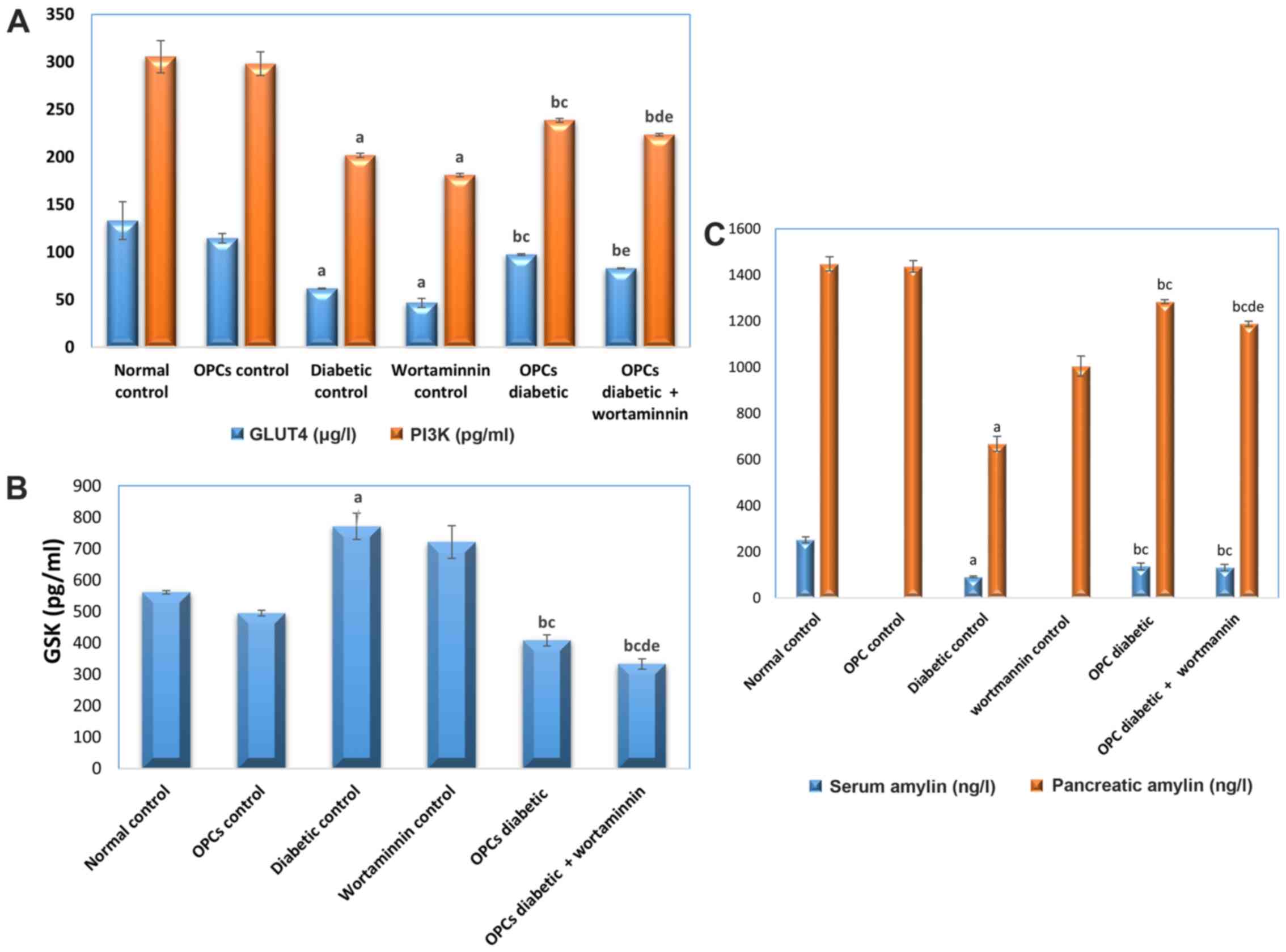 | Figure 1Effect on (A) muscular PI3K and GLUT4
(B) liver GSK (C) serum and pancreatic amylin levels in the
different studied groups. Data are presented as the mean ± SEM,
n=8-10 rats per group. The small letters on the top of the bars in
the figure indicate significant differences as follows: a,
significant difference vs. normal control (P<0.001); b,
significant difference vs. OPC control (P<0.001); c, significant
difference vs. diabetic control (P<0.001); d, significant
difference vs. OPC diabetic group (P<0.001); e, significant
difference vs. wortmannin control (P<0.001). OPC, oligomeric
proanthocyanidin; PI3K, phosphatidylinositol 3-kinase; GLUT4,
glucose transporter 4; GSK-3, glycogen synthase kinase-3. |
Effect of OPC on liver GSK-3 in the
studied groups
The diabetic control group exhibited a significant
increase in the liver GSK-3 level compared with the normal control
group. The OPC diabetic group exhibited a significant decrease
(P<0.001) in the GSK-3 level (-17.53 and -48.08%, respectively)
compared with the OPC control and diabetic control groups. The OPC
diabetic + wortmannin group exhibited a significant decrease
(P<0.001) in the GSK-3 level (-32.64, -56.78 and -53.76%,
respectively) compared with the OPC, diabetic and wortmannin
control groups (Fig. 1B).
Effect of OPC on serum and pancreatic
amylin levels in the studied groups
The diabetic control group exhibited a significant
decrease (P<0.001) in the serum amylin level (-63.78%) compared
with the normal control group. Both the OPC diabetic and OPC
diabetic + wortmannin groups exhibited an increase (P<0.001) in
the serum amylin level (+49.95 and +44.81%, respectively) compared
with the diabetic control group (Fig.
1C).
Compared with the normal control and wortmannin
control groups, the diabetic control group exhibited a significant
decrease (P<0.001) in the amylin level in pancreatic tissue
(-53.87 and -33.49%, respectively). The OPC diabetic group
exhibited a significant decrease (P<0.001) in the pancreatic
amylin level (-10.63%) compared with the OPC control group and a
significant increase (P<0.001) in the pancreatic amylin level
(+92.33%) compared with the diabetic control group. The OPC
diabetic + wortmannin group exhibited a significant increase
(P<0.001) in the pancreatic amylin level (+77.97 and +18.36%,
respectively) compared with the diabetic control and wortmannin
control group and a significant decrease (P<0.001; -17.31 and
-7.46%, respectively) compared with the OPC control and OPC
diabetic groups (Fig. 1C).
Discussion
Diabetes mellitus is one of the most prevalent
chronic disease. It develops due to metabolic dysregulation,
impaired insulin production, or insensitivity of insulin receptors
(24). It is characterized by
hyperglycemia, abnormal lipid profile and inappropriate consumption
of glucose in addition to increased reactive oxygen species (ROS)
production and altered insulin signaling and ROS-induced cellular
damage that leads to severe microvascular and macrovascular
secondary complications (25).
However, the use of oral anti-hyperglycemic agents
for glycemic control has several limitations and severe adverse
effects; this has led diabetic patients to use natural products
with valuable therapeutic efficacy without side-effects (26). The present study investigated the
anti-diabetic and anti-hyperlipidemic properties of
proanthocyanidins in rats with STZ-induced diabetes and aimed to
elucidate the underlying molecular mechanisms of proanthocyanidins
regarding its effects on glucose, lipid metabolism and insulin
signaling.
In the present study, T2D was induced in rats by an
i.p. injection of STZ to mimic the metabolic characteristics of
T2D. The findings revealed a significant increase in blood glucose,
TC, TG and LDL-C levels, along with a significant decrease in
insulin level and HDL-C levels in the diabetic control group, as
previously reported (17).
The dysregulation of lipid metabolism is common
among diabetic patients where the excessive production of free
fatty acids and the induction of endocrine factors leads to a
decrease in the biological activity of insulin and insulin
sensitivity. Therefore, a disruption in lipid profiles is
considered a predictor of T2D development (27). The findings of the present study
demonstrated a significant increase in TC, TG and LDL-C levels,
along with a significant decrease in HDL-C levels in the diabetic
control group. By contrast, an improved lipid profile was observed
in the OPC diabetic group, which exhibited a significant decrease
in TC, TG, LDL-C, and a significant increase in HDL-C compared with
the normal control, OOC control and diabetic control groups.
The physiological activities of insulin are exerted
through the post-insulin receptor cascade after binding insulin to
its receptors on the surface of cell membranes of target cells. The
PI3K/Akt signaling pathway is the dominant regulator where the
activation of PI3K, an essential protein involved in glucose
metabolism, increases the levels of phosphorylated Akt, which
activates downstream signaling molecules, such as GSK-3β and GLUT4.
Therefore, the PI3K/Akt signaling pathway mediates glucose uptake
and intracellular glycogen synthesis in skeletal muscle tissues,
glucose dysplasia and glucose output in the liver. The inhibition
of the PI3K signaling pathway blocks glucose transportation to the
plasma membrane (28). It has been
previously reported that the dysregulation of the PI3K/Akt
signaling pathway can lead to impaired glucose and lipid metabolism
and eventually, to insulin resistance (29).
Wortmannin is a potent inhibitor of PI3K, which
suppresses the PI3K/Akt signaling pathway. It has also been
previously reported that the insulin-mediated induction of GSK-3β
and insulin receptor substrate-1 is suppressed by wortmannin
(30). In the present study,
wortmannin was used to investigate the molecular mechanisms of OPCs
as anti-diabetic agents in insulin signaling in the presence or
absence of wortmannin, where the levels of glucose
metabolism-associated proteins, including GSK-3β, PI3K and GLUT4
were evaluated.
The present study demonstrated that the OPC +
wortmannin group exhibited a significant increase in HDL-C levels,
and a decrease in glucose, TG, TC and LDL-C levels compared with
the wortmannin control group. The wortmannin control group
exhibited a significant increase in glucose, TG, TC and LDL-C
levels, and a decrease in HDL-C levels compared with the normal
control group. The OPC + wortmannin group rats exhibited a
significant increase in PI3K and GLUT4 levels compared with the
wortmannin control group.
The data of the present study also demonstrated that
the diabetic control group exhibited a significant increase in
liver GSK-3 level compared with the normal control group. The OPC
diabetic group exhibited a significant decrease in the liver GSK-3
level compared with OPC control and diabetic control groups. The
OPC diabetic + wortmannin group exhibited a significant decrease
compared to the OPC control, diabetic control and wortmannin
control groups.
The mechanisms underlying the effects of OPC extract
on the insulin signaling pathway were also investigated. The
results revealed that OPC exerted its hypoglycemic effects through
the activation of the PI3K/Akt pathway and the stimulation of GLUT4
translocation, which in turn enhanced glucose transport and
cellular uptake, as well as lipid metabolism in addition to reduced
glycogenesis (GSK-3β), which is in line with the findings of
previous studies (31-33).
Amylin is co-secreted with insulin by pancreatic
β-cells and contributes to blood glucose homeostasis along with
insulin through complex neuronal, as well as endocrine pathways.
The present study revealed a significant decrease in the amylin
level in the diabetic group vs. the normal control group; however,
the OPC diabetic groups with or without wortmannin treatment
exhibited a significant increase in amylin levels compared to the
diabetic group. These results are supported by the findings of
previous studies that demonstrated that the induction of diabetes
by STZ in rats resulted in the loss of the ability to secrete
amylin (34,35).
The present study had some limitations. First, the
effect of proanthocyanidins on the phosphorylation of Akt need to
be investigated. Second, the present study only evaluated the
expression of components on insulin signal pathway, but did not
evaluate their activities. Therefore, additional studies are
required to determine the underlying mechanism responsible for the
anti-diabetic action of OPC extract on the insulin signaling
pathway.
In conclusion, the findings of the present study
provided a biochemical basis for the beneficial use of OPCs for
glycemic control in diabetics. In the present study, treatment with
OPC showed anti-diabetic action by significantly decreasing the
blood glucose level and increasing the insulin level through the
regulation of the PI3K signaling pathway, increasing insulin
receptor downstream proteins, and decreasing glycogenesis (GSK-3).
Moreover, OPC also exerted anti-hyperlipidemic effects by
decreasing lipid profile levels. These results thus suggest that
OPC as a cinnamon extract has potential for use in diabetic
patients.
Acknowledgements
Not applicable.
Funding
Funding: No funding was received.
Availability of data and materials
The datasets used and/or analyzed during the current
study are available from the corresponding author on reasonable
request.
Authors' contributions
NEEA contributed to the conception and the design of
the study, supervised the practical work, and revised and edited
the manuscript. EGK was involved in the conception and design of
the study, revised the manuscript and analyzed the data. NHA
conducted the experiments for the study. AOI was involved in the
writing of the manuscript, and in the statistical analysis of the
data. NHA and AOI verify the authenticity of the raw data. All
authors have read and approved the final manuscript.
Ethics approval and consent to
participate
The present study was conducted following the
institutional guidelines for care and use of laboratory animals
approved by the local Ethics Committee of the Faculty of Pharmacy,
Tanta University, Tanta, Egypt (approval no. 18112014).
Patient consent for publication
Not applicable.
Competing interests
The authors declare that they have no competing
interests.
References
|
1
|
Petersmann A, Nauck M, Müller-Wieland D,
Kerner W, Müller UA, Landgraf R, Freckmann G and Heinemann L:
Definition, classification and diagnosis of diabetes mellitus. Exp
Clin Endocrinol Diabetes. 126:406–410. 2018.PubMed/NCBI View Article : Google Scholar
|
|
2
|
Burgos-Morón E, Abad-Jiménez Z, Marañón
AM, Iannantuoni F, Escribano-López I, López-Domènech S, Salom C,
Jover A, Mora V, Roldan I, et al: Relationship between oxidative
stress, er stress, and inflammation in type 2 diabetes: The battle
continues. J Clin Med. 8(1385)2019.PubMed/NCBI View Article : Google Scholar
|
|
3
|
Moldogazieva NT, Mokhosoev IM, Mel'nikova
TI, Porozov YB and Terentiev AA: Oxidative stress and advanced
lipoxidation and glycation end products (ALEs and AGEs) in aging
and age-related diseases. Oxid Med Cell Longev.
2019(3085756)2019.PubMed/NCBI View Article : Google Scholar
|
|
4
|
Asthana S, Mallick B, Alexandrescu AT and
Jha S: IAPP in type II diabetes: Basic research on structure,
molecular interactions, and disease mechanisms suggests potential
intervention strategies. Biochim Biophys Acta Biomembr.
1860:1765–1782. 2018.PubMed/NCBI View Article : Google Scholar
|
|
5
|
Hieronymus L and Griffin S: Role of amylin
in type 1 and type 2 diabetes. Diabetes Educ. 41 (Suppl 1):47S–56S.
2015.PubMed/NCBI View Article : Google Scholar
|
|
6
|
Takenaka N, Araki N and Satoh T:
Involvement of the protein kinase Akt2 in insulin-stimulated Rac1
activation leading to glucose uptake in mouse skeletal muscle. PLoS
One. 14(e0212219)2019.PubMed/NCBI View Article : Google Scholar
|
|
7
|
Chadt A and Al-Hasani H: Glucose
transporters in adipose tissue, liver, and skeletal muscle in
metabolic health and disease. Pflugers Arch. 472:1273–1298.
2020.PubMed/NCBI View Article : Google Scholar
|
|
8
|
Pearce NJ, Arch JR, Clapham JC, Coghlan
MP, Corcoran SL, Lister CA, Llano A, Moore GB, Murphy GJ, Smith SA,
et al: Development of glucose intolerance in male transgenic mice
overexpressing human glycogen synthase kinase-3beta on a
muscle-specific promoter. Metabolism. 53:1322–1330. 2004.PubMed/NCBI View Article : Google Scholar
|
|
9
|
MacAulay K and Woodgett JR: Targeting
glycogen synthase kinase-3 (GSK-3) in the treatment of Type 2
diabetes. Expert Opin Ther Targets. 12:1265–1274. 2008.PubMed/NCBI View Article : Google Scholar
|
|
10
|
Salehi B, Ata A, V Anil Kumar N, Sharopov
F, Ramírez-Alarcón K, Ruiz-Ortega A, Abdulmajid Ayatollahi S, Tsouh
Fokou PV, Kobarfard F, Amiruddin Zakaria Z, et al: Antidiabetic
potential of medicinal plants and their active components.
Biomolecules. 9(551)2019.PubMed/NCBI View Article : Google Scholar
|
|
11
|
Dorri M, Hashemitabar S and Hosseinzadeh
H: Cinnamon (Cinnamomum zeylanicum) as an antidote or a protective
agent against natural or chemical toxicities: A review. Drug Chem
Toxicol. 41:338–351. 2018.PubMed/NCBI View Article : Google Scholar
|
|
12
|
Cao H, Ou J, Chen L, Zhang Y, Szkudelski
T, Delmas D, Daglia M and Xiao J: Dietary polyphenols and type 2
diabetes: Human study and clinical trial. Crit Rev Food Sci Nutr.
59:3371–3379. 2019.PubMed/NCBI View Article : Google Scholar
|
|
13
|
Sun C, Zhao C, Guven EC, Paoli P,
Simal-Gandara J, Ramkumar KM, Wang S, Buleu F, Pah A, Turi V, et
al: Dietary polyphenols as antidiabetic agents: Advances and
opportunities. Food Frontiers. 1:18–44. 2020.
|
|
14
|
Wang TK, Xu S, Li S and Zhang Y:
Proanthocyanidins should be a candidate in the treatment of cancer,
cardiovascular diseases and lipid metabolic disorder. Molecules.
25(5971)2020.PubMed/NCBI View Article : Google Scholar
|
|
15
|
Koutsos A, Riccadonna S, Ulaszewska MM,
Franceschi P, Trošt K, Galvin A, Braune T, Fava F, Perenzoni D,
Mattivi F, et al: Two apples a day lower serum cholesterol and
improve cardiometabolic biomarkers in mildly hypercholesterolemic
adults: A randomized, controlled, crossover trial. Am J Clin Nutr.
111:307–318. 2020.PubMed/NCBI View Article : Google Scholar
|
|
16
|
Yokozawa T, Cho EJ, Park CH and Kim JH:
Protective effect of proanthocyanidin against diabetic oxidative
stress. Evid Based Complement Alternat Med.
2012(623879)2012.PubMed/NCBI View Article : Google Scholar
|
|
17
|
Mabhida SE, Johnson R, Ndlovu M, Louw J,
Opoku A and Mosa RA: Molecular basis of the anti-hyperglycemic
activity of RA-3 in hyperlipidemic and streptozotocin-induced type
2 diabetes in rats. Diabetol Metab Syndr. 11(27)2019.PubMed/NCBI View Article : Google Scholar
|
|
18
|
Cheung DY, Kim JI, Park SH and Kim JK:
Proanthocyanidin from grape seed extracts protects
indomethacin-induced small intestinal mucosal injury. Gastroenterol
Res Pract. 2014(618068)2014.PubMed/NCBI View Article : Google Scholar
|
|
19
|
Qin B, Nagasaki M, Ren M, Bajotto G,
Oshida Y and Sato Y: Cinnamon extract (traditional herb)
potentiates in vivo insulin-regulated glucose utilization via
enhancing insulin signaling in rats. Diabetes Res Clin Pract.
62:139–148. 2003.PubMed/NCBI View Article : Google Scholar
|
|
20
|
Trinder P: Determination of blood glucose
using an oxidase-peroxidase system with a non-carcinogenic
chromogen. J Clin Pathol. 22:158–161. 1969.PubMed/NCBI View Article : Google Scholar
|
|
21
|
Savoldi R, Prandini BD and Donisi C:
Enzymatic determination of total serum cholesterol by
4-aminophenazone-phenol: Manual and automatic method (author's
translation). Quad Sclavo Diagn. 12:238–247. 1976.PubMed/NCBI(In Italian).
|
|
22
|
Nagele U, Hagele EO, Sauer G, Wiedemann E,
Lehmann P, Wahlefeld AW and Gruber W: Reagent for the enzymatic
determination of serum total triglycerides with improved lipolytic
efficiency. J Clin Chem Clin Biochem. 22:165–174. 1984.PubMed/NCBI View Article : Google Scholar
|
|
23
|
Austin GE, Maznicki E and Sgoutas D:
Comparison of phosphotungstate and dextran sulfate-Mg2+
precipitation procedure for determination of high density
lipoprotein cholesterol. Clin Biochem. 17:166–169. 1984.PubMed/NCBI View Article : Google Scholar
|
|
24
|
Silva JAD, Souza ECF, Echazú Böschemeier
AG, Costa CCMD, Bezerra HS and Feitosa EELC: Diagnosis of diabetes
mellitus and living with a chronic condition: Participatory study.
BMC Public Health. 18(699)2018.PubMed/NCBI View Article : Google Scholar
|
|
25
|
Jiang S, Young JL, Wang K, Qian Y and Cai
L: Diabetic-induced alterations in hepatic glucose and lipid
metabolism: The role of type 1 and type 2 diabetes mellitus
(Review). Mol Med Rep. 22:603–611. 2020.PubMed/NCBI View Article : Google Scholar
|
|
26
|
Zonszein J and Groop PH: Strategies for
diabetes management: Using newer oral combination therapies early
in the disease. Diabetes Ther. 7:621–639. 2016.PubMed/NCBI View Article : Google Scholar
|
|
27
|
I S, Sobczak A, A Blindauer C and J
Stewart A: Changes in plasma free fatty acids associated with
type-2 diabetes. Nutrients. 11(2022)2019.PubMed/NCBI View Article : Google Scholar
|
|
28
|
Huang X, Liu G, Guo J and Su Z: The
PI3K/AKT pathway in obesity and type 2 diabetes. Int J Biol Sci.
14:1483–1496. 2018.PubMed/NCBI View Article : Google Scholar
|
|
29
|
Yang H, Cao Q, Xiong X, Zhao P, Shen D,
Zhang Y and Zhang N: Fluoxetine regulates glucose and lipid
metabolism via the PI3K-AKT signaling pathway in diabetic rats. Mol
Med Rep. 22:3073–3080. 2020.PubMed/NCBI View Article : Google Scholar
|
|
30
|
Maffei A, Lembo G and Carnevale D:
PI3Kinases in diabetes mellitus and its related complications. Int
J Mol Sci. 19(4098)2018.PubMed/NCBI View Article : Google Scholar
|
|
31
|
Yogalakshmi B, Bhuvaneswari S, Sreeja S
and Anuradha CV: Grape seed proanthocyanidins and metformin act by
different mechanisms to promote insulin signaling in rats fed high
calorie diet. J Cell Commun Signal. 8:13–22. 2014.PubMed/NCBI View Article : Google Scholar
|
|
32
|
Santos HO and da Silva GAR: To what extent
does cinnamon administration improve the glycemic and lipid
profiles? Clin Nutr ESPEN. 27:1–9. 2018.PubMed/NCBI View Article : Google Scholar
|
|
33
|
Sun Q, Jia N, Li X, Yang J and Chen G:
Grape seed proanthocyanidins ameliorate neuronal oxidative damage
by inhibiting GSK-3β-dependent mitochondrial permeability
transition pore opening in an experimental model of sporadic
Alzheimer's disease. Aging (Albany NY). 11:4107–4124.
2019.PubMed/NCBI View Article : Google Scholar
|
|
34
|
Ogawa A, Harris V, McCorkle SK, Unger RH
and Luskey KL: Amylin secretion from the rat pancreas and its
selective loss after streptozotocin treatment. J Clin Invest.
85:973–976. 1990.PubMed/NCBI View Article : Google Scholar
|
|
35
|
Zhang XX, Pan YH, Huang YM and Zhao HL:
Neuroendocrine hormone amylin in diabetes. World J Diabetes.
7:189–197. 2016.PubMed/NCBI View Article : Google Scholar
|


