Introduction
Latent autoimmune diabetes in adults (LADA) is an
adult-onset form of autoimmune diabetes combining features of both
type 1 and type 2 diabetes. Defined by islet autoantibodies, most
commonly glutamic acid decarboxylase (GAD) antibodies, it presents
later in life and progresses more slowly toward insulin dependence,
leading to frequent misclassification as type 2 diabetes mellitus
(T2DM). LADA is not rare, accounting for 2-12% of adult-onset
diabetes' however, it remains under-recognised; diagnostic delays
result in prolonged hyperglycaemia, inappropriate treatment and
accelerated β-cell decline. Adults with atypical glycaemic
patterns, the rapid loss of glycaemic control, or coexisting
autoimmune disease should be evaluated using islet autoantibodies
and C-peptide testing (1). LADA
exists on a spectrum from classic autoimmune diabetes to a
T2DM-like phenotype, rendering diagnosis challenging, particularly
in individuals retaining β-cell function at onset or presenting
with obesity or insulin resistance (2). Early insulin initiation may preserve
endogenous secretion (3), and
guidelines support C-peptide-guided treatment decisions (4).
The present case report describes the case of a
39-year-old man with long-standing, severely uncontrolled diabetes
of initially undetermined type who presented with chronic watery
diarrhoea, significant unintentional weight loss, persistent
hyperglycaemia and a pruritic vesicular rash, a constellation that
prompted evaluation for neuroendocrine tumour, Crohn's disease and
malabsorption syndromes before the underlying autoimmune diabetes
was recognised.
Case report
A 39-year-old man with diabetes of unknown type,
hypertension and chronic medication non-adherence presented at the
Wellstar Spalding Medical Center (Griffin, GA, USA) with a 3-month
history of diffuse watery diarrhoea, nausea, vomiting and severe
cramping abdominal pain. Symptoms had worsened over the preceding
year, accompanied by profound unintentional weight loss from 113.4
kg (250 lb) to 57.2 kg (126 lb) over a period of 12 months,
accelerating over the final 6 months. Collateral history from his
mother confirmed he had been well until ~1 year earlier. He
reported a pruritic vesicular rash beginning on the buttocks that
spread to the left leg before partially resolving and persistent
angular lip fissures. He denied fevers, chest pain, dyspnoea and
substance use.
Of note, 1 year earlier, he had been hospitalised
with similar symptoms, treated as colitis with intravenous fluids
and a 7-day course of intravenous antibiotics and corticosteroids.
His HbA1c was then >15%, and he was discharged on insulin. His
family history included a father with severe uncontrolled diabetes
and a brother with thyroid disease. Home medications were insulin
glargine at 20 units, sliding-scale insulin, pantoprazole,
ondansetron, cetirizine and hyoscyamine. He has a documented
allergy to penicillins.
Upon admission he was afebrile (37.0˚C), with a
blood pressure of 108/74 mmHg, a heart rate of 80 beats/min, a
respiratory rate of 15/min and an oxygen saturation of 100% in room
air. His height was 1.778 m and his weight was 57.2 kg, with a body
mass index of 18.1 kg/m2, reduced from an estimated 35.9
kg/m2 at his pre-illness weight and representing a loss
of ~50% of total body weight. He appeared thin, cachectic and ill,
but non-toxic, alert and oriented. The oral mucosa was dry with
bilateral angular fissures and no intra-oral lesions. A cardiac
examination revealed normal heart sounds without murmurs, 2+
pulses, and no oedema; the lungs were clear. The abdomen was soft
with diffuse mild-to-moderate tenderness, no rebound or guarding
and normal bowel sounds. A skin examination revealed erythematous
plaques over the buttocks with scattered vesicles, crusted lesions
and excoriations without cellulitis; the left leg showed no active
rash. Acanthosis nigricans was absent. A neurological examination
was intact, and he was tearful with a depressed affect, but
cooperative.
A timeline summarising the key clinical events from
initial symptom onset through hospitalisation and discharge is
presented in Table I.
 | Table ITimeline of key clinical events. |
Table I
Timeline of key clinical events.
| Timeline | Clinical event |
|---|
| Age, 30s | Onset of diabetes
symptoms; classified as type 2 diabetes mellitus; started on oral
hypoglycaemic agents |
| 1 year before index
admission | First
hospitalisation: one month of crampy abdominal pain, nausea,
vomiting, diarrhoea; treated for colitis with IV fluids,
antiemetics, analgesics, 7-day course of IV antibiotics and
corticosteroids; HbA1c >15%; discharged on insulin regimen
(glargine 20 units plus sliding scale) |
| 6 months befor index
admission | Acceleration of
progressive unintentional weight loss (113.4 kg → 57.2 kg; 250 lb →
126 lb over 12 months, with marked acceleration over the final 6
months) |
| 3 months before index
admission | Onset of diffuse
watery diarrhoea, worsening nausea, vomiting, and severe cramping
abdominal pain; pruritic vesicular rash appears on buttocks,
spreads to left leg, then partially resolves; persistent angular
fissures at lip corners |
| Day 0 (Index
admission) | Admitted with
hyperosmolar hyperglycaemic state: glucose 740 mg/dl (41.1 mmol/l),
HbA1c 14.7%, β-hydroxybutyrate 0.80 mmol/l, normal anion gap,
sodium 124 mmol/l (pseudohyponatremia); started on IV insulin
infusion at 0.1 units/kg/h and isotonic fluids |
| Days 1-3 | Endocrine workup:
C-peptide 0.12 nmol/l, glucagon <6 ng/l, proinsulin-to-C-peptide
ratio 6.4%; islet autoantibodies positive (GAD-65 33 IU/ml, ICA
1:8, ZnT8 20 U/ml); thyroid, parathyroid, and adrenal studies
normal; faecal elastase 47 µg/g; tumour markers unremarkable except
mildly elevated CEA; short trial of octreotide initiated
(transiently lowered glucose; discontinued when hormonal workup did
not support neuroendocrine tumour) |
| Days 3-5 | CT abdomen/pelvis:
diffuse sigmoid wall thickening suggestive of prior colitis,
nonspecific small bowel wall thickening; CT enterography:
nondistended small bowel with mild non-specific thickening; MRCP
and MRI abdomen: no biliary, pancreatic, or mass lesion |
| Days 5-7 | Colonoscopy with
biopsy: Mild nonspecific active terminal ileitis, otherwise normal
colonic biopsies; stool studies (GI multiplex PCR, C. difficile
toxin, ova and parasites): negative; Saccharomyces IgA/IgG:
positive; faecal calprotectin 38 µg/g; CRP <0.3 mg/dl |
| Days 5-10 | Gastroenterology
commenced empirical therapy for presumptive Crohn's disease: IV
methylprednisolone followed by oral prednisone taper, mesalamine
2.4 g daily, dicyclomine as needed, amitriptyline 50 mg nightly;
pancrelipase continued |
| Days 7-10 | Transitioned from IV
insulin to subcutaneous NPH and regular insulin 40 units twice
daily; diarrhoea improved with hydration and glycaemic control;
rash resolved with topical emollients; nutritional counselling,
diabetes education, and home glucose monitoring instructions
provided |
| Discharge | Diagnosis confirmed:
LADA presenting with hyperosmolar hyperglycaemic state; glucose
values 150-250 mg/dl on NPH/regular insulin; improved appetite and
energy; discharged on NPH/regular insulin 40 units twice daily and
supportive medications |
| Post-discharge | Outpatient follow-up
arranged for reassessment of glycaemic control, repeat HbA1c,
weight monitoring, and evaluation of GI symptoms; patient did not
attend; no long-term data obtainable |
Investigation
The initial evaluation revealed severe
hyperglycaemia (glucose, 740 mg/dl; 41.1 mmol/l); HbA1c, 14.7%; and
sodium, 124 mmol/l consistent with pseudohyponatremia.
β-hydroxybutyrate was mildly elevated (0.80 mmol/l) with a normal
anion gap, consistent with a hyperosmolar hyperglycaemic state
(HHS) without significant ketoacidosis. An endocrine workup
revealed markedly reduced C-peptide (0.12 nmol/l), low glucagon
(<6 ng/l) and positive islet autoantibodies (GAD-65, 33 IU/ml;
islet cell, 1:8; ZnT8, 20 U/ml), confirming autoimmune aetiology.
The proinsulin-to-C-peptide ratio of 6.4% indicated stressed, yet
persisting β-cell function. Thyroid, parathyroid and adrenal tests
yielded normal results, and 21-hydroxylase antibody was negative.
Stool pancreatic elastase was low (47 µg/g). The analyses of tumour
markers did not reveal any notable findings, apart from mildly
elevated carcinoembryonic antigen and a borderline vasoactive
intestinal peptide obtained following octreotide exposure. The
levels of inflammatory markers were low, with C-reactive protein
<0.3 mg/dl and faecal calprotectin at 38 µg/g. Despite positive
Saccharomyces cerevisiae IgA and IgG, gastrointestinal
evaluation revealed no definitive evidence of inflammatory bowel
disease. Infectious studies (gastrointestinal multiplex polymerase
chain reaction, Clostridioides difficile toxin, ova and
parasites, human immunodeficiency virus, hepatitis serologies,
QuantiFERON-TB Gold Plus) and immunoglobulin G4 were negative. The
laboratory findings are summarised in Table II.
| Table IISummary of laboratory findings and
infectious aetiology workup. |
Table II
Summary of laboratory findings and
infectious aetiology workup.
| Test | Result (SI
units) | Reference range (SI
units) | Interpretation |
|---|
| Sodium | 124 mmol/l | 135-145 mmol/l | Low |
| Potassium | 3.8 mmol/l | 3.5-5.0 mmol/l | Normal |
| Chloride | 96 mmol/l | 98-107 mmol/l | Low |
| Bicarbonate | 19 mmol/l | 23-30 mmol/l | Low |
| WBC |
5.92x10⁹/l |
4-10x109/l | Normal |
| Glucose | 41.1 mmol/l | <11.1 mmol/; | High |
| Anion Gap | 13 mmol/l | 12-16 mmol/; | Normal |
|
Beta-hydroxybutyrate | 0.80 mmol/l | <0.5 mmol/l | High |
| HbA1c | 14.7% | <5.7% | High |
| C-reactive
protein | <0.3 mg/dl | <0.5 mg/dl | Normal |
| Albumin | 3.3 g/dl | 3.5-5.0 g/dl | Low |
| Fecal
calprotectin | 38 µg/g | <50 µg/g | Normal |
| Glucagon | <6 ng/l | 11-78 ng/l | Low |
| C-peptide | 0.12 nmol/l | 0.26-1.28 nmol/l | Low |
| Proinsulin | 7.7 pmol/l | ≤18.8 pmol/l | Normal |
|
Proinsulin-to-C-peptide ratio | 6.4% | - | Residual β-cell
function |
| GAD-65 Ab | 33 IU/ml | <5 IU/ml | High |
| Islet Cell Ab | 1:8 | <1:4 | High |
| ZnT8 Ab | 20 U/ml | <10 U/ml | High |
| Calcitonin | <2 ng/l | ≤10 ng/l | Normal |
| Chromogranin A | Normal | Per laboratory
reference | Normal |
| Gastrin | Normal | Per laboratory
reference | Normal |
| CA 19-9 | 30.5 U/ml | <47 U/ml | Normal |
| CEA | 7.9 ng/ml | <3 ng/ml | High |
| IgA (total) | 3.99 g/l | 0.7-4.0 g/l | Normal |
| AFP | 1.9 ng/ml | <10 ng/ml | Normal |
| VIP | 47 ng/l | <36 ng/l | High |
| Pancreatic elastase
(stool) | 47 µg/g | >200 µg/g | Low |
| NSE | 8 ng/ml | <14 ng/ml | Normal |
| IGF-1 | 52 µg/l | 53-331 µg/l | Low |
| ACTH | 18 ng/l | 6-50 ng/l | Normal |
| Prolactin | Normal | Per laboratory
reference | Normal |
| Gliadin IgA | 2 U/ml | <7 U/ml | Normal |
| ANA | Negative | Negative | Normal |
| ANCA | Negative | Negative | Normal |
| 21-Hydroxylase
Ab | Negative | Negative | Normal |
| Immunoglobulin
G4 | Negative | Per laboratory
reference | Normal |
| TSH | 2.75 mIU/l | 0.4-4.0 mIU/l | Normal |
| PTH | 25 pg/ml | 10-55 pg/ml | Normal |
| Vitamin D
25-OH | 67.5 nmol/l | 75-250 nmol/l | Low |
| Vitamin B12 | 729 ng/l | 139-619 ng/l | High |
| Folate | >45 nmol/l | 7-34 nmol/l | High |
| Zinc | 5.96 µmol/l | 9.2-20 µmol/l | Low |
| Saccharomyces
cerevisiae Ab IgA | 66.8 U/ml | >25 U/ml | Elevated |
| Saccharomyces
cerevisiae Ab IgG | 67.5 U/ml | >30 U/ml | Elevated |
| HIV 1/2
Antigen/Antibody | Non-reactive | Non-reactive | Normal |
| Hepatitis A (IgM)
Ab | Non-reactive | Non-reactive | Normal |
| Hepatitis B Core
(IgM) Ab | Non-reactive | Non-reactive | Normal |
| Hepatitis B Surface
Ag | Non-reactive | Non-reactive | Normal |
| Hepatitis C Ab | Non-reactive | Non-reactive | Normal |
| GI Multiplex
PCR | No pathogens
detected | Negative | Normal |
| Clostridioides
difficile toxin PCR | Negative | Negative | Normal |
| Stool ova and
parasites | Negative | Negative | Normal |
| Stool
leukocytes | Negative | Negative | Normal |
| QuantiFERON-TB Gold
Plus | Negative | Negative | Normal |
Imaging
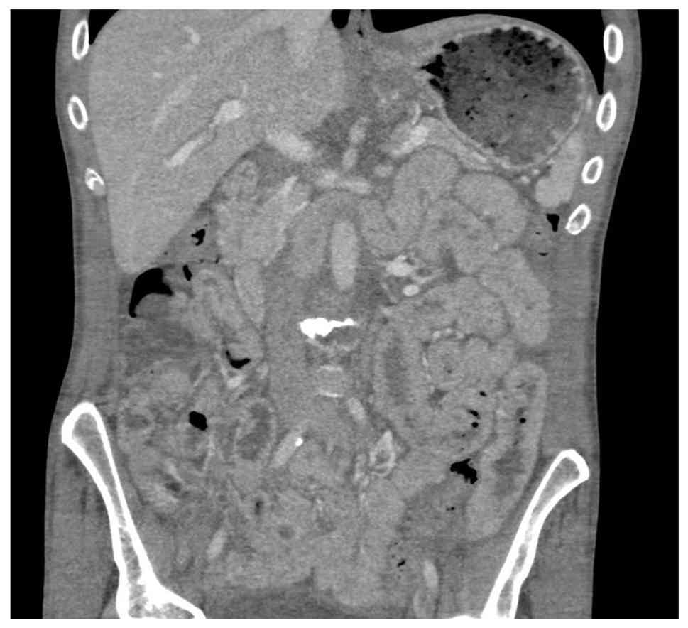
A computed tomography scan demonstrated diffuse
sigmoid colonic wall thickening suggestive of prior colitis with
non-specific small bowel wall thickening, as shown in Fig. 1. Computed tomographic enterography
revealed a non-distended small bowel with mild non-specific small
bowel and left colonic thickening. Magnetic resonance
cholangiopancreatography revealed no biliary or pancreatic
abnormality, and magnetic resonance imaging of the abdomen revealed
no mass. A colonoscopic biopsy of the terminal ileum demonstrated
mild non-specific active ileitis with otherwise normal colonic
biopsies. Prior endoscopic biopsies revealed normal duodenal and
colonic mucosa, with squamocolumnar junction inflammation and
intestinal metaplasia without dysplasia.
Differentials
Chronic watery diarrhoea, ~50% weight loss, a
vesicular rash and severe hyperglycaemia prompted the evaluation of
endocrine, gastrointestinal, infectious, inflammatory, and
neoplastic aetiologies (Table
III).
| Table IIIStructured differential diagnosis:
Supporting and opposing evidence. |
Table III
Structured differential diagnosis:
Supporting and opposing evidence.
| Diagnosis | Supporting
evidence | Opposing evidence
(Refs.) | Conclusion
(Refs.) |
|---|
| Type 2 diabetes
mellitus | Adult onset;
obesity at diagnosis (BMI 35.9 kg/m2); paternal history
of severe diabetes; initial non-insulin-requiring course | C-peptide 0.12
nmol/l (reference, 0.26-1.28); triple islet autoantibody positivity
(GAD-65 33 IU/ml, ICA 1:8, ZnT8 20 U/ml); progressive glycaemic
failure despite escalating therapy; no metabolic syndrome features
at presentation | Excluded |
| Classic type 1
diabetes mellitus | Triple islet
autoantibody positivity; near-absent C-peptide; insulin dependence;
sibling with thyroid disease | Obesity at
diagnosis; documented non-insulin-requiring interval of several
years; hyperosmolar hyper-glycaemic state rather than ketoacidosis
(β-hydroxybutyrate 0.80 mmol/l, anion gap 13 mmol/l, urine ketones
negative, urine glucose 3+), indicating partially preserved insulin
action; proinsulin-to-C-peptide ratio 6.4% | Excluded; initial
insulin-independent phase discriminates LADA per Buzzetti consensus
criteria (7) |
| Crohn's
disease | ASCA IgA 66.8 U/ml
and IgG 67.5 U/ml (both elevated); crampy abdominal pain with
weight loss; CT small-bowel and sigmoid wall thickening; mild
nonspecific active ileitis on terminal ileal biopsy; prior
admission treated as colitis; empirical corticosteroid and
mesalamine therapy administered | Faecal calprotectin
38 µg/g and CRP <0.3 mg/dl, both normal; no ulceration,
cobblestoning, or skip lesions at colonoscopy; no granulomas or
chronic architectural distortion; random colonic biopsies normal on
two occasions; ANCA negative; CT enterography revealed non-specific
thickening without stricture or fistula; ASCA has limited positive
predictive value in isolation | Not established;
empirical treatment given on the basis of biopsy-proven mild
ileitis. Because immunosuppression was administered concurrently
with insulin, relative contribution of each to symptom improvement
cannot be separated |
| Neuroendocrine
tumour (VIPoma, carcinoid, gastrinoma, glucagonoma) | Secretory-pattern
high-volume watery diarrhoea; ~50% weight loss; VIP mildly elevated
at 47 ng/l (reference, <36); CEA 7.9 ng/ml (reference, <3);
transient symptomatic response to octreotide | Glucagon <6
ng/l, gastrin normal, chromogranin A normal, calcitonin <2 ng/l,
NSE 8 ng/ml, CA 19-9 30.5 U/ml, AFP 1.9 ng/ml, ACTH 18 ng/l,
proinsulin 7.7 pmol/l all normal or non-diagnostic; no flushing,
peptic ulceration, or necrolytic migratory erythema; CT, CT
enterography, MRCP, and MRI abdomen without pancreatic or luminal
lesion | Excluded; modest
VIP elevation attributed to dehydration, catabolic state, and prior
octreotide exposure (somatostatin receptor imaging not obtained;
repeat VIP after adequate washout not performed) |
| Exocrine pancreatic
insufficiency/ malabsorption | Chronic diarrhoea;
~50% weight loss; faecal elastase 47 µg/g (severe range, reference
>200); angular cheilitis; zinc 5.96 µmol/l (low); vitamin D
25-OH 67.5 nmol/l (low); albumin 3.3 g/dl; IGF-1 52 µg/l (low) | No steatorrhea;
MRCP revealed no ductal stone or biliary abnormality; CT without
pancreatic parenchymal change; no history of pancreatitis, alcohol,
or tobacco use; IgG4 negative; vitamin B12 729 ng/l and folate
>45 nmol/l both elevated, arguing against generalised
small-bowel malabsorption; watery stool falsely lowers faecal
elastase through dilution (13,14) | Not primary
pancreatic disease; low elastase attributed to stool dilution and
loss of islet–acinar trophic support from profound insulinopenia;
pancrelipase continued empirically |
| Celiac disease/
dermatitis herpetiformis | Chronic diarrhoea;
weight loss; angular cheilitis; pruritic vesicular buttock
rash | Normal duodenal
histology at two separate endoscopies; anti-gliadin IgA 2 U/ml
(negative); total IgA 3.99 g/l (normal), excluding IgA-deficiency
false negatives | Excluded (tissue
transglutaminase IgA not obtained; rash not biopsied, so dermatitis
herpetiformis cannot be formally excluded) |
| Infectious
enteritis | Chronic diarrhoea;
prior ciprofloxacin and metronidazole exposure; pro-found weight
loss | GI multiplex PCR
negative for all pathogens; stool ova and parasites negative;
Clostridioides difficile toxin PCR negative; stool leukocytes
negative; HIV-1/2 antigen–antibody non-reactive; hepatitis A IgM,
hepatitis B core IgM, HBsAg, and HCV non-reactive; QuantiFERON-TB
Gold Plus negative | Excluded |
| Microscopic
colitis | Chronic watery
diarrhoea | Random colonic
biopsies normal on two separate occasions, with no lymphocytic or
collagenous changes | Excluded |
|
Hyperthyroidism | Diarrhoea with
weight loss | TSH 2.75 mIU/l
(normal) | Excluded |
Classifying the diabetes was central. Type 2
diabetes, presumed given adult onset, obesity at diagnosis (body
mass index, 35.9 kg/m2) and an initial
non–insulin-requiring course, was excluded by C-peptide at 0.12
nmol/l, triple autoantibody positivity and progressive glycaemic
failure despite escalating therapy. Classic type 1 diabetes was
inconsistent: The patient remained insulin-independent for years
and presented with HHS rather than ketoacidosis (β-hydroxybutyrate,
0.80 mmol/l; anion gap, 13 mmol/l; urine ketones, negative),
indicating partially preserved insulin action. Adult onset,
autoantibody positivity and an initial insulin-independent phase
satisfy the Buzzetti consensus criteria for LADA Crohn's disease,
neuroendocrine tumour, exocrine pancreatic insufficiency, celiac
disease, infectious enteritis, microscopic colitis, and
hyperthyroidism were each considered, with supporting and opposing
evidence presented in Table
III.
Treatment
The patient was treated with intravenous isotonic
fluids and an insulin infusion at 0.1 units/kg/h, with the
correction of pseudohyponatremia. During an evaluation for a
possible secretory neuroendocrine tumour, a short trial of
octreotide transiently lowered glucose, but was discontinued when
hormonal markers (glucagon, gastrin, chromogranin A) and imaging,
including CT enterography and MRI of the abdomen (images not
available) did not support that diagnosis.
Given biopsy-proven mild active terminal ileitis
with compatible symptoms, gastroenterology concurrently treated
presumptive Crohn's disease with intravenous methylprednisolone
followed by an oral prednisone taper and mesalamine 2.4 g daily,
with dicyclomine as needed and amitriptyline 50 mg nightly for
visceral pain. Pancreatic enzyme replacement with pancrelipase was
continued given the low levels of faecal elastase.
Following metabolic stabilisation, he was
transitioned to subcutaneous neutral protamine Hagedorn (NPH) and
regular insulin 40 units twice daily, with improved glycaemic
control. Diarrhoea improved with hydration and glycaemic
correction, and the rash resolved with topical emollients. He
received nutritional counselling, diabetes education and home
glucose monitoring instructions before discharge.
Outcome and follow-up
At discharge after 12 days of inpatient care,
glucose values were consistently 150-250 mg/dl on NPH/regular
insulin, with improved appetite and energy. Outpatient follow-up
was arranged for the reassessment of glycaemic control, repeat
HbA1c, weight monitoring and the evaluation of gastrointestinal
symptoms; however, the patient did not attend, and no long-term
data were obtainable.
Discussion
The present case report highlights the diagnostic
challenge of LADA, which is frequently misclassified as T2DM. The
patient described herein presented with adult-onset diabetes and a
family history suggestive of T2DM and was managed accordingly until
deteriorating glycaemic control prompted re-evaluation, revealing
islet autoantibody positivity. Progressive β-cell failure following
years of presumed T2DM is a well-described trajectory (4).
Under updated American Diabetes Association
guidance, LADA is classified within the type 1 diabetes mellitus
(T1DM) rubric, reflecting autoimmune β-cell destruction (5). Pathogenesis is primarily cell-mediated:
Peripheral blood mononuclear cells suppress insulin secretion, and
dysfunctional regulatory T-cells contribute to loss of immune
tolerance (6). Patients may test
positive for one or more islet autoantibodies, including GAD,
IA-2A, insulin, ZnT8A, or tetraspanin 7 antibodies (7). Consensus criteria generally include
adult-onset diabetes, typically after 30 years of age, islet
autoantibody positivity, and an initial non-insulin-requiring
course. Compared with T2DM, patients generally exhibit fewer
metabolic syndrome features and more rapid C-peptide decline,
although β-cell loss is typically slower than in classic T1DM
(8). The patient described herein
satisfied these criteria.
The antibody panel obtained, GAD-65, islet cell
antibodies and ZnT8, reflects diagnostic yield and local
availability. GAD antibodies are the most sensitive single marker
of adult-onset autoimmune diabetes and may persist longer than
several other islet autoantibodies. Islet cell antibodies, although
less standardized, provide complementary evidence of islet-directed
autoimmunity. ZnT8 antibodies may provide incremental diagnostic
sensitivity and contribute to identification of
multiple-autoantibody positivity, a phenotype associated with more
rapid β-cell decline and earlier insulin requirement. IA-2A was not
obtained; its inclusion could have further refined immunologic
characterization as IA-2A positivity has been shown to be
associated with a more rapidly progressive phenotype and a higher
prevalence of coexisting celiac disease (9,10).
Nonetheless, positivity for three distinct islet autoantibodies
together with a C-peptide of 0.12 nmol/l strongly supports an
autoimmune basis, as multiple-autoantibody positivity provides
greater specificity than any single marker alone.
As gastrointestinal and cutaneous manifestations
predominated rather than classical hyperglycaemic symptoms, several
alternative diagnoses were entertained. Diagnostic confusion arose
as diabetes had already been established and presumed to be type 2;
consequently, the constellation of cachexia, diarrhoea and rash was
initially interpreted as a separate coexisting process, rather than
potentially related to profound insulin deficiency (11). An abrupt classic T1DM presentation
was less consistent with the multi-year non-insulin-requiring
course and presentation with HHS rather than ketoacidosis. The
level of β-hydroxybutyrate was only 0.80 mmol/l and the anion gap
was normal, suggesting sufficient residual insulin action to
suppress substantial ketogenesis. As LADA lies along the autoimmune
T1DM spectrum, the distinction largely reflects the tempo of β-cell
loss and the timing of insulin dependence rather than fundamentally
different pathology.
A neuroendocrine tumour was considered due to weight
loss, secretory-type diarrhoea, mildly elevated carcinoembryonic
antigen and a transient response to octreotide. However, glucagon,
gastrin and chromogranin A levels were normal, imaging demonstrated
no corresponding lesion, and the octreotide response could reflect
its anti-secretory effects rather than tumour-directed activity.
Hyperglycaemia was also better accounted for by insulin deficiency
than by counter-regulatory hormone excess. Crohn's disease was
considered due to positive anti-Saccharomyces antibodies,
crampy abdominal pain, prior colitis, and biopsy-proven mild
ileitis and was treated empirically. However, these antibodies have
limited positive predictive value in isolation, the levels of
inflammatory markers were low, and no skip lesions, ulceration,
granulomata, or transmural inflammation were identified.
Malabsorption syndromes, including celiac disease, were also
considered due to weight loss, diarrhoea, low levels of faecal
elastase and angular cheilitis; however, normal total IgA, negative
gliadin antibodies, normal duodenal histology and the absence of
steatorrhea argued against celiac disease.
A critical caveat applies when interpreting the
clinical improvement. As empirical corticosteroid and mesalamine
therapy was administered concurrently with insulin, improvement
cannot be attributed to glycaemic correction alone. Several
findings nevertheless favour a predominantly metabolic explanation:
Faecal calprotectin and C-reactive protein levels were low,
colonoscopic and histologic abnormalities were mild and
non-specific without diagnostic features of Crohn's disease, and
the degree of cachexia was consistent with profound insulin
deficiency.
Gastrointestinal manifestations are not hallmark
features of LADA, although autoimmune comorbidities, including
thyroid disease, celiac disease and dermatitis herpetiformis, have
been reported, reflecting shared autoimmune predisposition rather
than a direct causal relationship (9,11).
Reported associations are summarized in Table IV. Despite chronic diarrhoea and a
vesicular rash, evaluation did not confirm celiac disease or
another specific enteropathy in the patient in the present case
report.
| Table IVReported associations between
autoimmune disorders and LADA. |
Table IV
Reported associations between
autoimmune disorders and LADA.
| Authors | Linked autoimmune
disorder | Evidence of
association with LADA | Overlapping
pathophysiology |
Autoantibodies/immune markers
reported | (Refs.) |
|---|
| Santoso, et
al | Thyroid autoimmune
disease (Hashimoto thyroiditis, Graves disease) | Thyroid dysfunction
confers approximately 1.88-fold higher odds of LADA compared with
T2DM | Shared genetic
susceptibility within the HLA/MHC region with over-lapping B-cell
and T-cell mediated immune mechanisms | Frequency of
thyroid peroxidase or thyroglobulin antibody positivity
significantly higher in LADA cohorts than in T2DM or controls | (10) |
| Kucera, et
al | Gastrointestinal
auto-immunity, including celiac and gluten-related disease | Increased frequency
of gliadin and endomysial antibodies in LADA compared with
T2DM | Shared
immuno-genetic background affecting both gut and islet
autoimmunity | Anti-gliadin IgA
and IgG, and occasionally endomysial antibodies, in addition to
islet autoantibodies | (11) |
| Gougourelas, et
al | Other
organ-specific autoimmune disease, including inflammatory bowel
disease | Inflammatory bowel
disease associated with increased odds of LADA (odds ratio
approximately 1.78) | LADA may represent
one manifestation of a broader tendency toward autoimmunity; shared
HLA/MHC and other immuno-genetic loci with environmental triggers
may underlie this polyautoimmunity | Islet
autoantibodies (GAD, IA-2, sometimes ZnT8) withadditional
nonpancreatic autoantibodies such as thyroid or celiac markers | (8) |
Progression to HHS is atypical; however, it has been
reported and may occur when progressive β-cell destruction produces
severe insulin deficiency without sufficient absolute insulin
deficiency to generate marked ketogenesis (12).
The mechanism of chronic diarrhoea also merits
consideration. Sustained hyperglycaemia may produce
gastrointestinal dysfunction through altered autonomic regulation
and intestinal motility. The low faecal elastase of 47 µg/g falls
within a range conventionally interpreted as severe exocrine
pancreatic insufficiency; however, several considerations argue
against primary pancreatic disease in this case. Faecal elastase-1
measures concentration, rather than total output and can yield
falsely low values in watery stool due to dilution. In one series,
11% of diarrheal samples with values <200 µg/g normalized
following correction for water content, supporting repeat testing
on a formed stool specimen before definitive interpretation
(13,14). Imaging, including MRCP (images not
available) demonstrated no ductal dilatation, calcification,
pancreatic atrophy, or mass and steatorrhea was absent. Reduced
exocrine pancreatic function has also been described in
longstanding insulin-deficient diabetes (15), potentially reflecting the loss of
insulin-mediated trophic effects on acinar tissue. Thus, the low
elastase in the present case may reflect stool dilution and
diabetes-associated exocrine hypofunction, rather than independent
pancreatic pathology.
At a pathophysiologic level, the autoimmune nature
of LADA is consistent with predominantly cell-mediated β-cell
injury, including impaired immune tolerance and abnormalities in
regulatory T-cell function (15).
This progressive autoimmune destruction explains the transition
from an initially non–insulin-requiring phenotype to profound
endogenous insulin deficiency.
C-peptide declines more slowly in LADA than in
classic T1DM, although generally more rapidly than in T2DM.
Management can therefore be guided in part by residual β-cell
function: C-peptide values <0.3 nmol/l support treatment as
insulin-deficient T1DM, values of 0.3-0.7 nmol/l represent an
intermediate range in which insulin should be considered alongside
other therapies, and values >0.7 nmol/l may permit a modified
T2DM-based strategy with continued monitoring (8,15,16). At
0.12 nmol/l, the patient described herein had profound endogenous
insulin deficiency and was clearly insulin-requiring. Agents,
including dipeptidyl peptidase-4 (DPP-4) inhibitors, glucagon-like
peptide-1 (GLP-1) receptor agonists and thiazolidinediones have
been investigated for potential β-cell-preserving effects, whereas
sulfonylureas may accelerate the deterioration of β-cell function
and sodium-glucose transport protein 2 (SGLT2) inhibitors require
caution due to the risk of ketoacidosis (17).
Compared with T2DM, LADA has been associated with
poorer glycaemic control and differences in long-term microvascular
outcomes, while higher GAD antibody titres have been associated
with cardiovascular events in some cohorts (18). These observations support maintaining
a high index of suspicion for autoimmune diabetes in adults whose
clinical course is atypical for T2DM and considering islet
autoantibody assessment when classification remains uncertain
(19-21).
The present case report has certain limitations
which should be mentioned. The absence of longitudinal follow-up
precludes the documentation of progressive β-cell failure and
sustained symptom resolution. As immunosuppression was administered
concurrently with insulin, their relative contributions cannot be
separated. As the rash was not biopsied, dermatitis herpetiformis
cannot be formally excluded, and IA-2A and stimulated C-peptide
were not obtained.
In conclusion, the present case report emphasizes
that profound insulin deficiency may present with systemic
manifestations that mimic malignancy, inflammatory bowel disease,
or malabsorptive disorders and may therefore be overlooked when
diabetes has already been presumed to be type 2.
Acknowledgements
Not applicable.
Funding
Funding: No funding was received.
Availability of data and materials
The data generated in the present study may be
requested from the corresponding author.
Authors' contributions
AB and PP assisted with the laboratory
interpretation of the patient, contributed to the literature
search, contributed to the differential diagnosis and discussion,
reviewed all sections for clinical accuracy, and drafted the
manuscript. RD reviewed the manuscript for intellectual content,
obtained medical images, analysed patient data and was also
involved in the drafting of the manuscript. PA was involved in the
conception of the case report, the acquisition of clinical data, in
the literature review, the drafting of the manuscript, preparation
of the tables, critical revision of the final version. NP
contributed to the literature search and to the differential
diagnosis and discussion of the patient's case. KL provided
oversight of the clinical case management, guided manuscript
development, ensured the accuracy of the medical details. All
authors have read and approved the final manuscript. PA and AB
confirm the authenticity of all the raw data.
Ethics approval and consent to
participate
Written informed consent for his inclusion in the
present case report was obtained from the patient.
Patient consent for publication
Written informed consent for publication of his
clinical details and/or clinical images was obtained from the
patient.
Competing interests
The authors declare that they have no competing
interests.
References
|
1
|
Shaikh K and Mathew N: Latent autoimmune
diabetes in adults: A case report. Oman Med J.
39(e596)2024.PubMed/NCBI View Article : Google Scholar
|
|
2
|
Jones AG, McDonald TJ, Shields BM,
Hagopian W and Hattersley AT: Latent autoimmune diabetes of adults
(LADA) is likely to represent a mixed population of autoimmune
(type 1) and nonautoimmune (type 2) diabetes. Diabetes Care.
44:1243–1251. 2021.PubMed/NCBI View Article : Google Scholar
|
|
3
|
Brophy S, Davies H, Mannan S, Brunt H and
Williams R: Interventions for latent autoimmune diabetes (LADA) in
adults. Cochrane Database Syst Rev. 2011(CD006165)2011.PubMed/NCBI View Article : Google Scholar
|
|
4
|
Buzzetti R, Maddaloni E, Gaglia J, Leslie
RD, Wong FS and Boehm BO: Adult-onset autoimmune diabetes. Nat Rev
Dis Primers. 8(63)2022.PubMed/NCBI View Article : Google Scholar
|
|
5
|
American Diabetes Association Professional
Practice Committee. 2. Diagnosis and classification of diabetes:
Standards of care in diabetes-2025. Diabetes Care. 48 (Suppl
1):S27–S49. 2025.PubMed/NCBI View Article : Google Scholar
|
|
6
|
Hu J, Zhang R, Zou H, Xie L, Zhou Z and
Xiao Y: Latent autoimmune diabetes in adults (LADA): From
immunopathogenesis to immunotherapy. Front Endocrinol (Lausanne).
13(917169)2022.PubMed/NCBI View Article : Google Scholar
|
|
7
|
Buzzetti R, Tuomi T, Mauricio D,
Pietropaolo M, Zhou Z, Pozzilli P and Leslie RD: Management of
latent autoimmune diabetes in adults: A consensus statement from an
international expert panel. Diabetes. 69:2037–2047. 2020.PubMed/NCBI View Article : Google Scholar
|
|
8
|
Gougourelas D, Tsentidis C, Koufadaki AM,
Koutsovasilis A, Gougourela E, Karanasios S, Sotiropoulos A,
Bousboulas S and Karavanaki KA: Associated autoimmunity in type 1
diabetes and latent autoimmune diabetes in adults: The role of
glutamic-acid decarboxylase autoantibodies. Diabetes Res Clin
Pract. 175(108847)2021.PubMed/NCBI View Article : Google Scholar
|
|
9
|
Falorni A and Calcinaro F: Autoantibody
profile and epitope mapping in latent autoimmune diabetes in
adults. Ann N Y Acad Sci. 958:99–106. 2002.PubMed/NCBI View Article : Google Scholar
|
|
10
|
Santoso C, Wei Y, Ahlqvist E, Tuomi T and
Carlsson S: Autoimmune diseases and the risk and prognosis of
latent autoimmune diabetes in adults. Diabetologia. 68:331–341.
2025.PubMed/NCBI View Article : Google Scholar
|
|
11
|
Kucera P, Nováková D, Behanová M, Novak J,
Tlaskalová-Hogenová H and Andel M: Gliadin, endomysial and thyroid
antibodies in patients with latent autoimmune diabetes of adults
(LADA). Clin Exp Immunol. 133:139–143. 2003.PubMed/NCBI View Article : Google Scholar
|
|
12
|
Li X, Chen Y, Xie Y, Xiang Y, Yan X, Huang
G and Zhou Z: Decline pattern of beta-cell function in adult-onset
latent autoimmune diabetes: An 8-year prospective study. J Clin
Endocrinol Metab. 105(dgaa205)2020.PubMed/NCBI View Article : Google Scholar
|
|
13
|
Fischer B, Hoh S, Wehler M, Hahn EG and
Schneider HT: Faecal elastase-1: Lyophilization of stool samples
prevents false low results in diarrhoea. Scand J Gastroenterol.
36:771–774. 2001.PubMed/NCBI View Article : Google Scholar
|
|
14
|
Jones AM: The clinical and laboratory
impact of reporting faecal elastase-1 in watery stool samples. Ann
Clin Biochem. 60:72–74. 2023.PubMed/NCBI View Article : Google Scholar
|
|
15
|
Brooks-Worrell BM, Juneja R, Minokadeh A,
Greenbaum CJ and Palmer JP: Cellular immune responses to human
islet proteins in antibody-positive type 2 diabetic patients.
Diabetes. 48:983–988. 1999.PubMed/NCBI View Article : Google Scholar
|
|
16
|
Hals IK: Treatment of latent autoimmune
diabetes in adults: What is best? Curr Diabetes Rev. 15:188–193.
2019.PubMed/NCBI View Article : Google Scholar
|
|
17
|
Yin W, Luo S, Xiao Z, Zhang Z, Liu B and
Zhou Z: Latent autoimmune diabetes in adults: A focus on β-cell
protection and therapy. Front Endocrinol (Lausanne).
13(959011)2022.PubMed/NCBI View Article : Google Scholar
|
|
18
|
Koufakis T, Vas P and Kotsa K: Treating
latent autoimmune diabetes in adults in the era of cardiovascular
outcomes trials: Old dog should learn new tricks. Diabet Med.
38(e14496)2021.PubMed/NCBI View Article : Google Scholar
|
|
19
|
Wei Y, Herzog K, Ahlqvist E, Andersson T,
Nyström T, Zhan Y, Tuomi T and Carlsson S: All-cause mortality and
cardiovascular and microvascular diseases in latent autoimmune
diabetes in adults. Diabetes Care. 46:1857–1865. 2023.PubMed/NCBI View Article : Google Scholar
|
|
20
|
Maddaloni E, Coleman RL, Agbaje O,
Buzzetti R and Holman RR: Time-varying risk of microvascular
complications in latent autoimmune diabetes of adulthood compared
with type 2 diabetes in adults: A post-hoc analysis of the UK
prospective diabetes study 30-year follow-up data (UKPDS 86).
Lancet Diabetes Endocrinol. 8:206–215. 2020.PubMed/NCBI View Article : Google Scholar
|
|
21
|
Chandran L, Singh SA and Vellapandian C:
Diagnostic dilemmas and current treatment approaches in latent
onset autoimmune diabetes in adults: A concise review. Curr
Diabetes Rev. 19(e240322202561)2023.PubMed/NCBI View Article : Google Scholar
|