Introduction
Brain-derived neurotrophic factor (BDNF) is a highly
conserved member of the neurotrophin family and a key regulator of
mammalian development. Traditionally, BDNF has been recognised for
its essential role in neuronal survival, differentiation, synaptic
plasticity and the maintenance of neural networks within both the
central and peripheral nervous systems. However, increasing
evidence indicates that its biological functions extend far beyond
neural tissues, highlighting its critical role in reproductive
physiology and foetal development. The developmental origins of
health and disease (DOHaD) paradigm emphasises the critical role of
the intrauterine environment in long-term health outcomes
throughout lifespan. Within this framework, BDNF has emerged as a
key molecular mediator linking maternal, placental and foetal
physiology. In addition to its well-established neurotrophic
functions, BDNF is widely expressed in reproductive tissues,
including the uterus and placenta, where it participates in
implantation, placental morphogenesis, angiogenesis and foetal
neurodevelopment (1,2). Consequently, BDNF is increasingly
recognised as a central component of the maternal-placental-foetal
signalling network involved in developmental programming.
First discovered in 1982 and subsequently isolated
in 1987, BDNF belongs to the neurotrophin family, which also
comprises nerve growth factor (NGF), neurotrophin-3 (NT-3) and
neurotrophin-4/5 (NT-4/5) (3). The
biological versatility of BDNF is determined by its complex
synthesis, post-translational processing and interactions with
specific receptors. In a landmark experimental study, Kawamura
et al (4) were the first to
demonstrate that both BDNF and its high-affinity receptor,
tropomyosin receptor kinase B (TrkB), are expressed in blastocyst
trophectoderm cells. The activation of the BDNF-TrkB signalling
pathway promotes trophoblast invasion through phosphatidylinositol
3-kinase-dependent mechanisms and enhances trophoblast survival,
thereby implicating BDNF in the earliest stages of implantation and
placental development (4).
Historically, BDNF was recognised for its crucial
role in the central nervous system, where it regulates neuronal
differentiation, synaptic plasticity and cell survival, and has
more recently emerged as a key molecular transducer of the
intrauterine environment (5,6). The clinical study by Antonakopoulos
et al (7) confirmed that
second-trimester BDNF levels in amniotic fluid serve as a predictor
of foetal growth restriction. The identification and quantitative
characterisation of BDNF in second-trimester amniotic fluid have
established a new avenue for investigating foetal neurodevelopment
and pregnancy-related pathophysiology. The disruption of this
neurotrophic regulatory network, resulting from genetic factors,
maternal mental health conditions, or environmental toxicants, has
been implicated in the development of adverse pregnancy outcomes
and persistent neurodevelopmental disturbances in offspring
(8).
BDNF is currently recognized as a systemic factor
with profound implications in reproductive physiology and obstetric
outcomes. The findings reported in the study by D'Souza et
al (9). indicate that reduced
maternal BDNF concentrations are linked to abnormalities in
placental development and function; this suggests that BDNF may
serve as a valuable biomarker for the identification of pregnancy
complications (10,11). These complications, including
preeclampsia (PE), gestational diabetes mellitus (GDM) and foetal
growth restriction (FGR), represent a triad of gestational
disorders often linked to placental insufficiency and metabolic
stress. When BDNF bioavailability is compromised, the compensatory
mechanisms of the placenta may fail, leading to endothelial
dysfunction, impaired glucose homeostasis and suboptimal foetal
growth (12). Mapping the existing
literature is therefore essential to identify key biomarkers,
understand the causal associations between neurotrophic deficit and
obstetric pathology, and highlight the gaps in current clinical
research that prevent the widespread adoption of BDNF as a
predictive tool for maternal-foetal health.
The primary objectives of the present study were to
provide a high-level scoping review of the molecular mechanisms
governing BDNF action in gestation, delineate its fluctuations in
normal vs. pathological pregnancies, and assess its utility as a
biomarker. By synthesizing evidence from clinical trials, animal
models and longitudinal cohort studies, the present scoping review
aimed to provide a nuanced understanding of how neurotrophic
support at the maternal-foetal interface dictates the trajectory of
human life.
Data and methods
The synthesis of evidence presented in the present
scoping review follows a meticulous scoping review framework, in
accordance with the Preferred Reporting Items for Systematic
Reviews and Meta-Analyses extension for Scoping Reviews
(PRISMA-ScR) recommendations, designed to map the breadth and depth
of research regarding BDNF and gestational complications. Given the
multidisciplinary nature of the available evidence, encompassing
basic science, clinical obstetrics and psychiatric research, this
approach provides an effective strategy for mapping the existing
knowledge base. The scoping review methodology was implemented to
ensure transparency in study selection, consistency in data
extraction, and comprehensive coverage of the literature.
Search strategy and evidence
identification
A comprehensive literature search was performed
using the PubMed, Scopus and Web of Science databases, including
studies published from January, 2010 to January, 2026. These
databases were selected to capture a broad spectrum of evidence,
ranging from fundamental molecular and signalling studies to
translational research, clinical investigations, and
population-based epidemiological cohorts. To maximise the retrieval
of relevant literature and minimise the risk of publication
omission, a backward citation-tracking (snowballing) strategy was
additionally applied through manual screening of the reference
lists of key review articles and original studies identified during
the search process. Search strings were constructed using Boolean
operators to combine terms related to neurotrophins, pregnancy and
specific pathologies. The search strategy incorporated combinations
of free-text terms and Medical Subject Headings (MeSH) reflecting
obstetric pathology and the neurotrophic protein BDNF, including,
but not limited to: ‘Brain-Derived Neurotrophic Factor’, ‘BDNF’,
‘Pregnancy’, ‘Gestational’, ‘Maternal-Foetal Interface’,
‘Preeclampsia’, ‘Hypertensive Disorders of Pregnancy’, ‘Gestational
Diabetes Mellitus’, ‘GDM’, ‘Insulin Resistance’, ‘Foetal Growth
Restriction’, ‘FGR’, ‘Small for Gestational Age’, ‘Perinatal
Depression’, ‘Neurodevelopmental Outcomes’. Logical operators
AND/OR were used to construct the search queries, including
combined queries of the following type: (‘Pregnancy’ OR
‘Gestational’ OR ‘Maternal-Foetal Interface’ OR ‘Preeclampsia’ OR
‘Hypertensive Disorders of Pregnancy’ OR ‘Gestational Diabetes
Mellitus’ OR ‘GDM’ OR ‘Insulin Resistance’ OR ‘Foetal Growth
Restriction’ OR ‘FGR’ OR ‘Small for Gestational Age’ OR ‘Perinatal
Depression’ OR ‘Neurodevelopmental Outcomes’) AND ‘BDNF’.
Study selection and quantitative
results
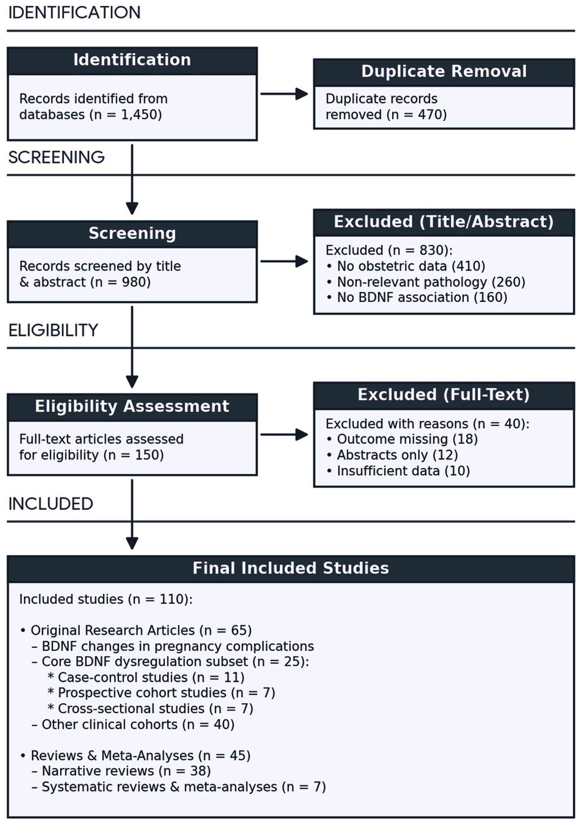
The initial search retrieved 1,450 records.
Following duplicate removal, 980 articles remained for screening.
During title and abstract screening, 830 records were excluded as
irrelevant due to the absence of obstetric data, focus on
non-relevant pathologies, or lack of association with the
neurotrophic protein BDNF. A total of 150 articles underwent
full-text assessment. After applying predefined inclusion and
exclusion criteria, 110 peer-reviewed publications were retained
for the final analysis. These included: original clinical studies;
reviews addressing the obstetrics-BDNF axis; studies examining
psychiatric complications during pregnancy in relation to BDNF;
research involving cohorts of pregnancies complicated by growth
restriction; publications exploring associations between BDNF and
metabolic disorders in pregnancy. A simplified PRISMA-ScR flow
diagram illustrating the study selection process is presented in
Fig. 1.
Inclusion and exclusion criteria
Eligible studies comprised peer-reviewed
publications in English or Russian, including original clinical
research (cohort studies, case-control studies and case series), as
well as narrative and systematic reviews and clinical guidelines
that examined the association between BDNF and physiological
changes during pregnancy, obstetric complications, or maternal
mental health. Sources were excluded if they lacked peer review
(except official international clinical guidelines), including
conference abstracts, non-reviewed preprints, and publications that
did not report obstetric-related data.
Data extraction and synthesis of
results
Study selection was conducted in a multi-stage
process, beginning with the screening of titles and abstracts,
followed by full-text assessment of potentially eligible articles.
From the included studies, data were extracted on study design,
population characteristics (including gestational age), type of
gestational complication (e.g., PE, GDM, preterm birth, foetal
growth restriction), methods used to quantify BDNF levels, as well
as reported associations, proposed pathophysiological mechanisms
and clinical implications.
In accordance with the PRISMA-ScR framework, a
formal assessment of methodological quality and risk of bias was
not performed, reflecting the exploratory nature of a scoping
review and representing an inherent limitation of the present
study. Current evidence suggests a consistent association between
disrupted BDNF regulation, maternal physiological responses and
gestational disorders; however, differences in methodological
frameworks across studies complicate direct comparisons and may
contribute to variability in reported findings. Variations in
biological sample sources (serum, plasma, cord blood, placental
tissue, and amniotic fluid), assay methodologies and the timing of
specimen collection may lead to inconsistencies in reported BDNF
concentrations and limit direct comparisons between studies.
Additional sources of potential bias include the predominance of
studies conducted in large academic and research centres, a
geographical concentration of evidence from high-income and
English-speaking countries, and the possible underrepresentation of
studies reporting null or negative findings. To address these
limitations, particular attention was paid to the consistency and
reproducibility of findings across independent investigations and
diverse study populations. Despite these methodological challenges,
the collective evidence identifies BDNF as a central biological
mediator linking placental function, maternal adaptation and foetal
neurodevelopment, highlighting its potential role as both a
biomarker and a mechanistic contributor to obstetric
pathophysiology.
Data synthesis was performed narratively, with
emphasis on epidemiological associations between BDNF levels and
gestational complications, the exploration of underlying
pathophysiological pathways (including neuroendocrine regulation,
inflammatory processes, and placental dysfunction), and
characterisation of clinical phenotypes. The findings were
integrated into a conceptual, clinically oriented framework
outlining the potential role of BDNF in the development and
progression of pregnancy-related complications.
Source type delineation
The evidence base for the present scoping review
consisted exclusively peer-reviewed scientific literature from
obstetrics, neonatology, endocrinology, reproductive biology and
related biomedical disciplines. These publications were
systematically analysed to characterise the epidemiological
relevance, biological functions, and clinical implications of BDNF
across normal and complicated pregnancies. Particular attention was
paid to its involvement in placental development, maternal
adaptation to pregnancy, foetal growth, neurodevelopmental
programming and the pathogenesis of major obstetric disorders,
including PE, GDM and FGR.
Results
From the 110 articles identified in the search
(1-110),
38 were reviews, and 7 were systematic reviews (meta-analyses). Of
the 65 remaining articles, the majority described changes in BDNF
levels in major pregnancy complications, focusing primarily on
maternal and neonatal outcomes. Several studies (n=25) linked
altered BDNF concentrations to pregnancy pathologies, suggesting
that BDNF dysregulation contributes to their pathobiology.
Methodologically, this core subset comprised 10 case-control
studies (26,41,42,46,47,49,59,60,62,68,93), 8
prospective longitudinal cohort studies (11,17,26,27,73,74,100)
and 7 cross-sectional studies (3,6,12,14,28,32,101).
The studies were derived from all regions of the world including,
Asia, Europe and America. Research studies were derived from last
16 years, i.e., 2010-2026. The studies were obtained from Google
Scholar (50%), Science Direct (35%) and other (15%) databases,
i.e., PubMed. The key clinical and experimental studies that shaped
the understanding of BDNF as a pivotal factor in gestational
vascular biology and pregnancy pathologies are detailed and
summarized in Table I.
 | Table IThe landmark clinical and
experimental studies that shaped the understanding of BDNF. |
Table I
The landmark clinical and
experimental studies that shaped the understanding of BDNF.
| Study no. | First author, year
of publication | Study design | Study
objective | Size and
characteristics of population | Conclusions | (Refs.) |
|---|
| 1 | D'Souza, 2014 | Case-control
study | To examine the
levels of maternal and cord BDNF in women with PE | 106 Full-term and
46 preterm with PE and 95 normotensive women delivering at
term | Maternal BDNF
levels were lower in PE when compared to normotensive women. Cord
BDNF levels were higher in women with PE delivering at term, while
it was lower in women delivering preterm. | (46) |
| 2 | Briana, 2018 | Case-control
study | To determine BDNF,
NGF and NT-4 are expressed in cord blood samples at birth in LGA,
IUGR and AGA offspring of diabetic mothers | 80 cord blood
samples from 15 LGA, 12 IUGR and 33 AGA diabetic, as well as from
AGA 20 normal singleton full-term pregnancies | BDNF is
down-regulated in the fetus exposed to GDM, independently of the
fetal growth pattern/ | (59) |
| 3 | Perucci, 2017 | Case-control
study | To investigate BDNF
plasma concentrations in PE and to compare with BDNF concentrations
from normotensive PW | 38 women with PE
(17 early-onset and 21 late-onset) and in 20 normotensive PW | Lower BDNF plasma
concentrations and cross-talk between BDNF and AnxA1 signaling
pathways might be involved in PE pathogenesis. | (47) |
| 4 | Ardiani, 2019 | Case-control
study | To determine
differences in BDNF and moderately mature MMP-9 between normal
birth weight and IUGR. | Umbilical cord
blood of appropriate gestational age neonate with normal birth
weight (31 neonates) and IUGR (31 neonates) | BDNF of moderately
mature neonates was significantly higher in normal birth weight
compared to IUGR, and the moderately high MMP-9 neonates were
significantly higher in IUGR compared with normal birth
weight. | (68) |
| 5 | Sonmez, 2019 | Case control
study | To assess the
association between cord blood BDNF concentration and maternal
depression during pregnancy | 48 PW, 23 women
with depression during pregnancy and the control group included 25
PW without depression during pregnancy | Cord blood BDNF
concentration was significantly lower in babies born to mothers
with major depression as compared with those in the control
group. | (98) |
| 6 | Guan, 2020 | Case-control
study | Correlation of
Val66Met variation in BDNF and PE | 1,138 Cases of the
PE group and 1342 cases of the control group | The analysis of
maternal-fetal pairing showed the transitivity of BDNF Val66Met
have no significant difference between the case group and the
control group | (49) |
| 7 | Kondratiuk,
2021 | Case-control
study | To investigate
quantitative serum brain-derived neurotrophic factor as a possible
predictor of the severity of PE and its complications | 88 Pregnant women
at 22 to 42 weeks of gestation were examined, 77 of them with
hyper-tensive disorders | In severe
complications of hypertensive disorders, a reliable increase of
BDNF in blood serum was revealed. | (41) |
| 8 | Pascual-Mancho,
2022 | Case-control
study | To analyze the
blood levels of BDNF in the cord blood from fetuses with FGR in
comparison to those with weight appropriate for gestational age
(AGA) | 130 Subjects were
recruited: 91 in AGA fetuses; 39 (16 FGR fetuses with exclusively
middle cerebral artery pulsatility index <5th percentile and 23
with umbilical artery >95th percentile). | Significant
decrease in cord blood levels of BDNF in FGR and more prominently
in those with UA PI >95th percentile in comparison to AGA. | (42) |
| 9 | Huang, 2024 | Case-control
study | To investigate the
roles of BDNF and the Val66Met polymorphism in MCI in patients with
type 2 diabetes mellitus (T2DM). | 169 Patients with
T2DM were involved and divided into long-term (>10 years) and
short-term (≤10 years) diabetes, and in each group, the patients
were separated as MCI and the control | Elevated peripheral
BDNF level associated with declined islet function, when combined
with its Val66Met polymorphism, may forecast memory dysfunction in
patients with long-term T2DM. | (62) |
| 10 | Firdaws, 2025 | Case-control
study | To investigate the
association between poor neonatal outcomes of MSAF and serum
BDNF. | 90 PW. The case
group-45 PW who underwent cesarean section due to fetal distress
with MSAF, and the control group-45 PW cesarean section due to
fetal distress without MSAF. | The maternal BDNF
cutoff for predicting meconium-stained liquor fetal distress was
1.76 ng/ml, showing an acceptable validity finding with sensitivity
100%, specificity 95.6% and accuracy 98.5%. | (93) |
| 11 | Su, 2021 | Longitudinal | To investigate the
possible relationship between the serum BDNF levels of IMGD and
neuro-developmental outcomes using the BSID-III. | 24 Candidates,
including 8 IMGD and 16 healthy infants | The BDNF content
was significantly higher in the non-IMGD than in the IMGD group at
12 months of age. | (60) |
| 12 | Cattaneo, 2010 | Longitudinal | Functional
polymorphism (Val66Met) in the pro-region of BDNF | In amniotic fluids
(AF) obtained from 139 healthy women during 15-17 weeks of
pregnancy | Significant BDNF
protein levels reduction in 55 Met carriers as compared to 84 non
carriers, and no effect of fetus gender, maternal age or gestation
week on BDNF levels. | (26) |
| 13 | Fung, 2015 | Longitudinal | BDNF in the
pathophysiology of depression | 968 Pregnant
women | Lower maternal
serum BDNF levels in early pregnancy are associated with antepartum
depression. | (100) |
| 14 | Gao, 2016 | Longitudinal | To determine
whether there is an association between serum BDNF and PPD | 340 women were
enrolled: 37 women (10.9%) were considered as meeting criteria for
PPD. | Serum BDNF levels
in women without PPD were significantly higher than those in women
with PPD. | (74) |
| 15 | Christian,
2016 | Longitudinal | The predictive
value of serum BDNF in pregnancy | During each
trimester and 4-11 weeks postpartum among 139 women (77 of African
origin, 62 Caucasian). | Serum BDNF declines
considerably across pregnancy in women of African origin and
Caucasians, with overall higher levels in those of African
origin. | (11) |
| 16 | Ghassabian,
2017 | Longitudinal | Mothers rated
children's development in five domains repeatedly through age 3
years | BDNF measured in
2,637 newborns dried blood spots | BDNF levels in
neonates may be impacted by maternal lifestyle
characteristics. | (17) |
| 17 | Dingsdale,
2022 | Longitudinal | Serum BDNF levels
in mothers and their newborns correlate with sex-specific outcomes
at 1 year. | 355 Women with
children | Reduced serum BDNF
levels at birth are linked to an increased risk for
neuro-developmental disorders. | (73) |
| 18 | Zhang, 2025 | Longitudinal | Depressive symptoms
in the first and third trimester | Depressive symptoms
in the first and third trimester | Serum BDNF shows
promise as a predictive biomarker for antenatal depressive symptoms
across all trimesters. | (27) |
| 19 | Saenen, 2015 | Cross-sectional
study | Adaptive responses
of the placenta to the maternal environment may influence central
nervous system development | Placental tissue of
90 mother-infant pairs | Placental
expression of BDNF and SYN1, two genes implicated in
normal neuro-developmental trajectories, decreased with increasing
in utero exposure to PM2.5. | (101) |
| 20 | Yang, 2017 | Cross-sectional
study | To examine the
correlates of early pregnancy serum BDNF concentrations | 982 Women attending
prenatal care | Serum BDNF
concentrations were positively associated with maternal age and
early pregnancy BMI, but inversely correlated with gestational age
at sample collection and C-reactive protein concentrations. | (6) |
| 21 | Zaw, 2019 | Cross-sectional
study | metals [lead (Pb),
mercury (Hg), cadmium (Cd) and arsenic (As)] were measured in
plasma BDNF level | 108 with Myanmar
migrant pregnancy with a single viable fetus of 0 to 14 gestation
weeks | Women with a higher
blood total arsenic level were more likely to have lower BDNF in
early pregnancy. Heavy metals could aggravate the BDNF level. | (32) |
| 22 | Mayuliani,
2021 | Cross-sectional
study | To analyze the
correlation between BDNF and perinatal outcomes in PE. | 73 Pregnant women
with PE. | BDNF level in PE
was lower than normal pregnancy, positive correlation between BDNF
and perinatal outcomes. | (28) |
| 23 | Flöck, 2022 | Cross-sectional
study | The importance of
BDNF acting at the foeto-placental interface. | 109 Maternal
blood-amniotic fluid pairs (including 66 maternal
blood-fetal-blood-amniotic fluid trios | Correlate AF-BDNF
samples with the corresponding maternal and fetal blood-BDNF
samples. | (14) |
| 24 | Jaskolski,
2022 | Cross-sectional
study | The influence of
dietary (dGDM) and insulin-dependent (iGDM) gestational diabetes
(GDM) on BDNF blood levels | Blood samples from
293 maternal-neonatal pairs were analyzed. | Maternal and
neonatal BDNF levels were lowest in the iGDM group, followed by the
dGDM group and healthy controls. | (12) |
| 25 | Granitzer,
2024 | Cross-sectional
study | Determine whether
BDNF and KISS-1 levels can be used as biomarkers | 65 Pregnant
women | Pb exposure and
iron status modulate BDNF and KISS-1 levels. The correlations
between maternal BDNF and KISS-1 levels, placental gene
expression. | (3) |
Biomarker matrices and molecular
targets
The selected studies displayed distinct biological
matrices and analytical targets, as follows:
Protein quantification. In total, 21 of 25
studies quantified total BDNF [representing unseparated pro-BDNF
and mature BDNF (mBDNF) isoforms] in serum (6,26-28,41,60,73,74,93,100),
plasma (32,47), or cord blood (42,46,59,68,98),
paired maternal-neonatal blood (12,14),
alternative biological matrices: blood spots (11) and amniotic fluid (26).
Genetics and epigenetics. A total of 4
studies evaluated single nucleotide polymorphisms (SNPs) of the
BDNF gene (e.g., Val66Met/rs6265) in cord blood or oral
swabs (3,26,49,62), and
3 studies examined BDNF mRNA expression or DNA methylation
patterns in cord blood or placental tissue (3,14,101).
Clinical pregnancy pathologies and
perinatal outcomes
BDNF dysregulation was strongly implicated across
major obstetric, metabolic and psychiatric pathologies:
Hypertensive disorders and PE. Maternal serum
and plasma BDNF concentrations were significantly reduced in PE
compared to normotensive pregnancies (28,46,47),
with cord blood BDNF exhibiting gestational age-dependent variation
(elevated in term PE, reduced in preterm PE) (46). Severe hypertensive complications
demonstrated a compensatory surge in serum BDNF (41). Maternal-foetal pairing analysis of
the Val66Met polymorphism revealed no transmission disequilibrium
in PE (49).
Foetal growth and metabolic pathologies. Cord
blood BDNF expression was downregulated in GDM regardless of foetal
growth pattern (59), exhibiting a
downward gradient from healthy controls to dietary GDM and
insulin-dependent GDM (12).
FGR/intrauterine growth restriction (IUGR) was significantly
associated with reduced cord BDNF levels (42,68),
particularly in foetuses presenting with an umbilical artery
pulsatility index exceeding the 95th percentile (42).
Perinatal mental health. Declining maternal
serum BDNF levels across trimesters were associated with antepartum
(27,100) and postpartum depression (74). Furthermore, maternal major depressive
disorder significantly reduced offspring cord blood BDNF
concentrations (98).
Environmental toxicants and
gene-environment interactions
Exposomic investigations demonstrated a high
sensitivity of BDNF signalling to environmental stressors:
Air pollution. Prenatal exposure to fine
particle air pollution with a diameter ≤2.5 µm (PM2.5) reduced
placental BDNF expression (101).
Heavy metals. Heavy metal exposures,
including arsenic and lead, modulated maternal and cord blood BDNF
concentrations (3,32).
Diagnostic significance. BDNF demonstrated
utility as a predictive clinical biomarker; a maternal serum BDNF
cut-off level of 1.76 ng/ml accurately predicted meconium-stained
amniotic fluid (MSAF)-associated foetal distress (sensitivity,
100%; specificity, 95.6%) (93).
Discussion
BDNF in normal gestation
In a population-based cohort study that applied a
comprehensive mother-placenta-foetus sampling approach, Flöck et
al (14) reported dynamic
changes in maternal circulating BDNF concentrations throughout
pregnancy. Mean maternal serum BDNF levels were 11.3±3.5 pg/ml
during the first trimester, decreased to 10.6±3.8 pg/ml in the
second trimester and remained relatively stable at 11.0±4.1 pg/ml
in the third trimester. Analysis of matched maternal blood,
amniotic fluid and foetal blood samples further demonstrated a
positive association between amniotic fluid BDNF (AF-BDNF) levels
and corresponding maternal and foetal circulating concentrations,
suggesting coordinated regulation of neurotrophic signalling across
the maternal-foetal interface (14).
Notably, Flöck et al (14) also identified that pre-pregnancy
maternal BMI exhibits a strong inverse association with maternal
BDNF levels. Furthermore, maternal obesity, particularly when
accompanied by lower baseline BDNF concentrations, was associated
with a greater probability of AF-BDNF levels falling below the
analytical detection threshold (14). Together, these empirical findings
highlight the potential influence of maternal metabolic status on
the intrauterine neurotrophic environment and suggest that maternal
adiposity may contribute to alterations in foetal
neurodevelopmental programming through modulation of BDNF
availability (13,14).
A thorough understanding of the involvement of BDNF
in pregnancy-related disorders requires an examination of its
physiological dynamics during uncomplicated gestation. Under normal
reproductive conditions, BDNF functions as a key regulatory
molecule from the earliest stages of development, contributing to
folliculogenesis, oocyte maturation, embryo implantation and
placental formation (4,15). In a longitudinal cohort study
involving 139 women from diverse racial backgrounds, Christian
et al (11) observed a
progressive decline in maternal serum BDNF concentrations across
pregnancy, from the first trimester (12.0±1.89 weeks) to the second
(21.7±2.2 weeks) and third trimesters (30.3±2.0 weeks), followed by
a marked increase during the postpartum period (4-11 weeks
following delivery). Several physiological mechanisms have been
proposed to explain this temporal pattern, including gestational
haemodilution, increasing placental uptake and utilization of
neurotrophic factors, and the modulatory effects of
pregnancy-associated hormones, such as progesterone and oestradiol
(11,15,16).
Notably, BDNF is capable of crossing the uteroplacental barrier,
and experimental evidence indicates a close association between
maternal circulating BDNF concentrations and foetal brain levels.
These findings suggest that the maternal neurotrophic status may
directly influence foetal neurodevelopment and support the concept
of BDNF as a critical mediator of maternal-foetal communication
during pregnancy (13).
Determinants of maternal and neonatal BDNF levels
are complex and encompass socioeconomic, lifestyle and demographic
factors. In population-based cohorts, lower neonatal BDNF measured
via dried blood spots has been found to be associated with maternal
smoking and alcohol consumption during pregnancy, as well as
pre-pregnancy obesity (17-19).
It is synthesized within the trophectoderm of the blastocyst and
remains a persistent factor in the development of both the maternal
decidua and the foetal placental structures throughout the three
trimesters (20). Furthermore, BDNF
serves as a potent pro-angiogenic factor, promoting the survival of
endothelial cells and facilitating the complex process of uterine
spiral artery remodelling (21,22).
Molecular architecture and genetic
regulation of BDNF
The BDNF gene locus is characterized by high
evolutionary complexity and conservation of the coding sequence;
however, the structure of its regulatory elements has undergone
significant sophistication in primates and humans compared to
rodents (23). Located on chromosome
11p13, the gene consists of 11 exons and utilizes nine distinct
promoters that are regulated in a tissue-specific and
activity-dependent manner (3,24). The
recent study by Esvald et al (25) substantially refined the
transcriptional landscape of the BDNF gene in cultured rat
cortical and hippocampal neurons.
According to Granitzer et al (3), BDNF is a highly conserved neurotrophin
synthesized as the precursor protein pro-BDNF (~32 kDa), which is
subsequently cleaved into the biologically active mBDNF form. This
maturation process contributes to the functional versatility of
BDNF in various physiological processes. This 32-kDa protein is
synthesized in the endoplasmic reticulum and subsequently
transported to the Golgi apparatus, where it undergoes proteolytic
cleavage by enzymes, such as furin or proprotein convertases to
generate the 14-kDa mBDNF isoform (3). During pregnancy, the balance between
these two isoforms is essential for maintaining placental health
and guiding the delicate processes of brain morphogenesis.
The seminal study by Cattaneo et al (26) provided critical insight into the
molecular consequences of the BDNF Val66Met polymorphism,
elucidating how this functional genetic variant alters
intracellular trafficking and activity-dependent release of BDNF.
Carriers of the Met allele exhibit reduced neurotrophin secretion
in response to neuronal stimulation, which has been associated with
structural alterations in brain regions involved in learning,
memory, and emotional regulation, particularly the hippocampus.
These neurobiological changes have also been linked to an increased
vulnerability to a range of neuropsychiatric conditions. During
pregnancy, the Val66Met polymorphism may further modulate
maternal-foetal neurotrophic signalling. Evidence suggests that the
presence of the Met allele in either the mother or the foetus can
influence BDNF concentrations within the intrauterine environment
and may modify the impact of maternal psychological stress on the
development of foetal limbic structures, including the amygdala and
hippocampus (26).
In addition, extravillous trophoblasts, which are
responsible for maternal decidual invasion and spiral artery
transformation, also express components of the BDNF signalling
pathway. This observation supports the hypothesis that BDNF may
regulate trophoblast migration and invasion through both autocrine
and paracrine mechanisms. As gestation advances, the distribution
of BDNF receptors undergoes substantial changes. Near term, the
expression of TrkB and p75NTR within trophoblastic layers declines,
whereas receptor localization becomes increasingly prominent within
the villous stromal compartment of the placenta, suggesting a shift
in the functional role of neurotrophin signalling during late
pregnancy (27). In their study,
Mayuliani et al (28) further
emphasized the importance of BDNF in placental development by
demonstrating that inadequate cytotrophoblast invasion and
incomplete remodelling of the maternal spiral arteries contribute
to placental ischemia and persistent hypoxic stress at the
maternal-foetal interface. These findings indicate that the
oxidative environment characteristic of PE suppresses placental
BDNF gene expression throughout gestation, providing a potential
link between impaired neurotrophin signalling with the
pathophysiology of placental dysfunction and hypertensive disorders
of pregnancy (28,29).
According to D'Souza et al (29), maternal plasma BDNF levels are
altered prior to the clinical onset of PE, establishing the
potential of BDNF as an early gestational biomarker. They connected
the placental neurotrophin axis to one-carbon metabolism and
maternal nutrition. They demonstrated that maternal micronutrient
imbalances [such as folate and vitamin В12 deficiencies combined
with low docosahexaenoic acid (DHA)] disrupt placental global DNA
methylation, thereby epigenetically downregulating the expression
of placental BDNF and TrkB; this leads to impaired placental
development and intrauterine growth restriction (29,30).
As demonstrated by Dingsdale et al (64), there is a sex-specific
maternal-foetal a involving maternal anxiety and serum BDNF levels.
In a large cohort of pregnant women, they found that maternal
symptoms of anxiety correlate with significantly elevated serum
BDNF exclusively in mothers carrying male foetuses, while maternal
depression shows no such correlation. To determine the
physiological path of this elevated BDNF, they developed a novel
mouse model that demonstrated that maternal BDNF does not cross the
placental barrier into the foetal circulation (64). Based on these findings, it was
concluded that the placenta serves as an active barrier, shielding
the developing foetus from fluctuating maternal serum neurotrophin
levels that could otherwise interfere with controlled and
endogenous foetal neurodevelopment (31).
The longitudinal profile of serum BDNF in healthy
pregnancies reveals a characteristic and statistically significant
decline as gestation progresses. According to Christian et
al (11), serum BDNF
concentrations declined progressively from the first to the third
trimester of pregnancy, followed by a rapid rebound in the
postpartum period. They documented that women of African origin
exhibit significantly higher circulating BDNF during the first two
trimesters and postpartum compared to Caucasian women, as well as
lower corresponding cortisol levels. In addition, they demonstrated
that lower maternal serum BDNF in late pregnancy was strongly
associated with high levels of maternal depressive symptoms and a
significantly increased risk of delivering low-birth-weight
(<2,500 g) infants, emphasizing the clinical value of
within-race prenatal monitoring (11). For the developing foetus, BDNF is
critically required starting from the third week of gestation, when
active differentiation of neural progenitor cells occurs (32,33).
As the pregnancy advances into the second and third
trimesters, these levels decline considerably, often reaching a
nadir in late pregnancy that is 30 to 50% lower than levels
observed in non-pregnant women (27). Of note, the postpartum period is
marked by a rapid and substantial rebound in maternal serum BDNF
levels, typically measured between 4 and 11 weeks after delivery.
This rebound may be associated with the neurobiological recovery
and remodelling required for the transition to maternal caregiving
(34).
BDNF and gestational
complications
PE remains a primary driver of maternal and neonatal
morbidity, affecting ~2-8% of pregnancies globally (35).
In a landmark study, D'Souza et al (9) demonstrated that women with PE exhibit
substantially reduced BDNF concentrations in maternal plasma,
placental tissue and umbilical cord blood, with levels ~40-50%
lower than those observed in normotensive pregnancies. These
reductions were closely associated with the severity of endothelial
dysfunction, indicating a critical role for BDNF in the
pathophysiology of PE. Notably, this decrease was associated with
both the severity of maternal hypertension and the extent of
placental ischemic injury, suggesting a disruption of compensatory
angiogenic pathways that are essential for maintaining placental
function (9). Under hypoxic
conditions, the ischemic placenta overproduces and secretes soluble
decoy receptors into the maternal bloodstream. Of note, Liao et
al (36) proposed a
‘self-defence’ hypothesis, demonstrating that foetal trophoblasts
overexpress soluble fms-like tyrosine kinase-1 (sFlt-1) to shield
the foetus from excessive maternal decidual VEGF, though this
feedback loop ultimately compromises the maternal vasculature.
There is also evidence of a regulatory interplay between BDNF and
established angiogenic markers, such as placental growth factor
(PlGF) and sFlt-1(37).
According to Nelson et al (38), reduced BDNF availability may
intensify placental dysfunction by compromising endothelial cell
proliferation, vascular remodelling and vasoregulatory mechanisms
that are essential for maintaining adequate uteroplacental
perfusion. As a result, a pathogenic feedback loop may emerge
whereby placental ischemia reduces BDNF production, while decreased
neurotrophic signalling further aggravates vascular impairment and
hypoxic stress. Ultimately, this disruption of the placental
neurotrophic environment may restrict the delivery of critical
developmental signals to the foetus during vulnerable stages of
brain maturation, which may increase susceptibility to
hypoxia-related neurodevelopmental injury (38).
Further findings reported by Cinkajzlova et
al (39) indicated that in
pregnancies complicated by both PE and diabetes mellitus, reduced
BDNF levels are accompanied by elevated concentrations of
P-selectin and interferon gamma receptor 1, contributing to the
progression of placental microvascular dysfunction. Similarly,
Meakin et al (40)
demonstrated that reduced BDNF concentrations in PE may not simply
result from placental ischemia, but may also predict early
postnatal neurocognitive impairment in offspring, probably due to
the reduced capacity of the placenta to buffer foetal exposure to
hypoxic stress. The aetiology of PE is closely linked to defective
trophoblast invasion and inadequate remodelling of the maternal
spiral arteries, leading to placental hypoxia and the systemic
release of anti-angiogenic factors. Research indicates that BDNF
plays a pivotal role in this process, with significant alterations
in its maternal serum levels and placental expression observed in
preeclamptic pregnancies (29,41).
This reduction in local BDNF availability may contribute to the
shallow trophoblast invasion observed in PE by failing to
sufficiently stimulate matrix metalloproteinase (MMP)-9 production
and other pro-invasive signalling pathways (29,42). In
PE, the increase in the levels of sFlt-1, a decoy receptor that
sequesters VEGF and PlGF, leads to an anti-angiogenic state. The
concurrent reduction in BDNF levels further compromises the
angiogenic potential of the placenta, reinforcing a state of
chronic hypoxia and endothelial stress (9,29,43).
The clinical phenotypes of PE differ substantially,
as demonstrated by Marzioni et al (44), who highlighted the critical
regulatory role of the STAT3 signalling pathway. Early-onset PE
(developing prior to 34 weeks of gestation) is strongly associated
with deficient spiral artery remodelling, severe placental hypoxia
and FGR. Conversely, late-onset PE (manifesting at or after 34
weeks) is more closely linked to pre-existing maternal metabolic or
cardiovascular predispositions and primary endothelial
susceptibility (44-49).
The interplay between BDNF and endothelial cells is highly
cooperative and relies on autocrine and paracrine signalling loops.
Healthy vascular endothelial cells express both the ligand BDNF and
its high-affinity TrkB receptor. Under normal blood flow
conditions, physiological fluid shear stress stimulates the
vesicular exocytosis of BDNF from endothelial cells (50).
The maintenance of endothelial barrier integrity
also depends on nitric oxide (NO). Under normal conditions, the
generation of NO via endothelial nitric oxide synthase (eNOS)
phosphorylation at Ser1177 serves as a key driver of BDNF synthesis
and release in the vascular network. Maternal endothelial
dysfunction is the ultimate common pathway in PE, characterized by
downregulated eNOS expression, NO deficiency, reduced prostacyclin
synthesis, elevated vasoconstrictor production (such as
endothelin-1) and the accumulation of reactive oxygen species
(ROS). Experimental models indicate that the pharmacological
blockade of eNOS via L-NAME markedly decreases endothelial BDNF
production, whereas the application of NO donors, such as glyceryl
trinitrate completely restores BDNF expression (51). In PE, this feed-forward loop is
broken. Excessive anti-angiogenic factors (such as sFlt-1) lead to
the downregulated expression of eNOS and reduced NO
bioavailability, leading to endothelial BDNF depletion and vascular
wall breakdown.
To systematically evaluate the degree of systemic
endothelial cell activation and stress, the Endothelial Activation
and Stress Index (EASIX) has been proposed (n=986): EASIX=LDH x
creatinine/platelets. This index has exhibited strong prognostic
power in stratifying maternal and perinatal risk, particularly in
patients diagnosed with early-onset PE (51).
Recent neuropathophysiological concepts classify
severe PE as a gestational proteinopathy with impaired autophagy,
sharing common pathways with neurodegenerative disorders.
Endothelial injury and anti-angiogenic stress cause blood-brain
barrier leakage, neuroinflammation and the loss of cerebral
autoregulation. Indeed, >70% of maternal PE deaths stem from
acute neurological complications, such as eclampsia, cerebral
oedema and stroke (52). Against
this clinical background, serum BDNF serves as a powerful
predictive biomarker. In mild or moderate PE, circulating BDNF is
systemically reduced due to endothelial depletion and impaired
placental synthesis.
Metabolic complications: Insulin
resistance and BDNF in GDM
GDM is one of the most common medical complications
of pregnancy, affecting ~14 to 16.5% of pregnancies worldwide, with
an increasing prevalence due to the global epidemics of maternal
obesity and delayed childbearing (54). In a normoglycemic gestation, maternal
pancreatic β-cells undergo robust compensatory hypertrophy and
hyperplasia to increase insulin secretion by 60 to 70% (53). However, in pregnancies predisposed by
genetic susceptibility, an advanced maternal age, or pre-existing
obesity, this compensatory capacity fails, culminating in
persistent hyperglycaemia, foetal hyperinsulinemia and a state of
chronic subclinical systemic inflammation (12,54).
During the first trimester, elevated concentrations
of oestrogen and human chorionic gonadotropin enhance peripheral
insulin sensitivity by ~20%, encouraging maternal lipid and
glycogen deposition. Beyond the 10th to 12th weeks of gestation, as
the placenta assumes dominant endocrine control, a significant
increase in maternal insulin resistance occurs. By the third
trimester, peak levels of human placental lactogen, human placental
growth hormone, cortisol and progesterone cause a 40 to 50%
decrease in systemic insulin sensitivity (55,56). In
patients with GDM, chronic maternal hyperglycaemia leads to a
persistent, systemic depletion of circulating BDNF, which deprives
peripheral tissues of its insulin-mimetic, protective functions and
accelerates maternal metabolic decay (12,57).
Jaskolski et al (12) pioneered the first matched cohort
analysis of corresponding maternal-neonatal pairs in pregnancies
complicated by GDM. Utilizing multiple regression models and 2:1
propensity score matching (adjusting for gestational age, infant
sex, and mode of delivery), they established that neonatal cord
blood log-BDNF is significantly suppressed in GDM pregnancies, with
the most profound deficit observed in insulin-dependent patients.
Their findings demonstrated that GDM is associated with a marked
reduction in neonatal neurotrophic support, which occurs
independently of maternal systemic BDNF levels and highlights the
vulnerability of the foetus to maternal metabolic dysregulation.
While maternal log-BDNF levels in GDM exhibited a downward trend
(mean reduction of 79.6 pg/ml), this difference did not reach
statistical significance after adjustment. This indicates that the
foetal-placental compartment undergoes more severe and immediate
BDNF suppression under conditions of maternal hyperglycaemia than
the maternal system, posing a direct threat to intrauterine foetal
brain development (12).
An Israeli research group [Chermon and Birk
(58)] carried out a large genetic
cohort study by analysing data from 4,025 women to investigate the
association of common BDNF SNPs with GDM susceptibility.
They identified a significant association between the BDNF
rs925946 Tag-SNP and GDM risk. Additionally, their study associated
the BDNF rs6265 (Val66Met) polymorphism with pre-pregnancy
obesity in patients with GDM, linking genetic neuroendocrine
disruption to clinical metabolic phenotypes (58).
Briana et al (59) prospectively evaluated cord blood
neurotrophic factors (BDNF, NGF and NT-4) in 80 term gestations
categorized by foetal growth patterns, including large for
gestational age (LGA), IUGR and appropriate-for-gestational-age
(AGA) infants born to GDM and control mothers. They also
established that IUGR infants of mothers with GDM suffer from a
concurrent deficiency in NGF, further compounding their risk for
long-term neurological sequelae (59).
Su et al (60)
conducted a longitudinal and prospective study that tracked infants
of mothers with GDM (IMGD) at birth, 6 months and 12 months of age,
and correlated serial serum BDNF levels with neurodevelopmental
outcomes using the Bayley Scales of Infant Development III
(BSID-III). They demonstrated that by 12 months of age, IMGD
demonstrated significantly lower systemic BDNF concentrations than
healthy control infants. Crucially, this postnatal BDNF deficiency
was accompanied by significantly lower language composite scores on
the BSID-III in GDM offspring. This longitudinal research was the
first to establish a direct clinical link between gestational BDNF
downregulation and functional neurodevelopmental deficits,
specifically delayed language acquisition, in human infants
(60). Complementing these clinical
findings, experimental research by Valle et al (61) demonstrated that early metabolic
stress during gestation induces severe central and peripheral BDNF
deficiency, leading to programmed insulin resistance and metabolic
dysfunction in adult offspring.
Huang et al (62) analysed the clinical intersection of
peripheral BDNF, pancreatic islet function and cognitive decline.
They concluded that, as pancreatic β-cell function declines, a
compensatory increase in peripheral BDNF occurs. However, in
patients carrying the Met allele of the BDNF Val66Met
polymorphism, this compensatory loop is insufficient, which leads
to accelerated cognitive decline and impaired verbal logical memory
compared to Val/Val homozygotes. These findings established
peripheral BDNF and its genetic variants as key predictive
biomarkers for diabetic cognitive impairment (62). In Russian perinatal medicine,
Evsyukova established GDM as a major pathogenetic risk factor for
neurological and psychiatric disorders in offspring. That study
demonstrated that GDM-associated maternal insulin resistance,
hyperleptinemia, hyperinsulinemia and placental insufficiency
combine to cause chronic foetal hypoxia (63).
The reduction in neonatal BDNF in GDM is
particularly noteworthy, as it remains significantly lower even
after adjusting for factors such as maternal BMI and mode of
delivery. This deficiency in the foetal circulation may have
long-term implications for the neurodevelopment of children born to
diabetic mothers, potentially predisposing them to metabolic and
psychiatric disorders later in life (65).
Foetal development issues: BDNF in
neonates who are small for gestational age (SGA) and FGR
FGR or IUGR, represents a pathological state in
which the foetus fails to achieve its genetically predetermined
growth potential (66). According to
Pascual-Mancho et al (42),
cord blood BDNF levels are consistently found to be lower in
neonates with FGR who are SGA compared to those with weight
AGA.
Under chronic hypoxic conditions and nutrient
deprivation, the foetus initiates a highly conserved circulatory
adaptation known as the ‘brain-sparing’ effect. Briana et al
(59) further explained these
mechanisms by demonstrating that BDNF concentrations in the cord
blood of foetuses with FGR are significantly lower than those
observed in appropriately grown foetuses. This finding was
attributed to the ‘brain-sparing effect’, a compensatory adaptive
response in which chronic foetal hypoxia leads to preferential
redistribution of blood flow toward the central nervous system.
However, despite this circulatory centralisation, the absolute
deficiency of BDNF appears to limit the effectiveness of
neuroprotective mechanisms, thereby contributing to microstructural
injury of the developing white matter (59,67).
This phenomenon is characterised by the selective vasodilation of
the foetal middle cerebral artery (MCA) in response to a drop in
cerebral oxygen tension, ensuring a redistribution of cardiac
output to vital organs-primarily the brain, myocardium and adrenal
glands-at the expense of mesenteric, renal and peripheral
perfusion.
At the tissue level, placental hypoperfusion
stimulates a compensatory angiogenic response driven by the
synergistic action of BDNF and MMP-9. BDNF functions as a direct
angiogenic factor within the placental vasculature, promoting
endothelial cell migration and capillary tubulogenesis.
Concurrently, MMP-9 degrades extracellular matrix components,
facilitating the sprouting of new capillary networks and maternal
trophoblast invasion (68).
According to the comparative clinical trial
conducted by Ardiani et al (68) in their Padang-based cohort, healthy
and symmetrically growing neonates (appropriate for gestational
age, n=31) demonstrated baseline cord blood concentrations of mBDNF
of 1.58 ng/ml (standard deviation of 0.23), coupled with
physiological MMP-9 levels of 1.09 ng/ml (standard deviation of
0.20). According to their interpretation, this biochemically
balanced state reflects stable physiological angiogenesis, adequate
trophoblastic invasion, and controlled extracellular matrix
degradation. Conversely, as shown in their results, in moderately
mature neonates diagnosed with intrauterine growth restriction
(n=31), a statistically significant depletion of cord blood BDNF to
1.25 ng/ml (standard deviation of 0.35) was observed (P=0.001).
According to the researchers, this neurotrophic deficiency was
accompanied by a significant compensatory upregulation of MMP-9 to
1.25 ng/ml (standard deviation of 0.35) (P=0.03), which
demonstrates the systemic impact of chronic hypoxia and ischemic
stress on placental architecture and tissue remodelling (68). Mechanistically, they proposed that
BDNF deficiency directly impairs placental angiogenesis, while the
elevated MMP-9 levels reflect a compensatory but ultimately
pathological tissue remodelling response. As stated by Ardiani
et al (68), inadequate
trophoblast invasion is met with a compensatory release of MMP-9 to
degrade the extracellular matrix, attempting to facilitate
placental expansion, though it fails to overcome the primary
neurotrophic and foeto-placental deficit (68).
Similarly, as documented in the amniotic fluid study
by Flöck et al (14), the
biochemical composition of the amniotic fluid compartment reveals a
separate and highly sensitive adaptational response along the
spectrum of foetal growth (14).
According to Malamitsi-Puchner et al (67), this elevated concentration serves as
an endogenous adaptive response, wherein the foetus mobilises its
neurotrophic reserves to accelerate cerebral development and
counter the hostile and nutrient-deprived intrauterine environment
(67).
The 2025 study by Kan et al (69) (n=45) explained that a significant
decrease in the expression of both pro-BDNF and pro-NGF within
maternal foetal neuronal exosomes (FNEs) occurs in the early-onset
FGR cohort. Crucially, mature neurotrophin proteins were not
detected in the FNE cargo, confirming that the vesicle cargo
primarily reflects the non-processed precursor forms. Furthermore,
Kan et al (69) established a
direct clinical correlation: Downregulations in pro-BDNF levels
within maternal-blood FNEs were specifically linked to the
subsequent occurrence of neonatal intraventricular haemorrhage in
late-onset FGR newborns. As stated by the authors, this discovery
introduced a diagnostic ‘liquid biopsy’ of the foetal brain,
permitting the early prenatal identification of foetuses at risk of
severe cerebral ischemic injury (69).
Maternal obesity, sexual dimorphism
and placental dysfunction
Pre-pregnancy maternal obesity (defined as a
pre-pregnancy) acts as a state of chronic low-grade inflammation,
oxidative stress and hypoxia, which profoundly impairs the
molecular machinery of the BDNF/TRKB pathway within the placenta
(70). Circulating maternal blood
BDNF levels are known to be significantly and negatively correlated
with pre-pregnancy BMI. Flöck et al (14) analysed mother-foetus trios and
demonstrated that maternal obesity restricts the availability of
BDNF in the amniotic fluid. In pregnancies where amniotic fluid
BDNF fell below the lower limit of quantitation (<1.19 pg/ml),
patients had a significantly higher pre-pregnancy BMI (26.3±6.7
kg/m² compared to 23.8±4.5 kg/m² in the detectable group) and
significantly lower maternal circulating blood BDNF concentrations
(510.6±554.7 pg/ml compared to 910.1±690.1 pg/ml in the detectable
group; P<0.0001), suggesting that maternal adiposity impairs the
transplacental transfer of neurotrophic support (14).
The placental molecular response to maternal
obesity and high-fat diet exposure is characterised by a striking
sexual dimorphism (71). The
capacity of the placenta to adapt to an adverse intrauterine
environment and compensate for maternal metabolic perturbations
varies significantly depending on foetal sex, with male foetuses
exhibiting a pronounced vulnerability to prenatal insults. In their
investigation of maternal obesity, Muralimanoharan et al
(72) identified sex-specific
mitochondrial dysfunction in the placenta mediated by miR-210. They
discovered that the inflammatory intrauterine environment of
maternal obesity selectively upregulates miR-210 in female
placentas, but not in male placentas. TNF drives this
female-specific upregulation by stimulating NF-B1 binding to the
placental miR-210 promoter (72).
A striking feature of neonatal BDNF biology is the
presence of significant sex-specific differences. At birth, male
infants naturally possess serum BDNF levels that are ~10 to 20%
lower than those of female infants. This disparity has led
researchers to propose the ‘male vulnerability’ hypothesis, which
suggests that boys may be at an increased risk of developing
neurodevelopmental disorders as they have a lower baseline of
neurotrophic protection. The ‘Grown in Wales Study’ provided
empirical support for this hypothesis by correlating cord serum
BDNF levels with neurodevelopmental outcomes at 1 year of age
(73). While no association was
found between BDNF levels and general cognitive or language scores
across the entire group, a significant sex-specific association
emerged for attention. In boys specifically, higher cord serum BDNF
was linked to a greater intensity of facial interest during a
sustained attention task at 12 to 14 months (73).
These findings suggest that male and female
foetuses may utilize different molecular strategies to adapt to
maternal metabolic stress, which may contribute to the observed sex
differences in the prevalence of neurodevelopmental disorders such
as autism spectrum disorder (ASD) and attention deficit
hyperactivity disorder (ADHD) (65).
Mental health and neurodevelopment:
The perinatal connection
In alignment with the neurotrophin hypothesis of
depression, which links depleted BDNF to altered hippocampal
neurogenesis and subsequent affective pathology, reductions in
circulating maternal BDNF are directly implicated in the
pathogenesis of perinatal mood disorders. During early pregnancy,
depleted maternal serum BDNF functions as a sensitive biomarker for
the development of antepartum depression (27).
At the clinical and epidemiological level, the
understanding of the longitudinal behaviour of maternal circulating
BDNF across gestation was greatly advanced by Christian et
al (11), who mapped serum
neurotrophin levels across all three trimesters and postpartum,
revealing a physiological decline in BDNF concentrations toward
late pregnancy and its association with prenatal depression and
risk for low birth weight. As gestation progresses, maternal BDNF
levels in the second trimester continue to show a strong,
predictive and negative association with late-pregnancy depressive
symptoms (11). Reduced maternal
serum BDNF concentrations around the time of childbirth have
emerged as a promising biomarker for the prediction of postpartum
depression (PPD). A growing evidence indicates that women who
subsequently develop PPD exhibit significantly lower circulating
BDNF levels both at delivery and during the early postpartum
period, typically between 6 and 11 weeks after birth. The clinical
study by Gao et al (74)
further demonstrated the prognostic value of BDNF measurements at
parturition. In particular, a maternal serum BDNF concentration
≤12.0 ng/ml has been proposed as a clinically relevant threshold
for identifying women at elevated risk of postpartum depression.
Using this cut-off value, individuals with BDNF levels below the
threshold were found to have more than a sevenfold increased
likelihood of developing PPD within the first 3 months following
delivery (OR, 7.243; 95% CI, 3.883-12.746; P<0.0001) (74). Collectively, these findings suggest
that peripartum BDNF assessment may offer valuable opportunities
for the early identification of vulnerable women and the
implementation of targeted preventive interventions.
The association between BDNF and mood is further
complicated by the influence of the hypothalamic-pituitary-adrenal
(HPA) axis. High levels of cortisol, the primary stress hormone,
are concurrently associated with lower serum BDNF levels,
particularly during the second trimester. This cortisol-BDNF
interplay suggests that chronic maternal stress may suppress
neurotrophic support for the brain of the mother, contributing to
depressive phenotypes and potentially affecting foetal brain
development through altered placental transfer or paracrine effects
(11,75). High maternal distress is associated
with reduced hippocampal and cerebellar volumes and altered
functional connectivity in the offspring. Chronic variations in
glucocorticoids affect BDNF-TrkB function in the prefrontal cortex,
contributing to stress-induced cognitive alterations. Conventional
antidepressants have been shown to restore BDNF levels,
highlighting the role of this factor as both a biomarker and a
therapeutic target (76).
Maternal mental health and sleep quality also
significantly influence the neurotrophic milieu. In migrant
pregnancies, poor sleep quality has been associated with
significantly lower maternal BDNF levels. By contrast, women with
mild to moderate depression in the first trimester had higher BDNF
levels, suggesting a possible early compensatory response that may
later fail as stress becomes chronic (77).
Preterm birth and developmental
domains
Preterm birth is associated with a profound
disruption of neurotrophic support during a critical period of
foetal development. The molecular analyses conducted by Dhobale
et al (78) demonstrated that
the placental mRNA expression of both BDNF and NGF was reduced by
~2.5-fold in cases of spontaneous preterm delivery, potentially
depriving the immature foetal brain of essential trophic signals
required for neuronal survival and maturation (78). The clinical research by Rao et
al (79) established a positive
association between gestational age and umbilical cord blood BDNF
concentrations; preterm infants are born with significantly lower
BDNF concentrations compared to term infants, and both groups have
lower levels than adults (79). A
rigorous systematic meta-analysis confirmed that preterm birth was
associated with a significant decrease in baseline BDNF
[standardized mean difference (SMD), -0.32; P=0.014] and NT-3 (SMD,
-0.31; P=0.004) compared to term infants, although NGF and NT-4 did
not exhibit no significant variations (80). Heterogeneity within these
meta-analytical findings is primarily moderated by the type of
biospecimen analysed, such as placental tissue, cerebrospinal
fluid, peripheral blood, or umbilical cord blood (80).
Following delivery, there is a sharp and abrupt
withdrawal of maternal and placental neurotrophic support, causing
a rapid decline in postnatal BDNF levels. In term infants, this
drop is transient, recovering by the fourth postnatal day. However,
preterm infants experience a prolonged neurotrophic deficiency,
with BDNF concentrations remaining depressed through the first
weeks of life before gradually rising by discharge (79). Preterm birth remains a primary risk
factor for adverse neurological outcomes, with complications, such
as periventricular leukomalacia, diffuse white matter injury and
cerebral palsy (CP) occurring at rates inversely proportional to
gestational age (79,81). In extremely preterm neonates born
before 28 weeks of gestation, circulating BDNF concentrations are
markedly reduced due to the interruption of the critical period of
neurotrophin transfer and endogenous synthesis that normally peaks
during the third trimester of pregnancy. Additional clinical
evidence highlights the broader implications of BDNF deficiency in
preterm infants. In a retrospective cohort study involving 400
preterm neonates, Sahay et al (13) identified a significant association
between BDNF concentrations measured within the first 24 h of life
and the subsequent development of severe retinopathy of prematurity
(13).
Moreover, D'Souza et al (46) proposed that a substantial decline in
maternal systemic BDNF levels may contribute to the pathogenesis of
preterm premature rupture of membranes through the activation of
pro-inflammatory cytokine pathways, including TNF-α and IL-1β
(46). Integrating biochemically
non-invasive markers into clinical care remains a key research
priority. In a prospective study conducted in Brazil, 40 preterm
neonates (GA 28 to 32 weeks) were evaluated across four time points
(83). Preterm infants with
below-expected motor development (evaluated using the Test of
Infant Motor Performance) had significantly elevated urinary
concentrations of BDNF and glial cell line-derived neurotrophic
factor compared to those with typical development (83).
Analysing long-term neurodevelopmental outcomes is
further complicated by changes in standardized assessment tools.
While early editions such as the BSID-II relied on combined
developmental indices (the Mental Developmental Index and
Psychomotor Developmental Index), the Bayley-III separated these
into distinct Cognitive, Language and Motor scales (84,85).
This structural separation helps identify specific language
deficits independently of overall cognitive performance, but
comparative studies have identified a systematic overestimation of
scores (a Flynn-like effect) when transitioning to the
Bayley-III.
Inconsistencies in autism
research
According to a previous meta-analysis, peripheral
blood BDNF levels are moderately, yet significantly elevated in
children with ASD compared to typically developing controls
(86). In that study, the
researchers compared this consistently elevated neurotrophic state
with the biomarkers of classic psychotic-affective disorders
(86). According to that study, this
pattern is highly concordant with the ‘diametric model of the
social brain’ proposed by Crespi and Badcock (87). In their study, thety present an
evolutionary and developmental framework which posits that autism
and psychotic-affective disorders represent opposite extremes of
human social-cognitive development (87).
As previously demonstrated, the elevated BDNF
levels observed in ASD represent a primary growth-signalling driver
that aligns with the macrocephaly and cortical overgrowth
frequently documented in young autistic cohorts, whereas depleted
BDNF levels in schizophrenia correspond directly to progressive
cortical thinning and volume loss (86-90).
The association between neonatal BDNF and ASD remains controversial
and inconsistent (17).
Clinical utility in acute obstetric
management
Observational clinical studies demonstrate that
maternal circulating plasma BDNF concentrations are significantly
lower in women with PE than in gestational age-matched normotensive
controls. Depressed plasma BDNF is negatively associated with
anti-inflammatory mediators, such as Annexin A1 and positively with
clinical severity indicators, including diastolic blood pressure
and pre-pregnancy BMI. The reduction in maternal circulating plasma
BDNF during early and mid-gestation reflects an underlying
impairment in placental synthesis alongside progressive endothelial
cell injury (47,107).
During parturition, acute clinical events can cause
sudden placental and foetal hypoxia, initiating cellular
depolarization, metabolic acidosis and excitotoxic cascades in the
developing brain (91). MSAF is a
common clinical sign of acute or chronic foetal stress,
complicating up to 27.1% of post-term deliveries and placing the
neonate at risk for meconium aspiration syndrome and subsequent
cerebral palsy (92). According to a
recent clinical evaluation by Jihad and Mohammed (93), maternal and umbilical vein BDNF
concentrations were significantly lower in pregnancies complicated
by MSAF compared to healthy controls (P<0.001), reflecting a
state of acute neurotrophic exhaustion under hypoxic stress. This
diagnostic performance suggests that maternal BDNF can distinguish
between benign meconium passage and severe, progressive
intrauterine asphyxia, offering clinicians an objective
biochemically based screening tool to guide the timing of operative
delivery (93).
According to the study by Lorain et al
(94), the development of
moderate-to-severe hypoxic-ischemic encephalopathy (HIE) in infants
with severe intrapartum acidosis (pH ≤7.0 at birth) is
independently associated with specific acute obstetric factors.
Within this clinical cohort, multi-variable analyses confirm that
the occurrence of an acute intrapartum event (aOR, 6.4; 95% CI,
1.8-22.5), maternal fever (aOR, 3.5; 95% CI, 1.0-11.9) and thick
meconium during labour (aOR, 2.9; 95% CI, 1.0-8.6) are the primary
independent risk factors for HIE. Elevated maternal and foetal BDNF
levels, along with high 25-hydroxyvitamin D [25(OH)D]
concentrations, act as significant protective factors against such
brain injury (95).
Gusar et al (96) demonstrated that isolation of foetal
neuronal vesicles (FNVs) from maternal blood, effectively serving
as a ‘foetal brain liquid biopsy’. These vesicles allow for the
early and non-invasive detection of neuronal dysfunction in
pregnancies complicated by FGR. In FGR cohorts, distinct protein
expression patterns have been identified within FNVs, including
significant alterations in pro-BDNF and presynaptic proteins such
as synaptophysin. These changes often precede clinical
manifestations and are associated with a higher risk of
intraventricular haemorrhage in newborns (96).
The primary question asked by this team was whether
FNEs isolated from maternal blood could serve as a clinical
surrogate to detect early and substructural signs of foetal brain
damage in utero before permanent neurological deficits manifest
postnatally. They questioned the safety of the classic
‘brain-sparing effect’ observed during Doppler ultrasonography.
While macrovascular Doppler studies reveal blood flow
redistribution to preserve the foetal brain during placental
insufficiency, Gusar et al (97) investigated whether these hemodynamic
shifts provide true neuroprotection or paradoxically aggravate
neuronal damage through oxidative stress and reperfusion
injury.
Explanatory hypotheses for discrepant
and paradoxical BDNF dynamics
A major challenge in perinatal neuroendocrinology
is explaining compartmental BDNF paradoxes, most notably the
finding where AF-BDNF concentrations are elevated or sustained in
growth-restricted pregnancies, while corresponding umbilical cord
blood BDNF levels are severely depleted. Similarly, maternal
peripheral serum BDNF increases in severe PE despite marked
downregulation within preeclamptic placental tissue (1,11,42). To
account for these compartmental divergences, four explanatory
hypotheses are systematically evaluated below:
Hypothesis 1: The compensatory brain-sparing
mechanism and central neuroprotective transudation. Under chronic
placental insufficiency and intrauterine hypoxia, the foetus
triggers an adaptive cardiovascular mechanism known as the
‘brain-sparing effect’ (97). This
response redistributes cardiac output to prioritize blood flow to
the brain, myocardium and adrenal glands, mediated by MCA
vasodilation and increased systemic vascular resistance (41). Amniotic fluid functions as an
integrative reservoir, capturing cumulative foetal excretory
products and resulting in elevated AF-BDNF levels. By contrast,
umbilical cord blood BDNF reflects immediate and real-time output
from the placental factory and foetal peripheral endothelial beds.
Cord blood BDNF concentrations fall because the hypoxic placenta
suffers structural damage and downregulation of local BDNF
expression. The elevated AF-BDNF serves as a marker of central
neuronal stress and compensatory brain-sparing, whereas the low
cord blood BDNF signals peripheral placental vascular exhaustion
(1,42).
Hypothesis 2: Impaired proteolytic cleavage,
pro-BDNF accumulation and receptor switching. A second explanation
focuses on post-translational processing impairments induced by
local tissue inflammation and oxidative stress. In pathological
states, such as in severe PE, FGR and maternal immune activation,
elevated levels of ROS and pro-inflammatory cytokines (such as IL-6
and TNF-α) suppress the enzymatic activity of intracellular furin
and extracellular convertases, including plasmin, MMP-2 and MMP-9.
This enzymatic inhibition blocks the cleavage of pro-BDNF into
mBDNF, altering the pro-BDNF/mBDNF ratio (3,11,41).
Hypothesis 3: Compartmental sequestration, exosomal
trafficking and blood-brain barrier trapping. A third mechanism
involves vesicular packaging and exosomal transport across the
maternal-foetal-placental unit. Central foetal neurons package
neuroprotective factors, post-translational modifiers (such as
sumoylation proteins SUMO2/3/4 and neddylation proteins
NAE1/UBC12), and neurotrophic precursors into FNEs or FNVs. Assays
that analyse un-fractionated serum or plasma record low free BDNF
levels in cord blood, even as FNE-packaged neurotrophic cargo
surges as an endogenous protective response to cerebral
hypoperfusion (42,96).
Hypothesis 4: Vitamin D depletion, epigenetic
vitamin D receptor (VDR) deregulation and attenuated BDNF
transcription. An additional mechanistically distinct hypothesis
links maternal and foetal BDNF deficits to systemic vitamin D
deficiency (VDD) and disrupted VDR signalling. Active
1,25-dihydroxyvitamin D3 [1,25(OH)2D3] functions as a potent
steroid hormone that readily crosses the placenta and blood-brain
barrier (109). Maternal VDD,
combined with placenta-derived oxidative stress, downregulates
placental VDR and CYP27B1 [the enzyme synthesizing active
1,25(OH)2D3], blunting local autocrine and paracrine vitamin D
signalling, where the absence of sufficient active VDR-RXR complex
binding leads to the epigenetic silencing at the BDNF locus.
Consequently, tissues fail to mount an adequate transcriptional
BDNF response, compromising trophoblast invasion, foetal vascular
endothelial repair and hippocampal neurogenesis. Supplementation
with vitamin D has been shown to restore VDR-mediated BDNF gene
transcription, mitigate HPA axis hyperactivation, and attenuate
hypoxia-induced neurodevelopmental deficits (17,110).
The foetal programming hypothesis suggests that
prenatal exposure to maternal psychological distress can
‘reprogram’ foetal development, leading to an increased risk of
behavioural and psychiatric disorders later in life. BDNF is a
central player in this process, as its levels in both maternal and
foetal serum are directly influenced by the stress hormone cortisol
(8). When a pregnant woman
experiences chronic stress or acute trauma, the activation of her
HPA axis results in elevated circulating glucocorticoids. These
hormones can cross the placental barrier and interfere with BDNF
signalling through several pathways, including the downregulation
of BDNF mRNA and the induction of epigenetic modifications
(23,34). According to Christian et al
(11), the optimal cut-off for serum
BDNF as an indicator of postpartum depression was estimated to be
12.0 ng/ml, yielding a sensitivity of 82.8% and a specificity of
72.6% (11). This reduction in
foetal circulating BDNF may serve as a biological marker for the
negative effects of maternal emotional stress on the developing
brain (75,98,99).
According Fung et al (100),
women whose early-pregnancy serum BDNF levels fall in the lowest
three quartiles (<17.32 ng/ml) exhibit a 1.61-fold increased
odds of developing antepartum depression compared to those in the
highest quartile (>25.31 ng/ml; 95% CI, 1.13-2.30). Furthermore,
serum BDNF measured at parturition serves as a potent predictive
biomarker for PPD (100).
One of the most compelling mechanisms by which
maternal experience is biologically embedded in the offspring is
through DNA methylation of the BDNF gene. DNA methylation typically
involves the addition of a methyl group to CpG sites within
promoter regions, which often leads to transcriptional silencing
(34). Studies of mothers exposed to
extreme trauma, such as war stress in the Democratic Republic of
Congo, have identified specific BDNF methylation patterns in
maternal blood, placental tissue and umbilical cord blood (23). The tissue-specificity of these
effects is high; for example, trauma may be associated with
hypomethylation in maternal blood; however, hypermethylation at
different sites in the placenta or cord blood. This suggests that
the epigenetic response to stress is highly nuanced and tailored to
the specific functional requirements of each tissue during
development (23,101).
Genetic and environmental modulators
of BDNF in pregnancy
Maternal exposure to environmental contaminants,
such as per- and polyfluoroalkyl substances (PFAS) and heavy metals
[lead (Pb), mercury (Hg), cadmium (Cd) and arsenic (As)], has also
been linked to alterations in the BDNF signalling pathway. Research
PFAS has shown that certain compounds, specifically
perfluorononanoic acid, can disrupt BDNF signalling in placental
trophoblast cells. These environmental endocrine disruptors readily
cross the placental barrier and are associated with adverse
neurobehavioral outcomes in children, likely through the
suppression of neurotrophic support during critical periods of
brain development (20). Exposure to
heavy metals during the first trimester has been significantly
associated with lower circulating BDNF levels, highlighting the
vulnerability of this neurotrophic system to industrial pollutants.
Blood As concentrations have been positively associated with a
2.6-fold increased risk of low plasma BDNF levels in early
pregnancy. Pb exposure has been inversely correlated with placental
and umbilical cord levels of kisspeptin-1 and BDNF, while Cd has
been shown to increase the levels of the pro-apoptotic pro-BDNF
isoform in trophoblast cells. These environmental stressors disrupt
the ‘internal exposome’, potentially leading to long-term
neurodevelopmental vulnerabilities in the offspring (3,27,32). Zaw
et al (32) thoroughly
investigated the toxicological control aspect of neurotrophin
levels. Studying a cohort of pregnant migrant women exposed to
industrial pollutants, they revealed the mechanisms of BDNF
suppression under the influence of heavy metals, specifically
arsenic (32). Their study confirmed
that toxic environmental pressure can completely disrupt the
natural trajectory of BDNF as early as the first trimester of
pregnancy, creating conditions for aberrant development of the
foetal cerebral cortex (33).
BDNF has been investigated as a biomarker for
several major gestational complications, with varying levels of
clinical utility and predictive performance. In PE, BDNF appears to
be a potential predictor of disease severity; however, its
diagnostic accuracy is currently considered moderate and requires
further validation. For GDM, BDNF levels exhibit an inverse
association with maternal glucose concentrations. Studies have
reported a strong association between altered BDNF levels and GDM,
indicating that BDNF may be particularly useful for identifying
more severe cases of the disorder (53-60).
BDNF has also shown relevance in FGR and SGA pregnancies.
Measurements in cord blood have been proposed as indicators of
foetal growth impairment, and significant associations have been
observed in Doppler-based assessments. These findings suggest that
BDNF may possess considerable prognostic value in monitoring foetal
growth outcomes (66,67).
Given the associations between BDNF and major
gestational complications, there is a clear impetus for evaluating
BDNF as a clinical biomarker and as a target for therapeutic
intervention. Integrating biochemical markers like BDNF with
biophysical parameters (such as uterine artery Doppler) and
maternal characteristics using artificial intelligence and machine
learning models has shown promise in achieving detection accuracies
as high as 93.4% (43,102).
Mechanisms of correction: Lifestyle
and pharmacology
Maternal lifestyle factors can enhance BDNF
bioavailability and improve offspring outcomes. Maternal physical
exercise during pregnancy increases BDNF levels and neuronal cell
numbers in the hippocampal formation of the offspring, leading to
improved cognitive performance in non-associative and associative
memory tasks. According to Gomes da Silva et al (104), physical activity during pregnancy
has been identified as the most effective lifestyle intervention
for increasing BDNF levels and reducing the risk of complications,
such as GDM. Structured exercise, specifically combined aerobic and
resistance training, leads to favourable changes in metabolic
markers and anti-inflammatory cytokines (104). Animal models further demonstrate
that maternal exercise increases hippocampal cell numbers and BDNF
levels in offspring, directly improving their cognitive performance
in spatial learning tasks (104-106).
Similarly, nutritional supplementation with omega-3 fatty acids,
particularly DHA, can protect against the neurotrophic deficits
caused by micronutrient-imbalanced diets. Nutritional strategies,
including the optimization of omega-3 fatty acid intake, may also
support the BDNF axis, as DHA is known to regulate neurotrophin
expression (45,104,107).
Pharmacological strategies are also being explored.
Since BDNF has a short half-life and poor blood-brain barrier
permeability, small-molecule BDNF mimetics such as
7,8-dihydroxyflavone are being investigated in preclinical models
for their ability to activate TrkB signalling and mitigate
neurodegenerative and metabolic diseases (16). As reviewed by Escobar et al
(82) сlinical trials are also
assessing the use of nutraceutical supplements, such as
anthocyanins and alpha-cyclodextrins, to reduce oxidative stress
and preserve metabolic health in pregnancies affected by GDM
(82). According to the Numakawa
et al (16), small molecules
like coumarin derivatives (LM-031) and flavonoids (7,8-DHF) have
exhibited potential in activating the TrkB pathway and protecting
neurons from toxicity in preclinical studies (16). These compounds could eventually
provide a therapeutic strategy for severe BDNF deficiencies in
pregnancy, although their safety profile for human gestation is yet
to be established. By monitoring maternal BDNF levels alongside
other biomarkers such as sFlt-1/PlGF or Vitamin D, clinicians can
better predict the onset of PE, GDM and perinatal depression
(27,108).
Knowledge gaps and future
directions
Several gaps remain in the current understanding of
BDNF in pregnancy:
Longitudinal stability: The majority of studies
focus on specific snapshots. To establish true prognostic value,
prospective longitudinal studies with serial sampling starting in
early first trimester (weeks 11-13) are urgently required.
Evaluating early BDNF trajectory deviations prior to symptom onset
is critical to identify early predictive windows for PE and FGR
(11,41,68).
Isoform distinction: Although the majority of
current ELISAs do not differentiate between pro-BDNF and mBDNF,
these isoforms have diametrically opposite biological effects. As a
result, these assays measure total BDNF rather than distinguishing
between precursor and cleaved isoforms. A patient may maintain a
stable total BDNF levels, while experiencing a pathological
transition toward elevated pro-BDNF and reduced mBDNF, masking
early placental ischemia (20,52).
Biological and patient-level confounding variables:
Peripheral BDNF expression is modulated by several maternal
physiological, genetic and environmental factors that must be
accounted for to ensure accurate biomarker interpretation: Maternal
pre-pregnancy BMI, systemic inflammation and acute-phase reactants
[systemic inflammatory states, characterized by elevated levels of
C-reactive protein or pro-inflammatory cytokines (IL-6 and TNF-α)],
genetic and epigenetic variants (the SNP rs6265 within the
BDNF gene substitutes methionine for valine at codon 66
within the 5' pro-domain region), metabolic dynamics and glycemic
flux, racial disparities (12,20,26,33,60,65).
Environmental interactions: The impact of
environmental pollutants such as PFAS, maternal smoking,
nutritional status and chronic psychological stress induce
epigenetic modifications, specifically altering DNA methylation at
the BDNF promoter IV region (12,20,26).
Pre-analytical bottlenecks and methodological
standardization: A major practical obstacle to implementing BDNF in
routine clinical diagnostics is its extreme sensitivity to
pre-analytical variables (7,14,102).
For diagnostic purposes, serum is currently recommended for its
higher stability and sensitivity to systemic changes, provided that
clotting and centrifugation protocols are standardized (3,19,83).
The lack of gestational age-specific reference
intervals: From a practical standpoint, a biomarker cannot be
utilized at the bedside without clear normative ranges. Developing
these baseline gestational trajectories is an essential
prerequisite for clinicians to distinguish physiological
fluctuations from true pathological deviations in PE or FGR.
Overcoming these obstacles requires standardized
sampling protocols, multi-marker panel integration, liquid biopsy
technology and point-of-care testing platforms. Establishing
clinical utility requires standardized pre-analytical handling
protocols across research and diagnostic networks: Standardized
centrifugation protocols should enforce a primary low-speed
separation (for 10 min at 4˚C) within 30 min of phlebotomy,
followed immediately by a secondary high-speed centrifugation (for
10 min at 4˚C) to clear residual platelets (76,102).
Overcoming diagnostic non-specificity via
multi-marker integration: To realize the diagnostic potential of
BDNFs, future clinical strategies should integrate BDNF (or the
pro-BDNF/mBDNF ratio) into composite multi-marker diagnostic
algorithms alongside established obstetric parameters, such as the
sFlt-1/PlGF ratio, pregnancy-associated plasma protein-A and
uterine artery Doppler pulsatility index. Machine learning-driven
multi-marker panels represent the most viable pathway toward high
sensitivity and specificity in early risk stratification (3,11,104).
Extracellular vesicle isolation and foetal liquid
biopsy: Isolating cell-specific extracellular vesicles from
maternal blood provides a promising approach to bypass maternal
systemic noise, such as BMI variations, platelet contamination and
acute-phase inflammatory responses. Isolating these vesicles using
cell-specific surface markers enables direct analysis of their
internal neurotrophic protein cargo (including pro-BDNF and mBDNF).
This vesicle-bound pool remains protected from plasma protease
degradation and avoids confounding by maternal systemic
inflammation or adiposity, delivering a direct ‘liquid biopsy’ of
foetal brain and placental state (97).
Assay standardization and point-of-care
translation: Current research heavily relies on benchtop ELISA
kits, which exhibit high inter-assay variability and lack
cross-platform standardization. Furthermore, traditional ELISAs
cannot differentiate between neuroprotective mBDNF and
pro-apoptotic pro-BDNF. The development of standardized, automated
immunoassay platforms and point-of-care microfluidic devices
capable of rapidly quantifying BDNF isoforms at the bedside
represents a critical technological milestone for future clinical
translation. These point-of-care platforms enable near-patient risk
stratification during routine prenatal visits, facilitating early
intervention for hypertensive or metabolic gestational disorders
(102).
In conclusion, the evidence presented in the
present scoping review establishes BDNF as a dual-purpose master
regulator at the maternal-foetal interface. Its roles span from the
microscopic, which means facilitating implantation and trophoblast
survival, to the systemic, where it orchestrates vascular
remodelling and metabolic homeostasis. The synthesis of high-impact
research indicates that BDNF is not merely a bystander in pregnancy
but a fundamental determinant of gestational outcome.
The role of BDNF in the development of gestational
complications is multifaceted and profound. BDNF is an essential
component of the molecular architecture that ensures successful
implantation, robust placental growth, and systemic metabolic
adaptation during pregnancy. Its dysregulation is a common thread
running through PE, GDM and FGR, as well as perinatal mood
disorders. The evidence suggests that a deficiency in BDNF early in
gestation can set the stage for placental insufficiency and
maternal metabolic failure, while the foetal system attempts to
compensate through adaptive increases in amniotic fluid
neurotrophins.
The mapping of obstetric complications reveals a
consistent ‘neurotrophic deficit’ associated with PE, GDM and FGR.
In each of these conditions, the reduction of BDNF bioavailability
compromises the ability of the placenta to support the foetus,
leading to immediate obstetric risks and long-term
neurodevelopmental challenges for the child, specifically an
increased risk of ASD and ADHD.
The present scoping review of BDNF in the context
of pregnancy and neurodevelopmental programming underscores its
role as an indispensable regulator of early human life. The
synthesized evidence demonstrates that BDNF serves as a critical
proxy for maternal-foetal health, reflecting the cumulative impact
of genetic, psychological and environmental factors. However, its
successful adoption as a clinical biomarker for gestational
complications requires addressing clear methodological and
physiological challenges.
To realize the diagnostic potential of BDNF in
clinical obstetrics, the field must pursue a structured
translational roadmap: First, diagnostic networks must implement
standardized serum processing protocols. Second, commercial assay
development must transition from non-specific ‘total BDNF’
measurements to isoform-specific platforms capable of quantifying
the proBDNF/mBDNF ratio. Third, clinical trials must validate
multi-marker diagnostic panels that combine BDNF with sFlt-1/PlGF,
Kisspeptin-1 and maternal clinical covariates (BMI, gestational
age, genotype). Finally, research is required to focus on
validating non-invasive foetal neuronal and placental extracellular
vesicle isolation to query foetal-placental status while bypassing
maternal systemic confounders directly. Future research must
prioritize the standardization of longitudinal protocols and
investigate the clinical efficacy of lifestyle interventions, such
as structured resistance and aerobic exercise, which have shown
promise in modulating metabolic and inflammatory biomarkers.
Additionally, further research is warranted to investigate the
dynamics of BDNF during the very early stages of pregnancy,
including the pre-implantation and implantation periods, as these
windows may dictate the long-term neurotrophic programming of the
offspring. Ultimately, the transition of BDNF from a research
target to a formal clinical screening tool offers a transformative
opportunity to optimize maternal health and improve neonatal
neurodevelopmental trajectories.
Acknowledgements
Not applicable.
Funding
Funding: The present study was supported of a research project
in the field of the Ministry of Science and Higher Education of the
Republic of Kazakhstan (grant no. AP32728929).
Availability of data and materials
The data generated in the present study may be
requested from the corresponding author.
Authors' contributions
The present scoping review was prepared by all the
authors (GS, GT and MS) who conceived and designed the study, wrote
the manuscript and contributed to its composition, editing and
critical revision. All authors (GS, GT and MS) participated in the
revision of the manuscript to ensure that it contained important
intellectual content. All authors have read and approved the final
manuscript. GT and MS confirm the authenticity of all the raw
data.
Ethics approval and consent to
participate
Not applicable.
Patient consent for publication
Not applicable.
Competing interests
The authors declare that they have no competing
interests.
Authors' information
The ORCID IDs of the authors are as follows: GS
(https://orcid.org/0000-0001-6562-5421), GT
(https://orcid.org/0000-0001-9531-5950) and MS
(https://orcid.org/0000-0002-5009-7387).
References
|
1
|
Tanysheva GA, Berikkhanova KE,
Sarkuchikova GZH, Zhaksylykova ZK and Shegenov GA: The significance
of brain-derived neurotrophic factor (BDNF) in pregnancy pathology:
A literature review. Reproductive Med (Central Asia). 2:36–43.
2025.
|
|
2
|
Pisani A, Paciello F, Del Vecchio V,
Malesci R, De Corso E, Cantone E and Fetoni AR: The role of BDNF as
a biomarker in cognitive and sensory neurodegeneration. J Pers Med.
13(652)2023.PubMed/NCBI View Article : Google Scholar
|
|
3
|
Granitzer S, Widhalm R, Atteneder S,
Fernandez FM, Mustieles V, Zeisler H, Hengstschläger M and
Gundacker C: BDNF and KISS-1 levels in maternal serum, umbilical
cord, and placenta: The potential role of maternal levels as effect
biomarker. Expo Health. 16:445–461. 2024.
|
|
4
|
Kawamura K, Kawamura N, Sato W, Fukuda J,
Kumagai J and Tanaka T: Brain-derived neurotrophic factor promotes
implantation and subsequent placental development by stimulating
trophoblast cell growth and survival. Endocrinology. 150:3774–3782.
2009.PubMed/NCBI View Article : Google Scholar
|
|
5
|
Arévalo JC and Deogracias R: Mechanisms
controlling the expression and secretion of BDNF. Biomolecules.
13(789)2023.PubMed/NCBI View Article : Google Scholar
|
|
6
|
Yang N, Levey E, Gelaye B, Zhong QY,
Rondon MB, Sanchez SE and Williams MA: Correlates of early
pregnancy serum brain-derived neurotrophic factor in a Peruvian
population. Arch Womens Ment Health. 20:777–785. 2017.PubMed/NCBI View Article : Google Scholar
|
|
7
|
Antonakopoulos N, Iliodromiti Z,
Mastorakos G, Iavazzo C, Valsamakis G, Salakos N, Papageorghiou A,
Margeli A, Kalantaridou S, Creatsas G, et al: Association between
Brain-derived neurotrophic factor (BDNF) Levels in 2nd trimester
amniotic fluid and fetal development. Mediators Inflamm.
2018(8476217)2018.PubMed/NCBI View Article : Google Scholar
|
|
8
|
Afridi A, Adam M, Pathan S, Ali K, Ahsan
N, Sarwer A and Ali A: Brain derived neurotrophic factor in
pregnancy: Stress responses and Fetal Neurodevelopment: BDNF in
Pregnancy. Pakistan J Health Sci. 5(12)2024.
|
|
9
|
D'Souza V, Patil V, Randhir K, Joshi A,
Mehendale S, Wagh G, Gupte S and Joshi S: Levels of brain derived
neurotrophic factors across gestation in women with preeclampsia.
Int J Dev Neurosci. 37:36–40. 2014.PubMed/NCBI View Article : Google Scholar
|
|
10
|
Chow R, Wessels JM and Foster WG:
Brain-derived neurotrophic factor (BDNF) expression and function in
the mammalian reproductive Tract. HumReprod Update. 26:545–564.
2020.PubMed/NCBI View Article : Google Scholar
|
|
11
|
Christian LM, Mitchell AM, Gillespie SL
and Palettas M: Serum brain-derived neurotrophic factor (BDNF)
across pregnancy and postpartum: Associations with race, depressive
symptoms, and low birth weight. Psychoneuroendocrinology. 74:69–76.
2016.PubMed/NCBI View Article : Google Scholar
|
|
12
|
Jaskolski MR, Diedrich AK, Odainic A,
Schmidt SV, Schmitz MT, Strizek B, Gembruch U, Merz WM and Flöck A:
Brain-derived neurotrophic factor in gestational diabetes: Analysis
of maternal serum and cord blood pairs and comparison of Dietary-
and Insulin-dependent GDM. Metabolites. 12(482)2022.PubMed/NCBI View Article : Google Scholar
|
|
13
|
Sahay AS, Sundrani DP, Wagh GN, Mehendale
SS and Joshi SR: Neurotrophin levels in different regions of the
placenta and their association with birth outcome and blood
pressure. Placenta. 36:938–943. 2015.PubMed/NCBI View Article : Google Scholar
|
|
14
|
Flöck A, Odainic A, Dolscheid-Pommerich R,
Jaskolski RM, Diedrich AK, Puth MT, Schmidt S, Stoffel-Wagner B,
Strizek B, Gembruch U, et al: High maternal BMI and low maternal
blood BDNF may determine the limit of detection of amniotic fluid
BDNF throughout gestation: Analysis of mother-fetus trios and
literature review. PLoS One. 17(e0265186)2022.PubMed/NCBI View Article : Google Scholar
|
|
15
|
Kim TW, Park SS and Kim SH, Kim MK, Shin
MS and Kim SH: Exercise before pregnancy exerts protective effect
on prenatal stress-induced impairment of memory, neurogenesis, and
mitochondrial function in offspring. J Exerc Rehabil. 20:2–10.
2024.PubMed/NCBI View Article : Google Scholar
|
|
16
|
Numakawa T and Kajihara R: The role of
Brain-derived neurotrophic factor as an essential mediator in
neuronal functions and the therapeutic potential of its mimetics
for neuroprotection in neurologic and psychiatric disorders.
Molecules. 30(848)2025.PubMed/NCBI View Article : Google Scholar
|
|
17
|
Ghassabian A, Sundaram R, Chahal N, McLain
AC, Bell E, Lawrence DA and Yeung EH: Determinants of neonatal
brain-derived neurotrophic factor and association with child
development. Dev Psychopathol. 29:1499–1511. 2017.PubMed/NCBI View Article : Google Scholar
|
|
18
|
Rodríguez-Carrillo A, Verheyen VJ, Van
Nuijs ALN, Fernández MF and Remy S: Brain-derived neurotrophic
factor (BDNF): An effect biomarker of neurodevelopment in human
biomonitoring programs. Front Toxicol. 5(1319788)2024.PubMed/NCBI View Article : Google Scholar
|
|
19
|
Polyakova M, Schlögl H, Sacher J,
Schmidt-Kassow M, Kaiser J, Stumvoll M, Kratzsch J and Schroeter
ML: Stability of BDNF in human samples stored Up to 6 months and
correlations of serum and EDTA-plasma concentrations. Int J Mol
Sci. 18(1189)2017.PubMed/NCBI View Article : Google Scholar
|
|
20
|
Marchese MJ, Li S, Liu B, Zhang JJ and
Feng L: Perfluoroalkyl substance exposure and the BDNF pathway in
the placental trophoblast. Front Endocrinol (Lausanne).
12(694885)2021.PubMed/NCBI View Article : Google Scholar
|
|
21
|
Hsiao EY and Patterson PH: Placental
regulation of maternal-fetal interactions and brain development.
Dev Neurobiol. 72:1317–1326. 2012.PubMed/NCBI View Article : Google Scholar
|
|
22
|
Oancea M, Strilciuc S, Zanoaga O, Ciocan
C, Malutan A, Păunescu I, Boitor D, Braicu C and Mihu D:
Trophoblast function in preeclampsia: Decoding the mechanistic
roles of coding and Non-Coding genes. Int J Mol Sci.
26(11709)2025.PubMed/NCBI View Article : Google Scholar
|
|
23
|
Kertes DA, Bhatt SS, Kamin HS, Hughes DA,
Rodney NC and Mulligan CJ: BNDF methylation in mothers and newborns
is associated with maternal exposure to war trauma. Clin Epigenet.
9(68)2017.PubMed/NCBI View Article : Google Scholar
|
|
24
|
Treble-Barna A, Heinsberg LW, Stec Z,
Breazeale S, Davis TS, Kesbhat AA, Chattopadhyay A, VonVille HM,
Ketchum AM, Yeates KO, et al: Brain-derived neurotrophic factor
(BDNF) epigenomic modifications and brain-related phenotypes in
humans: A systematic review. Neurosci Biobehav Rev.
147(105078)2023.PubMed/NCBI View Article : Google Scholar
|
|
25
|
Esvald EE, Moistus A, Lehe K, Avarlaid A,
Šubina A, Kuusemets L, Tuvikene J and Timmusk T: Stimulus-dependent
expression of BDNF is mediated by ATF2, MYT1L, and EGR1
transcription factors. J Neurosci. 45(e0313242025)2025.PubMed/NCBI View Article : Google Scholar
|
|
26
|
Cattaneo A, Bocchio-Chiavetto L, Zanardini
R, Marchina E, Bellotti D, Milanesi E, Moraschi S, Calabrese F,
Barlati S, Riva MA and Gennarelli M: BDNF Val66Met polymorphism and
protein levels in amniotic fluid. BMC Neurosci.
11(16)2010.PubMed/NCBI View Article : Google Scholar
|
|
27
|
Zhang Y, Liu Y, Wu X, Yan Y, Lou Y and Ni
J: Association of brain-derived neurotrophic factor levels at
different trimesters and new-onset depressive symptom in the third
trimester among pregnant women: A longitudinal study. Front
Psychiatry. 16(1618041)2025.PubMed/NCBI View Article : Google Scholar
|
|
28
|
Mayuliani M, Yusrawati Y and Defrin D:
Correlation of brain derived neurotrophic factors (BDNF) with
maternal and perinatal outcome in preeclampsia. AOJ. 5:148–160.
2021.
|
|
29
|
D'Souza V, Rani A, Patil V, Pisal H,
Randhir K, Mehendale S, Wagh G, Gupta S and Joshi S: Increased
oxidative stress from early pregnancy in women who develop
preeclampsia. Clin Exp Hypertens. 38:225–232. 2016.PubMed/NCBI View Article : Google Scholar
|
|
30
|
Mayeur S, Silhol M, Moitrot E, Barbaux S,
Breton C, Gabory A, Vaiman D, Dutriez-Casteloot I, Fajardy I,
Vambergue A, et al: Placental BDNF/TrkB signaling system is
modulated by fetal growth disturbances in rat and human. Placenta.
31:785–791. 2010.PubMed/NCBI View Article : Google Scholar
|
|
31
|
Kepinska AP, Smout S, Robakis T, Cohen LE,
Mahjani CG, Skalkidou A, Bergink V and Mahjanet B: Association of
parental prenatal mental health on offspring neurodevelopmental
disorders: A systematic review and Meta-analysis. medRxiv: Sep 13,
2024 (Epub ahead of print). doi: 10.1101/2024.09.12.24313571.
|
|
32
|
Zaw YH and Taneepanichskul N: Blood heavy
metals and brain-derived neurotrophicfactor in the first trimester
of pregnancy among migrant workers. PLoS One.
14(e0218409)2019.PubMed/NCBI View Article : Google Scholar
|
|
33
|
Mercado L, Rose S, Escalona-Vargas D,
Dajani N, Siegel ER, Preissl H and Eswaran H: Correlating maternal
and cord-blood inflammatory markers and BDNF with human fetal brain
activity recorded by magnetoencephalography: An exploratory study.
Brain Behav Immun Health. 39(100804)2024.PubMed/NCBI View Article : Google Scholar
|
|
34
|
Provenzi L, Villa M, Mambretti F, Citterio
A, Grumi S, Bertazzoli E, Biasucci G, Decembrino L, Gardella B,
Giacchero R, et al: Is Brain-derived neurotropic factor methylation
involved in the association between prenatal stress and maternal
postnatal anxiety during the COVID-19 pandemic? Front Psychiatry.
13(950455)2022.PubMed/NCBI View Article : Google Scholar
|
|
35
|
Papapanagiotou A, Daskalaki MA,
Gargalionis AN, Margoni A, Domali A, Daskalakis G and Papavassiliou
AG: The role of angiogenetic factors in preeclampsia. Int J Mol
Sci. 26(10431)2025.PubMed/NCBI View Article : Google Scholar
|
|
36
|
Liao L, Liu M, Yin Y, Xu Q, Yuan L, Xie S
and Zhou R: Glycosylated fibronectin in preeclampsia: A systematic
review and meta-analysis. BMC Pregnancy Childbirth.
25(228)2025.PubMed/NCBI View Article : Google Scholar
|
|
37
|
Jena MK, Sharma NR, Petitt M, Maulik D and
Nayak NR: Pathogenesis of preeclampsia and therapeutic approaches
targeting the placenta. Biomolecules. 10(953)2020.PubMed/NCBI View Article : Google Scholar
|
|
38
|
Nelson KB, Grether JK, Croen LA, Dambrosia
JM, Dickens BF, Jelliffe LL, Hansen RL and Phillips TM:
Neuropeptides and neurotrophins in neonatal blood of children with
autism or mental retardation. Ann Neurol. 49:597–606.
2001.PubMed/NCBI
|
|
39
|
Cinkajzlová A, Anderlová K, Hornová M,
Parizek A, Mraz M, Krsek M, Haluzik M and Simjak P: Novel
predictors of preeclampsia and pregnancy complications: The Impact
of type 1 diabetes mellitus on maternal and fetal circulatory
levels. Physiol Res. 74:403–417. 2025.PubMed/NCBI View Article : Google Scholar
|
|
40
|
Meakin CJ, Martin EM, Santos HP, Mokrova
I, Kuban K, O'Shea TM, Joseph RM, Smeester L and Fry RC: Placental
CpG methylation of HPA-axis genes is associated with cognitive
impairment at age 10 among children born extremely preterm. Horm
Behav. 101:29–35. 2018.PubMed/NCBI View Article : Google Scholar
|
|
41
|
Kondratiuk IV, Karakulova YV, Padrul MM
and Turova EV: Cerebral neurotrophic factor as a predictor of the
severity of hypertensive complications in pregnancy. Ural Med J.
20:21–26. 2021.
|
|
42
|
Pascual-Mancho J, Pintado-Recarte P,
Morales-Camino JC, Romero-Román C, Hernández-Martin C, Bravo C,
Bujan J, Alvarez-Mon M, Ortega MA and De León-Luis J: Brain-derived
neurotrophic factor levels in cord blood from growth restricted
fetuses with doppler alteration compared to adequate for
gestational age fetuses. Medicina (Kaunas). 58(178)2022.PubMed/NCBI View Article : Google Scholar
|
|
43
|
Das DB, Patra KK and Samanta AP: The role
of placental growth factor in predicting preeclampsia: Diagnostic
accuracy and clinical applications. Dialogues Cardiovasc Med.
3:49–56. 2025.
|
|
44
|
Marzioni D, Piani F, Di Simone N,
Giannubilo S, Ciavattini A and Tossetta G: Importance of STAT3
signaling in preeclampsia (review). Int J Mol Med.
55(58)2025.PubMed/NCBI View Article : Google Scholar
|
|
45
|
Chiang YT, Seow KM and Chen KH: The
pathophysiological, genetic, and hormonal changes in preeclampsia:
A systematic review of the molecular mechanisms. Int J Mol Sci.
25(4532)2024.PubMed/NCBI View Article : Google Scholar
|
|
46
|
D'Souza VA, Kilari AS, Joshi AA, Mehendale
SS, Pisal HM and Joshi SR: Differential regulation of Brain-derived
neurotrophic factor in term and preterm preeclampsia. Reprod Sci.
21:230–235. 2014.PubMed/NCBI View Article : Google Scholar
|
|
47
|
Perucci LO, Vieira ÉLM, Teixeira AL, Gomes
KB, Dusse LM and Sousa LP: Decreased plasma concentrations of
brain-derived neurotrophic factor in preeclampsia. Clin Chim Acta.
464:142–147. 2017.PubMed/NCBI View Article : Google Scholar
|
|
48
|
Arutjunyan AV, Kerkeshko GO, Milyutina YP,
Shcherbitskaia AD, Zalozniaia IV, Mikhel AV, Inozemtseva DB,
Vasilev DS and Kovalenko AA: Imbalance of angiogenic and growth
factors in the placenta in maternal hyperhomocysteinemia.
Biochemistry (Mosc). 88:243–262. 2023.PubMed/NCBI View Article : Google Scholar
|
|
49
|
Guan C, Zhang M, Zhang L, Hou B, Zhang Y,
Jiang H and Liu S: BDNF Val66Met was associated with the
susceptibility of PE but irrespective of neonatal poor prognosis in
a Han population: A case-control study. Authorea: Sep 20, 2020
(Epub ahead of print). doi: 10.22541/au.160091416.66804734.
|
|
50
|
Ishii T, Warabi E and Mann GE: Mechanisms
underlying unidirectional laminar shear stress-mediated Nrf2
activation in endothelial cells: Amplification of low shear stress
signaling by primary cilia. Redox Biol. 46(102103)2021.PubMed/NCBI View Article : Google Scholar
|
|
51
|
Scholz AS, Kussner A, Elsässer M, Luft T,
Wallwiener S and Von Au A: Endothelial activation and stress index
is associated with adverse maternal and perinatal outcomes in
preeclampsia. J Am Heart Assoc. 14(e041059)2025.PubMed/NCBI View Article : Google Scholar
|
|
52
|
Taskina ES, Kibalina IV, Mudrov VA and
Davydov SO: Cerebral markers of neuroinflammation and
neurodegeneration in hypertensive disorders during pregnancy. Med
Immunol. 27:713–722. 2025.
|
|
53
|
Rozanska O, Uruska A and
Zozulinska-Ziolkiewicz D: Brain-derived neurotrophic factor and
diabetes. Int J Mol Sci. 21(841)2020.PubMed/NCBI View Article : Google Scholar
|
|
54
|
Du R, Wang F, Li L and Wang Q: The
Double-Edged sword of gestational insulin resistance: Navigating
maternal adaptation and its risks for pregnancy and offspring
health. Obes Rev. 27(e70048)2026.PubMed/NCBI View Article : Google Scholar
|
|
55
|
Kaza M, Paltoglou G, Rodolaki K, Kakleas
K, Karanasios S and Karavanaki K: Gestational diabetes and obesity:
Immediate and late sequelae for offspring. Children (Basel).
12(1263)2025.PubMed/NCBI View Article : Google Scholar
|
|
56
|
Duo Y, Song S, Zhang Y, Qiao X, Xu J,
Zhang J, Peng Z, Chen Y, Nie X, Sun Q, et al: Predictability of
HOMA-IR for gestational diabetes mellitus in early pregnancy based
on different first trimester BMI values. J Pers Med.
13(60)2022.PubMed/NCBI View Article : Google Scholar
|
|
57
|
Varthaliti A, Lygizos V, Fanaki M,
Pergialiotis V, Papapanagiotou A, Pappa K, Theodora M, Daskalaki
MA, Antsaklis P and Daskalakis G: The role of IL-6 and TNF-α as
early biomarkers in the prediction and diagnosis of gestational
diabetes mellitus. Biomedicines. 13(1627)2025.PubMed/NCBI View Article : Google Scholar
|
|
58
|
Chermon D and Birk R: Association of BDNF
polymorphism with gestational diabetes mellitus risk: A novel
insight into genetic predisposition. J Perinat Med. 52:611–616.
2024.PubMed/NCBI View Article : Google Scholar
|
|
59
|
Briana DD, Papastavrou M, Boutsikou M,
Marmarinos A, Gourgiotis D and Malamitsi-Puchner A: Differential
expression of cord blood neurotrophins in gestational diabetes: The
impact of fetal growth abnormalities. J Matern Fetal Neonatal Med.
31:278–283. 2018.PubMed/NCBI View Article : Google Scholar
|
|
60
|
Su CH, Liu TY, Chen IT, Ou-Yang MC, Huang
LT, Tsai CC and Chen CC: Correlations between serum BDNF levels and
neurodevelopmental outcomes in infants of mothers with gestational
diabetes. Pediatr Neonatol. 62:298–304. 2021.PubMed/NCBI View Article : Google Scholar
|
|
61
|
Valle A, Castillo P, García-Rodríguez A,
Palou A, Palou M and Picó C: Brain-derived neurotrophic factor as a
potential mediator of the beneficial effects of Myo-inositol
supplementation during suckling in the offspring of
Gestational-Calorie-Restricted rats. Nutrients.
16(980)2024.PubMed/NCBI View Article : Google Scholar
|
|
62
|
Huang X, Xie Z, Wang C and Wang S:
Elevated peripheral brain-derived neurotrophic factor level
associated with decreasing insulin secretion may forecast memory
dysfunction in patients with Long-Term type 2 diabetes. Front
Physiol. 12(686838)2022.PubMed/NCBI View Article : Google Scholar
|
|
63
|
Evsyukova II: Gestational diabetes
mellitus as a risk factor for neuropsychiatric pathology in
offspring. J Obstet Women's Dis. 73:101–111. 2024.PubMed/NCBI View Article : Google Scholar
|
|
64
|
Dingsdale H, Nan X, Garay SM, Mueller A,
Sumption LA, Chacón-Fernández P, Martinez-Garay I, Ghevaert C,
Barde YA and John RM: The placenta protects the fetal circulation
from anxiety-driven elevations in maternal serum levels of
brain-derived neurotrophic factor. Transl Psychiatry.
11(62)2021.PubMed/NCBI View Article : Google Scholar
|
|
65
|
Prince CS, Maloyan A and Myatt L: Maternal
obesity alters brain derived neurotrophic factor (BDNF) signaling
in the placenta in a sexually dimorphic manner. Placenta. 49:55–63.
2017.PubMed/NCBI View Article : Google Scholar
|
|
66
|
Shcherbakova EA, Baranov AN, Revako PP,
Istomina NG and Burenkov GM: Fetal growth restriction: Ways to the
solution of the problem. A literature review. J Obstet Women's Dis.
71:83–95. 2023.PubMed/NCBI View Article : Google Scholar
|
|
67
|
Malamitsi-Puchner A, Nikolaou KE and
Puchner K: Intrauterine growth restriction, brain-sparing effect,
and neurotrophins. Ann N Y Acad Sci. 1092:293–296. 2006.PubMed/NCBI View Article : Google Scholar
|
|
68
|
Ardiani Y, Defrin D and Yetti H:
Differences in Brain-derived neurotrophic factor and matrix
Metalloproteinase-9 between appropriate neonates between normal
birth weight and intrauterine growth restriction. Open Access Maced
J Med Sci. 7:736–741. 2019.PubMed/NCBI View Article : Google Scholar
|
|
69
|
Kan NE, Leonova AA, Gusar VA, Chagovets
VV, Tyutyunnik VL, Volochaeva MV, Soldatova EE, Ryzhova KO and
Serebriakova AP: Criteria for assessing fetal neurogenesis
dysfunction in early-onset growth restriction using extracellular
vesicles. Akush i Ginekol. 3:56–64. 2025.
|
|
70
|
Prince CS, Maloyan A and Myatt L:
Tropomyosin receptor kinase B Agonist, 7,8-dihydroxyflavone,
improves mitochondrial respiration in placentas from obese women.
Reprod Sci. 25:452–462. 2018.PubMed/NCBI View Article : Google Scholar
|
|
71
|
Bale TL: The placenta and
neurodevelopment: Sex differences in prenatal vulnerability.
Dialogues Clin Neurosci. 18:459–464. 2016.PubMed/NCBI View Article : Google Scholar
|
|
72
|
Muralimanoharan S, Guo C, Myatt L and
Maloyan A: Sexual dimorphism in miR-210 expression and
mitochondrial dysfunction in the placenta with maternal obesity.
Int J Obes. 39:1274–1281. 2015.PubMed/NCBI View Article : Google Scholar
|
|
73
|
Dingsdale H, Garay SM, Tyson HR, Savory
KA, Sumption LA, Kelleher JA, Langley K, Goozen SV and John RM:
Cord serum brain-derived neurotrophic factor levels at birth
associate with temperament outcomes at one year. J Psychiatr Res.
150:47–53. 2022.PubMed/NCBI View Article : Google Scholar
|
|
74
|
Gao X, Wang J, Yao H, Cai Y and Cheng R:
Serum BDNF concentration after delivery is associated with
development of postpartum depression: A 3-month follow up study. J
Affect Disord. 200:25–30. 2016.PubMed/NCBI View Article : Google Scholar
|
|
75
|
Wu Y, De Asis-Cruz J and Limperopoulos C:
Brain structural and functional outcomes in the offspring of women
experiencing psychological distress during pregnancy. Mol
Psychiatry. 29:2223–2240. 2024.PubMed/NCBI View Article : Google Scholar
|
|
76
|
Worthen RJ and Beurel E: Inflammatory and
neurodegenerative pathophysiology implicated in postpartum
depression. Neurobiol Dis. 165(105646)2022.PubMed/NCBI View Article : Google Scholar
|
|
77
|
Kriengtuntiwong T, Zaw YH and
Taneepanichskul N: Brain-derived neurotrophic factor (BDNF)
depression and subjective sleep quality in the first trimester of
pregnancy among migrant workers in Thailand. J Multidiscip Healthc.
14:2549–2556. 2021.PubMed/NCBI View Article : Google Scholar
|
|
78
|
Dhobale MV, Wadhwani N, Mehendale SS,
Pisal HR and Joshi SR: Reduced levels of placental long chain
polyunsaturated fatty acids in preterm deliveries. Prostaglandins
Leukot Essent Fatty Acids. 85:149–153. 2011.PubMed/NCBI View Article : Google Scholar
|
|
79
|
Rao R, Mashburn CB, Mao J, Wadhwa N, Smith
GM and Desai NS: Brain-derived neurotrophic factor in infants
<32 weeks gestational age: Correlation with antenatal factors
and postnatal outcomes. Pediatr Res. 65:548–552. 2009.PubMed/NCBI View Article : Google Scholar
|
|
80
|
Krey FC, Stocchero BA, Creutzberg KC,
Heberle BA, Tractenberg SG, Xiang L, Wei W, Kluwe-Schiavon B and
Viola TW: Neurotrophic factor levels in preterm infants: A
systematic review and Meta-Analysis. Front Neurol.
12(643576)2021.PubMed/NCBI View Article : Google Scholar
|
|
81
|
Simpson SL, Grayson S, Peterson JH, More
JJ, Mhana MJ, Perez MK, Rezaee F and Piedimote G: Serum
neurotrophins at birth correlate with respiratory and
neurodevelopmental outcomes of premature infants. Pediatric
Pulmonology. 54:303–312. 2019.PubMed/NCBI View Article : Google Scholar
|
|
82
|
Escobar GMV, Li L, Cheng W, Li Z, Gao Q,
Sun J and Hao L: Anthocyanins: A comprehensive review of their
chemical properties and health impacts on glucose metabolism in
Gestational Diabetes Mellitus. Food Nutr Health. 2(28)2025.
|
|
83
|
Gabriel LS, Ferreira Júnior VD, Pereira
MOA, Marques DGDM, Vallejos VMR and Barros-Pinheiro M: Inflammatory
biomarkers and neurotrophic factors in preterm newborns as
predictors of motor development: A systematic review. Pediatr Rep.
18(7)2026.PubMed/NCBI View Article : Google Scholar
|
|
84
|
Hammerl M, Zimmermann M, Posod A, Peglow
UP, Hock M, Griesmajer E, Kjechi-Kohlendorfer U and Neubauer V:
Comparative analysis of developmental outcomes in very preterm
infants: BSID-II versus Bayley-III German norms. PLoS One.
20(e0318263)2025.PubMed/NCBI View Article : Google Scholar
|
|
85
|
Flynn RS, Huber MD and DeMauro SB:
Predictive value of the BSID-II and the Bayley-III for early school
age cognitive function in very preterm infants. Glob Pediatr
Health: Nov 20, 2020 (Epub ahead of print). doi:
10.1177/2333794X20973146.
|
|
86
|
Armeanu R, Mokkonen M and Crespi B:
Meta-analysis of BDNF levels in autism. Cell Mol Neurobiol.
37:949–954. 2017.PubMed/NCBI View Article : Google Scholar
|
|
87
|
Crespi B and Badcock C: Psychosis and
autism as diametrical disorders of the social brain. Behav Brain
Sci. 31:241–261. 2008.PubMed/NCBI View Article : Google Scholar
|
|
88
|
Saghazadeh A and Rezaei N: Brain-derived
neurotrophic factor levels in autism: A systematic review and
Meta-analysis. J Autism Dev Disord. 47:1018–1029. 2017.PubMed/NCBI View Article : Google Scholar
|
|
89
|
Liu SH, Shi XJ, Fan FC and Cheng Y:
Peripheral blood neurotrophic factor levels in children with autism
spectrum disorder: A meta-analysis. Sci Rep. 11(15)2021.PubMed/NCBI View Article : Google Scholar
|
|
90
|
Qin XY, Feng JC, Cao C, Wu HT, Loh YP and
Cheng Y: Association of peripheral blood levels of Brain-derived
neurotrophic factor with autism spectrum disorder in children: A
systematic review and Meta-analysis. JAMA Pediatr.
170(1079)2016.PubMed/NCBI View Article : Google Scholar
|
|
91
|
Hee Chung E, Chou J and Brown KA:
Neurodevelopmental outcomes of preterm infants: A recent literature
review. Transl Pediatr. 9 (Suppl 1):S3–S8. 2020.PubMed/NCBI View Article : Google Scholar
|
|
92
|
Kadioglu N, Sert UY, Topfedaisi Ozkan N
and Celen S: Clinical Importance of serum BDNF (Brain-Derived
Neurotrophic Factor) level for the management of pregnancies
complicated with meconium-stained amniotic fluid. Cureus.
15(e43354)2023.PubMed/NCBI View Article : Google Scholar
|
|
93
|
Jihad FM and Mohammed EA: Clinical
Importance of Serum BDNF (Brain Derived Neurotrophic Factor) Level
for the Management of Pregnancies Complicated with Meconium-Stained
Amniotic Fluid. djm. 29:97–110. 2025.DOI: https://djm.uodiyala.edu.iq/index.php/djm/article/view/1578/version/1551.
|
|
94
|
Lorain P, Bower A, Gottardi E, Dommergues
M, Foix L'Helias L, Guellec I and Kayem G: Risk factors for
hypoxic-ischemic encephalopathy in cases of severe acidosis: A
case-control study. Acta Obstet Gynecol Scand. 101:471–478.
2022.PubMed/NCBI View Article : Google Scholar
|
|
95
|
Huang D, Zhou R, He Y, Sun Y, He Y and Zhu
M: Multivariate risk assessment and analysis of brain injury in
high-risk infants in neonatal intensive care unit. Front Pediatr.
14(1831483)2026.PubMed/NCBI View Article : Google Scholar
|
|
96
|
Gusar V, Kan N, Leonova A, Chagovets V,
Tyutyunnik V, Khachatryan Z, Yarotskaya E and Sukhikh G:
Non-Invasive assessment of neurogenesis dysfunction in fetuses with
Early-onset growth restriction using fetal neuronal exosomes
isolating from maternal blood: A pilot study. Int J Mol Sci.
26(1497)2025.PubMed/NCBI View Article : Google Scholar
|
|
97
|
Gusar V, Kan N, Leonova A, Chagovets V,
Tyutyunnik V, Zolotareva A, Tyutyunnik N, Yarotskaya E and Sukhikh
G: Fetal neuronal vesicles in the assessment of perinatal brain
dysfunction and Late-onset growth restriction: A pilot study. Int J
Mol Sci. 27(679)2026.PubMed/NCBI View Article : Google Scholar
|
|
98
|
Sonmez EO, Uguz F, Sahingoz M, Sonmez G,
Kaya N, Camkurt MA, Gokmen Z, Basaran M, Gezginc K, Erdem SS, et
al: Effect of maternal depression on Brain-derived neurotrophic
factor levels in fetal cord blood. Clin Psychopharmacol Neurosci.
17:308–313. 2019.PubMed/NCBI View Article : Google Scholar
|
|
99
|
Romero Infante XC, Uriel M, Porras Ramírez
A and Rincón Franco S: Comparison of preclampsia and fetal growth
restriction screenings at first trimester in a high-risk
population. J Obstet Gynaecol Res. 47:765–773. 2021.PubMed/NCBI View Article : Google Scholar
|
|
100
|
Fung J, Gelaye B, Zhong QY, Rondon MB,
Sanchez SE, Barrios YV, Hevner K, Qiu C and Williams MA:
Association of decreased serum brain-derived neurotrophic factor
(BDNF) concentrations in early pregnancy with antepartum
depression. BMC Psychiatry. 15(43)2015.PubMed/NCBI View Article : Google Scholar
|
|
101
|
Saenen ND, Plusquin M, Bijnens E, Janssen
BG, Gyselaers W, Cox B, Fierens F, Molenberghs G, Penders J,
Vrijens K, et al: In Utero fine particle air pollution and
placental expression of genes in the brain-derived neurotrophic
factor signaling pathway: An ENVIRONAGE Birth Cohort Study. Environ
Health Perspect. 123:834–840. 2015.PubMed/NCBI View Article : Google Scholar
|
|
102
|
Liang H, Zhao X, Zhang Y, Wu Y, Wu H,
Zhang Z and He Y: A comprehensive first-trimester predictive model
for preeclampsia based on multi-indicators and machine learning: A
retrospective single-center study. Medicine (Baltimore).
104(e45555)2025.PubMed/NCBI View Article : Google Scholar
|
|
103
|
Baydyuk M and Xu B: BDNF signaling and
survival of striatal neurons. Front Cell Neurosci.
8(254)2014.PubMed/NCBI View Article : Google Scholar
|
|
104
|
Gomes Da Silva S, De Almeida AA, Fernandes
J, Lopim GM, Cabral FR, Scerni DA, Virgínia De Oliveira-Pinto A,
Lent R and Arida RM: Maternal exercise during pregnancy increases
BDNF levels and cell numbers in the hippocampal formation but not
in the cerebral cortex of adult rat offspring. PLoS One.
11(e0147200)2016.PubMed/NCBI View Article : Google Scholar
|
|
105
|
Na X, Andres A, Ouyang L, Bellando J,
Whiteside M, Glasier CM and Ou X: Maternal physical activity during
pregnancy is associated with changes of brain cortical development
and executive function in 8-year-old children. Front Hum Neurosci.
20(1779836)2026.PubMed/NCBI View Article : Google Scholar
|
|
106
|
Sable PS, Dangat KD, Joshi AA and Joshi
SR: Maternal omega 3 fatty acid supplementation during pregnancy to
a micronutrient-imbalanced diet protects postnatal reduction of
brain neurotrophins in the rat offspring. Neuroscience. 217:46–55.
2012.PubMed/NCBI View Article : Google Scholar
|
|
107
|
Goldfield GS, Walsh J, Sigal RJ, Kenny GP,
Hadjiyannakis S, Lisio MD, Ngu M, Prud'homme D, Alberga AS,
Doucette S, et al: Associations of the BDNF Val66Met polymorphism
with body composition, cardiometabolic risk factors, and energy
intake in youth with obesity: Findings from the HEARTY study. Front
Neurosci. 15(715330)2021.PubMed/NCBI View Article : Google Scholar
|
|
108
|
Tusa BS, Alati R, Betts K, Ayano G and
Dachew B: Associations of maternal perinatal depressive disorders
with autism spectrum disorder in offspring: Findings from a
data-linkage cohort study. Aust N Z J Psychiatry. 59:282–292.
2025.PubMed/NCBI View Article : Google Scholar
|
|
109
|
Tahir H, Munir N, Iqbal SS, Bacha U, Amir
S, Umar H, Riaz M, Tahir IM, Shah SMA, Shafiq A, et al: Maternal
vitamin D status and attention deficit hyperactivity disorder
(ADHD), an under diagnosed risk factor; A review. Eur J Inflamm:
Feb 28, 2023 (Epub ahead of print).
|
|
110
|
Saidi L, Hammou H, Sicard F, Landrier JF
and Mounien L: Maternal vitamin D deficiency and brain functions: A
never-ending story. Food Funct. 14:6290–6301. 2023.PubMed/NCBI View Article : Google Scholar
|