Introduction
Tuberculosis (TB), caused by Mycobacterium
tuberculosis (M. tuberculosis), remains one of the
leading infectious diseases worldwide, despite decades of global
control efforts. According to the World Health Organization Global
Tuberculosis Report, ~10.8 million individuals developed TB and
almost 1.25 million related deaths were reported worldwide in 2023,
rendering TB the leading cause of mortality from a single
infectious agent following the COVID-19 pandemic (1). The growing burden of
multidrug-resistant (MDR) strains continues to threaten global TB
elimination strategies (1-3).
The emergence of drug resistance, prolonged treatment duration,
adverse drug reactions and poor patient compliance has markedly
reduced the effectiveness of currently available anti-TB treatments
(2-5).
The standard treatment regimen for drug-susceptible
TB consists of isoniazid, rifampicin, pyrazinamide and ethambutol
(4). Highly effective, prolonged
administration is frequently associated with hepatotoxicity,
peripheral neuropathy, gastrointestinal disturbances,
hypersensitivity reactions, nephrotoxicity and the emergence of
drug-resistant strains (4,5). Resistance to first-line anti-TB drugs
primarily results from chromosomal mutations in genes encoding drug
targets; however, intrinsic resistance mechanisms, including
reduced cell-wall permeability, efflux pump activity and
antibiotic-inactivating enzymes, also contribute substantially to
treatment failure (5,6). These challenges emphasize the urgent
need to identify alternative therapeutic strategies capable of
enhancing antimicrobial efficacy while minimizing toxicity and
delaying the emergence of resistance.
Natural products have historically served as
valuable sources of antimicrobial agents, and medicinal plants
continue to provide structurally diverse bioactive compounds with
significant therapeutic potential (7). Numerous phytochemicals, including
alkaloids, flavonoids, phenolic compounds, terpenoids and
glycosides, have demonstrated antimycobacterial activity against
both drug-susceptible and drug-resistant M. tuberculosis
(8-10).
Moreover, several medicinal plants possess antioxidant and
hepatoprotective properties that may help reduce drug-induced liver
injury during prolonged anti-TB therapy (7,11).
Combination therapy involving natural products has attracted
considerable attention as synergistic interactions between
phytochemicals may improve antimicrobial activity, lower effective
drug concentrations, reduce toxicity and minimize the development
of resistance (12,13).
Piper longum L. (P. longum; family
Piperaceae), commonly known as long pepper or Pippali, is a major
medicinal plant widely used in Ayurveda for the treatment of
respiratory disorders (14). Its key
bioactive constituent, piperine, possesses antimicrobial,
anti-inflammatory, antioxidant, immunomodulatory and
hepatoprotective properties (14,15).
Previous research has also demonstrated that piperine enhances the
activity of conventional antimycobacterial agents and may inhibit
bacterial efflux pumps, thereby increasing intracellular
accumulation of antimicrobial compounds (15). These characteristics render P.
longum a promising candidate for the development of adjunctive
therapies against TB.
Similarly, Cressa cretica L. (C.
cretica; family Convolvulaceae), commonly known as Rudravanti,
has long been used in traditional medicine for the treatment of
respiratory and inflammatory disorders (16). The plant exhibits antibacterial,
antioxidant, anti-inflammatory, analgesic, antipyretic and
hepatoprotective activities, and has also been demonstrated to
possess antimycobacterial potential (10,16).
Although the individual antimycobacterial activities of P.
longum and C. cretica have been reported, limited
information is available regarding their combined effects against
M. tuberculosis or their influence on molecular pathways
associated with bacterial survival.
Among the essential genes required for the growth
and survival of M. tuberculosis, rpoB, gyrA
and gyrB encode critical proteins involved in transcription
and DNA replication (17,18). The rpoB gene encodes the
β-subunit of DNA-dependent RNA polymerase, the primary target of
rifampicin, whereas gyrA and gyrB encode the two
subunits of DNA gyrase, an essential type II topoisomerase
responsible for DNA supercoiling, replication, transcription and
chromosome segregation (17,18). Alterations in the expression or
function of these genes are closely associated with bacterial
adaptation and responses to antimicrobial stress (5,6).
The present study investigated the synergistic
antimycobacterial activity of methanolic extracts of P.
longum and C. cretica against M. tuberculosis
H37Rv using a two-dimensional checkerboard assay. The present study
further evaluated the effects of the individual plant extracts on
the expression profiles of rpoB, gyrA and gyrB
using reverse transcription-quantitative PCR (RT-qPCR) to obtain
preliminary insight into the molecular responses of M.
tuberculosis following exposure to these medicinal plant
extracts.
Materials and methods
Plant collection and extraction
Taxonomically verified plant fresh leaves of P.
longum (Voucher no. JN-BOT-1426) and C. cretica (Voucher
no. JN-BOT-6532) were collected from the Herbal Garden of
Jawaharlal Nehru Agricultural University, Jabalpur, Madhya Pradesh,
India. The botanical nomenclature of both species was verified
using the Tropicos database (https://www.tropicos.org) (19).
The collected leaves were washed thoroughly with
distilled water, shade-dried at room temperature and pulverized
into a fine powder using a mechanical grinder. Methanolic extracts
were prepared using a Soxhlet extraction apparatus according to the
method described in the study by Redfern et al (19). The extracts were concentrated under
reduced pressure using a rotary evaporator and stored at 4˚C until
further use.
Qualitative phytochemical
screening
Preliminary qualitative phytochemical analysis of
the methanolic extracts of P. longum and C. cretica
was carried out according to the standard phytochemical screening
procedures described in the study by Ali et al (20). The extracts were screened for the
presence of alkaloids, flavonoids, phenolic compounds, tannins,
glycosides, carbohydrates, proteins, sterols, terpenoids, steroids
and anthraquinones using established qualitative chemical tests
(21).
Bacterial strain and culture
conditions
All microbiological experiments were performed in
the Biosafety Level III (BSL-III) Tuberculosis Containment
Laboratory of the Division of Communicable Diseases, ICMR-National
Institute of Research in Tribal Health (ICMR-NIRTH), Jabalpur,
India.
The reference strain M. tuberculosis H37Rv
(ATCC 25618) was used throughout the study. The organism was
cultured in Middlebrook 7H9 broth supplemented with 10% Oleic
Acid-Albumin-Dextrose-Catalase (OADC; HiMedia Laboratories Private
Limited) and 1% glycerol. Cultures were incubated at 37˚C under
standard laboratory conditions until the required growth phase was
achieved.
Preparation of plant extract stock
solutions
Stock solutions of the methanolic extracts were
prepared in 1% methanol (cat. no. 320390, MilliporeSigma)
containing 0.05% dimethyl sulfoxide (DMSO; cat. no. GRM5856,
HiMedia Laboratories, LLC), as appropriate, to obtain a final
concentration of 10 mg/ml (Table I).
The solutions were sterilized by filtration through sterile 0.22-µm
syringe filters (MilliporeSigma) and stored at 4˚C until use.
 | Table IExtract preparation and
dilutions. |
Table I
Extract preparation and
dilutions.
| | | Extract
dilution | |
|---|
| Extract/weight | Solvent/volume | Stock (mg/ml) | Stock (µl) | 7H9 (µl) | Working
solution |
|---|
| P. longum
100 mg | 1% methanol 10
ml | 10 | 300 | 1,200 | 2 mg/ml |
| C. cretica
100 mg | 0.05% DMSO 10
ml | 10 | 300 | 1,200 | 2 mg/ml |
Determination of minimum inhibitory
concentration (MIC)
The antimycobacterial activity of the plant extracts
was evaluated using the Resazurin Microtiter Assay (REMA), also
referred to as the Colorimetric Redox Indicator Assay, following
the standard protocol described in the study by Singh et al
(22).
Briefly, 2-fold serial dilutions of each extract,
ranging from 1.0 to 0.0625 mg/ml, were prepared in sterile 96-well
microtiter plates. The bacterial inoculum was standardized to
McFarland 1.0 and diluted 1:20 prior to inoculation. Each well
received equal volumes of extract solution and bacterial
suspension. Positive (bacterial growth) and negative (culture
medium only) controls were included in every experiment.
Following incubation at 37˚C for 7 days, 30 µl of
0.02% resazurin solution were added to each well and the plates
were incubated for an additional 24 h at 37˚C. A color change from
blue to pink indicated bacterial growth, whereas the retention of
the blue colour indicated the complete inhibition of growth. The
MIC was defined as the lowest concentration of extract that
prevented a change in color. All experiments were performed in
triplicate and repeated independently.
Evaluation of synergistic activity by
checkerboard assay
The synergistic interaction between P. longum
and C. cretica extracts was evaluated using a
two-dimensional checkerboard microdilution assay, as previously
described by Chen et al (12).
Serial two-fold dilutions of P. longum
extract were dispensed along the horizontal axis of sterile 96-well
microplates, whereas serial dilutions of C. cretica extract
were arranged along the vertical axis, thereby generating various
concentration combinations. Subsequently, 100 µl of a standardized
bacterial suspension (1:20 dilution of McFarland 1.0 culture) were
added to each well.
The final two columns of each plate served as
positive (bacterial growth without extract) and negative (only
media) controls. Wells containing individual extracts were included
to determine their respective MIC values under identical
experimental conditions.
Following incubation at 37˚C for 7 days, 30 µl of
0.02% resazurin solution (cat. no. P8833, MilliporeSigma) was added
to each well, followed by an additional 24 h of incubation at 37˚C.
Wells that remained blue were considered to exhibit complete
inhibition of bacterial growth, whereas pink wells indicated
bacterial proliferation.
Fractional inhibitory concentration
index (FICI)
The interaction between the two plant extracts was
quantified by calculating the FICI according to Botelho (23). The FICI was calculated as
follows:
where the interaction was interpreted according to
Odds (24) as follows: FICI ≤0.5,
synergistic; 0.5 <FICI ≤4.0, additive or indifferent; and FICI
>4.0, antagonistic.
Gene expression analysis. RNA
extraction and reverse transcription
For gene expression analysis, M. tuberculosis
H37Rv cultures were grown in Middlebrook 7H9 broth (cat. no.
271310, BD DIFCO) supplemented with OADC. Individual plant extracts
were added separately at sub-inhibitory concentrations
corresponding to one-half of their respective MIC values. Untreated
cultures served as the controls. Cultures were incubated at 37˚C
and aliquots were collected at predefined time points (0, 6, 12, 24
and 72 h, as well as 7, 15 and 30 days) for RNA isolation.
Total RNA was extracted using TRIzol®
reagent (Invitrogen; Thermo Fisher Scientific, Inc.) according to
the manufacturer's instructions and the method described in the
study by Simões et al (25).
Residual genomic DNA was removed by DNase I treatment and purified
RNA was subsequently cleaned using RNeasy Mini spin columns
(Qiagen, Inc.). RNA concentration and purity were determined
spectrophotometrically. A total of 1.5 µg total RNA from each
sample was reverse transcribed into complementary DNA (cDNA) using
Sensiscript Reverse Transcriptase (cat. no. 205211, Qiagen, Inc.)
according to the manufacturer's instructions. The reverse
transcription conditions consisted of 70˚C for 5 min followed by
37˚C for 90 min. The synthesized cDNA was stored at -20˚C until
qPCR analysis.
qPCR
Gene expression analysis was performed using the
QuantStudio™ 5 Real-Time PCR System (Applied Biosystems; Thermo
Fisher Scientific, Inc.) with Luna Universal qPCR Master Mix (New
England BioLabs, Inc.). A total of five primer pairs were used in
the present study, including three target genes (rpoB,
gyrA and gyrB) and two endogenous reference genes
[16S rRNA and internal transcribed spacer (ITS); Table II]. Each 20-µL PCR reaction
contained the following: 10 µl of 2X Luna Universal qPCR Master Mix
(cat. no. M3003S, New England Biolabs), 1 µl each of forward and
reverse primers, 1 µl of cDNA template and nuclease-free water to a
final volume of 20 µl. Thermal cycling conditions consisted of an
initial denaturation at 95˚C for 1 min, followed by 45
amplification cycles of 95˚C for 15 sec and 60˚C for 30 sec. A
melt-curve analysis (65-95˚C) was performed following amplification
to verify primer specificity and sample purity.
| Table IIPrimers used to amplify and sequence
rpoB, gyrB, gyrA, 16SRNA and ITS. |
Table II
Primers used to amplify and sequence
rpoB, gyrB, gyrA, 16SRNA and ITS.
| Gene | Primer | Sequence
(5'→3') | Amplicon size
(bp) |
|---|
| rpoB | F |
ACACCGCAGACGTTGATCA | 369 |
| | R |
TCGTCGGCGGTCAGGTAC | |
| gyrB | F |
GGCGCCGAAGTCGGCAAGT | 157 |
| | R |
CCGGAAAAGTCCACGATGCTGCTA | |
| gyrA | F |
CGTCGTTGTCACTATCACCG | 207 |
| | R |
GATCAAATCGTGGGTGGAGC | |
| 16sRNA | F |
CCTTCGGGTTGTAAACCTCTTTC | 132 |
| | R |
GGACAACGCTCGCACC | |
| ITS | F |
TGCGGCTGGATCACCTCCTT | 101 |
| | R |
CCGGCTCTCGCCCACTACAG | |
Relative gene expression analysis
Relative gene expression was determined using
SYBR-Green (cat. no. M3003S, New England Biolabs) chemistry. All
reactions were performed in triplicate. The expression levels of
the target genes were normalized against the endogenous reference
genes (16S rRNA and ITS). Relative changes in gene expression
between treated and untreated cultures were calculated using the
comparative 2-ΔΔCq method as previously described by Li
et al (26,27).
Statistical analysis
All experiments were performed independently in
triplicate and statistical analyses were carried out using GraphPad
Prism 7 (Dotmatics). For the gene expression data analysis,
differences in relative gene expression were analyzed using two-way
analysis of variance (ANOVA), with treatment and time as the two
factors. Comparisons were made between the untreated control and
each plant-extract-treated group at the corresponding time points.
Post hoc comparisons were performed using Tukey's
multiple-comparisons test. A P-value <0.05 was considered to
indicate a statistically significant difference.
Results
Qualitative phytochemical screening of P.
longum and C. cretica. Preliminary qualitative
phytochemical screening demonstrated that both methanolic extracts
contained a diverse range of bioactive secondary metabolites
(Table III). Alkaloids, phenolic
compounds, tannins, flavonoids, glycosides, carbohydrates,
proteins, sterols, anthraquinones and terpenoids were detected in
both P. longum and C. cretica. The presence of these
phytochemical classes suggests that the observed antimycobacterial
activity may result from the combined action of multiple
biologically active constituents rather than a single compound.
| Table IIIResults of the phytochemical tests
done of the three selected medicinal plant extracts. |
Table III
Results of the phytochemical tests
done of the three selected medicinal plant extracts.
| Serial no. | Name of
metabolites | Piper
longum | Cressa
cretica |
|---|
| 1 | Phenols | - | + |
| 2 | Terpenoids | + | - |
| 3 | Sterols | + | + |
| 4 | Tannins | - | + |
| 5 | Flavonoids | - | + |
| 6 | Alkaloids | + | + |
| 8 | Carbohydrates | + | + |
| 9 | Glycosides | - | + |
| 10 | Protein | + | + |
| 11 | Anthraquinone | - | - |
| 12 | Phytosteroids | - | - |
MIC of the plant extracts
The antimycobacterial activity of the individual
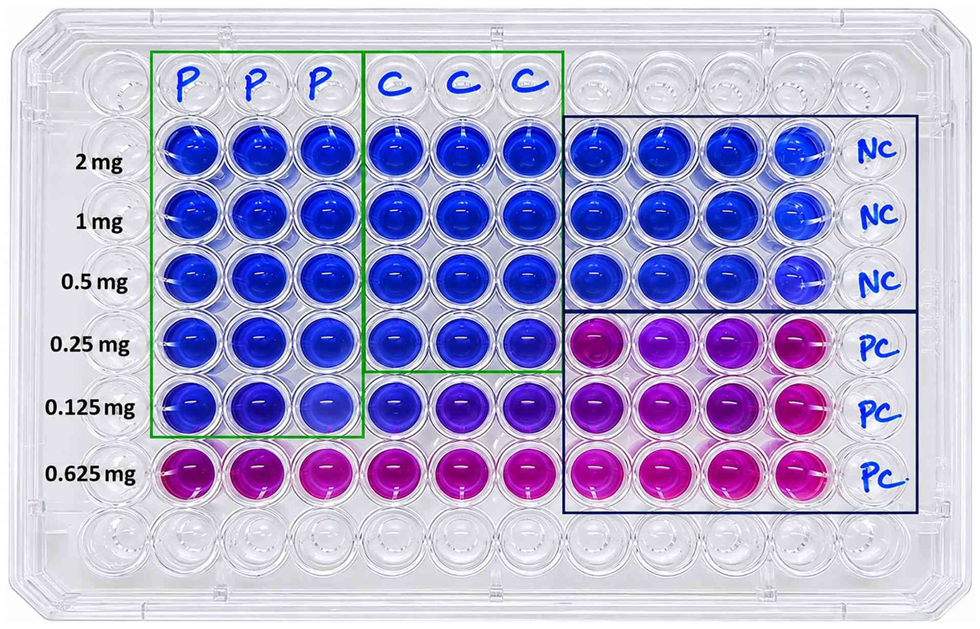
plant extracts was initially evaluated using the REMA. The MIC was
defined as the lowest concentration that completely inhibited the
color change of resazurin from blue to pink. The MIC values of the
methanolic extracts were determined to be 0.125 mg/ml (125 µg/ml)
for P. longum and 0.250 mg/ml (250 µg/ml) for C.
cretica against M. tuberculosis H37Rv (Fig. 1). These concentrations were
considered the effective inhibitory concentrations for subsequent
synergistic interaction studies.
Synergistic interaction between P.
longum and C. cretica
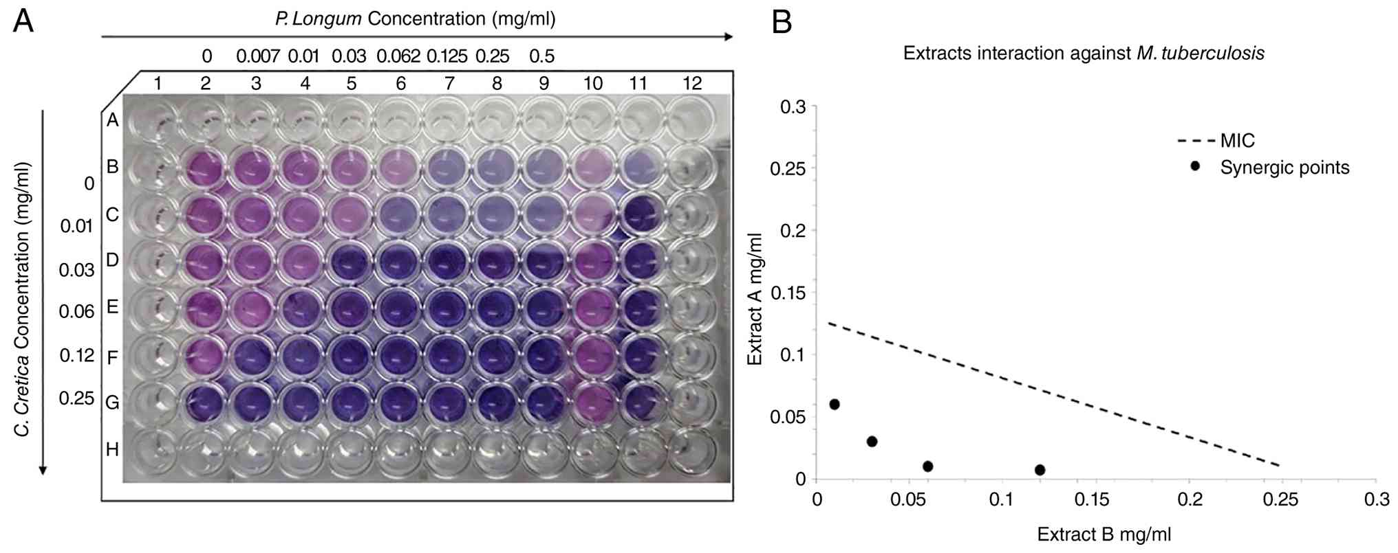
To evaluate the interaction between the two plant
extracts, a two-dimensional checkerboard assay was performed,
followed by isobologram analysis (Fig.
2). A synergistic interaction was observed between the extracts
(P. longum and C. cretica). In the presence of 0.125
mg/ml (125 µg/ml) C. cretica, the MIC of P. longum
decreased from 0.125 mg/ml (125 µg/ml) to 0.007 mg/ml (7 µg/ml),
representing an ~18-fold reduction. Likewise, the MIC of C.
cretica decreased from 0.250 mg/ml (250 µg/ml) to 0.010 mg/ml
(10 µg/ml) in the presence of 0.0625 mg/ml (62.5 µg/ml) P.
longum, corresponding to an ~25-fold reduction.
The calculated FICI was 0.096, which is well below
the accepted threshold for synergism (FICI ≤0.5). The isobologram
demonstrated that all inhibitory concentration combinations were
located below the line of additivity, confirming a strong
synergistic interaction between the two extracts (Fig. 2). These findings indicate that the
combination of P. longum and C. cretica enhanced the
antimycobacterial activity of the individual extracts and
substantially reduced the concentrations required to inhibit the
growth of M. tuberculosis.
Effect of plant extracts on the
expression of rpoB, gyrA and gyrB
To investigate the transcriptional response of M.
tuberculosis following exposure to the plant extracts, the
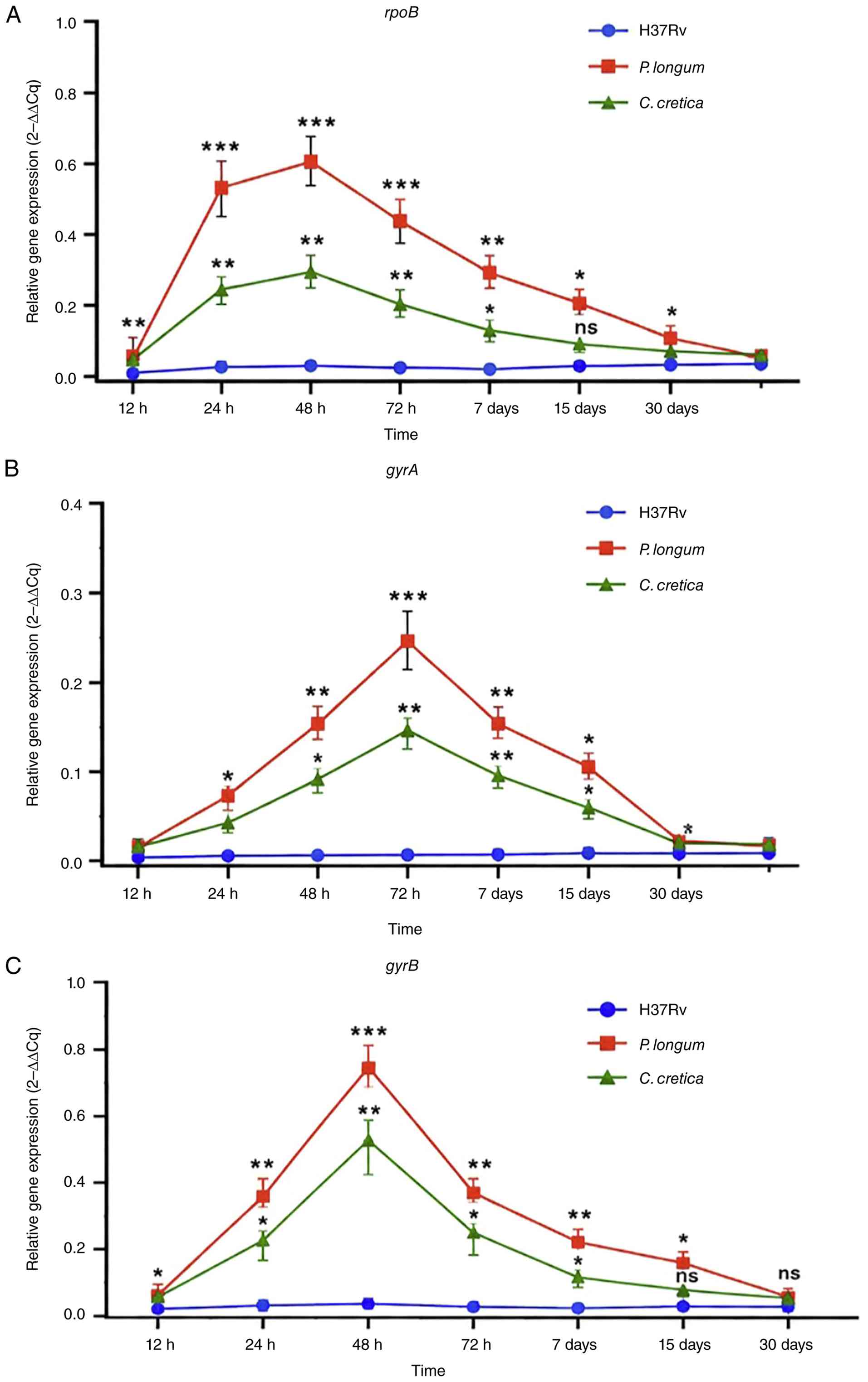
relative expression of rpoB, gyrA and gyrB was
analyzed using RT-qPCR at different time points following treatment
with the individual extracts (Fig.
3). Treatment with both P. longum and C. cretica
resulted in time-dependent changes in the expression profiles of
all three target genes. Changes in relative transcript abundance
were observed during the early stages of exposure, with pronounced
changes occurring between 24 and 72 h.
The expression profiles differed according to the
target gene, plant extract and duration of exposure. For
rpoB, treatment with P. longum resulted in a
pronounced change in relative expression, with the highest level
observed at 48 h, followed by a gradual decrease at later time
points. A similar temporal pattern was observed following treatment
with C. cretica, although the magnitude of the change was
lower than that observed with P. longum (Fig. 3A).
The expression of gyrA also exhibited a
time-dependent pattern, with the highest relative expression
observed at 72 h following treatment with both extracts. The
expression subsequently decreased at 7 and 15 days and approached
the control level at 30 days (Fig.
3B). For gyrB, the most notable change was observed at
48 h, followed by a progressive decrease during the subsequent time
points. The response to P. longum was generally greater than
that observed with C. cretica (Fig. 3C).
Two-way ANOVA demonstrated significant effects of
treatment and time on the expression of rpoB, gyrA
and gyrB, with a significant treatment vs. time interaction
for all three genes. Post hoc comparisons using Tukey's
multiple-comparisons test identified significant differences
between the treatment groups at several time points, as shown in
Fig. 3.
The temporal expression patterns indicate that
exposure to the plant extracts was associated with changes in the
transcription of genes involved in essential cellular processes,
including transcription (rpoB) and DNA replication
(gyrA and gyrB). The differences in the magnitude and
timing of these responses suggest that the transcriptional response
varied according to both the extract and the duration of exposure.
These changes in transcript abundance represent a molecular
response to phytochemical exposure.
Discussion
TB continues to represent a major global public
health challenge despite the availability of effective treatment
(4). The emergence of MDR and
extensively drug-resistant (XDR) M. tuberculosis strains,
together with prolonged treatment regimens, poor patient compliance
and drug-associated toxicity, has considerably reduced the
effectiveness of current therapeutic strategies (1-4).
Consequently, there is increasing interest in identifying novel
antimycobacterial agents from natural sources and developing
combination therapies capable of enhancing antimicrobial efficacy
while reducing the risk of resistance development (26).
The present study demonstrated that the methanolic
extracts of P. longum and C. cretica possess
significant antimycobacterial activity against M.
tuberculosis H37Rv when tested individually. More notably, the
checkerboard assay revealed a pronounced synergistic interaction
between the two extracts, resulting in substantial reductions in
the MICs of both plant extracts. Specifically, the MIC of P.
longum decreased from 125 to 7 µg/ml, whereas the MIC of C.
cretica decreased from 250 to 10 µg/ml, yielding a FICI of
0.096. According to the interpretative criteria proposed by Odds
(23), an FICI value ≤0.5 is
indicative of synergism. The exceptionally low FICI obtained in the
present study indicates a potent synergistic interaction and
suggests that the combination of these extracts is considerably
more effective than either extract alone.
The potential of plant-derived compounds to act
synergistically has attracted considerable attention in
antimicrobial research as phytochemicals frequently target multiple
bacterial pathways simultaneously (26,27).
Unlike conventional antibiotics, which generally interact with a
single molecular target, crude plant extracts contain structurally
diverse bioactive constituents capable of producing complementary
or additive biological effects (27). Such multitarget activity is
particularly advantageous against M. tuberculosis, whose
lipid-rich cell envelope, intrinsic resistance mechanisms and
metabolic adaptability often limit the efficacy of single-agent
therapies (27). Therefore, the
marked reduction in MIC observed in the present study supports the
concept that combining medicinal plants may enhance
antimycobacterial efficacy while reducing the concentrations
required for bacterial inhibition.
Preliminary phytochemical analysis demonstrated that
both extracts contained several classes of secondary metabolites,
including alkaloids, flavonoids, phenolic compounds, tannins,
terpenoids, glycosides, sterols, carbohydrates and proteins. Many
of these phytochemical classes have previously been reported to
exhibit antimycobacterial activity through diverse mechanisms,
including the disruption of the mycobacterial cell envelope,
interference with nucleic acid synthesis, the inhibition of
essential enzymes, the induction of oxidative stress and the
modulation of bacterial metabolic pathways (28). Since crude plant extracts comprise
numerous bioactive constituents, the synergistic activity observed
in the present study is likely attributable to the combined action
of multiple phytochemicals rather than a single active
compound.
Among the phytochemicals identified in P.
longum, piperine is considered a principal bioactive alkaloid
associated with several of the pharmacological properties of the
plant. Piperine exhibits antimicrobial, antioxidant,
anti-inflammatory, immunomodulatory and hepatoprotective activities
(14,15). Piperine has been shown to inhibit
bacterial efflux pumps, alter membrane permeability and enhance the
intracellular accumulation of antimicrobial agents, thereby
potentiating their antibacterial efficacy (15,29).
These mechanisms were not directly investigated in the present
study; the marked reduction in MIC observed during combination
treatment suggests that similar complementary interactions between
constituents of P. longum and C. cretica may
contribute to the enhanced antimycobacterial activity.
Likewise, C. cretica contains diverse
phenolic compounds, flavonoids and other secondary metabolites that
possess antimicrobial, antioxidant, anti-inflammatory and
hepatoprotective activities (16,28).
These phytochemicals may complement the activity of piperine by
targeting distinct bacterial pathways, thereby producing additive
or synergistic biological effects. Such multitarget interactions
are particularly advantageous for chronic bacterial infections as
simultaneous interference with multiple physiological processes
reduces the probability of resistance development (27,30).
The precise mechanism responsible for the observed
synergistic interaction remains to be elucidated. The enhanced
activity may result from increased membrane permeability, the
inhibition of bacterial efflux systems, the disruption of cell wall
integrity, interference with essential metabolic pathways or the
simultaneous action of multiple phytochemicals on distinct
molecular targets (30).
Alternatively, one extract may facilitate the intracellular
availability or activity of bioactive constituents present in the
other extract. Nevertheless, these mechanisms remain speculative
and require confirmation through bioactivity-guided fractionation,
metabolomic profiling, enzyme inhibition studies and transcriptomic
analyses.
To further investigate the molecular response of
M. tuberculosis to phytochemical exposure, the expression of
the essential genes rpoB, gyrA and gyrB was
evaluated using RT-q PCR. These genes encode proteins that play
indispensable roles in bacterial transcription and DNA replication
and are well-established molecular targets of first-line and
second-line anti-TB drugs, including rifampicin and
fluoroquinolones (31).
Consequently, changes in the expression of these genes may provide
valuable insight into the bacterial response following exposure to
antimicrobial agents.
The present study demonstrated that treatment with
the methanolic extracts of P. longum and C. cretica
resulted in time-dependent changes in the expression of
rpoB, gyrA and gyrB. The most pronounced
changes were observed between 24 and 72 h following treatment,
followed by a gradual reduction in relative transcript abundance at
later time points. The magnitude and timing of the response
differed among the three genes and between the two extracts, with
P. longum generally producing more notable changes in
relative expression than C. cretica. These findings indicate
that exposure to the plant extracts elicited measurable
transcriptional responses in genes associated with essential
cellular processes. The significant effects of treatment and time,
together with the treatment vs. time interaction observed, further
indicate that the transcriptional response was dependent on both
the extract and the duration of exposure.
The changes in transcript abundance should not be
interpreted as direct evidence of inhibition of RNA polymerase or
DNA gyrase activity. Instead, they are more likely to reflect
adaptive molecular responses to phytochemical-induced stress.
Bacterial exposure to antimicrobial agents is known to trigger
transcriptional reprogramming involving genes associated with DNA
replication, transcription, stress adaptation and metabolic
homeostasis as part of a coordinated physiological response to
environmental stress (29,32). Therefore, the differential expression
profiles observed in the present are consistent with a
transcriptional response of M. tuberculosis to phytochemical
exposure rather than providing direct evidence of enzyme
inhibition. Further investigations integrating transcriptomics,
proteomics and enzyme activity assays will be required to determine
the functional significance of these transcriptional
alterations.
The temporal changes in gene expression observed in
the present study indicate an early molecular response to
phytochemical exposure. The marked changes observed between 24 and
72 h, followed by reduced transcript abundance at later time
points, suggest that the transcriptional response may vary during
prolonged exposure. Bacterial exposure to antimicrobial agents can
involve early transcriptional reprogramming followed by downstream
physiological effects on cellular growth and survival (29,32).
Thus, the gene expression profiles observed herein provide
preliminary evidence of a time-dependent molecular response to the
individual plant extracts. However, the association between these
transcriptional changes and the observed antimycobacterial activity
requires further investigation.
The present study has several limitations which
should be mentioned. Gene expression analysis was performed using
the individual methanolic extracts of P. longum and C.
cretica rather than their synergistic combination, and
phytochemical analysis was limited to qualitative screening without
identification of the active constituents. Future studies are
required to evaluate the transcriptional effects of the extract
combination and employ bioactivity-guided fractionation with HPLC,
LC-MS, GC-MS and NMR to identify the compounds responsible for the
observed synergistic activity. The antimycobacterial activity was
evaluated only against the laboratory strain M. tuberculosis
H37Rv. Validation using clinical MDR/XDR isolates, together with
cytotoxicity, hepatoprotective and in vivo studies, is
required to establish the therapeutic efficacy and safety of this
plant combination. Despite these limitations, the marked reduction
in MIC values and the observed modulation of essential bacterial
genes support the potential of the P. longum-C. cretica
combination as a promising phytotherapeutic candidate for TB,
warranting further mechanistic and preclinical investigations.
In conclusion, the present study demonstrates that
the methanolic extracts of P. longum and C. cretica
possess significant antimycobacterial activity against M.
tuberculosis H37Rv and exhibit strong synergism in combination
(FICI, 0.096), with marked reductions in the MICs of both extracts.
Phytochemical screening identified alkaloids, flavonoids,
phenolics, tannins, terpenoids and glycosides, which may
collectively contribute to the observed synergistic effect. The
RT-qPCR analysis revealed the time-dependent modulation of the
essential rpoB, gyrA and gyrB genes following
treatment with the individual extracts, with the most pronounced
transcriptional changes observed during the early exposure period.
These findings provide preliminary molecular evidence of a
transcriptional response to phytochemical exposure. However, the
observed changes in transcript abundance do not establish the
direct inhibition of RNA polymerase or DNA gyrase activity. Further
studies involving the combined extracts, clinical M.
tuberculosis isolates, the identification of active
phytochemicals, protein-level analysis and functional enzyme assays
are required to elucidate the mechanisms underlying the observed
synergistic antimycobacterial activity.
Acknowledgements
The authors would like to express their gratitude
to Dr Aparup Das, Director ICMR-NIRTH, Jabalpur, India for his
support and guidance during this study. The authors also sincerely
acknowledge Mrs. Canina Luke, Mrs. Priyanka Raizada, Mrs. Mala
Prajapati, and Mr. Abhinav Pathak, Tuberculosis Laboratory
ICMR-National Institute of Research in Tribal Health (NIRTH),
Jabalpur, India for their respective assistance with the laboratory
work and experimental procedures associated with this study. The
manuscript has been approved by the Publication Screening Committee
of ICMR-NIRTH, Jabalpur, and assigned the no.
ICMR-NIRTH/PSC/04/2023.
Funding
Funding: The present study is part of the PhD program funded by
the Indian Council of Medical Research, New Delhi, India, in the
form of a Senior Research Fellowship.
Availability of data and materials
The data generated in the present study may be
requested from the corresponding author.
Authors' contributions
VKT, JB and RS conceived and designed the study.
VKT and NSP carried out the experiments. VKT, JB and RS were
involved in drafting and revision of the manuscript. VKT and JB
confirm the authenticity of all the raw data. All authors have read
and approved the final manuscript.
Ethics approval and consent to
participate
The Institutional Ethical Committee (IEC) of
ICMR-NIRTH, Jabalpur approved the study with a reference number
NIRTH/IEC/539/2018 dated 5/06/2018. The IEC review was obtained for
the overall proposed PhD/SRF research protocol, including the
laboratory investigations involving the antimycobacterial activity
of plant extracts against M. tuberculosis. The
approval/reference was required as part of the institutional
requirements for registration of the PhD research work and the
ICMR-SRF proposal. Taxonomically verified plants (P. longum,
voucher no. JN-BOT-1426 and 12 Page 16 of 21 ESub: 326588
21-07-2025 287 288 289 290 291 292 293 294 295 C. cretica
voucher no. JN-BOT-6532) were collected from Jawaharlal Nehru
Agricultural University, Jabalpur, Madhya Pradesh, India, in
July-August, 2019, with approval from the Faculty of Botanical
Garden, Department of Plant Physiology, Jawaharlal Nehru
Agricultural University, Jabalpur, India, following institutional
guidelines and regulations. The nomenclature of the selected plants
was compared with the Tropicos database (https://www.tropicos.org).
Patient consent for publication
Not applicable.
Competing interests
The authors declare that they have no competing
interests.
References
|
1
|
World Health Organization (WHO): Global
tuberculosis report 2024. WHO, Geneva, 2024.
|
|
2
|
Espinal MA: The global situation of
MDR-TB. Tuberculosis (Edinb). 83:44–51. 2003.PubMed/NCBI View Article : Google Scholar
|
|
3
|
Seung KJ, Keshavjee S and Rich ML:
Multidrug-resistant tuberculosis and extensively drug-resistant
tuberculosis. Cold Spring Harb Perspect Med.
5(a017863)2015.PubMed/NCBI View Article : Google Scholar
|
|
4
|
Nahid P, Dorman SE, Alipanah N, Barry PM,
Brozek JL, Cattamanchi A, Chaisson LH, Chaisson RE, Daley CL,
Grzemska M, et al: Official American Thoracic Society/Centers for
Disease Control and Prevention/Infectious Diseases Society of
America clinical practice guidelines: Treatment of drug-susceptible
tuberculosis. Clin Infect Dis. 63:e147–e195. 2016.PubMed/NCBI View Article : Google Scholar
|
|
5
|
Umar F, Hatta M, Husain DR, Natzir R,
Dwiyanti R, Junita AR and Primaguna MR: The effect of
anti-tuberculosis drug therapy on mRNA efflux pump gene expression
of Mycobacterium tuberculosis. New Microbes New Infect.
32(100609)2019.PubMed/NCBI View Article : Google Scholar
|
|
6
|
Silva PE, Bigi F, Santangelo MP, Romano
MI, Martín C, Cataldi A and Aínsa JA: Characterization of P55, a
multidrug efflux pump in Mycobacterium bovis and
Mycobacterium tuberculosis. Antimicrob Agents Chemother.
45:800–804. 2001.PubMed/NCBI View Article : Google Scholar
|
|
7
|
Jiménez-Arellanes MA, Gutiérrez-Rebolledo
GA, Meckes-Fischer M and León-Díaz R: Medicinal plant extracts and
natural compounds with a hepatoprotective effect against damage
caused by antitubercular drugs: A review. Asian Pac J Trop Med.
9:1141–1149. 2016.PubMed/NCBI View Article : Google Scholar
|
|
8
|
Gupta R, Thakur B, Singh P, Singh HB,
Sharma VD, Katoch VM and Chauhan SVS: Anti-tuberculosis activity of
selected medicinal plants against multidrug-resistant
Mycobacterium tuberculosis isolates. Indian J Med Res.
131:809–813. 2010.
|
|
9
|
Mishra SK, Tripathi G, Kishore N, Singh
RK, Singh A and Tiwari VK: Drug development against tuberculosis:
Impact of alkaloids. Eur J Med Chem. 137:504–544. 2017.PubMed/NCBI View Article : Google Scholar
|
|
10
|
Mazlun MH, Sabran SF, Mohamed M, Bakar MFA
and Abdullah Z: Phenolic compounds as promising drug candidates in
tuberculosis therapy. Molecules. 24(2449)2019.PubMed/NCBI View Article : Google Scholar
|
|
11
|
Kerantzas CA and Jacobs WR Jr: Origins of
combination therapy for tuberculosis: Lessons for future
antimicrobial development and application. mBio. 8:e01586–e0116.
2017.PubMed/NCBI View Article : Google Scholar
|
|
12
|
Chen P, Gearhart J, Protopopova M, Einck L
and Nacy CA: Synergistic interactions of SQ109, a new ethylene
diamine, with front-line antitubercular drugs in vitro. J
Antimicrob Chemother. 58:332–337. 2006.PubMed/NCBI View Article : Google Scholar
|
|
13
|
Fauziyah PN, Sukandar EY and Ayuningtyas
DK: Combination effect of antituberculosis drugs and ethanolic
extract of selected medicinal plants against multi-drug resistant
Mycobacterium tuberculosis isolates. Sci Pharm.
85(14)2017.PubMed/NCBI View Article : Google Scholar
|
|
14
|
Sunila ES and Kuttan G: Immunomodulatory
and antitumor activity of Piper longum Linn and piperine. J
Ethnopharmacol. 90:339–346. 2004.PubMed/NCBI View Article : Google Scholar
|
|
15
|
Hegeto LA, Caleffi-Ferracioli KR, Souza
JP, Almeida AL, Vasconcelos SSN, Barros ILE and Cardoso RF:
Promising antituberculosis activity of piperine combined with
antimicrobials: A systematic review. Microb Drug Resist.
25:120–126. 2019.PubMed/NCBI View Article : Google Scholar
|
|
16
|
Chaudhary S, Khosa RL, Priyank and
Rani S: A report on pharmacognostical and quality control
parameters of stem and root of Cressa cretica Linn.,
Convolvulaceae. J Pharm Res. 5:616–621. 2012.
|
|
17
|
Miotto P, Cabibbe AM, Borroni E, Degano M
and Cirillo DM: Role of disputed mutations in the rpoB gene
for rifampicin resistance in Mycobacterium tuberculosis. J
Clin Microbiol. 56:e01578–e0117. 2018.PubMed/NCBI View Article : Google Scholar
|
|
18
|
Aubry A, Pan XS, Fisher LM, Jarlier V and
Cambau E: Mycobacterium tuberculosis DNA gyrase: Interaction
with quinolones and correlation with antimycobacterial drug
activity. Antimicrob Agents Chemother. 48:1281–1288.
2004.PubMed/NCBI View Article : Google Scholar
|
|
19
|
Redfern J, Kinninmonth M, Burdass D and
Verran J: . Using soxhlet ethanol extraction to produce and test
plant material (essential oils) for their antimicrobial properties.
J Microbiol Biol Educ. 15:45–46. 2014.PubMed/NCBI View Article : Google Scholar
|
|
20
|
Ali S, Khan MR, Irfanullah Sajid M and
Zahra Z: Phytochemical investigation and antimicrobial appraisal of
Parrotiopsis jacquemontiana (Decne) Rehder. BMC Complement Altern
Med. 18(43)2018.PubMed/NCBI View Article : Google Scholar
|
|
21
|
Tamrakar VK, Bhat J, Parihar NS, Thakur H
and Rajasubramaniam S: In vitro, evaluation of anti-mycobacterial
active plant extracts against Mycobacterium tuberculosis
using BACTEC mycobacteria growth indicator tube 960 automated
system. J Pharmacogn Phytochem. 12:63–65. 2023.
|
|
22
|
Singh S, Kumar P, Sharma S, Mumbowa F,
Martin A and Durier N: Rapid identification and drug susceptibility
testing of Mycobacterium tuberculosis: Standard operating
procedure for non-commercial assays: Part 3: Colorimetric redox
indicator assay v1.3.12. J Lab Physicians. 4:120–126.
2012.PubMed/NCBI View Article : Google Scholar
|
|
23
|
Botelho MG: Fractional inhibitory
concentration index of combinations of antibacterial agents against
cariogenic organisms. J Dent. 28:565–570. 2000.PubMed/NCBI View Article : Google Scholar
|
|
24
|
Odds FC: Synergy, antagonism, and what the
chequerboard puts between them. J Antimicrob Chemother.
52(1)2003.PubMed/NCBI View Article : Google Scholar
|
|
25
|
Simões AE, Pereira DM, Amaral JD, Nunes
AF, Gomes SE, Rodrigues PM, Lo AC, D'Hooge R, Steer CJ, Thibodeau
SN, et al: Efficient recovery of proteins from multiple source
samples after TRIzol(®) or TRIzol(®)LS RNA
extraction and long-term storage. BMC Genomics.
14(181)2013.PubMed/NCBI View Article : Google Scholar
|
|
26
|
Li G, Zhang J, Guo Q, Jiang Y, Wei J, Zhao
LL, Zhao X, Lu J and Wan K: Efflux pump gene expression in
multidrug-resistant Mycobacterium tuberculosis clinical
isolates. PLoS One. 10(e0119013)2015.PubMed/NCBI View Article : Google Scholar
|
|
27
|
Livak KJ and Schmittgen TD: Analysis of
relative gene expression data using real-time quantitative PCR and
the 2(-Delta Delta C(T)) method. Methods. 25:402–408.
2001.PubMed/NCBI View Article : Google Scholar
|
|
28
|
Kumar G and Amrutha C: Natural products
and their analogues acting against Mycobacterium
tuberculosis: A recent update. Drug Dev Res. 84:779–804.
2023.PubMed/NCBI View Article : Google Scholar
|
|
29
|
Singh V, Dziwornu GA and Chibale K: The
implication of Mycobacterium tuberculosis-mediated
metabolism of targeted xenobiotics. Nat Rev Chem. 7:340–354.
2023.PubMed/NCBI View Article : Google Scholar
|
|
30
|
Li S, Lv M and Xu H: Overview of piperine:
Bioactivities, total synthesis, structural modification, and
structure-activity relationships. Mini Rev Med Chem. 23:917–940.
2023.PubMed/NCBI View Article : Google Scholar
|
|
31
|
Murase LS, de Souza JVP, Meneguello JE,
Palomo CT, Milaré ÁCFHR, Negri M, Siqueira VLD, Demarchi IG,
Teixeira JJV and Cardoso RF: Antibacterial and immunological
properties of piperine evidenced by preclinical studies: A
systematic review. Future Microbiol. 18:1279–1299. 2023.PubMed/NCBI View Article : Google Scholar
|
|
32
|
Han J, Zhang S, He J and Li T: Piperine:
Chemistry and biology. Toxins(Basel). 15(696)2023.PubMed/NCBI View Article : Google Scholar
|