Introduction
In 2008, the estimated number of deaths from
malignant liver tumors was 47,830 in males; hepatic cancer is the
second most common cause of cancer-related mortality worldwide
(1). In 2010, cancers of the liver
and intrahepatic bile ducts were also the leading cause of
cancer-related mortality in males (36.8/100,000) in Taiwan
(2). As hepatocellular carcinoma
(HCC) is resistant to many of the available chemotherapeutic
agents, HCC remains a challenging disease to treat worldwide
(3). Sann-Joong-Kuey-Jian-Tang
(SJKJT), a traditional Chinese medicine, has been prescribed as
complementary medication for a number of types of solid cancer in
Taiwan. SJKJT consists of 17 species of medicinal herbs: Coptis
chinensis Franch, Cimicifuga heracleifolia Komar,
Scutellaria baicalensis Georgi, Gentiana scabra
Bunge, Trichosanthes cucumeroides Maxim, Phellodendron
amurense Rupr, Anemarrhena asphodeloides Bunge,
Platycodon grandiflorum, Laminaria japonica Aresch,
Bupleurum scorzoneri folium Willd (Bupleurum Chinese DC),
Glycyrrhiza uralensis Fisch, Sparganium toloniferum
Buch, Curcuma aeruginosa Roxb, Forsythia suspense
Vahl, Pueraria lobata Ohwi, Paeonia lactiflora Pall
and Angelica sinensis Diels (4). In our previous studies, it was shown
that SJKJT inhibits colon cancer colo 205 cells by inducing
autophagy in vitro(5) or
increasing TNF-α expression to induce apoptosis in vitro and
in vivo(6). However, the
anticancer effects of SJKJT in hepatic cancer have not been
understood. The present study focused on the anticancer effect and
molecular mechanisms of SJKJT in HCC, using human hepatocellular
carcinoma Hep-G2 cells.
Materials and methods
Materials
The crude extract of SJKJT was obtained from Chuang
Song Zong Pharmaceutical Co., Ltd. (Ligang Plant, Taiwan).
[3-(4,5-dimethylthiazol-2-y1)-2,5-diphenyltetrazolium bromide] MTT,
sodium deoxycholate, leupeptin, Triton X-100, Tris-HCl,
ribonuclease-A, sodium pyruvate, HEPES, dimethyl sulfoxide (DMSO)
and RPMI-1640 were obtained from Sigma-Aldrich (St. Louis, MO,
USA). The Hep-G2 human hepatic cancer cell line (BCRC number:
60025) was obtained from the Food Industry Research and Development
Institute (Hsinchu, Taiwan). Potassium phosphate and TE buffer were
purchased from Merck Co. (Darmstadt, Germany). Fetal bovine serum
(FBS) and glutamine were obtained from Gibco BRL (Grand Island, NY,
USA). Mouse anti-β-actin and penicillin-streptomycin were obtained
from Sigma-Aldrich. BioMax film was obtained from Kodak. The FADD
(no. 2782), Bax (no. 2774), Bcl-xL (no. 2764), Mcl-1 (no. 2764),
TCTP (no. 2764), Caspase-8 (no. 9502) and TNF-α (no. 3707)
antibodies were all obtained from Cell Signaling Technology Inc.
(Beverly, MA, USA). Fas (NB120-13550) and Caspase-3 (NB500-210)
antibodies were obtained from Novus Biologicals (Littleton, CO,
USA). Other materials and reagents not specified were obtained from
Sigma or Merck.
Cell culture
The Hep-G2 cells were obtained from the Food
Industry Research and Development Institute (Hsinchu, Taiwan), and
maintained in RPMI-1640 medium containing 10% FBS and 1%
penicillin/streptomycin (10,000 U/ml penicillin, 10 mg/ml
streptomycin) at 37°C in a humidified atmosphere containing 5%
CO2.
Cytotoxicity assay
The cells were plated in 96-well plates at a density
of 1×10⁴ cells/well for 16–20 h. Thereafter, the cells
were treated with various concentrations of SJKJT for 24, 48 and 72
h. Subsequently, the cells were incubated with 1 mg/ml of MTT in
fresh RPMI medium for 2 h. The surviving cells converted MTT to
formazan by forming a blue-purple color when dissolved in DMSO. The
intensity of formazan was measured at 590 nm using a microplate
reader. The relative percentage of cell viability was calculated by
dividing the absorbance of treated cells by that of the control in
each experiment.
Cell cycle analysis
Hep-G2 cells treated with various concentrations of
SJKJT for different durations were measured using a flow
cytometer.
Hep-G2 cells were treated with various
concentrations (0, 0.75, 1 and 1.5 mg/ml) of SJKJT for 48 h or with
various concentrations (0, 0.5, 0.75 and 1 mg/ml) of SJKJT for 72
h, and then collected and fixed with ice-cold ethanol (70%)
overnight at −20°C. The cell pellets were then treated with
propidium iodide (PI) solutions (containing 100 μg/ml RNase) for 30
min at 37°C. Subsequently, the samples were analyzed by a Cytomics™
FC500 Flow Cytometer (Beckman Coulter, Miami, FL, USA). A minimum
of 10,000 cells was analyzed to determine DNA content, and the
percentage of cells in each cell cycle phase was quantified.
Immunocytochemical staining
Hep-G2 cells were treated with various
concentrations (0, 0.75, 1 and 1.5 mg/ml) of SJKJT for 48 h or with
various concentrations (0, 0.5, 0.75 and 1 mg/ml) of SJKJT for 72
h, and then washed with PBS. Fixation with 50% acetone and 50%
methanol solution overnight at 4°C was performed, the cells were
washed three times with PBS, and non-specific binding sites were
blocked in PBS containing 0.1% BSA for 1 h at room temperature.
Thereafter, the cells were separately incubated with rabbit
anti-caspase 3 (1:20) antibody in PBS containing 0.1% BSA overnight
at 4°C, and washed three times with PBS. The cells were then
incubated with anti-rabbit FITC (1:200) in PBS containing 0.1% BSA
for 1 h at room temperature, and washed three times with PBS. The
nuclei were stained with 5 μg/ml PI, respectively. After staining,
the samples were immediately examined under an Olympus IX81
microscope (Olympus, Tokyo, Japan).
Western blotting
The effects of SJKJT on the protein expression of
Fas, TNF-α, FADD, Caspase-8, Bax, Caspase-3, Mcl-1, TCTP and Bcl-xl
in Hep-G2 cells was measured by western blotting.
Hep-G2 cells were treated with various
concentrations (0, 0.75, 1 and 1.5 mg/ml) of SJKJT for 48 h and the
protein expression levels of Fas, TNF-α, FADD, Caspase-8, Bax,
Caspase-3, Mcl-1, TCTP and Bcl-xl were evaluated by western
blotting.
Hep-G2 cells were treated with various
concentrations (0, 0.5, 0.75 and 1 mg/ml) of SJKJT for 72 h and the
protein expression levels of Fas, TNF-α, FADD, Caspase-8, Bax,
Caspase-3, Mcl-1, TCTP and Bcl-xl were evaluated by western
blotting.
Hep-G2 cells were treated with SJKJT (1 mg/ml) for
different durations (0, 24, 48 and 72 h) and then the protein
expression levels of Bcl-xl, Mcl-1, TCTP and Bax were evaluated by
western blotting.
Following the termination of drug treatment, the
cells were lysed in ice-cold whole cell extract buffer containing
the protease inhibitors. The lysate was vibrated for 30 min at 4°C
and centrifuged at 9.4 × g for 10 min. Protein concentration was
measured using a BCA protein assay kit (Pierce, Rockford, IL, USA).
Equal amounts of proteins were subjected to electrophoresis using
12% sodium dodecyl sulfate-polyacrylamide gels. To verify equal
protein loading and transfer, proteins were then transferred to
polyvinylidene difluoride membranes and the membranes were blocked
overnight at 4°C using blocking buffer [5% non-fat dried milk in
solution containing 50 mM Tris/HCl (pH 8.0), 2 mM CaCl2,
80 mM sodium chloride, 0.05% Tween 20 and 0.02% sodium azide]. The
membranes were then incubated for 2 h at 25°C with specific primary
antibody followed by anti-rabbit or anti-mouse immunoglobulin
G-horseradish peroxidase conjugated secondary antibodies. The
membranes were washed three times for 10 min with washing solution.
The protein bands were then visualized on X-ray film using the
enhanced chemiluminescence detection system (PerkinElmer Life and
Analytical Sciences, Boston, MA, USA).
Statistical analysis
Values were presented as the means ± standard
deviation (SD). The Student's t-test was used to analyze
statistical significance. P<0.05 was considered to indicate a
statistically significant difference for all the tests.
Results and Discussion
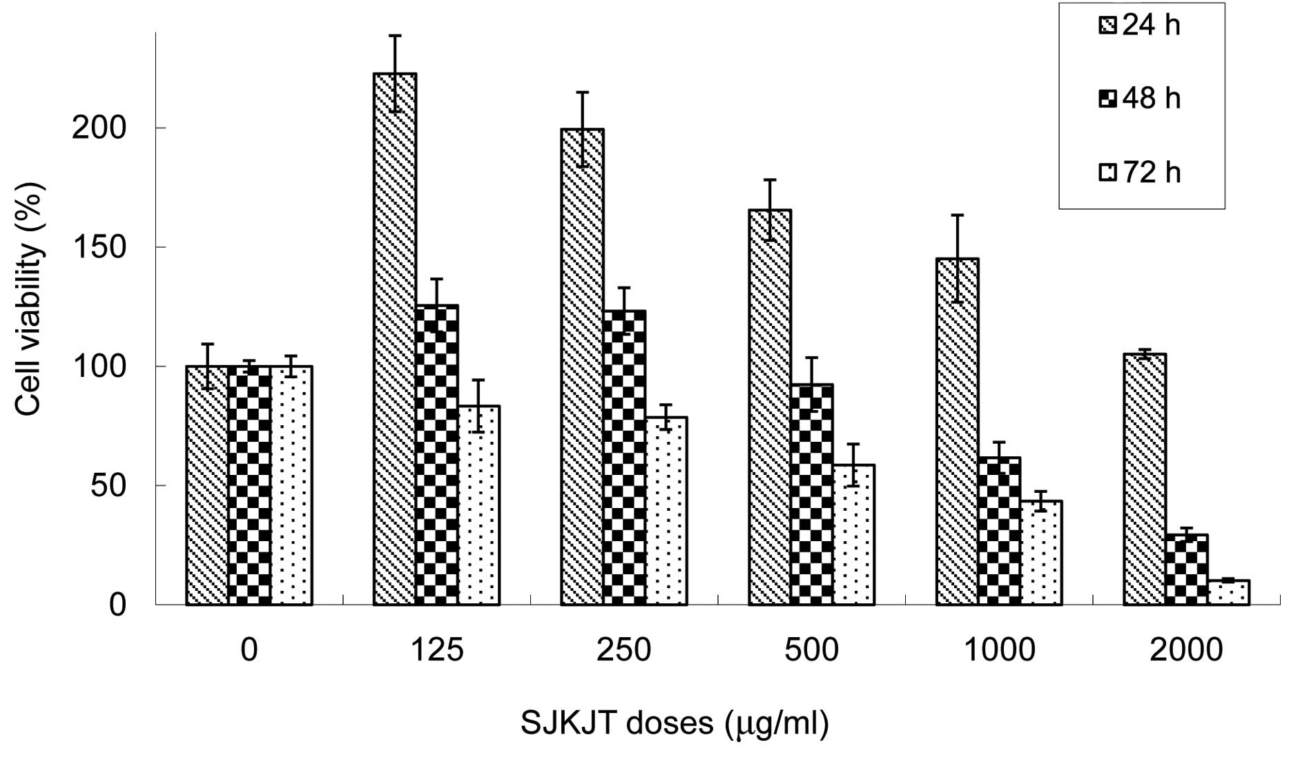
The effects of SJKJT on the viability of
Hep-G2 cells
Our results revealed that SJKJT inhibits the
proliferation of Hep-G2 cells in a time- and dose-dependent manner.
The half-maximum inhibitory concentration (IC50) was
1.48 and 0.94 mg/ml for 48 and 72 h, respectively (Fig. 1).
SJKJT induced apoptosis in Hep-G2
cells
The Hep-G2 cells were plated in 6 cm dishes at a
density of 1×10⁶ cells per dish and were then treated
with various concentrations of SJKJT for different durations (48
and 72 h). The cell cycles were analyzed by fluorescence-activated
cell sorting (FACS). When Hep-G2 cells were cultured with various
concentrations (0, 0.75, 1 and 1.5 mg/ml) of SJKJT for 48 h, the
sub-G1 phase percentages were 1.2±0.15, 4.9±0.59, 5.8±0.32 and
6.9±0.35%, respectively. When Hep-G2 cells were cultured with
various concentrations (0, 0.5, 0.75 and 1 mg/ml) of SJKJT for 72
h, the sub-G1 phase percentages were 1.5±0.10, 7.3±0.64, 13.7±1.4
and 13.0±0.78%, respectively (Fig. 2A
and B). The FACS results showed that treatment with SJKJT
induced apoptosis.
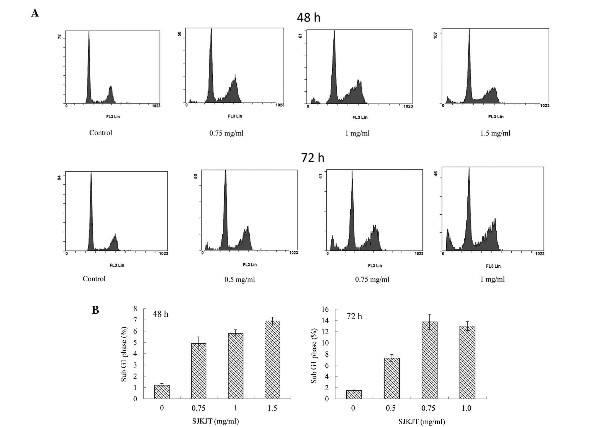 | Figure 2Effect of SJKJT on the cell cycle of
Hep-G2 cells. (A) The Hep-G2 cells were treated with SJKJT for
various durations. The cell cycles were analyzed by FACS. (B) When
Hep-G2 cells were cultured with SJKJT (0, 0.75, 1 and 1.5 mg/ml)
for 48 h, the sub-G1 phase percentages were 1.2±0.15, 4.9±0.59,
5.8±0.32 and 6.9±0.35%, respectively. Hep-G2 cells were cultured
with SJKJT (0, 0.5, 0.75 and 1 mg/ml) for 72 h, the sub-G1 phase
percentages were 1.5±0.10, 7.3±0.64, 13.7±1.4 and 13.0±0.78%,
respectively. SJKJT, Sann-Joong-Kuey-Jian-Tang; FACS,
fluorescence-activated cell sorting. |
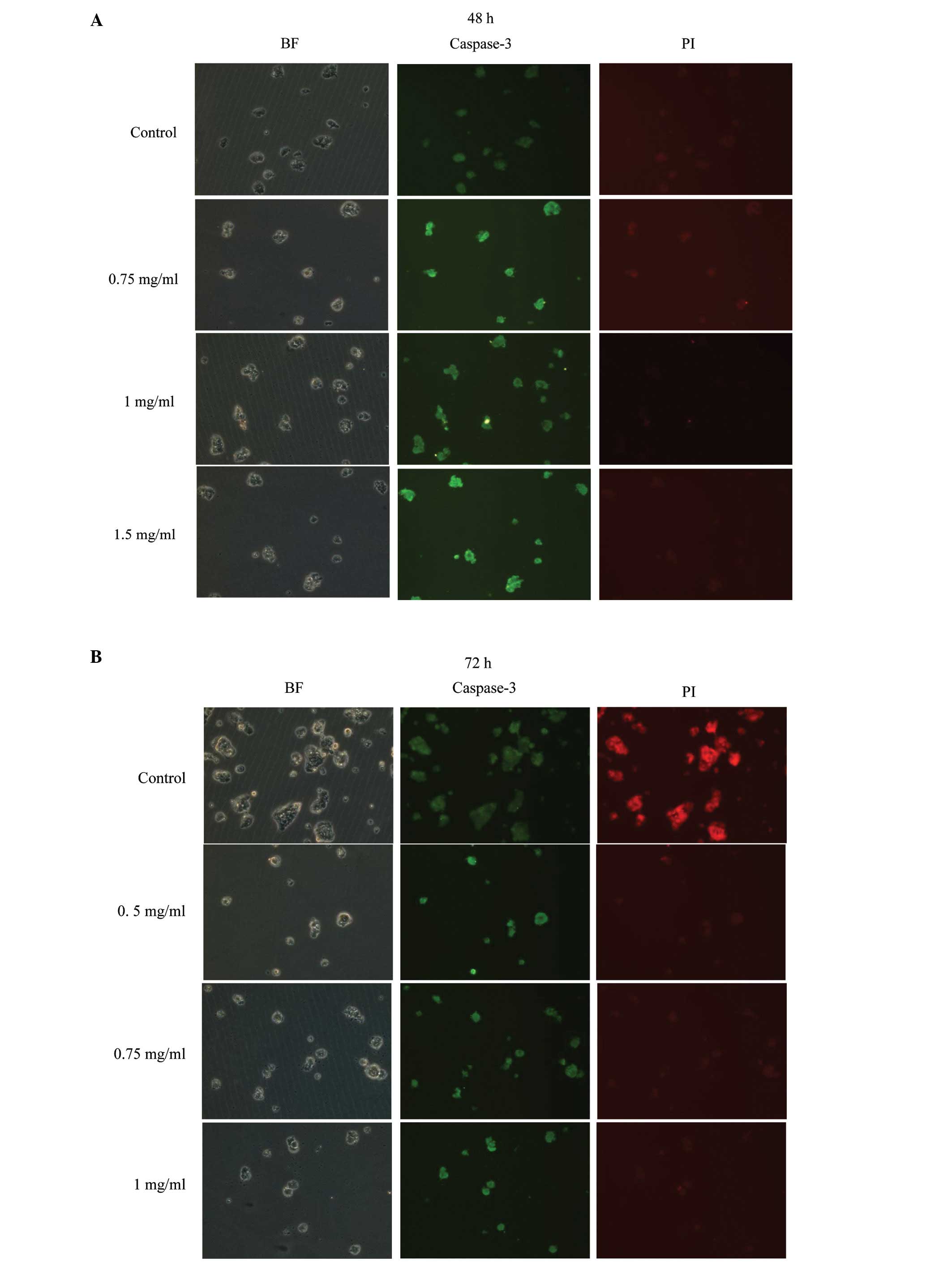
Immunocytochemical analysis
Hep-G2 cells were treated with various
concentrations (0, 0.75, 1 and 1.5 mg/ml) of SJKJT for 48 h or with
various concentrations (0, 0.5, 0.75 and 1 mg/ml) of SJKJT for 72
h, and then were fixed with 4% paraformaldehyde to allow for the
detection of Caspase 3 by staining with antibodies. These results
revealed that Hep-G2 cells treated with SJKJT have increased
expression of Caspase-3 at 48 h (Fig.
3A) and 72 h (Fig. 3B), and
the effect was dose-dependent. These results suggest that SJKJT
induces apoptosis in Hep-G2 cells.
The effects of SJKJT on the protein
expression of Fas, TNF-α, FADD, Caspase-8, Bax, Caspase-3, Mcl-1,
TCTP and Bcl-xl in Hep-G2 cells
Hep-G2 cells were treated with various
concentrations (0, 0.75, 1 and 1.5 mg/ml) of SJKJT for 48 h and the
protein expression levels of Fas, TNF-α, FADD, Caspase-8, Bax,
Caspase-3, Mcl-1, TCTP and Bcl-xl were evaluated by western
blotting. The results demonstrated that SJKJT can increase the
protein expression level of Fas (Fig.
4A), FADD (Fig. 4B), TNF-α
(Fig. 4C), Caspase-8 (Fig. 4D), Bax (Fig. 4E) and Caspase-3 (Fig. 4F), but the protein expression level
of Mcl-1, TCTP and Bcl-xl did not change significantly (Fig. 5). It is well documented that when
the Fas receptor binds to an activator, the receptor forms a
death-inducing signaling complex, resulting in the active form of
Caspase-8 cleaving and activating Caspase-3, leading to apoptosis
(7,8). Tumor necrosis factor α (TNF-α) is
produced by macrophages, and binds to TNF receptor type 1 and
activates to form the death-inducing signaling complex, resulting
in the recruitment of caspases and subsequently leading to cell
apoptosis (9,10).
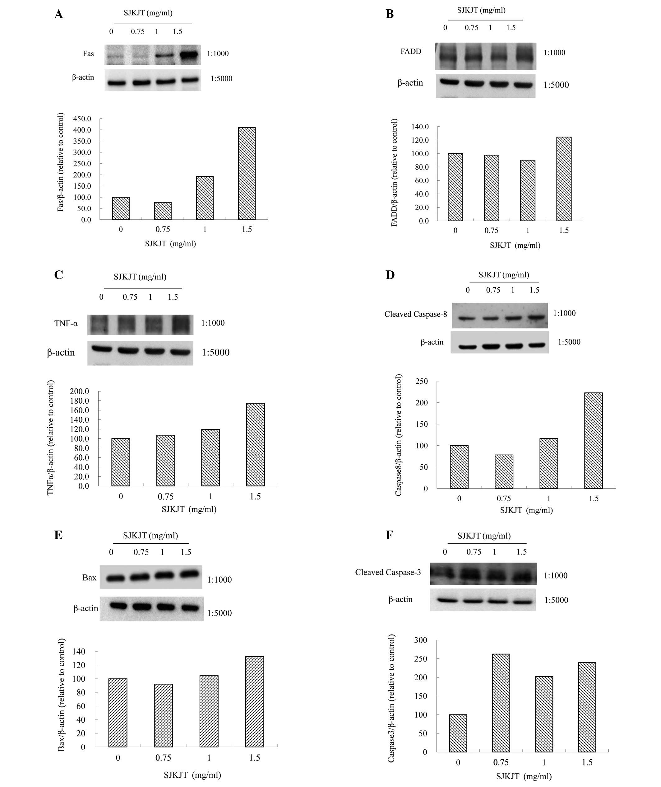 | Figure 4The effects of SJKJT on the protein
expression of Fas, TNF-α, FADD, Caspase-8, Bax and Caspase-3 in
Hep-G2 cells. Hep-G2 cells were treated with SJKJT (0, 0.75, 1 and
1.5 mg/ml) for 48 h and the protein expression was evaluated by
western blotting. The results showed that SJKJT can increase the
protein expression level of (A) Fas, (B) FADD, (C) TNF-α, (D)
Caspase-8, (E) Bax and (F) Caspase-3. SJKJT,
Sann-Joong-Kuey-Jian-Tang. |
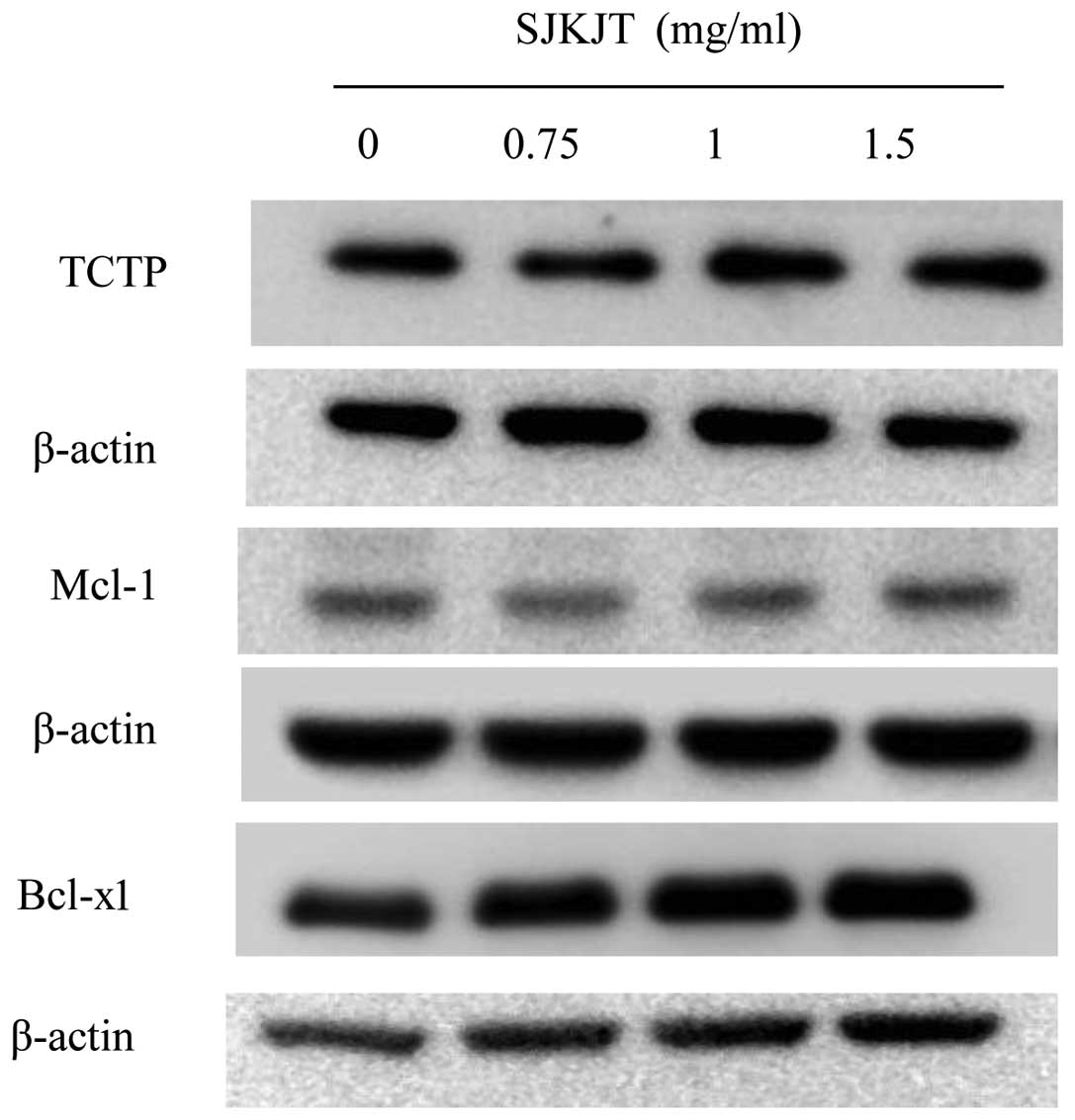
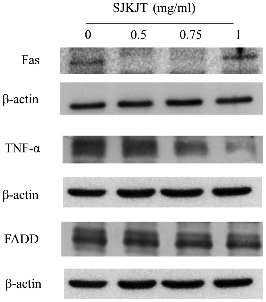
Hep-G2 cells were treated with various
concentrations (0, 0.5, 0.75 and 1 mg/ml) of SJKJT for 72 h and the
protein expression levels of Fas, TNF-α, FADD, Caspase-8, Bax,
Caspase-3, Mcl-1, TCTP and Bcl-xl were evaluated by western
blotting. The results showed that SJKJT can decrease the protein
expression level of MCl-1 (Fig.
6A) and TCTP (Fig. 6B). The
results also showed that the protein expression level of Fas was
increased, but TNF-α and FADD did not change significantly
(Fig. 7). It is well documented
that TCTP overexpression inhibits apoptosis by binding to Mcl-1 and
antagonizing Bax (4,11–13).
Our results showed that SJKJT inhibits TCTP and Mcl-1 expression in
Hep-G2 cells. One of the molecular mechanisms by which SJKJT
inhibits Hep-G2 cells may be through decreasing TCTP and Mcl-1
expression.
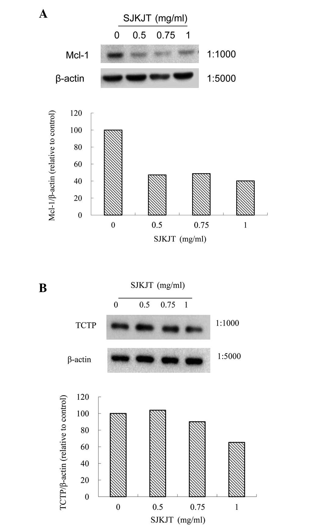
Hep-G2 cells were treated with SJKJT (1 mg/ml) for
different durations (0, 24, 48 and 72 h) and then the protein
expression levels of Fas, Caspase-8, Caspase-3, Bcl-xl, Mcl-1, TCTP
and Bax were evaluated by western blotting. The results revealed
that increasing the treatment duration of SJKJT can decrease the
protein expression level of Mcl-1 and TCTP (Fig. 8A), but increase the protein
expression level of Bcl-xl, Fas, Caspase-8, Caspase-3 and Bax
(Fig. 8B). SJKJT did not
demonstrate significant toxicity in normal cells and it has been
well documented to inhibit human breast cancer by inducing
apoptosis (14). Our results also
demonstrated that SJKJT induces apoptosis in Hep-G2 cells. The
molecular mechanisms may be through the downregulation of protein
expression of TCTP and Mcl-1, but upregulation of Fas, TNF-α,
Caspase-8, Caspase-3 and Bax expression. This is the first study to
demonstrate that SJKJT inhibits human hepatocellular carcinoma
Hep-G2 cells through the extrinsic and intrinsic pathways. The
therapeutic potential for SJKJT to treat human HCC requires further
study.
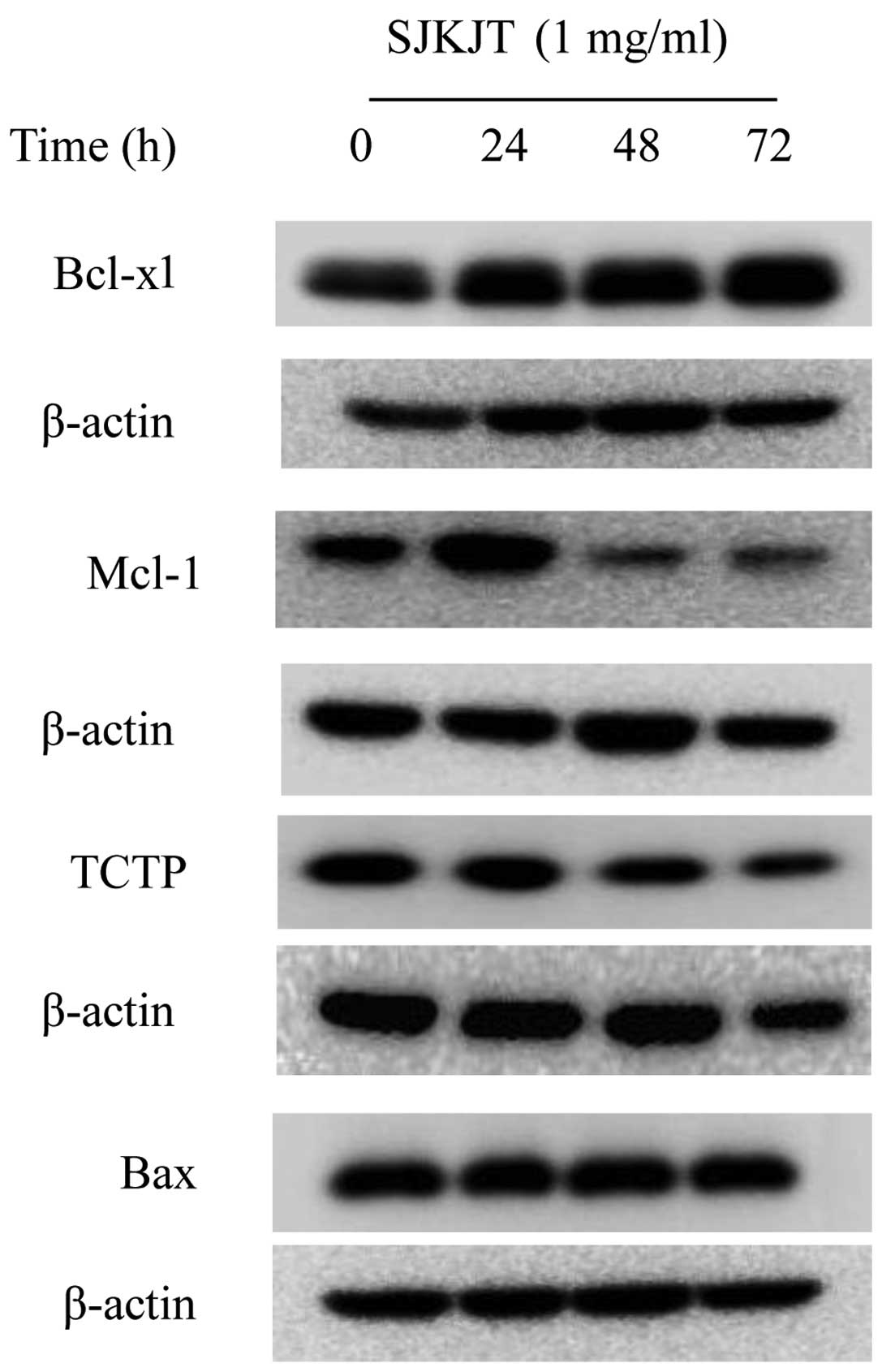 | Figure 8Effects of SJKJT on the protein
expression of Bcl-xl, Mcl-1, TCTP, Fas, Caspase-8, Caspase-3 and
Bax in Hep-G2 cells. Hep-G2 cells were treated with SJKJT (1 mg/ml)
for different durations (0, 24, 48 and 72 h) and then the protein
expression was evaluated by western blotting. The results showed
that SJKJT can decrease the protein expression level of (A) Mcl-1
and TCTP, but increase the protein expression level of (B) Bcl-xl,
Fas, Caspase-8, Caspase-3 and Bax. SJKJT,
Sann-Joong-Kuey-Jian-Tang. |
Acknowledgements
This study was supported by grant 100-CCH-ICO-06-3
from Changhua Christian Hospital.
References
|
1
|
Jemal A, Bray F, Center MM, Ferlay J, Ward
E and Forman D: Global cancer statistics. CA Cancer J Clin.
61:69–90. 2011. View Article : Google Scholar
|
|
2
|
Statistics of Causes of Death, 2010.
Department of Health, Executive Yuan; Taipei, Taiwan, R.O.C: 153.
2012
|
|
3
|
Leung TW and Johnson PJ: Systemic therapy
for hepatocellular carcinoma. Semin Oncol. 28:514–520. 2001.
View Article : Google Scholar : PubMed/NCBI
|
|
4
|
Hsu YL, Yen MH, Kuo PL, et al:
San-Zhong-Kui-Jian-Tang, a traditional Chinese medicine
prescription, inhibits the proliferation of human breast cancer
cells by blocking cell cycle progression and inducing apoptosis.
Biol Pharm Bull. 29:2388–2394. 2006. View Article : Google Scholar
|
|
5
|
Cheng CY, Lin YH and Su CC:
Sann-Joong-Kuey-Jian-Tang increases the protein expression of
microtubule-associated protein II light chain 3 in human colon
cancer colo 205 cells. Mol Med Rep. 2:707–711. 2009.
|
|
6
|
Cheng CY, Lin YH and Su CC:
Sann-Joong-Kuey-Jian-Tang up-regulates the protein expression of
Fas and TNF-α in colo 205 cells in vivo and in vitro.
Mol Med Rep. 3:63–67. 2010.PubMed/NCBI
|
|
7
|
Cohen CGM: Caspases, the executioners of
apoptosis. Biochem J. 326:1–16. 1997.
|
|
8
|
Lowe LSW, Cepero E and Evan G: Intrinsic
tumour suppression. Nature. 432:307–315. 2004. View Article : Google Scholar : PubMed/NCBI
|
|
9
|
Carswell EA, Old LJ, Kassel RL, et al: An
endotoxin-induced serum factor that causes necrosis of tumors. Proc
Natl Acad Sci USA. 72:3666–3670. 1975. View Article : Google Scholar : PubMed/NCBI
|
|
10
|
Gaur U and Aggarwal BB: Regulation of
proliferation, survival and apoptosis by members of the TNF
superfamily. Biochem Pharmacol. 66:1403–1408. 2003. View Article : Google Scholar : PubMed/NCBI
|
|
11
|
Graidist P, Phongdara A and Fujise K:
Antiapoptotic protein partners fortilin and MCL1 independently
protect cells from 5-fluorouracil-induced cytotoxicity. J Biol
Chem. 279:40868–40875. 2004. View Article : Google Scholar : PubMed/NCBI
|
|
12
|
Liu H, Peng HW, Cheng YS, et al:
Stabilization and enhancement of the antiapoptotic activity of
mcl-1 by TCTP. Mol Cell Biol. 25:3117–3126. 2005. View Article : Google Scholar : PubMed/NCBI
|
|
13
|
Zhang D, Li F, Weidner D, et al: Physical
and functional interaction between myeloid cell leukemia 1 protein
(MCL1) and Fortilin. The potential role of MCL1 as a fortilin
chaperone. J Biol Chem. 277:37430–37438. 2002. View Article : Google Scholar : PubMed/NCBI
|
|
14
|
Susini L, Besse S, Duflaut D, et al: TCTP
protects from apoptotic cell death by antagonizing bax function.
Cell Death Differ. 15:1211–1220. 2008. View Article : Google Scholar : PubMed/NCBI
|