Introduction
Osteosarcoma is an aggressive type of cancer
commonly observed in adolescents and children. With the
introduction of intensive chemotherapy, the cure rate of patients
with localized osteosarcoma has improved from 15 to 20%, and
achieved to ~70% with surgery alone (1). Nonetheless, approximately one-third
of osteosarcoma patients experience recurrent or progressive
disease, of which the majority of cases are due to the development
of resistance to chemotherapeutic drugs by the osteosarcoma cells
(2). Therefore, drug resistance
has been a hindrance in achieving improved cure rates.
Arsenic trioxide (As2O3)
compounds have been used in Traditional Chinese Medicine for
thousands of years. The therapeutic use of these compounds in acute
promyelocytic leukemia (APL) was described in the 1970s. In
addition to APL, As2O3 has been effective in
the treatment of certain solid tumors, such as gastric cancer, lung
cancer, breast cancer and hepatocellular carcinoma (3,4).
Studies have demonstrated that As2O3 induced
cytotoxic effects in these tumors in a dose- and time-dependent
manner (5). However, the detailed
mechanisms of As2O3 cytotoxicity remain to be
further elucidated.
Stathmin is a microtubule regulatory phosphoprotein
that is important in the assembly of the mitotic spindle. Stathmin
is a key regulator of the microtubule network and provides an
attractive therapeutic target in cancer treatment (6,7).
Previous studies have demonstrated that small interfering (si)RNA
or antisense-mediated downregulation of stathmin expression may
result in proliferation inhibition and chemosensitivity enhancement
in human osteosarcoma cells (8,9).
In the present study, the effect of
As2O3 on MG63 human doxorubicin-resistant
osteosarcoma cells was examined, along with the molecular
mechanisms of the effects. The present study was conducted by
analyzing the effects of As2O3 on cell
proliferation, cell apoptosis, and stathmin mRNA and protein
expression levels in MG63 and MG63/dox human osteosarcoma
cells.
Materials and methods
Regents
As2O3 was obtained from
ShuangLu Pharmaceutical Co., Ltd. (Beijing, China), doxorubicin
(ADM) was provided by Pfizer Pharmaceuticals Corporation (New York,
NY, USA), propidium iodide (PI) was purchased from Sigma-Aldrich
(St. Louis, MO, USA), Cell Counting Kit-8 (CCK-8) was obtained from
Dojindo Laboratories (Kumamoto, Japan), rabbit monoclonal antibody
against stathmin was purchased from Abgent (1:5,000, San Diego, CA,
USA) and antibody against β-actin was purchased from CoWin
Bioscience Co., Ltd. (Beijing, China).
Cell lines
The MG63 human osteosarcoma parental cell line was
obtained from the Institute of Biochemistry and Cell Biology,
Chinese Academy of Sciences (Shanghai, China). The MG63/dox human
multidrug-resistant (MDR) osteosarcoma cell line with
P-glycoprotein overexpression, which was selected in a step-wise
manner through exposing drug-sensitive MG63 cells to increasing
doses of ADM, was provided by Dr Yoshio Oda (Graduate School of
Medical Sciences, Kyushu University, Fukuoka, Japan). These cell
lines were grown in high-glucose Dulbecco’s modified Eagle’s medium
(DMEM; Hyclone, Logan, Utah, USA) supplemented with 10% fetal
bovine serum (FBS; Hyclone), 100 units/ml penicillin and 100 g/ml
streptomycin (Gibco-BRL, Carlsbad, CA, USA) at 37°C in a humidified
5% CO2 atmosphere.
Cell proliferation assay
The cells were diluted with standard culture medium
to a seeding density of 5×10³ cells/well, suspended in
96-well plates (100 μl/well) and incubated at 37°C for 6 h.
Subsequently, the cells were incubated for 24 or 48 h in the
absence or presence of various concentrations of ADM and
As2O3. Cells without any anticancer agent
treatment served as a negative control. Cell proliferation was
evaluated by CCK-8 assay. Briefly, 10 μl CCK-8 solution was added
to each well containing 100 μl DMEM. Following incubation at 37°C
for 4 h, the plates were analyzed on a Multiskan MK3 ELISA reader
(Thermo Scientific, Madison, WI, USA) at 450 nm.
Cell cycle analysis
The distribution of cells in the different phases of
the cell cycle was analyzed by PI staining of fixed whole cells.
The cells were incubated for 48 h with As2O3
(2 μM) and ADM (200 ng/ml) and as indicated. A total of
~1×10⁶ cells were harvested, washed and fixed in 70%
cold alcohol overnight at −20°C. The fixed cells were washed in
phosphate-buffered saline (PBS) and resuspended in 1 ml PI solution
(PBS containing 0.05 mg/ml PI and 1 mg/ml RNase). The cells were
incubated for 30 min at 37°C. DNA content was analyzed within 2 h
using a FACSCalibur flow cytometer (Becton-Dickinson, Franklin
Lakes, NJ, USA) at 488 nm single laser excitation. The cell-cycle
distribution was analyzed using Lysis II software
(Becton-Dickinson).
Cell apoptosis assay
Apoptotic cells were detected by flow cytometry with
Annexin V-fluorescein isothiocyanate (FITC)/PI dual staining
(Invitrogen Life Technologies, Carlsbad, CA, USA). The assay was
performed following the manufacturer’s instructions. The emitted
green Annexin V fluorescence and red PI fluorescence were detected
by the flow cytometer with an excitation wavelength of 488 nm and
emission wavelengths of 525 and 575 nm, respectively. For each
sample, 10,000 events were recorded. The quantities of cells in
early apoptosis, late apoptosis and necrosis were determined as the
percentages of Annexin V+/PI−, Annexin V+/PI+ and Annexin V−/PI+
cells, respectively.
Reverse transcription polymerase chain
reaction (RT-PCR)
The harvested cells were washed with PBS and total
RNA was extracted from cells using TRIzol reagent (Invitrogen Life
Technologies). RNA purity (A260/A280 >1.8) was verified using a
spectrophotometer, and RNA integrity was confirmed by visualization
of 28 S and 18 S bands (2:1) on a 1% agarose gel. RNA (1 μg) was
used to synthesize cDNA using Superscript First-Strand Synthesis
kit (Fermentas, Waltham, MA, USA) following the manufacturer’s
instructions. The stathmin mRNA expression levels were detected by
PCR using the following specific primers: Stathmin sense, 5′-TCC
AAT CTG CAT TGA TTA CCTG-3′; and antisense, 5′-CTT CCT TCC TAA GGT
CCC ACTT-3′. Human β-actin served as an internal loading control;
the primers used were as follows: β-actin sense, 5′-CCA GCC GAG CCA
CAT CGC TC-3′; and β-actin antisense, 5′-ATG AGC CCC AGC CTT CTC
CAT-3′.
Quantitative (q)PCR
Total RNA was extracted using TRIzol and quantified
by spectrophotometry. Following the reverse transcription reaction,
1 μl cDNA served as a template.
qPCR was then performed using Platinum SYBR Green
qPCR SuperMix (Takara, Dalian, China) and the ABI Prism 7500
Sequence Detection system. The primer sequences used were as
follows: β-actin sense, 5′-GGC GGC ACC ACC ATG TAC CCT-3′ and
antisense, 5′-AGG GGC CGG ACT CGT CAT ACT-3′. The stathmin primer
(P162495) was purchased from Shanghai Bioneer Company (Shanghai,
China). Amplification conditions were as follows: 95°C for 10 min,
and then 40 cycles at 95°C for 15 sec, 60°C for 60 sec and 72°C for
60 sec. The specificity of detected signals was confirmed by a
dissociation curve consisting of a single peak. All samples from
each experiment were run in duplicate. The fold change of stathmin
small interfering (si)RNA transcript levels between the indicated
group and the control equals 2−ΔΔCt, where ΔCycle
threshold (Ct) = Ct siRNA - Ct Actin and ΔΔCt = ΔCt indicated group
- ΔCt control.
Western blot analysis
The harvested cells were washed with PBS twice and
lysed in RIPA lysis buffer (Beyotime Institute of Biotechnology,
Jiangsu, China) on ice. The cell extracts were clarified by
centrifugation and protein concentrations were determined using an
Evolution 60S UV-Visible spectrophotometer (Thermo Scientific).
Each protein extract (30 μg) was purified by 12% SDS-PAGE followed
by western blot analysis using the antibodies against stathmin and
β-actin, respectively.
Indirect immunofluorescence assay
Slide-cultured MG63/dox cells were incubated with
ADM and As2O3 as indicated. At 48 h after
incubation, the cells were fixed in 4% paraformaldehyde at room
temperature for 30 min. The slides were washed in PBS and then
permeabilized by 0.1% Triton X-100 at 4°C for 20 min. Following
incubation with 2% FBS in PBS for 1 h, the cells were stained with
rabbit monoclonal antibody against alpha-tubulin (1:300; Epitomics,
Burlingame, CA, USA) at 4°C for 12 h. Subsequent to washing with
PBS three times, the slides were incubated with FITC-conjugated
anti-rabbit immunoglobulin (Ig)G (Proteintech group, Inc., Chicago,
IL, USA) at room temperature for 1 h. DNA was stained with DAPI at
room temperature for 5 min and slides were analyzed by fluorescence
microscopy using an Olympus CKX41 inverted microscope (Olympus,
Tokyo, Japan).
siRNA transient transfection
The cells were plated in six-well plates at
3×10⁵ cells per well and cultured overnight to achieve
50–70% confluence prior to transfection. The respective siRNA
molecules, namely stathmin sense, 5′-GUGUUGGUCUUUCUAAUGU-3′;
negative control sense, 5′-CCUACGCCACCAAUUUCGU-3′; and positive
control GAPDH sense, 5′-GUGUGAACCAUGAGAAGUA-3′ were transfected
into the cells with Lipofectamine 2000 transfection reagent
(Invitrogen Life Technologies) according to the manufacturer’s
instructions. The cells were harvested at 48 h post transfection
for PCR and western blot analysis.
Statistical analysis
Values are representative of triplicate
determinations in two or more experiments and statistical analyses
were performed with SPSS software version 17.0 (SPSS, Inc.,
Chicago, IL, USA). The results are presented as the mean ± standard
deviation. P<0.05 was considered to indicate a statistically
significant difference.
Results
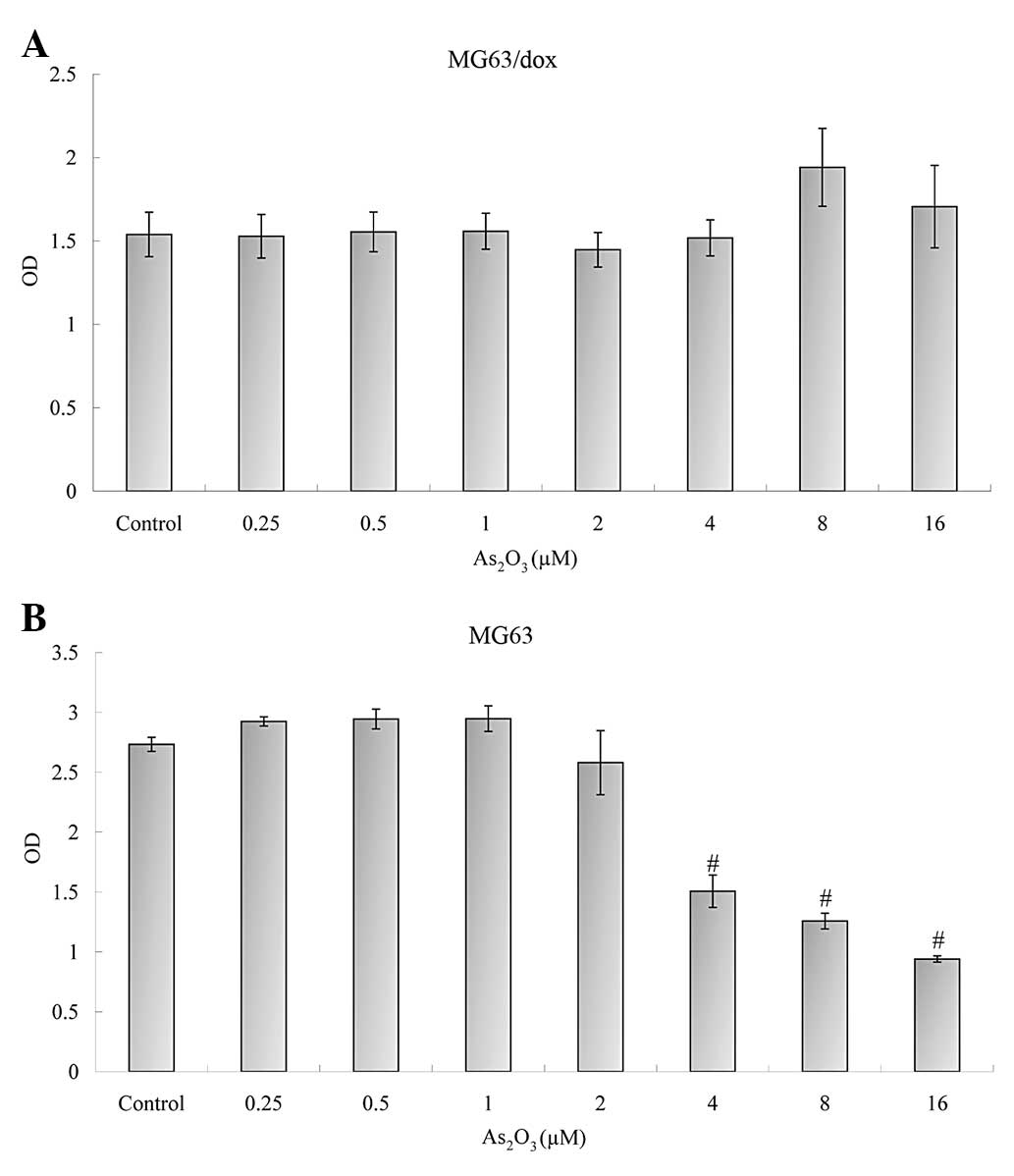
Effect of As2O3 and
ADM on parental MG63 and MG63/dox cell proliferation
To confirm that As2O3 and ADM
combination treatment inhibited cell growth, the parental MG63
cells and the resistant MG63/dox cells were quantified using the
CCK-8 assay. As2O3 alone did not inhibit MG63
or MG63/dox cell proliferation effectively at any of the various
doses (Fig. 1A), but
As2O3 was effective in significantly
inhibiting the growth of MG63 cells at 4, 8 and 16 μM
concentrations (P<0.01, Fig.
1B).
As a cytotoxic drug, As2O3 may
produce severe side effects in patients at increasing dosages or
extended treatment durations. The practical dose of
As2O3 in clinical therapy conversed to the
laboratory research should be 0.5–2 μM, and therefore, 2 μM was
selected as the dose of As2O3 in the
follow-up experiments (10).
In contrast to the various concentrations of ADM
alone, the combination of 2 μM As2O3 with
varying doses of ADM between 1 and 1,000 ng/ml resulted in a
significant reduction in the growth of MG63/dox cells (P<0.05,
Fig. 2A). Although combination
treatment also significantly restrained the growth of MG63 cells
compared with that of the control group (P<0.05), the addition
of As2O3 reduced the inhibitory effect of ADM
at 50, 100, 200, 400, 500 and 1,000 ng/ml doses (Fig. 2B). The results demonstrated that
combination treatment with As2O3 and ADM was
effective in inhibiting the growth of MDR osteosarcoma cells.
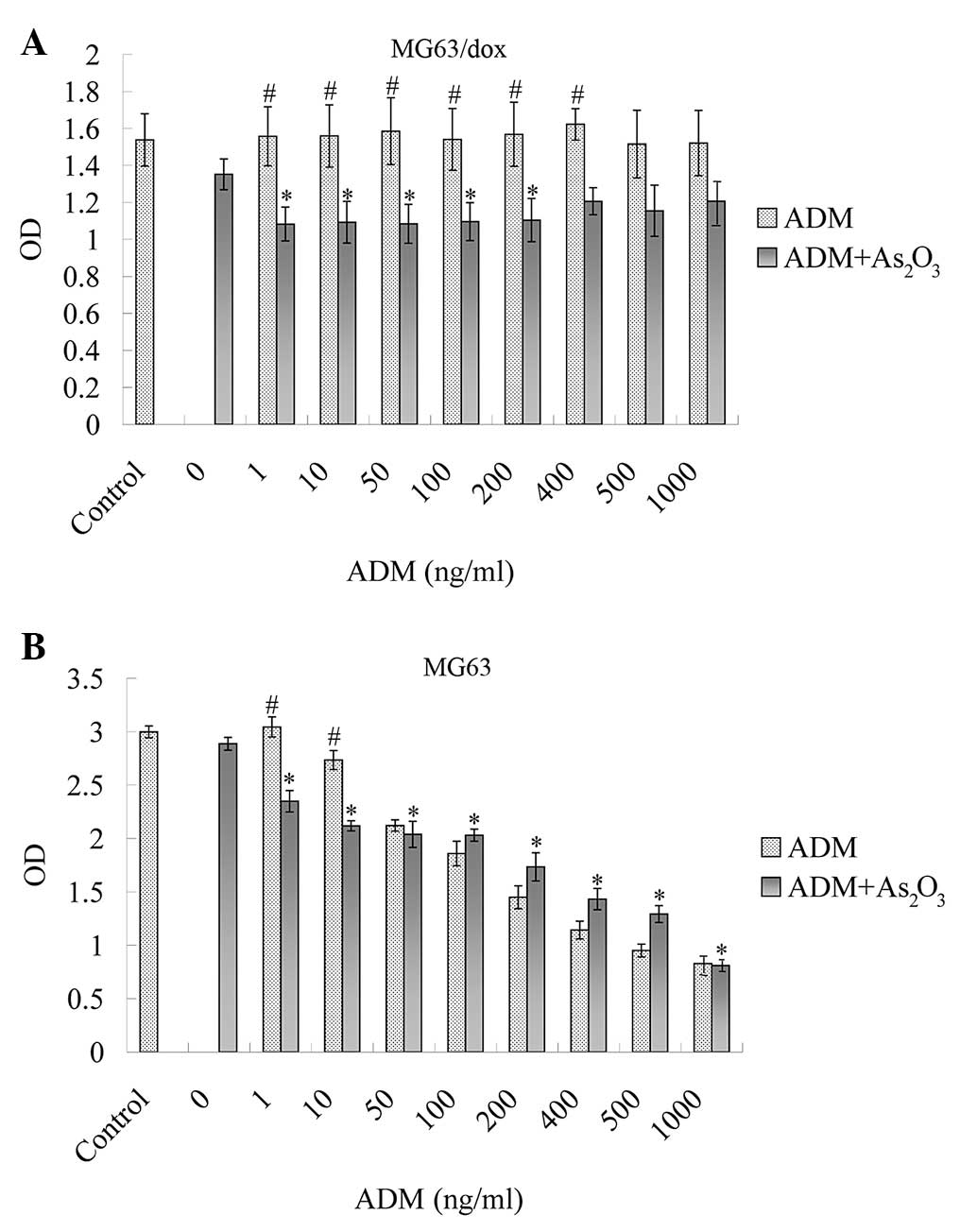 | Figure 2Effect of As2O3
and ADM combination treatment on (A) MG63 and (B) MG63/dox human
osteosarcoma cells. Cells were treated with 2 μM
As2O3 and various concentrations of ADM, and
incubated for 48 h. (A) Combination treatment inhibited cell
proliferation significantly compared with the control at 1, 10, 50,
100 and 200 ng/ml ADM (*P<0.05). In contrast to ADM
alone, combination treatment inhibited cell proliferation
distinctly at 1, 10, 50, 100, 200 and 400 ng/ml concentrations
(#P<0.05). (B) Combination treatment at various
concentrations inhibited cell proliferation significantly compared
with the control (*P<0.05), and reduced cell growth
at 1 and 10 ng/ml doses compared with ADM alone
(#P<0.05). The combined inhibitory effect declined at
high doses of ADM. ADM, doxorubicin; OD, optical density. |
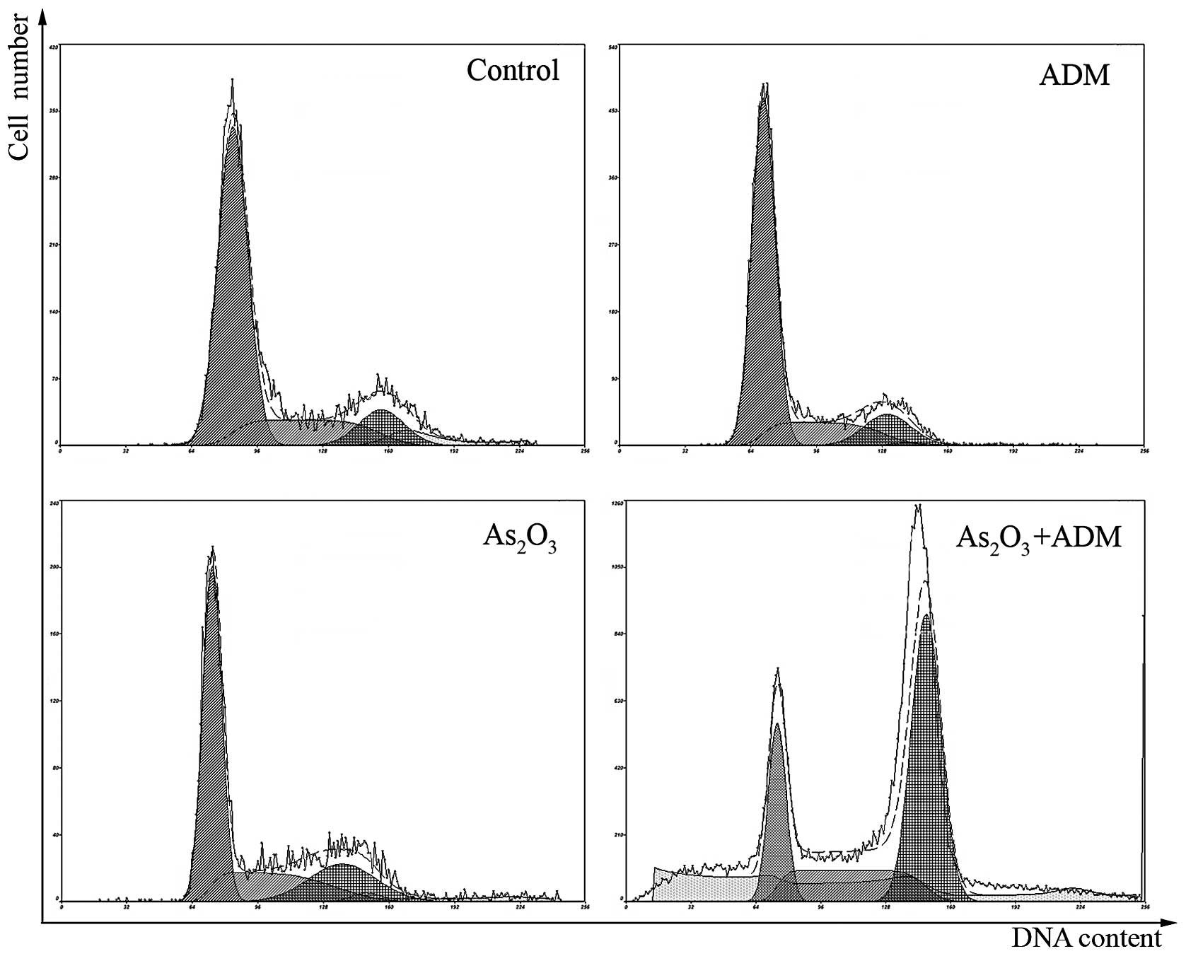
Effect of As2O3 and
ADM on MG63/dox cell cycle
Analysis of the cell-cycle phase distribution was
conducted to investigate the antiproliferative mechanism of
As2O3 and ADM on MG63/dox cells. The results
revealed significant differences in the percentages of combination
treatment cells in each cell cycle phase compared with the control
cells (P<0.01; Fig. 3 and
Table I). Increases in the
fraction of cells in the G2/M phase were detected following
As2O3 and ADM treatment, and a concurrent
reduction of the cell proportion in G0/G1 phase was observed. The
results demonstrated that As2O3 and ADM
combination treatment inhibited the proliferation of MG63/dox cells
through cell cycle arrest at the G2/M phase.
 | Table IEffect of As2O3
and ADM on MG63/dox cell cycle distribution. |
Table I
Effect of As2O3
and ADM on MG63/dox cell cycle distribution.
| Treatment | G0/G1 phase
(%) | S phase (%) | G2/M phase (%) |
|---|
| Control | 67.3±0.19 | 21.2±0.08 | 11.6±0.23 |
| ADM (200
ng/ml) | 68.2±0.12 | 19.2±0.33 | 12.6±0.45 |
|
As2O3 (2 μM) | 53.9±0.15 | 24.7±0.18 | 21.4±0.32 |
|
As2O3 and ADM | 20.7±0.19a | 24.1±0.09a | 55.2±0.26a |
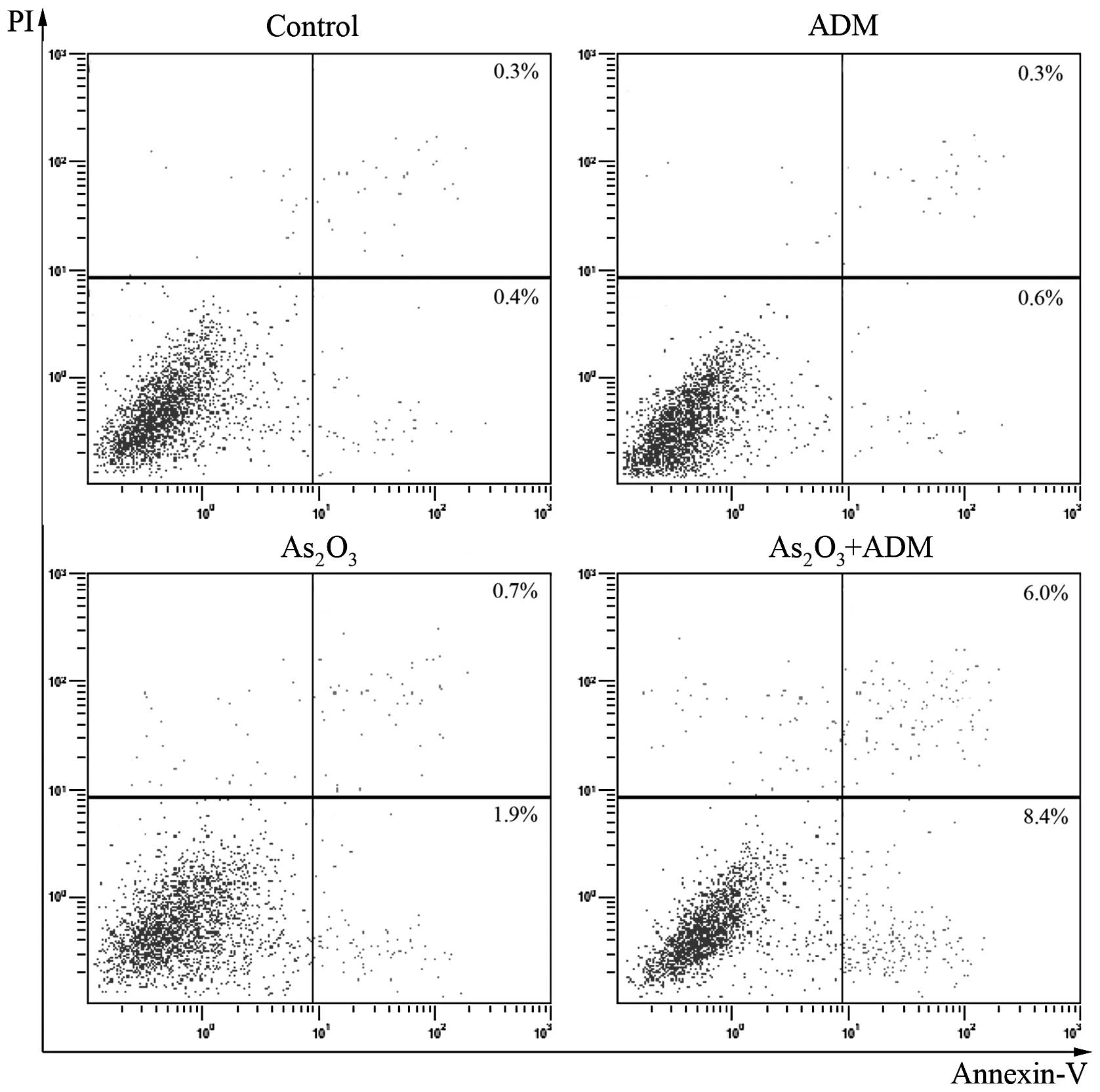
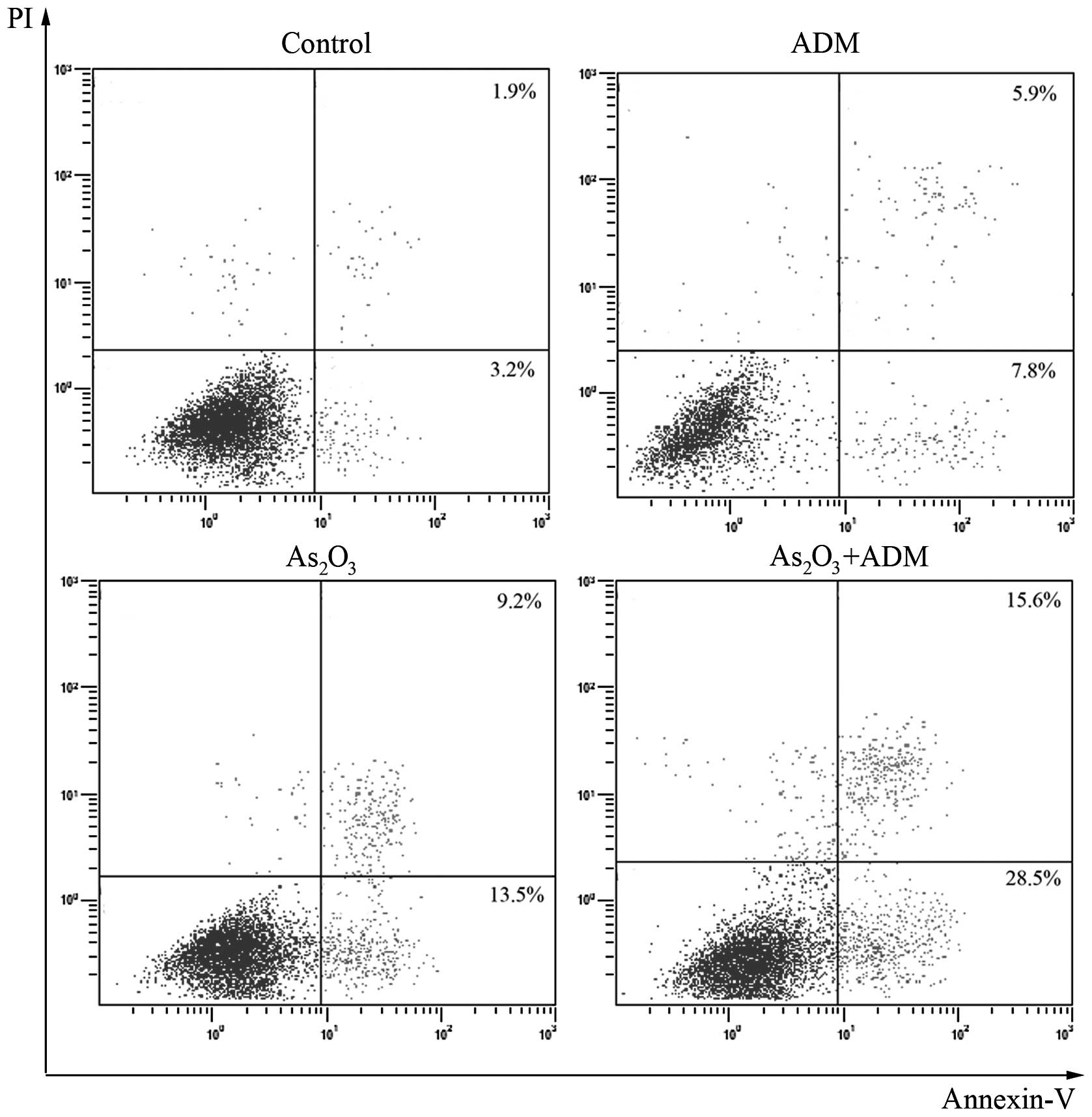
Induction of apoptosis in MG63/dox cells
by ADM and As2O3
As2O3 has been reported to
induce apoptosis in various human cancer cells (10). To determine whether
As2O3 induced apoptosis in MG63/dox cells,
the cells were treated with As2O3 and ADM for
48 h. The results of flow cytometric analysis with Annexin V-PI
staining revealed that ADM or As2O3 treatment
alone did not induce apoptosis; however, ADM and
As2O3 combination treatment increased the
percentage of apoptotic cells significantly (Fig. 4).
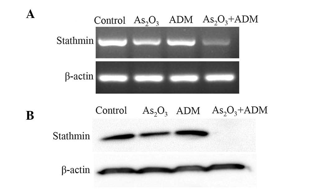
Effect of As2O3 and
ADM combination treatment on stathmin expression levels
Stathmin, a signal transduction regulatory factor,
is crucial in cell division and malignant development. Zhang et
al (8) observed that the
stathmin gene was expressed at high levels in osteosarcoma and may
become a novel target in osteosarcoma treatment. In order to
determine whether stathmin was involved in
As2O3 and ADM-induced apoptosis, MG63/dox
cells were incubated with As2O3 and ADM for
48 h, and stathmin expression levels were analyzed using PCR and
western blotting. The results demonstrated that incubation with
As2O3 and ADM resulted in significant
downregulation of stathmin expression in MG63/dox cells. Treatment
of the MDR cell line with As2O3 or ADM alone
did not induce distinctly reduced stathmin expression levels
(Fig. 5).
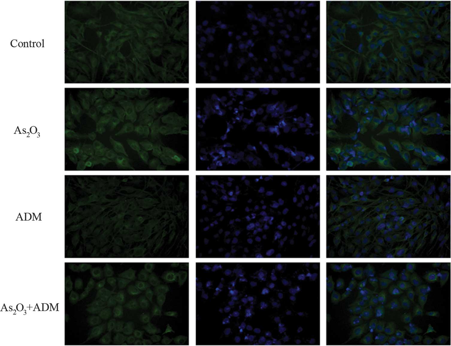
Effect of As2O3 and
ADM combination treatment on the cytoskeleton and morphology of
MG63/dox cells
Stathmin knockdown has been reported to alter the
phenotype of microtubules. In order to validate the above finding
that As2O3 and ADM combined treatment
downregulated stathmin expression, the cell microtubule network was
examined by immunofluorescent imaging of cells treated with
As2O3 and ADM (Fig. 6). Distinguished cell morphological
changes were observed using staining conditions and counting all
cells within multiple images. In particular, the cells treated with
ADM and As2O3 exhibited increased numbers of
short neurite-like extensions compared with the controls. Treatment
of the cells with As2O3 or ADM alone induced
marginal changes in the cytoskeleton and morphology. These data
indicated that As2O3 and ADM markedly altered
the organization of microtubule networks through stathmin
downregulation.
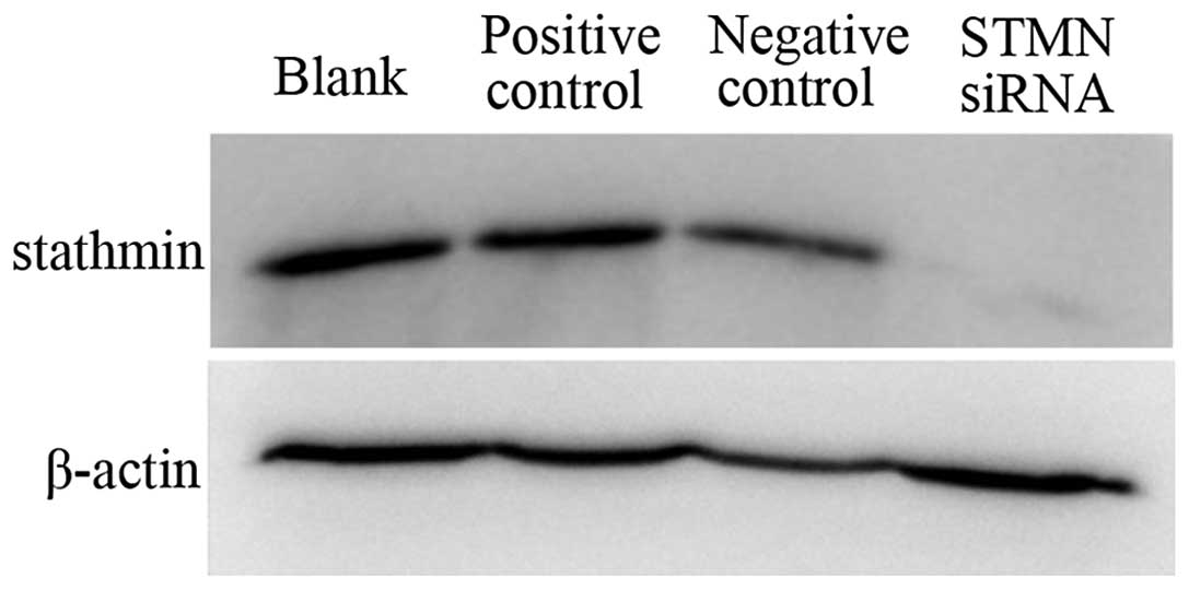
Effect of RNA interference targeting
stathmin on stathmin expression levels, cell proliferation and
apoptosis
To further investigate the role of stathmin in
As2O3 and ADM-induced apoptosis, siRNA
targeting stathmin was used to analyze the potential of these novel
therapeutic targets in the treatment of human osteosarcoma. PCR and
western blot analysis were used to determine the effect of siRNA
treatment on stathmin mRNA and protein expression levels in
MG63/dox cells. As shown in Fig.
7, western blotting indicated that the expression levels of
stathmin protein in MG63/dox cells were significantly reduced by
stathmin-siRNA. The fold change in stathmin siRNA transcript levels
between the cells transfected with stathmin-siRNA and the control
cells was calculated using the 2−ΔΔCt method (where Δ
cycle threshold (Ct) = CtsiRNA −CtActin and
ΔΔCt = ΔCtindicated group - ΔCtcontrol), and
~85.5% stathmin gene expression was found to be inhibited following
stathmin-siRNA transfection (Table
II). These results demonstrated that siRNA sequences targeting
the stathmin gene were effective in knocking down stathmin gene
expression.
| Table IIStathmin and β-actin Ct values in the
control and stathmin-siRNA transfected cells. |
Table II
Stathmin and β-actin Ct values in the
control and stathmin-siRNA transfected cells.
| Sample | Ct stathmin | Ct β-actin |
|---|
| Control | 18.93604 | 14.72279 |
| Stathmin-siRNA | 21.95575 | 14.95309 |
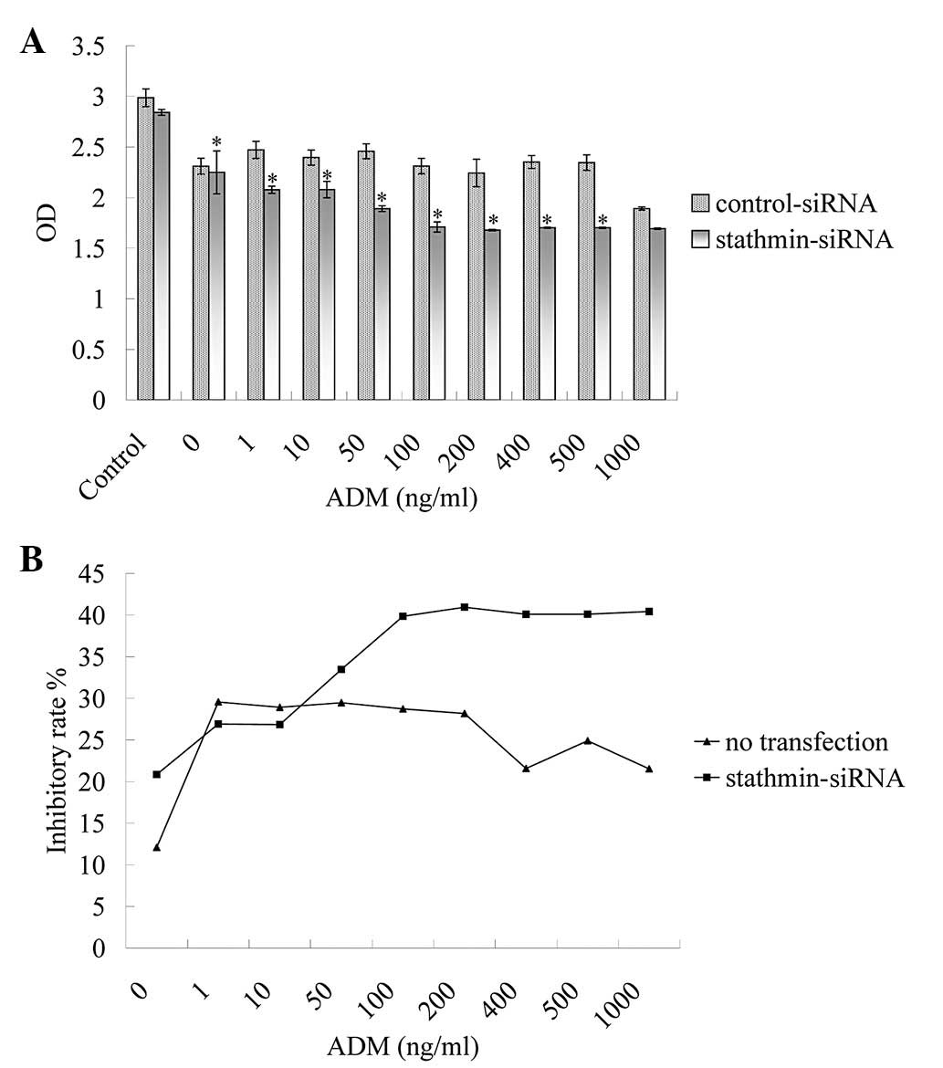
To analyze whether As2O3
reversed drug resistance through stathmin inhibition,
stathmin-siRNA-transfected MG63/dox cells were incubated with 2 μM
As2O3 and various concentrations of ADM for
48 h, and quantified by CCK-8 assay. Compared with the control
cells, stathmin-siRNA-transfected cells exhibited significantly
enhanced chemosensitivity, increasing the inhibitory rate of
As2O3 and ADM treatment by 12.77% (at 200
ng/ml ADM) to 18.91% (at 1,000 ng/ml ADM; Fig. 8). The effect of
As2O3 and ADM administration on inducing
apoptosis was also detected in the stathmin-siRNA-transfected
MG63/dox cells. As shown in Fig.
9, a higher apoptotic rate was demonstrated in
stathmin-siRNA-transfected cells compared with MG63/dox cells
following As2O3 and ADM treatment. The above
results indicated that As2O3 and ADM
suppressed cell proliferation and induced apoptosis, possibly
through inhibiting stathmin expression.
Discussion
Osteosarcoma is the most common type of primary
malignant bone tumor in children and adolescents; ~60% of primary
malignant bone tumors are diagnosed in the first two decades of
life (11). With the introduction
of intensive chemotherapeutics, including cisplatin, doxorubicin,
ifosphamide and high dose methotrexate, >60% of the patients are
cured (1). However, 30–40%
patients with localized osteosarcoma experience recurrent or
progressive disease. The majority of these patients are considered
to have MDR osteosarcoma cancer cells, resistant to one or more
chemotherapeutic agents (12).
Resistance to these agents remains a challenge in the treatment of
osteosarcoma and an obstacle to achieving improved cure rates.
Thus, novel drugs and therapeutic regimens are required for more
effective treatment of aggressive and recurrent MDR
osteosarcoma.
As2O3-based compounds are the
most widely used and analyzed arsenic-based cancer drugs. Results
of in vitro studies and clinical trials have revealed that
As2O3 is effective in inhibiting the growth
of APL cells (3). Recent studies
have found that As2O3 also exhibits an
anticancer effect in particular types of non-APL cancer, including
myeloid leukemia, gastric cancer, prostate and ovarian carcinomas,
and breast cancer (5). However,
studies regarding the effect of As2O3 on
osteosarcoma are rare. According to a study by Guo et al
(13), As2O3
combined with VP-16 and paclitaxel was an effective remedy in the
treatment of stage III osteosarcoma. A previous study also revealed
that As2O3 induced apoptosis in MG63 human
osteosarcoma cells (14). These
findings prompted the investigation of the effect of
As2O3 on MDR human osteosarcoma cells in the
present study. As2O3 has been reported to
downregulate P-glycoprotein expression in human leukemia cells
(15). In the present study, the
MG63/dox MDR osteosarcoma cell line, which is characterized by
upregulation of the MDR1 gene and overexpression of P-glycoprotein,
was used.
The results revealed that combination treatment with
As2O3 and ADM was effective in inhibiting
MG63 and MG63/dox cell proliferation. The MG63/dox drug-resistant
cells exhibited particular sensitivity to the combination
treatment. Furthermore, the combination treatment induced MG63/dox
apoptosis and cell-cycle arrest. This finding is of great
importance for evaluating the potential use of
As2O3 in treating patients with MDR.
A growing number of studies have reported that
stathmin was expressed at high levels in a wide variety of human
malignancies, including osteosarcoma. Stathmin is a key regulator
of the microtubule network and provides an attractive therapeutic
target in cancer treatment (6).
High levels of stathmin expression in cancer cells were observed to
correlate with cell proliferative potential and appear to be
required for the maintenance of the malignant phenotype (16).
The present study provided evidence that stathmin
may be involved in As2O3-induced MDR cell
apoptosis. As2O3 and ADM treatment not only
inhibited MG63/dox cell proliferation and induced apoptosis, but
also resulted in reduced stathmin expression levels, indicating
that reduced stathmin expression levels are associated with
As2O3 and ADM-induced apoptosis. Furthermore,
treatment with As2O3 and ADM altered the
cytoskeleton of MG63/dox cells, while stathmin knockdown has been
reported to alter the phenotype of microtubules. These results
demonstrated that stathmin may be crucial in the reversion of drug
resistance by As2O3.
To demonstrate the possible role of stathmin in the
As2O3-induced apoptotic pathway, siRNA
targeting stathmin was transfected into MG63/dox cells. The CCK-8
assay demonstrated that the combination treatment inhibited the
transfected MG63/dox cell proliferation markedly compared with the
non-transfection group. Flow cytometric analysis revealed that the
treatment with As2O3 and ADM induced marked
apoptosis. Stathmin is a ubiquitous cytoplasmic phosphoprotein that
regulates microtubule dynamics. Inhibiting stathmin expression may
influence the functions of either centrosomes or G2/M checkpoint
proteins at the centrosome, which may result in a G2/M block and
reduce the rate of cell proliferation (17). In the present study, stathmin was
demonstrated to be involved in As2O3-induced
apoptosis in human osteosarcoma cells and this finding may support
the use of stathmin as a therapeutic target in human osteosarcomas.
The exact molecular mechanism that accounts for the observed
interaction between stathmin inhibition and
As2O3 treatment appears complex and requires
further clarification.
In conclusion, the administration of
As2O3 together with ADM may be a useful novel
anticancer chemotherapy, particularly in MDR cases, as
As2O3 reversed ADM resistance in MG63/dox
cells through downregulation of stathmin. The results indicated
that As2O3 is a promising chemotherapeutic
agent for patients with drug-resistant osteosarcoma.
Acknowledgements
This study was supported by the National Natural
Science Foundation of China (grant no. 81172105).
References
|
1
|
Serra M, Scotlandi K, Manara MC, et al:
Establishment and characterization of multidrug-resistant human
osteosarcoma cell lines. Anticancer Res. 13:323–329.
1993.PubMed/NCBI
|
|
2
|
Rajkumar T and Yamuna M: Multiple pathways
are involved in drug resistance to doxorubicin in an osteosarcoma
cell line. Anticancer Drugs. 19:257–265. 2008. View Article : Google Scholar : PubMed/NCBI
|
|
3
|
Rego EM, He LZ, Warrell RP Jr, et al:
Retinoic acid (RA) and As2O3 treatment in
transgenic models of acute promyelocytic leukemia (APL) unravel the
distinct nature of the leukemogenic process induced by the
PML-RARalpha and PLZF-RARalpha oncoproteins. Proc Natl Acad Sci
USA. 97:10173–10178. 2000.PubMed/NCBI
|
|
4
|
Bachleitner-Hofmann T, Kees M and
Gisslinger H: Arsenic trioxide: acute promyelocytic leukemia and
beyond. Leuk Lymphoma. 43:1535–1540. 2002. View Article : Google Scholar : PubMed/NCBI
|
|
5
|
Miller WH Jr, Schipper HM, Lee JS, et al:
Mechanisms of action of arsenic trioxide. Cancer Res. 62:3893–3903.
2002.PubMed/NCBI
|
|
6
|
Niethammer P, Bastiaens P and Karsenti E:
Stathmin-tubulin interaction gradients in motile and mitotic cells.
Science. 303:1862–1866. 2004. View Article : Google Scholar : PubMed/NCBI
|
|
7
|
Rubin CI and Atweh GF: The role of
stathmin in the regulation of the cell cycle. J Cell Biochem.
93:242–250. 2004. View Article : Google Scholar : PubMed/NCBI
|
|
8
|
Zhang HZ, Wang Y, Gao P, et al: Silencing
stathmin gene expression by survivin promoter-driven siRNA vector
to reverse malignant phenotype of tumor cells. Cancer Biol Ther.
5:1457–1461. 2006. View Article : Google Scholar
|
|
9
|
Wang R, Dong K, Lin F, et al: Inhibiting
proliferation and enhancing chemosensitivity to taxanes in
osteosarcoma cells by RNA interference-mediated downregulation of
stathmin expression. Mol Med. 13:567–575. 2007. View Article : Google Scholar : PubMed/NCBI
|
|
10
|
Ling YH, Jiang JD, Holland JF and
Perez-Soler R: Arsenic trioxide produces polymerization of
microtubules and mitotic arrest before apoptosis in human tumor
cell lines. Mol Pharmacol. 62:529–538. 2002. View Article : Google Scholar : PubMed/NCBI
|
|
11
|
Schwartz CL, Gorlick R, Teot L, et al;
Children’s Oncology Group. Multiple drug resistance in osteogenic
sarcoma: INT0133 from the Children’s Oncology Group. J Clin Oncol.
25:2057–2062. 2007.PubMed/NCBI
|
|
12
|
Mimeault M, Hauke R, Mehta PP, et al:
Recent advances on the molecular mechanisms involved in the drug
resistance of cancer cells and novel targeting therapies. J Cell
Mol Med. 11:981–1011. 2007.PubMed/NCBI
|
|
13
|
Guo W, Tang XD, Tang S and Yang Y:
Preliminary report of combination chemotherapy including Arsenic
trioxide for stage III osteosarcoma and Ewing sarcoma. Zhonghua Wai
Ke Za Zhi. 44:805–808. 2006.(In Chinese).
|
|
14
|
Xiao T, Li KH and Fang JZ: Experimental
study on the apoptotic effect of arsenic trioxide on human
osteosarcoma MG-63 cells. Hunan Yi Ke Da Xue Xue Bao. 27:111–113.
2002.(In Chinese).
|
|
15
|
Wei H, Su H, Bai D, et al: Arsenic
trioxide inhibits p-glycoprotein expression in multidrug-resistant
human leukemia cells that overexpress the MDR1 gene. Chin Med J
(Engl). 116:1644–1648. 2003.
|
|
16
|
Belmont LD and Mitchison TJ:
Identification of a protein that interacts with tubulin dimers and
increases the catastrophe rate of microtubules. Cell. 84:623–631.
1996. View Article : Google Scholar : PubMed/NCBI
|
|
17
|
Mistry SJ and Atweh GF: Stathmin
inhibition enhances okadaic acid-induced mitotic arrest: a
potential role for stathmin in mitotic exit. J Biol Chem.
276:31209–31215. 2001. View Article : Google Scholar : PubMed/NCBI
|