Introduction
Ginseng has been widely used as a herbal medicine in
Eastern Asian countries, including Korea. Ginseng is a species of
perennial plant belonging to the genus Panax (P) in the family
Araliaceae. Ginseng species are found only in the Northern
hemisphere from Siberia to Vietnam. Among the ginseng species,
Asian ginseng (P. ginseng) and American ginseng (P.
quinquefolius) are the best characterized. Previously,
Asafu-Adjaye and Wong (1) have
demonstrated the quantitative and qualitative profiling of
ginsenosides (ginseng saponins) in dry root powder from P.
ginseng and P. quinquefolius, respectively. Evidence
strongly suggests that P. ginseng contains >30
pharmacologically active saponins, including Rh1, Rh2, Rg3 and Rg5
(2). There is, of further
importance, evidence strongly supporting that ginseng extract
and/or ginsenosides (Rg3, Rh2, Rb2, compound K and F2) have
anti-carcinogenic activities against numerous types of cancer,
including colon, liver and breast cancer (3–6). In
addition, the preventive activities of ginseng saponins by
inhibiting cancer cell growth and metastasis in vivo and
in vitro have also been previously proposed (7). In a recent study, it was demonstrated
that butanolic extract of mountain ginseng had anti-proliferative
and pro-apoptotic effects on A549 human lung cancer cells via
modulation of the expression and/or activity of p53, caspase-3 and
nuclear factor (NF)-κB (8).
Furthermore, a previous study by our group provided evidence that
Rb1, Rb2 and Rg1, three of the most abundant ginsenosides in the
ginseng extract, induced apoptosis in A549 cells through extrinsic
(death receptor-mediated) apoptotic pathways (9). In addition, there is clinical
evidence that red ginseng powder effectively enhances postoperative
immunity and survival in patients with stage III gastric cancer
(10), suggesting the potential
clinical applications of ginseng preparations against gastric
cancer.
Gastric cancer is the 4th most frequent cancer and
the second leading cause of cancer-associated mortality worldwide.
Although 50% of gastric tumors are detected at an early stage in
asymptomatic individuals, often when symptoms occur, the cancer has
already reached advanced stages and may proceed to metastasis,
leading to a poor prognosis for patients (11). At present, the current treatment
strategies for gastric cancer include surgery, chemotherapy and/or
radiotherapy (12). Among these,
radical surgery offers the only chance of cure for patients with
operable gastric cancer, but the outcomes remain generally poor due
to a high rate of relapse post gastric surgery. Therefore,
multimodality therapy using chemotherapy, radiation or a
combination of both has been evaluated in different parts of the
world to improve the outcomes from surgery alone (12,13).
Numerous chemotherapeutic drugs, including 5-fluorouracil,
doxorubicin, mitomycin C, cisplatin and taxotere, have been used
individually or in combination for the treatment of gastric cancer
(14). Furthermore, there is
evidence that everolimus, an inhibitor of the mammalian target of
rapamycin (mTOR) pathway, is also effective and well tolerated in
phase I/II studies of patients with chemotherapy-refractory
metastatic gastric cancer (15).
However, due to the poor long-term outcomes associated with current
chemotherapy treatment of patients with advanced gastric cancer,
there is still a need for the development of novel targeted agents
that may confer an improved survival benefit.
In the present study, the anti-cancer effect and
mechanism of a fortified ginseng extract (FGX), newly prepared from
P. ginseng root butanolic extract (GBX) by the treatment of
two enzymes, laminarinase (polysaccharides-degrading enzyme) and
pectinase (pectin-degrading enzyme), in KATO3 human gastric cancer
cells were investigated.
Materials and methods
Materials
Dulbecco’s modified Eagle’s medium (DMEM) was
purchased from Sigma-Aldrich (St. Louis, MO, USA). The 96-well
plates used were obtained from NUNC (Roskilde, Denmark). Fetal
bovine serum (FBS), phosphate-buffered saline (PBS),
penicillin-streptomycin and l-glutamate were obtained from
Gibco-BRL (Carlsbad, CA, USA). The Annexin-V-carboxyfluorescein
(FLUOS) staining kit was provided from Roche Diagnostics GmbH
(Mannheim, Germany). An antibody against nuclear factor of kappa
light polypeptide gene enhancer in B-cells inhibitor α (IκBα) was
purchased from Santa Cruz Biotechnology, Inc. (Santa Cruz, CA,
USA). B-cell lymphoma-2-associated X (Bax) and B-cell lymphoma-2
(Bcl-2) antibodies were obtained from Abcam (Cambridge, MA, USA).
Antibodies against protein kinase B (PKB), phosphorylated (p)-PKB,
mTOR and p-mTOR were purchased from Cell Signaling Technology, Inc.
(Beverly, MA, USA). Secondary antibodies of horseradish peroxidase
(HRP)-conjugated anti-rabbit immunoglobulin (Ig)G were purchased
from Invitrogen Life Technologies, Inc. (Carlsbad, CA, USA). Other
reagents, including mouse monoclonal anti-human actin antibody,
were purchased from Sigma-Aldrich.
Cell culture
KATO3, a human gastric cancer cell line, was
obtained from American Type Culture Collection (Rockville, MD,
USA). KATO3 cells were grown in DMEM supplemented with 10% (v/v)
FBS and 1% (w/v) penicillin-streptomycin in a cell culture chamber
at 37°C with 5% (v/v) CO2.
Preparation of an enzymatically FGX from
GBX
The root of regular ginseng (four years old) was
purchased from National Agricultural Cooperative Foundation
(Chuncheongnam-do, Korea). A total of 20 g pulverized ginseng root
powder was suspended in 380 ml distilled water and then sterilized
at 121°C for 15 min. To reduce the complexity of components in
cultivated regular ginseng root, the extract was fractionated via
extraction with various solvents, including water, methanol and
butanol. Among them, ginseng butanolic extract (GBX) contained
compounds with specific anti-gastric cancer activity. GBX was used
as a control of regular ginseng. In addition, the suspension was
treated with an aliquot of filter-sterilized commercial enzymes
(laminarinase, pectinase) at an equimolar ratio (1:1, specific
activity units), and the mixture was then incubated at 40°C for two
days and evaporated to dryness at 60°C. These enzyme-modified
ginseng powders were suspended in 400 ml 80% (v/v) methanol. The
suspension was treated in an ultrasound bath for 5 min and filtered
through Whatman no. 2 filter paper (Whatman International Ltd.,
Maidstone, UK). The wet powder on the filter paper was collected,
suspended, treated in an ultrasound bath and filtered in the same
manner once again. The two filtrates were combined and evaporated
to dryness at 50°C. The methanolic extract was dissolved in 200 ml
of distilled water and washed with 200 ml ethyl acetate in a
separation funnel and then extracted with 200 ml butanol. The
butanolic extract was evaporated to dryness at 50°C and redissolved
to the concentration of 10% (w/v) in 70% ethanol. This final
extract was termed as an enzymatically FGX herein.
High-performance liquid chromatography
(HPLC) analysis of the ginsenoside profiles in FGX and GBX
Standard ginsenosides, including Rg1, Re, Rf, Rh1,
Rb1, Rc, Rb2, f1, Rd, Rg3(S), Rg3(R), PPT, compound K, Rh2 and PPD,
were obtained from ChromaDex, Inc. (Irvine, CA, USA) and the
profiles were analyzed using an Acquity ultra (U)PLC system
(Waters, Milford, MA, USA) with an Acquity BEH C18 HPLC
column. The mobile phase consisted of solution A (CH3CN,
HPLC-grade; JT Baker, Center Valley, PA, USA) and solution B
(Milli-Q H2O; Millipore, Billerica, MA, USA). The flow
rate was set at 0.6 ml/min and the injection of volume was fixed at
2 μl. The separation of ginsenosides was performed using an
isocratic gradient of 100% solution A for 27 min. The column
temperature was maintained at room temperature during the
separation and the ultraviolet (UV) diode array detection was set
at 203 nm. For the confirmation of a reliable retention time of
sample ginsenoside, the individual ginsenoside was identified and
quantified by comparing it with the retention times of the standard
ginsenosides.
Cell viability assay
KATO3 cells (5×10³ cells/well) were
seeded in a 96-well plate overnight. The cells were then treated
with GBX or FGX at various concentrations for 24 h, followed by a
measurement of cell viability using the Cell Counting kit-8
(Dojindo Molecular Technologies, Inc., Kumamoto, Japan ), as
described previously (8). The cell
viability was determined by measuring the absorbance at 450 nm
using a microplate reader (Sunrise™-Basic; Tecan, Mannedorf,
Switzerland). Each data point was determined by triplicate
experiments.
Identification of apoptosis by
PI-Annexin-V staining
The cell apoptosis was assessed using an
Annexin-V-FLUOS Staining kit (Roche Diagnostics), according to the
manufacturer’s instructions. KATO3 cells were incubated with GBX or
FGX for 24 h, scraped and washed twice with PBS, and centrifuged at
930 × g for 5 min at 4°C. The cells were then incubated with 0.2
mg/ml Annexin-V-FLUOS and 1.4 mg/ml DNA stain propidium iodide (PI)
for 15 min at room temperature. The measurements were conducted on
a FACSCalibur flow cytometer (Becton Dickinson Immunocytometry
Systems, San Jose, CA, USA) at 488 nm excitation and 530/30 nm band
pass filter for fluorescence detection, and 670 nm high pass filter
for PI detection. The data were analyzed by WinMDI V2.9 software
(Joe Trotter, The Scripps Institute, La Jolla, CA, USA).
Western blot analysis
The expression levels of intracellular proteins from
GBX- and FBX-treated cells were examined by western blotting, as
described previously (8). The
denatured protein of 30 μg was separated by 12% SDS-PAGE and then
transferred onto a nitrocellulose membrane. The transferred
nitrocellulose membrane was stained with Ponceau S (Sigma-Aldrich)
to visualize the proteins. The blotted membrane was blocked for 1 h
with 5% (w/v) skimmed milk in TTBS [0.1% (v/v) Tween-20 and
Tris-buffered saline], followed by incubation with the respective
primary antibody for IκBα (1:1,000), Bcl-2 (1:1,000), Bax
(1:2,000), PKB (1:1,000), p-PKB (1:1,000), mTOR (1:2,000), p-mTOR
(1:1,000) and actin (1:5,000) at room temperature (RT) for 2 h or
at 4°C overnight. The membrane was washed three times for 5 min
with TTBS and further incubated with secondary antibody of
HRP-conjugated rabbit anti-goat IgG (1:2,000) in TTBS containing 5%
(w/v) skimmed milk at RT for 1 h. The membrane was then rinsed
three times with TTBS-0.1% (v/v) Tween-20 for 5 min. Then, the
Pierce enhanced chemiluminescence system (Thermo Scientific, San
Jose, CA, USA) was used to develop the proteins on an X-ray film
(Kodak, Seoul, Korea). The expression levels of each protein were
quantitatively analyzed, with those of actin protein used as an
internal control.
Statistical analyses
The GraphPad Prism version 5.0 software (GraphPad,
San Diego, CA, USA) was used for statistical analysis. A Student’s
t-test was used to assess the difference between the GBX and
FGX-treated group. The IC50 values were calculated by
non-linear curve fit at four data points and presented as the mean
± standard deviation. P<0.001 was considered to indicate a
statistically significant difference.
Results
Ginsenoside profiles in FGX and GBX
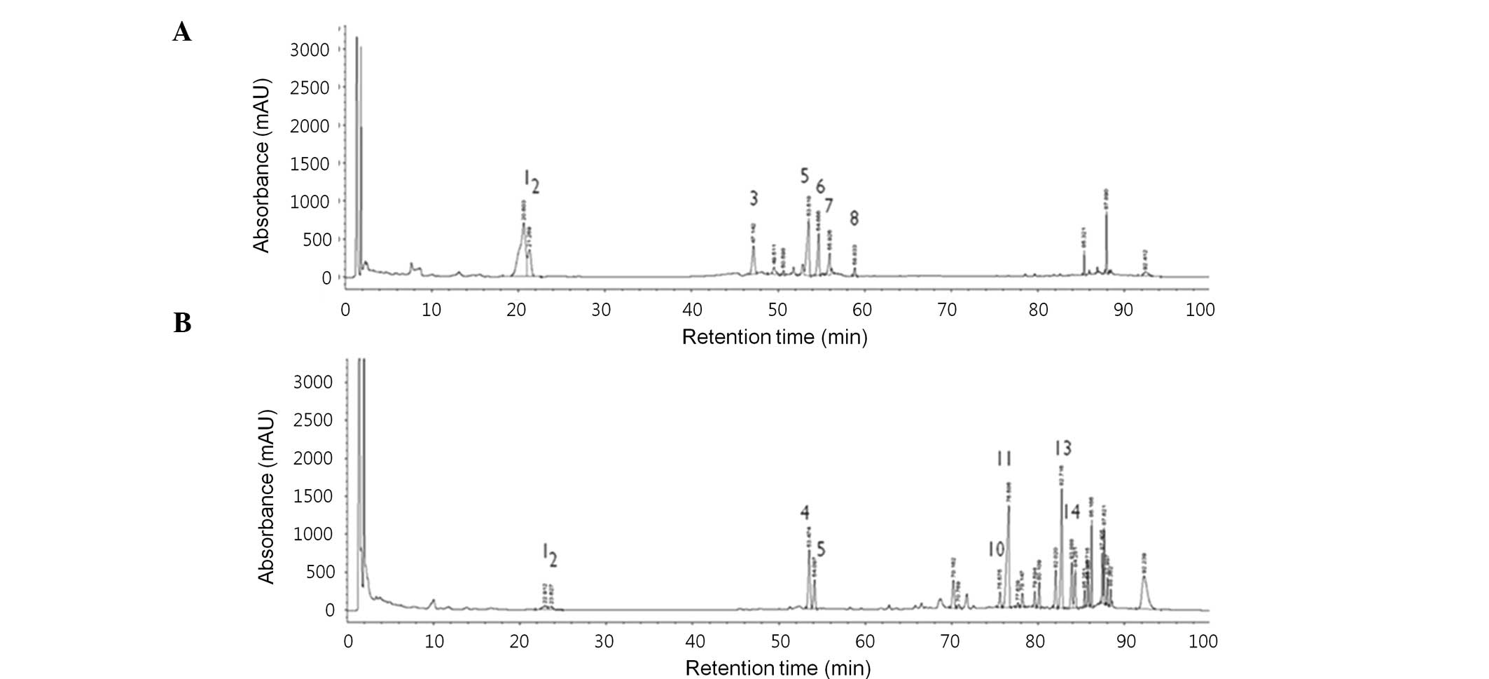
Primarily, HPLC-UV analysis was conducted to
generate a quantitative profile of the ginsenoside composition of
FGX and GBX. As demonstrated in Fig.
1, the prominent ginsenoside peaks were present in the
chromatograms of FGX and GBX. A total of seven ginsenosides,
including Rg1, Re, Rf, Rb1, Rc, Rb2 and F1, were presented in GBX.
By contrast, five ginsenosides, including Rh1, Rg3(S), Rg3(R),
compound K and Rh2, were exclusively detected in FGX. In
particular, in FGX, the most intensive HPLC peak was Rg3(R) in the
range of 75–80 min. Therefore, considering that Rg3, Rh2 and
compound K are major ginsenosides with anti-cancer activity, these
results suggested the enrichment of more ginsenosides with
anti-cancer activity in FGX compared with the ginsenosides in GBX.
The quantitative data of the ginsenoside profiles in FGX and GBX
are listed in detail in Table
I.
 | Table IQuantities of ginsenosides in GBX and
FGX. |
Table I
Quantities of ginsenosides in GBX and
FGX.
|
Quantity of
Ginsenoside (mg/g) |
|---|
|
|
|---|
| Group | Rg1 | Re | Rf | Rh1 | Rb1 | Rc | Rb2 | F1 | Rd | Rg3(S) | Rg3(S)R | PPT | ComK | Rh2 |
|---|
| GBX | 7.00 | 7.21 | 2.54 | - | 7.65 | 4.68 | 2.32 | 2.02 | - | - | - | - | - | - |
| FGX | 0.61 | 1.02 | - | 2.74 | 2.65 | - | - | - | - | 0.94 | 8.58 | - | 4.73 | 2.65 |
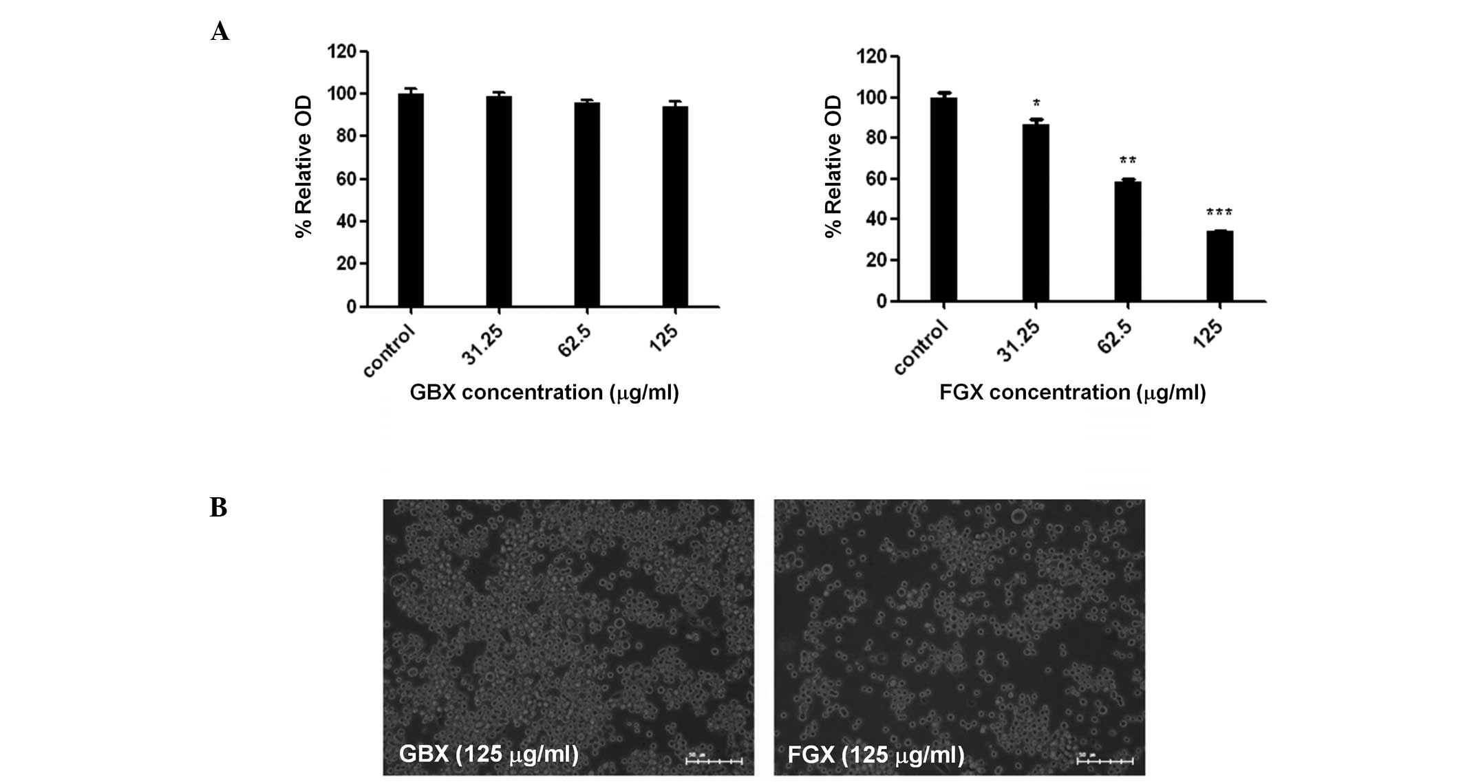
FGX treatment induces a marked loss of
KATO3 cells
Next, the effects of FGX and GBX on the
proliferation and the morphology of KATO3 human gastric cancer
cells was examined. As demonstrated in Fig. 2A, FGX inhibited the growth of KATO3
in a dose-dependent manner, while GBX exhibited no clear inhibition
at up to 125 μg/ml. Furthermore, KATO3 cells had round shapes but
there was no marked difference in the morphology of KATO3 cells
treated with FGX and GBX at 125 μg/ml for 24 h, respectively, as
revealed in Fig. 2B. However, the
number of KATO3 cells following treatment with FGX (125 μg/ml, 24
h) was significantly lower compared with that following treatment
with GBX (125 μg/ml, 24 h) under these experimental conditions.
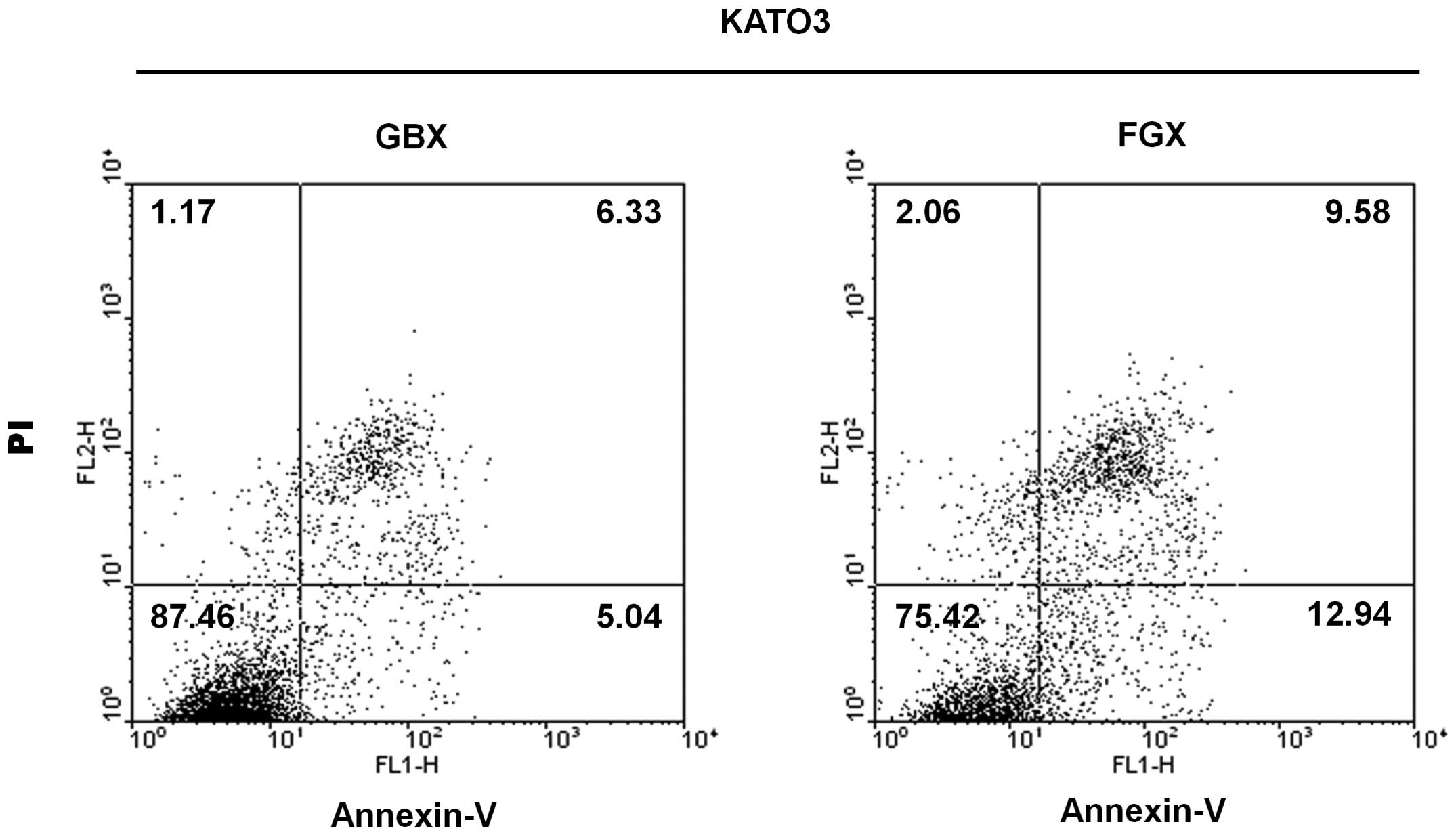
FGX treatment induces apoptosis of KATO3
cells
It was further investigated whether FGX induces
apoptosis of KATO3 cells by using Annexin V/PI staining-based flow
cytometric analysis, in which cells in early stages of apoptosis
(Annexin-stained, non-disrupted cells) were distinguished from
those necrotic cells (disrupted or lysed cells). The quantitative
data of the flow cytometric analysis, as revealed in Fig. 3, demonstrated that treatment with
FGX and GBX at 125 μg/ml for 24 h resulted in ~12.94 and 5.04%
cells at early stages of apoptosis, and ~9.58 and 6.33% of cells in
late apoptosis, respectively. These results confirmed the
FGX-induced apoptosis of KATO3 cells.
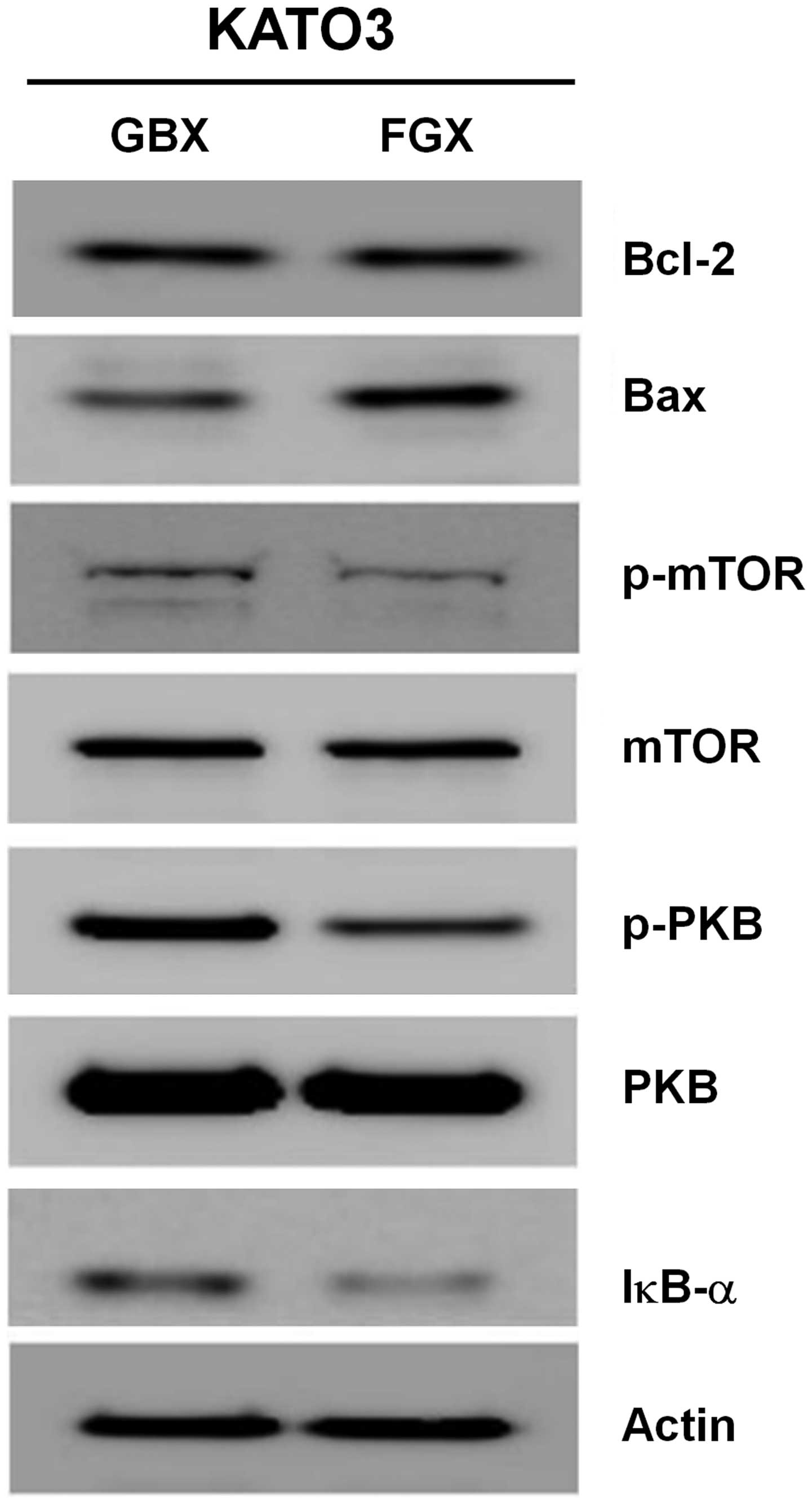
FGX treatment leads to upregulation of
Bax, dephosphorylation of mTOR and PKB and downregulation of IκBα
in KATO3 cells
Cell survival and/or apoptosis are largely affected
by the expression and/or activity of a variety of proteins,
including Bcl-2 family proteins, PKB and mTOR. Therefore, the
effects of FGX and GBX on the expression and/or activity
(phosphorylation) of those proteins in KATO3 cells were examined by
western blot analysis. As revealed in Fig. 4, compared with the effects of GBX
(125 μg/ml, 24 h), treatment with FGX (125 μg/ml, 24 h) resulted in
an increased expression of Bax, but did not affect Bcl-2 in KATO3
cells. Furthermore, compared with GBX, FGX further led to a
decrease in the phosphorylated forms of PKB and mTOR in KATO3
cells, without affecting their total protein expression. There is
further evidence that suggests the involvement of NF-κB, a
transcription factor, in cell survival and/or apoptosis. Activation
of NF-κB is strongly correlated to the proteolysis of IκBα, a
cytosolic NF-κB inhibitory protein. This led us promptly to
investigate the effect of FGX and GBX on the expression levels of
IκBα in KATO3 cells. Notably, there were lower expression levels of
IκBα in FGX-treated KATO3 cells than in the GBX-treated cells.
Discussion
The pharmacological actions of the majority of
ginseng preparations are attributed to ginsenosides (16). It is suggested that manufacturing
methods, including steaming or heating, may enhance the anti-cancer
activity of ginseng and/or ginsenosides over that of the original
ginseng powder (17). In the
present study, the effects of an enzymatically FGX and GBX,
original ginseng extract on KATO3 human gastric cancer cells were
compared. It was demonstrated that FGX had stronger
anti-proliferative and pro-apoptotic effects on KATO3 cells than
GBX. These data suggested that the FGX’s anti-proliferative and
pro-apoptotic effects on KATO3 cells appeared to be mediated via
modulation of the expression and/or activities of Bax, IκBα/NF-κB,
mTOR and PKB signaling agents.
Previously, it has been demonstrated that Rg3 and
Rh2 block cell cycle progression of cancer cells (18–20)
and compound K leads to cell cycle arrest in leukemia cells
(21). A previous study by our
group, also revealed that GBX, which contains high quantities of
Rb1, inhibits growth and induces apoptosis of A549 human lung
cancer cells (9), implying a
positive role of Rb1 on the GBX-induced anti-growth and
pro-apoptotic effects. In the present study, using HPLC-UV
analysis, it was demonstrated that five ginsenosides, including
Rh1, Rb1, Rg3(R), compound K and Rh2, were enriched in FGX compared
with the ginsenoside profile in GBX. Importantly, the present study
demonstrated lower numbers of KATO3 cells and more
Annexin-V-stained populations following treatment with FGX as
compared with the GBX-treated cells, suggesting that FGX had more
potent anti-growth and pro-apoptotic activity in KATO3 cells than
the original ginseng extract. These results may therefore indicate
that the enzymatic method used in the present study may be useful
for the production of a preparation of ginseng extract containing
more ginsenosides with anti-cancer activity.
The induction of apoptosis is mediated through two
main pathways; the intrinsic (mitochondrial) and the extrinsic
(death receptor-mediated) pathways. Central to both apoptosis
pathways are the caspases, a group of essential proteases required
for the execution of programmed cell death by apoptotic stimuli
(22,23). Previously, it has been demonstrated
that ginseng root extract induced mitochondrial-dependent apoptotic
pathways in tumor cells (24), as
deduced from the evidence that a caspase-3 inhibitor was able to
partially block the activation of caspase-3 and the subsequent
apoptotic cell death caused by ginseng root extract. Abundant data
have suggested that the mitochondrial-dependent apoptotic pathways
are largely affected by the expression and/or activity of several
mitochondrial proteins, including Bcl-2 and Bax. Bcl-2 is an
anti-apoptotic protein and participates in apoptosis initiation
and/or caspase activation by regulating mitochondrial membrane
integrity (25), while Bax is a
pro-apoptotic protein and has a central role in cancer cell death
by regulating mitochondrial outer-membrane permeabilization
(26). Therefore, the ratio of
Bax/Bcl-2 expression in the mitochondria is a key factor in the
regulation of apoptosis induction (27). In the present study, it was
demonstrated that treatment with FGX leads to upregulation of Bax
protein, but does not affect the expression levels of the Bcl-2
protein. Therefore, it was hypothesized that FGX-induced
upregulation of Bax may increase mitochondrial outer-membrane
permeabilization and thereby facilitate the mitochondrial-mediated
apoptotic pathways in KATO3 cells.
It has been reported that mTOR integrates multiple
signals reflecting the availability of growth factors, nutrients or
energy to promote cellular growth and/or to repress it (28). PKB is a well-established downstream
effector of mTOR and, as a protein kinase, has been demonstrated to
be involved in cell survival and/or apoptosis (29). Notably, in the present study, it
was demonstrated that while there were substantial levels of
phosphorylated mTOR and PKB in GBX-treated KATO3 cells, there was a
marked diminishment of mTOR and PKB phosphorylation in the
FGX-treated cells. It was thus hypothesized that FGX may interfere
with the mTOR/PKB signals in KATO3 cells, which may contribute to
FGX-induced reduction of cell survival and/or apoptosis of KATO3
cells.
Previously, it has been demonstrated that NF-κB has
dual functions, anti-apoptotic and pro-apoptotic, in cell survival
responses (30), and its
anti-apoptotic and/or pro-apoptotic function is largely dependent
on the nature of the apoptotic stimuli and/or the type of cell
(31). It is documented that
activation (nuclear localization) of NF-κB is well associated with
phosphorylation, ubiquitination and subsequent proteolytic
degradation of IκBα, a NF-κB inhibitory protein (32). With this in mind, it is important
to note the evidence of several earlier studies reporting that
ginsenosides, including Rh2, Rg3 and compound K, inhibited nuclear
translocation of NF-κB (33–35),
which suggested that these ginsenosides are able to inhibit NF-κB.
In the present study, however, a reduction in the expression levels
of IκBα protein in FGX-treated KATO3 cells as compared with the
GBX-treated cells (Fig. 4) was
demonstrated, which may imply FGX-induced proteolytic
downregulation of IκBα and thereby activation of NF-κB. At present,
the role of IκBα downregulation (NF-κB activation) in FGX-induced
apoptosis and/or reduction of survival of KATO3 cells, however,
remains elusive. Numerous studies have demonstrated that NF-κB may
exert its pro-apoptotic activity through transcriptional induction
of a number of pro-apoptotic genes, including death receptors, Fas
ligand or Bcl-2 extra small, which, respectively, have NF-κB
cis-acting elements in their promoter regions (36–38).
To elucidate the role of NF-κB/IκBα signals in FGX-induced
apoptosis and/or the reduction of survival of KATO3 cells, further
studies are required to investigate whether ectopic overexpression
and/or small interfering RNA-mediated knockdown of NF-κB affects
the FGX’s anti-proliferative and/or pro-apoptotic effects on KATO3
cells, and also which pro-apoptotic genes are differentially
regulated under these experimental conditions.
In conclusion, the present study demonstrated for
the first time, to the best of our knowledge, that FGX had stronger
anti-growth and pro-apoptotic effects on KATO3 cells than GBX, and
the FGX’s anti-growth and pro-apoptotic effects appear to be
associated with the upregulation of Bax, IκBα proteolysis-mediated
activation of NF-κB, and the inhibition of mTOR and PKB signals. It
is proposed that FGX may be used, as a single and/or combined
treatment, with known anti-cancer drugs, against human gastric
cancer cells.
Acknowledgements
This study was supported by the National Research
Foundation of Korea Grant funded by the Korean Government (MEST)
(2013, University-Institute cooperation program) and was also in
part supported by a Korea Basic Science Institute grant (D33403) to
J.S. Choi.
References
|
1
|
Asafu-Adjaye EB and Wong SK: Determination
of ginsenosides (ginseng saponins) in dry root powder from Panax
ginseng, Panax quinquefolius, and selected commercial
products by liquid chromatography: interlaboratory study. J AOAC
Int. 86:1112–1123. 2003.PubMed/NCBI
|
|
2
|
Yun TK, Lee YS, Lee YH, Kim SI and Yun HY:
Anticarcinogenic effect of Panax ginseng C.A. Meyer and
identification of active compounds. J Kor Med Sci. 16:S6–S18.
2001.
|
|
3
|
He BC, Gao JL, Luo X, Luo J, Shen J, Wang
L, Zhou Q, Wang YT, Luu HH, Haydon RC, Wang CZ, Du W, Yuan CS, He
TC and Zhang BQ: Ginsenoside Rg3 inhibits colorectal tumor growth
through the down-regulation of Wnt/-catenin signaling. Int J Oncol.
38:437–445. 2011.PubMed/NCBI
|
|
4
|
Kim do Y, Park MW, Yuan HD, Lee HJ, Kim SH
and Chung SH: Compound K induces apoptosis via CAMK-IV/AMPK pathway
in HT-29 colon cancer cells. J Agric Food Chem. 57:10573–10578.
2009.PubMed/NCBI
|
|
5
|
Park HM, Kim SJ, Kim JS and Kang HS:
Reactive oxygen mediated ginsenoside Rg3- and Rh2-induced apoptosis
in hepatoma cells through mitochondrial signaling pathways. Food
Chem Toxicol. 50:2736–2741. 2012. View Article : Google Scholar
|
|
6
|
Mai TT, Moon J, Song Y, Viet PQ, Phuc PV,
Lee JM, Yi TH, Cho M and Cho SK: Ginsenoside F2 induces apoptosis
accompanied by protective autophagy in breast cancer stem cells.
Cancer Lett. 321:144–153. 2012. View Article : Google Scholar : PubMed/NCBI
|
|
7
|
Shibata S: Chemistry and cancer preventing
activities of ginseng saponins and some related triterpenoid
compounds. J Kor Med. 16:S28–S37. 2001. View Article : Google Scholar : PubMed/NCBI
|
|
8
|
Hwang JW, Oh JH, Yoo HS, Lee YW, Cho CK,
Kwon KR, Yoon JH, Park J, Her S, Lee ZW, Jang IS and Choi JS:
Mountain ginseng extract exhibits anti-lung cancecr activity by
inhibiting the nuclear translocation of NF-κB. Am J Chin Med.
40:187–202. 2012.PubMed/NCBI
|
|
9
|
Lee DG, Jang SI, Hwang JW, Yang KE, Yoon
SJ, Lee ZW, An HJ, Jang IS, Choi JS and Yoo HS: Anti-proliferative
effects of ginsenosides extracted from mountain ginseng on lung
cancer. Chin J Integ Med. (In press).
|
|
10
|
Suh SO, Kroh M, Kim NR, Joh YG and Cho MY:
Effects of red ginseng upon postoperative immunity and survival in
patients with stage III gastric cancer. Am J Chin. 30:483–494.
2002. View Article : Google Scholar : PubMed/NCBI
|
|
11
|
Crew KD and Neugut AI: Epidemiology of
gastric cancer. World J Gastroenterol. 12:354–362. 2006.
|
|
12
|
Meyer HJ and Wilke H: Treatment strategies
in gastric cancer. Dtsch Arztebl Int. 108:698–706. 2011.
|
|
13
|
Jain VK, Cunningham D and Chau I:
Preoperative and postoperative chemotherapy for gastric cancer.
Surg Oncol Clin N Am. 21:99–112. 2012. View Article : Google Scholar : PubMed/NCBI
|
|
14
|
Scartozzi M, Galizia E, Verdecchia L,
Berardi R, Antognoli S, Chiorrini S and Cascinu S: Chemotherapy for
advanced gastric cancer: across the years for a standard of care.
Expert Op Pharmacother. 8:797–908. 2007.PubMed/NCBI
|
|
15
|
Al-Batran SE, Ducreux M and Ohtsu A: mTOR
as a therapeutic target in patients with gastric cancer. Int J
Cancer. 130:491–496. 2012. View Article : Google Scholar : PubMed/NCBI
|
|
16
|
Attele AS, Wu JA and Yuan CS: Ginseng
pharmacology: multiple consitituents and multiple actions. Biochem
Pharmacol. 58:1685–1693. 1999. View Article : Google Scholar : PubMed/NCBI
|
|
17
|
Christensen LP: Ginsenosides chemistry,
biosynthesis, analysis, and potential health effects. Adv Food Nutr
Res. 55:1–99. 2009.PubMed/NCBI
|
|
18
|
Kim SM, Lee SY, Cho JS, Son SM, Choi SS,
Yun YP, Yoo HS, Yoon do Y, Oh KW, Han SB and Hong JT: Combination
of ginsenoside Rg3 with docetaxel enhances the susceptibility of
prostate cancer cells via inhibition of NF-kappa B. Eur J
Pharmacol. 631:1–9. 2010. View Article : Google Scholar : PubMed/NCBI
|
|
19
|
Lee KY, Park JA, Chung E, Lee YH, Kim SI
and Lee SK: Ginsenoside-Rh2 blocks the cell cycle of SK-HEP-1 cells
at the G1/S boundary by selectively inducing the protein expression
of p27 (kip1). Cancer Lett. 110:193–200. 1996. View Article : Google Scholar : PubMed/NCBI
|
|
20
|
Park JA, Lee KY, Oh YJ, Kim KW and Lee SK:
Activation of caspase-3 protease via a Bcl-2 insensitive pathway
during the process of ginsenoside Rh2-induced apoptosis. Cancer
Lett. 121:73–81. 1997. View Article : Google Scholar : PubMed/NCBI
|
|
21
|
Kang KA, Kim YW, Kim SU, Chae S, Koh YS,
Kim HS, Choo MK, Kim DH and Hyun JW: G1 phase arrest of the cell
cycle by a ginseng metabolite, compound K, in U937 human monocytic
leukemia cells. Arch Pharm Res. 28:685–690. 2005. View Article : Google Scholar : PubMed/NCBI
|
|
22
|
Slee EA, Adrain C and Martin SJ: Serial
killers: ordering caspase activation events in apoptosis. Cell
Death Differ. 6:1067–1074. 1999. View Article : Google Scholar : PubMed/NCBI
|
|
23
|
Cohen GM: Caspases: the executioners of
apoptosis. Biochem J. 326:1–16. 1997.
|
|
24
|
Mohamad N, Gutiérrez A, Núñez M, Cocca C,
Martín G, Cricco G, Medina V, Rivera E and Bergoc R: Mitochondrial
apoptotic pathways. Biocell. 29:149–161. 2005.
|
|
25
|
Adams JM and Cory S: The Bcl-2 protein
family: arbiters of cell survival. Science. 281:1322–1326. 1998.
View Article : Google Scholar : PubMed/NCBI
|
|
26
|
Jourdain A and Martinou JC: Mitochondrial
outer-membrane permeabilization and remodelling in apoptosis. Int J
Biochem Cell Biol. 41:1884–1889. 2009. View Article : Google Scholar : PubMed/NCBI
|
|
27
|
Zinkel S, Gross A and Yang E: BCL2 family
in DNA damage and cell cycle control. Cell Death Differ.
13:1351–1359. 2006. View Article : Google Scholar : PubMed/NCBI
|
|
28
|
Fingar DC and Blenis J: Target of
rapamycin (TOR): an integrator of nutrient and growth factor
signals and coordinator of cell growth and cell progression.
Oncogene. 23:3151–3171. 2004. View Article : Google Scholar : PubMed/NCBI
|
|
29
|
Sarbassov DD, Guertin DA, Ali SM and
Sabatini DM: Phosphorylation and regulation of Akt/PKB by the
rictor-mTOR complex. Science. 307:1098–1101. 2005. View Article : Google Scholar : PubMed/NCBI
|
|
30
|
Graham B and Gibson SB: The two faces of
NFkB in cell survival responses. Cell Cycle. 4:1342–1345. 2005.
View Article : Google Scholar
|
|
31
|
Kaltschmidt B, Kaltschmidt C, Hofmann TG,
Hehner SP, Dröge W and Schmitz ML: The pro- or anti-apoptotic
function of NF-kappaB is determined by the nature of the apoptotic
stimulus. Eur J Biochem. 267:3828–3835. 2000. View Article : Google Scholar : PubMed/NCBI
|
|
32
|
Ghosh S and Baltimore D: Activation in
vitro of NF-kappa B by phosphorylation of its inhibitor I kappa B.
Nature. 344:678–682. 1990. View
Article : Google Scholar : PubMed/NCBI
|
|
33
|
Bi WY, Fu BD, Shen HQ, Wei Q, Zhang C,
Song Z, Qin QQ, Li HP, Lv S, Wu SC, Yi PF and Wei XB: Sulfated
derivative of 20(S)-ginsenoside Rh2 inhibits inflammatory cytokines
through MAPKs and NF-kappa B pathways in LPS-induced RAW264.7
macrophages. Inflammation. 35:1659–1668. 2012. View Article : Google Scholar : PubMed/NCBI
|
|
34
|
Kim ND, Kim EM, Kang KW, Cho MK, Choi SY
and Kim SG: Ginsenoside Rg3 inhibits phenylephrine-induced vascular
contraction through induction of nitric oxide synthase. Br J
Pharmacol. 40:661–670. 2003. View Article : Google Scholar : PubMed/NCBI
|
|
35
|
Ming Y, Chen Z, Chen L, Lin D, Tong Q,
Zheng Z and Song G: Ginsenoside compound K attenuates metastatic
growth of hepatocellular carcinoma, which is associated with the
translocation of nuclear factor-KB p65 and reduction of matrix
metalloproteinase-2/9. Planta Med. 77:428–433. 2011. View Article : Google Scholar
|
|
36
|
Matsui K, Fine A, Zhu B, Marshak-Rothstein
A and Ju ST: Identification of two NF-kappa B sites in mouse CD95
ligand (Fas ligand) promoter: functional analysis in T cell
hybridoma. J Immunol. 161:3469–3473. 1998.PubMed/NCBI
|
|
37
|
Ravi R, Bedi GC, Engstrom LW, Zeng Q,
Mookerjee B, Gélinas C, Fuchs EJ and Bedi A: Regulation of death
receptor expression and TRAIL/Apo2L-induced apoptosis by NF-κB. Nat
Cell Biol. 3:409–416. 2001.
|
|
38
|
Zheng Y, Ouaaz F, Bruzzo P, Singh V,
Gerondakis S and Beg AA: NF-κB RelA (p65) is essential for
TNF-a-induced fas expression but dispensable for both TCR-induced
expression and activation-induced cell death. J Immunol.
166:4949–4957. 2001.
|