Introduction
Osteosarcoma is the most common type of primary bone
malignancy, which arises from osteoid tissue and produces immature
bone. Osteosarcoma occurs mainly in infants and adolescents and has
an incidence of 4–5 cases/100,000,000 (1,2).
There has been a significant improvement in the 5 year survival
rate of patients with osteosarcoma to ~60–70% since combinatorial
chemotherapy was introduced (3).
However, the response to chemotherapy is poor in a significant
proportion of these patients, with the possibility of local relapse
or distant metastasis following curative resection of the primary
tumor and intensive chemotherapy (1–4). An
increased understanding of the pathogenesis of osteosarcoma is
required to improve treatment strategies, identify novel biomarkers
and develop chemotherapeutic agents.
MicroRNA (miRNA) is a type of short non-coding RNA,
which suppresses the expression of protein coding genes by partial
complementary binding, particularly to the 3′ untranslated regions
(UTRs) of messenger RNAs (5,6).
Alterations to the expression of miRNAs are involved in the
initiation, progression and metastasis of human cancer and it is
hypothesized that miRNAs function as tumor suppressors and as
oncogenes in cancer development (4,5).
Several studies have investigated the expression profile of
osteosarcoma tissues and a variety of miRNA expression has been
identified. Taulli et al (6) reported that, in mice xenografts,
myogenic differentiation is promoted by the miRNAs, miR-1 and
miR-206 to regulate skeletal muscle development and inhibit
rhabdomyosarcoma tumor growth. Subramanian et al (7) examined the miRNA expression profiles
in histological soft tissue samples, including 27 from synovial
sarcoma, liposarcoma, rhabdomyosarcoma, leiomyosarcoma and
gastrointestinal stromal tumor and seven from normal tissues. In
addition, analyses of the miRNA expression profile of 19 human
osteosarcoma cell lines by Namløs et al revealed 177 miRNAs
that were differentially expressed in osteosarcoma cell lines
compared with normal bone cells (8).
In order to contribute to the clarification of the
roles of miRNA during osteosarcoma pathogenesis, the expression of
eight candidate miRNAs was detected in a total of 13 paired soft
tissue sarcoma and normal tissue samples in the present study.
Following identification of significantly altered miRNAs in a
screen, one of their target genes, which was predicted by
bioinformatics tools, was selected for studying its function in the
migration and invasion ability of U2OS cells.
Materials and methods
Patients and tissue samples
The present study had been permitted by the Yidu
Central Hospital (Weifang, China) and Yantai Yuhuangding Hospital
(Yantai, China). Written informed consent had been obtained from
all patients prior to participation in the study. According to the
ethical and legal standards [NO. (2011)103 ethical and legal
standards of Yantai Yuhuangding Hospital], all specimens were made
anonymous. Thirteen pediatric patients who were diagnosed with
osteosarcoma were 10–16 (median 13) years old. Prior to neoadjuvant
therapy, the tumor biopsies were obtained, freshly frozen and
stored at −80°C, and histologically confirmed by pathologists.
Osteosarcoma tumor tissue and the corresponding normal bone tissue
samples from the same specimens were successively obtained in Yidu
Central Hospital or Yantai Yuhuangding Hospital in 2011 and
2012.
Quantitative reverse-transcription
polymerase chain reaction (RT-qPCR)
RT-qPCR analysis was used to determine the relative
expression level of eight candidate microRNAs (13). TRIzol reagent (Invitrogen Life
Technologies, Carlsbad, CA, USA; 1 ml TRIzol to 50–100 mg of
tissue), was used to extract total RNA from the osteosarcoma or
normal bone tissues, according to the manufacturer’s instructions.
The expression levels of eight candidate miRNAs were measured by
TaqMan miRNA RT-qPCR. Single-stranded cDNA for each miRNA was
synthesized with TaqMan MicroRNA Reverse Transcription kit (Applied
Biosystems, Foster City, CA, USA) referring to the manufacturer’s
instructions. TaqMan Universal PCR Master mix with miRNA-specific
TaqMan MGB probes (Applied Biosystems, Foster City, CA, USA) was
used to amplify the cDNA. U6 snRNA served as a normalizer. Primer
sequences were as follows: miR-542-3p forward, 5′-TGT GAC AGA TTG
ATA ACT-3′ and stem-loop RT primer, 5′-GTC GTA TCC AGT GCA GGG TCC
GAG GTA TTC GCA CTG GAT ACG ACC TGC GGT TTC AGT-3′; miR-21 forward,
5′-TAG CTT ATC AGA CTG AT-3′ and stem-loop RT primer, 5′-GTC GTA
TCC AGT GCA GGG TCC GAG GTA TTC GCA CTG GAT ACG ACC TGC GGT CAA
CAT-3′; miR-34a forward, 5′-TGG CAG TGT CTT AGC T-3′ and stem-loop
RT primer, 5′-GTC GTA TCC AGT GCA GGG TCC GAG GTA TTC GCA CTG GAT
ACG ACC TGC GGA CAA CCA-3′; miR-125a forward, 5′-TCC CTG AGA CCC
TTT AA-3′ and stem-loop RT primer, 5′-GTC GTA TCC AGT GCA GGG TCC
GAG GTA TTC GCA CTG GAT ACG ACC TGC GGT CAC TGG-3′; miR-132
forward, 5′-ACC GTG GCT TTC GAT TG-3′ and stem-loop RT primer,
5′-GTC GTA TCC AGT GCA GGG TCC GAG GTA TTC GCA CTG GAT ACG ACC TGC
GGC GAC CAT-3′; miR-143 forward, 5′-TGA GAT GAA GCA CTG T-3′ and
stem-loop RT primer, 5′-GTC GTA TCC AGT GCA GGG TCC GAG GTA TTC GCA
CTG GAT ACG ACC TGC GGG AGC TAC-3′; miR-199-3p forward, 5′-CCC AGT
GTT TAG ACT A-3′ and stem-loop RT primer, 5′-GTC GTA TCC AGT GCA
GGG TCC GAG GTA TTC GCA CTG GAT ACG ACC TGC GGG AAC AGA; miR-542-5p
forward, 5′-TCG GGG ATC ATC ATG TCA-3′ and stem-loop RT primer,
5′-GTC GTA TCC AGT GCA GGG TCC GAG GTA TTC GCA CTG GAT ACG ACC TGC
GGT CTC GTG-3′; common reverse, 5′-GTG CAG GGT CCG AGG T-3′. The
scramble control was the result of the random rearrangement of
miR-542-3P CAA UAG UUA GAC AGU and the sequence was GAC UAG UCA AGA
UUA. Total RNA (1 μg) was used for RT-qPCR. PCR reaction conditions
were as follows: Stage 1, 95°C for 15 sec; stage 2 (40 circles),
95°C for 5 sec, 64°C for 34 sec; stage 3, melting curve. All the
above experiments were performed in triplicate and repeated three
times. Data analysis was performed using Microsoft Excel (Microsoft
Corp., Redmont, WA, USA).
Cell culture
The U2OS cells (Cell Bank of Type Culture Collection
of Chinese Academy of Sciences, Shanghai, China) were cultured in
Dulbecco’s modified Eagle’s medium (DMEM) containing 10% fetal
bovine serum (HyClone, Logan, UT, USA), 100 IU/ml penicillin and 10
mg/ml streptomycin (HyClone, Logan, UT, USA). All cells were
maintained at 37°C under an atmosphere of 5% CO2.
3′-UTR luciferase reporter assays
Luciferase reporter assays were conducted according
to a published procedure (14).
The target sequence was amplified by PCR using the following
primers: Forward, 5′-CCG GTA CCG CTG AAT AGA TCC CTG AGG T-3′ and
reverse, 5′-CGC TCG AGG GGC CAG CAA ATT TTG CTC A-3′ and cloned
into pGL3-control vector through KpnI and XhoI sites. Mimic was
synthesized according to the sequence of miR-542-3p. A VNAGL2
3′-UTR segment (516 bp) containing the predicted miR-542-3p binding
site was cloned into the pGL3-control vector (Promega Corporation,
Madison, WI, USA) downstream of the firefly luciferase gene, after
which the 3′-UTR luciferase reporter was obtained. The control was
a mutant target site of miR-542-3p in 3′-UTR of VANGL2. The
miR-542-3p mimic and miR-542-3p inhibitor used in this study were
customized and synthesized by GenePharma Co., Ltd. (Shanghai,
China). Thymidine kinase promoter-Renilla luciferase
reporter plasmid (pRL-TK; Promega) was co-transfected into the U2OS
cells. The cells were seeded in 48-well plates and cultivated at
37°C. Together with the miR-542-3p mimic or the miR-542-3p
inhibitor, the luciferase reporter vectors (100 ng) were
co-transfected in the presence of 0.5 μl Lipofectamine 2000 (1
μg/ml; Invitrogen Life Technologies) and then incubated for 48 h in
5% CO2 at 37°C. Two days following transfection, the
cells were collected and evaluated by Dual-Luciferase assay
(Promega). Every treatment was performed in triplicate, and
independent experiments were performed. The results were presented
as the relative luciferase activity, and Renilla luciferase
was used to normalize the expression of firefly luciferase.
Western blot analysis
The protein extracts obtained from the cells of
osteosarcoma tissue of normal bone tissue samples were boiled in
SDS/β-mercaptoethanol sample buffer [containing Tris–HCl, SDS,
β-mercaptoethanol, bromophenol blue and glycerol (Owl Scientific,
Inc., Woburn, MA, USA)] at 55°C for 15 min, and 20 μg of each
extract was loaded into each lane of 8% polyacrylamide gels. The
proteins were separated by electrophoresis and the proteins in the
gels were blotted onto polyvinylidene difluoride membranes
(Amersham Pharmacia Biotech, St. Albans, UK) by electrophoretic
transfer. The membrane was incubated with goat anti-VANGL2
polyclonal antibody (Abcam, Cambridge, MA, USA) and mouse
anti-β-actin monoclonal antibody (Santa Cruz Biotechnology, Inc.,
Santa Cruz, CA, USA) for 1 h at 37°C. The specific protein-antibody
complex was then detected using horseradish peroxidase-conjugated
rabbit anti-goat or rabbit anti-mouse immunoglobulin (Ig)G (Cell
Signaling Technology, Inc., Danvers, MA, USA). After the membrane
was washed using PBS containing 0.1% Triton-X 100), the
determinands were developed using an enhanced chemiluminescence kit
(ECL; Pierce Biotechnology, Inc., Appleton, WI, USA) and exposed to
an X-ray film (X-Omat XBT-1; Kodak, Rochester, NY, USA) for 1 min.
The film was quantified using ImageJ software (version 2.1.4.7;
National Institutes of Health, Bethesda, MD, USA). β-actin signal
was used as a loading control.
Cell proliferation assay
The U2OS cells were seeded into 96-well plates at
low density (5×10³ cells/well) in DMEM culture overnight
for attachment to occur. The cells were then transfected with
either the miR-542-3p mimic or the scrambled miRNA (miRNA control).
MTT (5 mg/ml; 20 μl; Sigma, St. Louis, MO, USA) were added into
each well 48 h after transfection and the cells were incubated for
a further 4 h. The absorbance was recorded at A570 nm using a
96-well plate reader (BioTek Instruments, Inc., Burlington, VT,
USA) following the addition of 150 μl dimethyl sulfoxide
(Sigma).
In vitro migration assays
The U2OS cells transfected with the miR-542-3p
mimic, scramble miRNA, miR-542-3p inhibitor or anti-miR control
were harvested 48 h after transfection and subjected to the
following assays. For migration assays, the transfected cells
(0.5×10⁶ cells/ml) were seeded into the top of an 8.0-mm
pore membrane chamber (Corning Costar, Cambridge, MA, USA).
Following 12 h of incubation, the cells that had passed through the
membrane to attach to the bottom of membrane were fixed with
methanol and stained with hematoxylin and eosin (Sigma). The cells
were scraped and removed from the top of chamber, the membranes
were mounted on cover slides and the cell migration was quantified
by counting the number of cells that had passed through the pores
in five randomly selected fields per sample at magnification of
×100 under a microscope (Nikon Eclipse TE2000-U; Nikon, Tokyo,
Japan).
Statistical analysis
Data were analyzed using SPSS statistical software,
version 16 (SPSS, Inc., Chicago, IL, USA) followed by analysis with
the independent two-sample t-test. P<0.05 was considered to
indicate a statistically significant difference.
Results
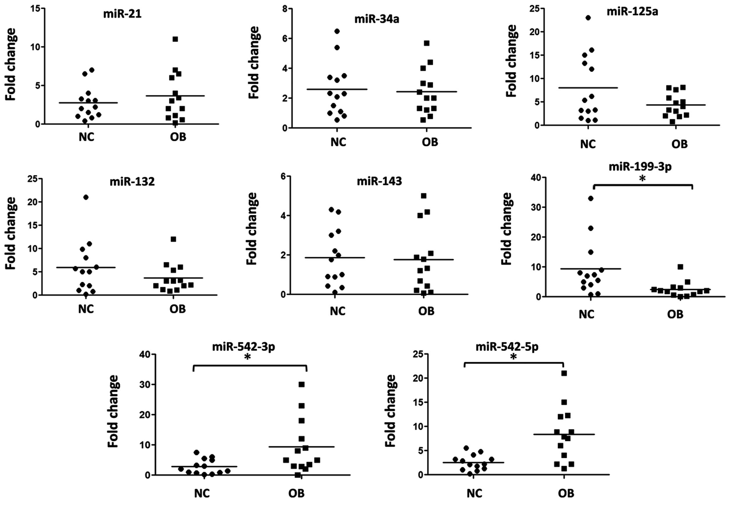
Disturbance of the microRNA expression
profile in osteosarcoma tissues
There is evidence that altered patterns of miRNA
expression correlate with various human diseases and particularly
several types of cancer (7,8). The
behavior of miRNAs is complex due to their regulation of hundreds
of targets, which can result in the downregulation of numerous
target genes, including oncogenes and tumor suppressor genes.
Therefore, examining their clinical potential is particularly
worthwhile.
Several studies have reported that the miRNA
expression profile is altered significantly in the progression of
osteosarcoma (9,10), however, further clarification is
required. In the present study, the expression profiles of eight
miRNAs, which were identified by another study as differentially
expressed in osteosarcoma tissues or osteosarcoma cell lines
(11), were determined by RT-qPCR.
As shown in Fig. 1, miR-542-3p and
miR-542-5p were significantly upregulated and miR-199-3p was
significantly downregulated in the osteosarcoma tissues.
VANGL2 expression is repressed by
miR-542-3p
The function of an miRNA is reflected mainly in its
repressive effects on the expression of its target genes (12,13).
miR-542-3p and miR-542-5p are products of the same molecule,
pre-miR-542, and miR-542-5p was considered to be the passenger
strand, therefore the present study investigated the target gene of
miR-542-3p using online bioinformatics tools. Using miRanda
(http://www.microrna.org/microrna/home.do) and
TargetScan (http://www.targetscan.org/; and Pictar, http://pictar.mdc-berlin.de/), VANGL2 was identified
as a possible target gene of miR-542-3p, the downregulation of
which is associated with enhanced cancer cell migration and
invasion ability.
To confirm whether VANGL2 was the target gene of
miR-542-3p, a 516 bp segment of the VANGL2 3′-UTR, containing the
interaction sites of miR-542-3p, was cloned into the pGL3 control
vector (pGL3-VANGL2) downstream of the firefly luciferase reporter
gene (Fig. 2A) to perform a dual
luciferase assay. The U2OS cells were cotransfected with
pGL3-VANGL2 and either the miR-542-3p mimic or inhibitor (Fig. 2B). Compared with the miRNA control,
luciferase activity was significantly decreased by miR-542-3p by
~47.8% (P<0.05). Furthermore, luciferase activity was
significantly increased by the miR-542-3p inhibitor compared with
the anti-miR control by ~23.5% (P<0.05). These results indicated
that miR-542-3p targets the 3′-UTR of VANGL2, leading to a change
in firefly luciferase translation.
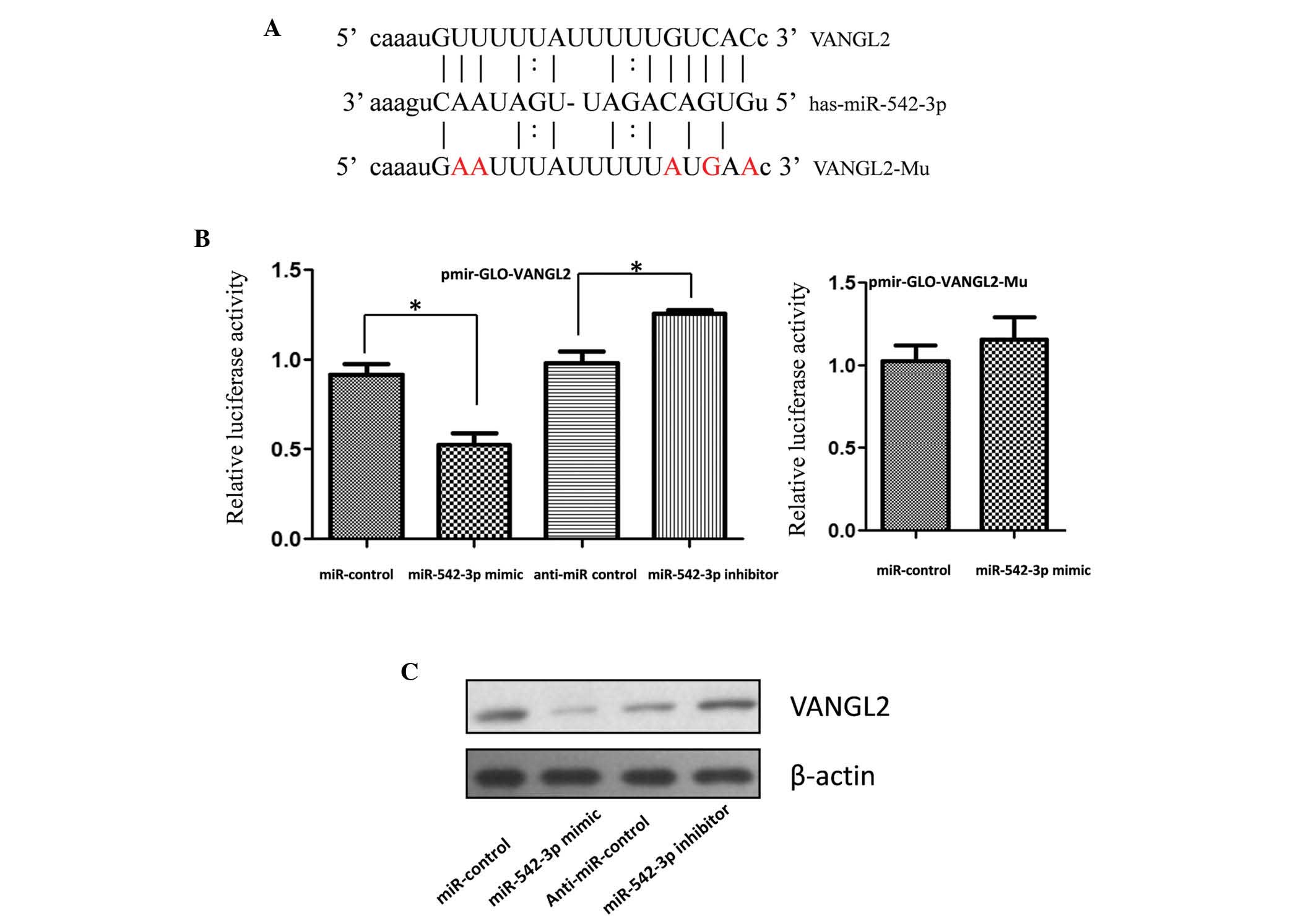 | Figure 2Expression of VANGL2 is suppressed by
miR-542-3p. (A) Schematic diagram for constructing the predicted
miR-542-3p binding site into the pGL3 control vector. (B)
Confirmation of the miR-542-3p target gene. U2OS cells were
co-transfected with the miRNA control, miR-542-3p mimic, anti-miR
control or miR-542-3p inhibitor and pGL3-VANGL2 for the
dual-luciferase assay. PRL-TK, containing RL, was cotransfected
with the 3′-UTR of VANGL2 for data normalization. (C) Mutation
analysis of the miR-542-3p binding site. Following mutation of five
nucleotides of the binding site of miR-542-3p in the 3′-UTR of
VANGL2 (pGL3-VANGL2-Mu), the luciferase activity was significantly
decreased in the U2OS cells cotransfected with the miR-542-3p
mimics and pGL3-VANGL2 compared with those cotransfected with
pGL3-VANGL2-Mu or pGL3. (D) VANGL2 protein levels in the miR-542-3p
mimic and inhibitor-treated U2OS cells were detected by western
blot analysis. miR, microRNA; VANGL2, Van Gogh-like 2; UTR,
untranslated region; Mu, mutant; PRL-TK, thymidine kinase
promoter-Renilla luciferase reporter plasmid. |
A seed sequence mutation clone was also used to
confirm the binding site for miR-542-3p (Fig. 2A). A vector, containing putative
miR-542-3p binding regions in the 3′-UTR of VANGL2 with five mutant
nucleotides (pGL3-VANGL2-Mu) was used and a wild type VANGL2 vector
was used as a control. As the histogram in Fig. 2B (right) shows, there were no
differences in enzyme activity between the cells cotransfected with
miR-542-3p mimics or pGL3-VANGL2-Mu compared with pGL3-VANGL2.
These data indicated that miR-542-3p may suppress the expression of
VANGL2 by binding to the seed sequence at the 3′-UTR of VANGL2.
miR-542-3p regulates the endogenous
expression of VANGL2 in U2OS cells
Although VANGL2 was identified as a target gene for
miR-542-3p, whether miR-542-3p was able to regulate the endogenous
expression of VANGL2 remained unclear. The U2OS cells were
transfected with the miR-542-3p mimic or inhibitor to determine
whether dysregulation of the expression of miR-542-3p affected the
endogenous expression of VANGL2. Compared with the corresponding
control, the protein expression of VANGL2 was significantly
suppressed by the miR-542-3p mimics and upregulated by the
miR-542-3p inhibitor (Fig.
2C).
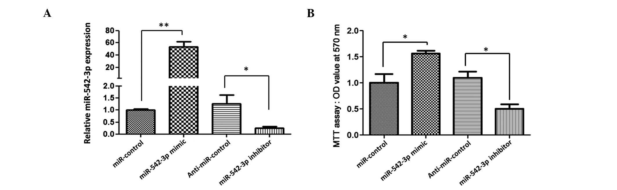
miR-542-3p overexpression enhances U2OS
cell viability
To determine the possible function of miR-542-3p in
the pathogenesis of osteosarcoma, the effect of miR-542-3p on the
growth of osteosarcoma cells was detected using an in vitro
cell line model.
The U2OS cells were transfected with either the
miR-542-3p mimic, miR control, miR-542-3p inhibitor or anti-miR
control. The results demonstrated that the expression level of
miR-542-3p in the U20S cells was increased in the cells transfected
with the miR-542-3p mimic compared with the pre-miR control
(P<0.01) and was decreased in the cells transfected with the
miR-542-3p inhibitor compared with the anti-miR control (P<0.05)
as shown in Fig. 3A.
Cell proliferation and viability were determined
using an MTT assay 48 h after transfection. As shown in Fig. 3B, the relative proliferation rates
in the U2OS cells transfected with miR-542-3p mimics were increased
by ~52.3% compared with the pre-miR control (P<0.05). The
relative proliferation rates in the U2OS cells transfected with the
miR-542-3p inhibitor decreased ~55.2% compared with the anti-miR
control (P<0.05). These results indicated that overexpression of
miR-542-3p significantly increased osteosarcoma cell viability,
while downregulation of miR-542-3p repressed osteosarcoma cell
proliferation.
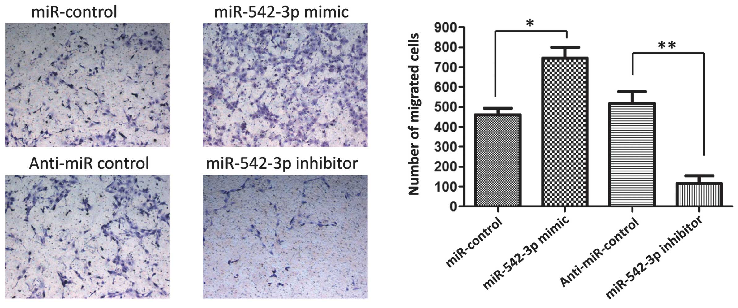
miR-542-3p modulates the migration
capacity of osteosarcoma cells in vitro
In order to investigate the role of miR-542-3p in
the metastasis of osteosarcoma cells, the present study then
analyzed the effects of miR-542-3p on the migratory behavior of
osteosarcoma cells (Fig. 4). The
results revealed that the migration capacity of the U2OS cells
transfected with the miR-542-3p mimic were significantly higher
compared with those transfected with the miR control (P<0.05).
Conversely, the migration capacity was significantly suppressed in
the U2OS cells transfected with the miR-542-3p inhibitor compared
with the anti-miR control (P<0.01). These findings suggested
that the level of miR-542-3p may be closely associated with the
metastasis of osteosarcoma cells.
Discussion
Previous evidence has demonstrated that altered
patterns of miRNA expression are associated with various human
diseases and, in particular, several types of cancer. The behavior
of miRNAs is complex as they regulate hundreds of targets, which
can result in the downregulation of numerous genes, including
oncogenes and tumor suppressors. Therefore, examining the clinical
potential of miRNAs is particularly useful.
In the present study, the expression of eight
candidate miRNAs was detected in 13 soft tissue sarcoma samples and
13 normal tissue samples by RT-qPCR. The miRNAs miR-542-3p and
miR-542-5p were significantly upregulated and miR-199-3p was
significantly downregulated. As miR-542-3p and miR-542-5p are
products of pre-miR-542, of which miR-542-3p is the main product,
the present study subsequently investigated the biological function
of upregulated miR-542-3p. The possible target genes of miR-542-3p
were predicted using online bioinformatics tools. Of the thousands
of predicted miR-542-3p target genes, the expression of VANGL2 was
found to be repressed by miR-542-3p. VANGL2 belongs to the
non-canonical WNT pathway, whose activation inhibits canonical WNT
signaling. Previous studies have demonstrated that the expression
of VANGL2 is significantly downregulated in several cancer cell
lines and primary tumors, and a low expression of VANGL2 is
associated with a significantly worse clinical outcome in
neuroblastoma (9–11). Additionally, siRNA experiments have
revealed that knock down of VANGL2 increases cell proliferation of
neuroblastoma cells (10). Wnt
transduction pathways function to modulate cell fate and
proliferation, and regulate cell growth and differentiation in a
variety of organ systems (16). In
osteosarcoma cells, as opposed to normal cells, several Wnt
ligands, receptors and coreceptors are highly expressed, while Wnt
inhibitors are downregulated; therefore, Wnt signaling is
aberrantly activated (17). VANGL2
has been proved to be structurally associated with the Wnt pathway,
and to be implicated in the development of osteosarcomas (18). However, as a target gene of
miR-542-3p, whether the osteosarcoma cell proliferation and
migration mediated by VANGL2 are regulated by miR-542-3p has
remained elusive. Therefore, the present study investigated the
impact of miR-542-3p overexpression on U2OS cell proliferation and
migration ability. As expected, miR-542-3p enhanced U2OS cell
proliferation and migration, which indicated that miR-542-3p may
act as an oncogene by repressing VANGL2 and, thus, preventing the
inhibition of the non-canonical WNT pathway.
In conclusion, the results of the present study
indicated that miR-542-3p may function as an oncogene by targeting
VANGL2 during osteosarcoma pathogenesis. These findings may provide
insight into the development of osteosarcoma and aid in the
development of novel diagnostic and therapeutic strategies for
osteosarcoma.
References
|
1
|
Hauben EI, Weeden S, Pringle J, Van Marck
EA and Hogendoorn PC: Does the histological subtype of high-grade
central osteosarcoma influence the response to treatment with
chemotherapy and does it affect overall survival? A study on 570
patients of two consecutive trials of the European Osteosarcoma
Intergroup. Eur J Cancer. 38:1218–1225. 2002. View Article : Google Scholar : PubMed/NCBI
|
|
2
|
Hamada K, Tomita Y, et al: Evaluation of
chemotherapy response in osteosarcoma with FDG-PET. Ann Nucl Med.
23:89–95. 2009. View Article : Google Scholar : PubMed/NCBI
|
|
3
|
Tabone MD, Kalifa C, Rodary C, et al:
Osteosarcoma recurrences in pediatric patients previously treated
with intensive chemotherapy. J Clin Oncol. 12:2614–2620.
1994.PubMed/NCBI
|
|
4
|
Longhi A, Errani C, et al: Primary bone
osteosarcoma in the pediatric age: state of the art. Cancer Treat
Rev. 32:423–436. 2006. View Article : Google Scholar : PubMed/NCBI
|
|
5
|
Lal A, et al: miR-24 Inhibits cell
proliferation by targeting E2F2, MYC, and other cell-cycle genes
via binding to ‘seedless’ 3′UTR microRNA recognition elements. Mol
Cell. 35:610–625. 2009. View Article : Google Scholar : PubMed/NCBI
|
|
6
|
Zhao Y, Samal E and Srivastava D: Serum
response factor regulates a muscle-specific microRNA that targets
Hand2 during cardiogenesis. Nature. 436:214–220. 2005. View Article : Google Scholar : PubMed/NCBI
|
|
7
|
Calin GA and Croce CM: MicroRNA-cancer
connection: the beginning of a new tale. Cancer Res. 66:7390–7394.
2006. View Article : Google Scholar : PubMed/NCBI
|
|
8
|
Ikeda S, Kong SW, Lu J, et al: Altered
microRNA expression in human heart disease. Physiol Genomics.
31:367–373. 2007. View Article : Google Scholar : PubMed/NCBI
|
|
9
|
Maire G, Martin JW, et al: Analysis of
miRNA-gene expression-genomic profiles reveals complex mechanisms
of microRNA deregulation in osteosarcoma. Cancer Genet.
204:138–146. 2011. View Article : Google Scholar : PubMed/NCBI
|
|
10
|
Jones KB, et al: miRNA signatures
associate with pathogenesis and progression of osteosarcoma. Cancer
Res. 72:1865–1877. 2012. View Article : Google Scholar : PubMed/NCBI
|
|
11
|
Namløs HM, Meza-Zepeda LA, Barøy T, et al:
Modulation of the osteosarcoma expression phenotype by microRNAs.
PLoS One. 7:e480862012. View Article : Google Scholar : PubMed/NCBI
|
|
12
|
Doench JG and Sharp PA: Specificity of
microRNA target selection in translational repression. Genes Dev.
18:504–511. 2004. View Article : Google Scholar : PubMed/NCBI
|
|
13
|
Lewis BP, Burge CB and Bartel DP:
Conserved seed pairing, often flanked by adenosines, indicates that
thousands of human genes are microRNA targets. Cell. 120:15–20.
2005. View Article : Google Scholar : PubMed/NCBI
|
|
14
|
Wang R, Wang HB, et al: MiR-101 is
involved in human breast carcinogenesis by targeting Stathmin1.
PLoS One. 7:e461732012. View Article : Google Scholar : PubMed/NCBI
|
|
15
|
Hu Y, Liu CM, et al: Two common SNPs in
pri-miR-125a alter the mature miRNA expression and associate with
recurrent pregnancy loss in a Han-Chinese population. RNA Biol.
8:861–872. 2011. View Article : Google Scholar : PubMed/NCBI
|
|
16
|
Paul S and Dey A: Wnt signaling and cancer
development: therapeutic implication. Neoplasma. 55:165–176.
2008.PubMed/NCBI
|
|
17
|
McQueen P, Ghaffar S, et al: The Wnt
signaling pathway: implications for therapy in osteosarcoma. Expert
Rev Anticancer Ther. 11:1223–1232. 2011. View Article : Google Scholar : PubMed/NCBI
|
|
18
|
Nishita M, Enomoto M, Yamagata K and
Minami Y: Cell/tissue-tropic functions of Wnt5a signaling in normal
and cancer cells. Trends Cell Biol. 20:346–354. 2010. View Article : Google Scholar : PubMed/NCBI
|