Introduction
Choroidal neovascularization (CNV) refers to the
abnormal growth of choroidal vessels, which extend through Bruch’s
membrane into the sub-retinal pigment epithelium (RPE) or
subretinal space (1). Its
formation is widely considered as the outcome of breaking the
balance between angiogenic and angiostatic factors. Among these
factors, vascular endothelial growth factor (VEGF), one of numerous
pro-angiogenic molecules, has been indicated as a critical factor
in CNV growth in a number of experimental and clinical studies
(2–4).
Endostar is a novel recombinant human endostatin,
which was approved by the State Food and Drug Administration of
China for the treatment of non-small-cell lung cancer in 2005
(5). It has a six-histidine tag at
the N-terminal of the protein, which may simplify purification and
improve the stability of the protein (6). Previous studies have indicated that
Endostar reduces micro blood vessel density and lymphatic micro
vessel density to control tumor growth in vivo via the
downregulation of VEGF-A and VEGF-C (7).
Considering the potential effect of endostatin on
experimental CNV (8,9), Endostar was administered to
laser-induced CNV in rats to determine whether this drug may
prevent CNV formation. In addition, the potential underlying
mechanisms were investigated.
Materials and methods
Animals
Male brown Norway rats (age, 8 weeks) obtained from
the Animal Supply Center of Zhejiang Academy of Medical Science
(Hangzhou, China) were used in accordance with the Association for
Research in Vision and Ophthalmology Statement for the Use of
Animals in Ophthalmic and Vision Research. The present study was
approved by the Ethical Committee of the School of Medicine,
Zhenjiang University (Hangzhou, China). Anesthesia was induced by
intraperitoneal injection of 1% sodium pentobarbital (45 mg/kg body
weight), and then the ocular surface was anesthetized with topical
instillation of oxybuprocaine hydrochloride. The pupils were
dilated with topical 1% tropicamide (Santen Pharmaceutical Co.,
Ltd., Osaka, Japan).
Induction of CNV and administration of
Endostar or vehicle
Eight burns of 532-nm diode laser photocoagulation
(50 μm, 0.05 sec, 300 mW) were delivered to each retina with the
slit lamp delivery system of a diode laser (OcuLight GL; Iridex,
Mountain View, CA, USA) to induce the CNV model. The laser spots
were applied between the major retinal vessels 2–4 disc diameters
from the optical disc. Ocular contact lenses (Ocular Instruments
Inc., Bellevue, WA, USA) filled with one drop of methylcellulose
were applied to the rat cornea. Production of a bubble at the time
of laser exposure was confirmed for each spot. A total of 40 rats
received laser photocoagulation treatment in each eye and were then
divided equally into two groups, the Endostar group and
phosphate-buffered saline (PBS) control group, at random. Each rat
received an intravitreal injection of 10 μl Endostar (5 mg/ml;
Yantai Sincere Machinery Co., Ltd., Yantai, China) or 10 μl PBS
bilaterally using a sterile 10 μl Hamilton syringe (Hamilton Co.,
Reno, NV, USA) with a 33G needle following laser photocoagulation
every other day for two weeks. This surgery was completed under
visualization of Zeiss OPMI 6SFR operating microscope (Zeiss
Humphrey Systems, Dublin, CA, USA). All examinations of the rats
were performed 14 days following the photocoagulation.
Fluorescein angiogram (FA)
The activity of CNV was evaluated by assessment of
late-phase FA, captured 8–10 min following intraperitoneal
injection of 0.1 ml of 10% fluorescein sodium (Alcon, Fort Worth,
TX, USA) at day 14 in the rat models. The anesthetized rats with
dilated pupils were observed using a digital imaging system
(Heidelberg Retina Angiograph II; Heidelberg Engineering,
Heidelberg, Germany). Leakage was defined as the presence of a
hyperfluorescent spot that increased in size with time. Angiograms
were graded by a previously established scheme (10) as follows: 0, no leakage; 1, slight
leakage (hyperfluorescent lesion without progressive increase in
size or intensity); 2, moderate leakage (hyperfluorescence
increasing in intensity but not in size); 3, prominent leakage
(hyperfluorescence increasing in intensity and in size). In order
to ensure the effectiveness and accuracy of the evaluation, the
angiograms were assessed by two independent graders, who were
blinded to the intervention.
Choroidal flat mounts
CNV area was measured with choroidal flat mounts in
accordance with the previously reported methods (11). The rats were anesthetized and
perfused through the left ventricle with 50 ml vehicle, followed by
20 ml of 5 mg/ml FITC-dextran (MW, 2×10⁶; Sigma, St.
Louis, MO, USA) in 10% (wt/vol) gelatin. The eyes were enucleated
and fixed in 4% paraformaldehyde for 1 h. Next, the anterior
segment and retina were removed from the eyecup. Four to six
relaxing radial incisions were made, and the remaining
RPE-choroid-sclera complex was coverslipped. Images of the
choroidal flat mounts were then captured under an epifluorescent
microscope (Leica Microsystems, Wetzlar, Germany). For measuring
the CNV lesion, the green hyperfluorescent area at the laser spot
in the flat mounts was measured by Image-Pro Plus software (Media
Cybernetics, Bethesda, MD, USA).
Immunofluorescent staining
Enucleated eyes from rats were immediately fixed in
4% paraformaldehyde at 4°C for 12 h. After the anterior segment and
lens were removed, the remaining eyecup was cytoprotected with 30%
sucrose and then embedded in Tissue-Tek OCT compound (Sakura
Finetech, Tokyo, Japan). The eyecups were cut into 7-μm sections.
Following blocking with 5% bovine serum albumin (BSA) for 1 h,
these cryo-sections were incubated with the primary rabbit
anti-CD31 monoclonal antibody (Abcam, Cambridge, UK) at 4°C
overnight. The sections were then incubated with secondary
antibody, FITC-conjugated goat-anti-rabbit IgG F(ab)2 fragment
(Santa Cruz Biotechnology, Santa Cruz, CA, USA) for 1 h. The slides
were viewed with an epifluorescent microscope (Leica Microsystems).
CNV thickness was then measured vertically from the adjacent RPE
layer to the top of the CNV. Consecutive sections were examined to
select maximal CNV thickness. The thickness was measured by
Image-Pro Plus software (Media Cybernetics).
Quantitative polymerase chain reaction
(qPCR)
Total RNA was extracted from six rat eyecups in each
group using TRIzol reagent and the cDNA was generated using the
SuperScript III First-Strand Synthesis system (both from Invitrogen
Life Technologies, Carlsbad, CA, USA). qPCR was performed using an
ABI Prism 7500™ instrument (Applied Biosystems, Carlsbad, CA, USA)
and the SYBR Premix Dimer Eraser (Takara Bio, Inc., Dalian, China)
according to the manufacturer’s instructions. The mRNA expression
of VEGF, hypoxia-inducible factor 1α (HIF-1α), chemokine C-X-C
motif ligand 1 (CXCL1), angiogenin (Ang), fms-related tyrosine
kinase 1 (Flt-1) and pigment epithelium-derived factor (PEDF) were
analyzed after normalization against the expression of GAPDH.
Primer sequences were as follows: Forward:
5′-AGGCAGCTTGAGTTAAACGAACGTA-3′ and reverse:
5′-AGGTCTAGTTCCCGACCCTGA-3′ for VEGF; forward:
5′-TCTAGTGAACAGGATGGAATGGAG-3′ and reverse:
5′-TCGTAACTGGGCTGTGGTAA-3′ for HIF-1α; forward:
5′-TGCACCCAAACCGTC-3′ and reverse: 5′-ACGCCA TCGGTGCAATCTA-3′ for
CXCL1; forward: 5′-CCAGTT GCAAGCATA-3′ and reverse:
5′-AGCCATTCTCACA GGCAATAACAAC-3′ for Ang; forward: 5′-GAGTGCAAA
TGGATGAAG-3′ and reverse: 5′-CAGCAATCCATG ATTTGGTA-3′ for Flt-1;
forward: 5′-GAGTGCCTC CAGAATTGTGTTTGA-3′ and reverse:
5′-CCTGCACCCAGT TGTTAATCTCC-3′ for PEDF; and forward: 5-CAAGTTCAA
CACAGTCA-3′ and reverse: 5′-CCATTTGATGTTAG CGGGAT-3′ for GAPDH. The
PCR conditions for various genes were as follows: 40 cycles of 95°C
for 2 min; 95°C for 5 sec, 55°C for 30 sec and 72°C for 34 sec.
Western blot analysis
The protein extracts were prepared from three
eyecups in each group. The samples were then assessed for protein
concentration (Bradford assay; Bio-Rad Laboratories, Munich,
Germany). Electrophoresis of proteins was performed with 10%
SDS-polyacrylamide gels. A total of 30 μg of protein were loaded on
each lane. The protein was electrotransferred to a polyvinylidene
difluoride membrane (Millipore, Bedford, MA, USA), which was
blocked in Tris-buffered saline with Tween-20 (TBST) containing 5%
(wt/vol) BSA for 1 h at room temperature and then probed overnight
at 4°C with rabbit polyclonal anti-VEGF (1:1,000; Santa Cruz
Biotechnology, Inc.) or rabbit polyclonal anti-β-actin antibody
(1:2,000; Affinity Bioreagents, Golden, CO, USA). The blots were
washed three times with TBST for 15 min per wash and then incubated
with anti-rabbit horseradish peroxidase-conjugated IgG secondary
antibody (1:1,000; Santa Cruz Biotechnology, Inc.) for 1 h at room
temperature. They were then washed again three times each for 5
min. Chemiluminescent detection was accomplished using the enhanced
chemiluminescence kit (Amersham Biosciences, Little Chalfont, UK)
and ChemiDoc XRS apparatus (Bio-Rad Laboratories, Hercules, CA,
USA). The band intensity was measured using densitometry. The
results were expressed as the density ratio relative to
β-actin.
Statistical analysis
The data are expressed as the mean ± standard error
of the mean. Student’s t-test was used for statistical comparison
of the means between the groups. The changes of leakage of CNV were
analyzed using Pearson’s χ2 test or Mann-Whitney U test,
where appropriate. Statistical analyses were performed using SPSS
software version 16.0 (SPSS, Inc., Chicago, IL, USA). All P-values
are two-sided, and P<0.05 was considered to indicate a
statistically significant difference.
Results
Endostar suppresses the activity and
volume of CNV
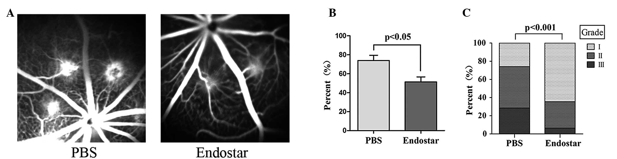
The FA in the two groups was compared at day 14
following treatment. The outcome revealed that Endostar-treated
rats had reduced CNV incidence and leakage grade compared with the
control. CNV was observed in 50.3% laser spots in Endostar-treated
rats (n=161 spots), while it was observed in a significantly higher
proportion in control rats (75.8%, n=153 spots; P<0.05, by
Pearson’s χ2 test). The extent of CNV leakage also
reduced markedly in Endostar-treated rats (P<0.001, by
Mann-Whitney U test; Fig. 1).
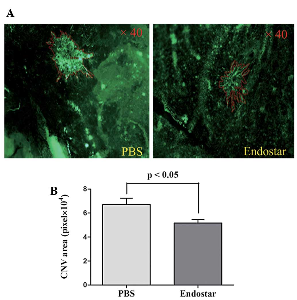
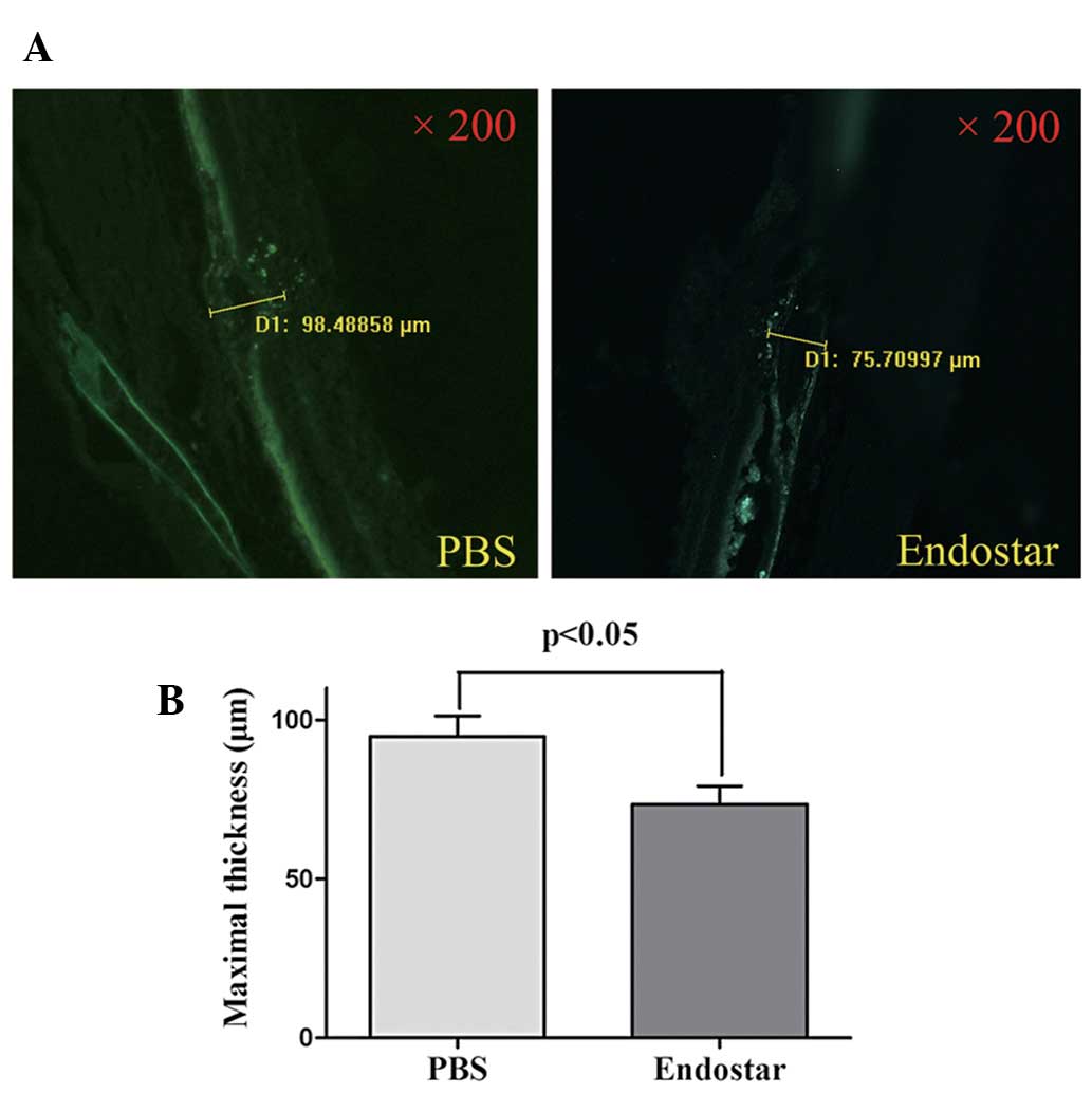
CNV area and the maximal thickness of each CNV were
quantified by analysis of fluorescence on day 14 following Endostar
or PBS treatment. The mean CNV area in Endostar-treated rats (n=39
spots) was significantly less than that in PBS-treated rats (n=42
spots; 51711±2880 vs. 67019±5407 pixel, P<0.05, by Student’s
t-test; Fig. 2). Furthermore, the
maximal thickness of CNV in the Endostar group was also reduced
significantly compared with the PBS group (73.5±5.6 μm vs. 94.8±6.4
μm, respectively; n=11 spots per group, P<0.05, by Student’s
t-test; Fig. 3).
Endostar downregulates
angiogenesis-associated molecules
To determine the effect of Endostar on
angiogenesis-associated molecules, eyecups of rat models at day 14
were collected, and qPCR was performed to detect the expression
changes of certain molecules between the two groups and the results
demonstrated that at the mRNA level, VEGF, HIF-1α and CXCL1 were
significantly downregulated in the Endostar group (n=6 per group;
P<0.05, by Student’s t-test). However, no significant changes
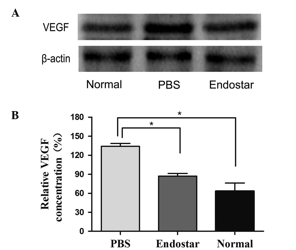
were observed in the expression of Ang, Flt-1 and PEDF (Fig. 4). Furthermore, as verified by
western blot analysis, Endostar significantly suppressed the
expression of VEGF at the protein level compared with the PBS group
(n=3 per group; P<0.05, by Student’s t-test), although the two
groups demonstrated upregulated expression of VEGF compared with
the control group without laser photocoagulation (Fig. 5).
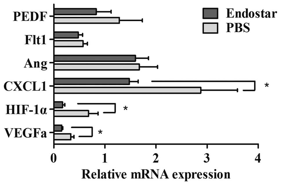 | Figure 4Gene regulation by Endostar in CNV as
determined with qPCR. qPCR demonstrated that angiogenic molecules,
VEGF, HIF-1α and CXCL1, were significantly downregulated in the
Endostar group compared with the PBS group (*P<0.05,
by Student’s t-test). CNV, choroidal neovascularization; qPCR,
quantitative polymerase chain reaction; PEDF, pigment
epithelium-derived factor; Flt1, fms-related tyrosine kinase 1;
Ang, angiogenin; HIF-1α, hypoxia-inducible factor 1α VEGF, vascular
endothelial growth factor; CXCL1, chemokine C-X-C motif ligand 1;
PBS, phosphate-buffered saline. |
Discussion
Endostatin is a broad-spectrum endogenous
angiostatic protein, which is proposed to inhibit E-cadherins,
matrix metalloproteinases (MMPs), the VEGF signaling pathway, cell
survival-related genes, STATs and other key molecules in
angiogenesis (12–15). Compared with endostatin, Endostar
has an improved function and longer half-life (16). The anti-vescularization properties
of Endostar are widely conisdered to be correlated with VEGF
(7,17).
In the present study, the angiostatic effects of
Endostar were investigated in a laser-induced CNV model. As
exhibited by FA, Endostar-treated rats had a significantly reduced
incidence of CNV and grade of fluorescence leakage than the PBS
group at day 14. To compare the volume of CNV, the choroidal flat
mounts and the maximal CNV thickness were detected separately. The
results indicated that Endostar treatment significantly decreased
the volume of CNV. qPCR and western blot analysis of eyecups in
each group confirmed the hypothesis that Endostar exerts its
angiostatic effect by regulating the transcription and expression
of VEGF, the vital molecule in angiogenesis. In addition, Endostar
may also affect other important factors in the angiogenic network.
A previous study suggested that RPE cell-derived HIF-1α is a
pivotal contributor to CNV (18).
The present results demonstrated an evident decrease in the HIF-1α
mRNA in the Endostar group, which may contribute to CNV
suppression. Neutrophil invasion was characteristic of early
inflammatory responses during laser-induced CNV, which promoted the
early development of CNV possibly via the secretion of angiogenic
growth factors (19). CXCR2 is
considered to have a crucial role in neutrophil infiltration, while
its ligand CXCL1 has potent angiogenic activity (20). Therefore, the downregulation of
CXCL1 in the Endostar group may also be conducive to the
suppression of CNV.
The present study does however have a number of
limitations. Although this CNV model shares histological and
angiographic features with the human disease, this acute
wound-healing model cannot absolutely reflect the chronic disease
found clinically. However, it remains useful to understand the
process of CNV development and investigate novel therapeutic agents
and strategies. Another limitation of the present study is the
absence of further exploration of the appropriate dosage of
Endostar for intravitreal injection. Although based on the
experience from other experiments, it was possible to observe CNV
suppression in the present rat model, further studies are required
to examine the efficiency and safety of Endostar and to determine
the optimal dosage for future application.
In conclusion, the present study demonstrated that
Endostar is able to suppress newly-formed CNV by reducing the
expression of VEGF and other angiogenic factors. Therefore, it has
potential for clinical CNV treatment alone or as an adjuvant
therapy.
Acknowledgements
This study was supported by the Youth Program of the
National Natural Science Foundation of China (grant no.
11104246/A040414), the Zhejiang Natural Science Foundation (grant
no. Y2100380), the Zhejiang Science and Technology Department
Public Project (grant no. 2010C33085), the Zhejiang Key Innovation
Team Project (grant no. 2009R50039), the Doctoral Fund of Ministry
of Education of China (grant no. 20100101120135) and the Key Lab
Fund of Zhejiang Province (grant no. 2011E10006).
References
|
1
|
Grossniklaus HE and Green WR: Choroidal
neovascularization. Am J Ophthalmol. 137:496–503. 2004. View Article : Google Scholar : PubMed/NCBI
|
|
2
|
Yu MJ, Shen WY, Lai MC, Constable IJ,
Papadimitriou JM and Rakoczy PE: The role of vascular endothelial
growth factor (VEGF) in abnormal vascular changes in the adult rat
eye. Growth Factors. 17:301–312. 2000. View Article : Google Scholar : PubMed/NCBI
|
|
3
|
Kwak N, Okamoto N, Wood JM and Campochiaro
PA: VEGF is major stimulator in model of choroidal
neovascularization. Invest Ophthalmol Vis Sci. 41:3158–3164.
2000.PubMed/NCBI
|
|
4
|
Rosenfeld PJ, Brown DM, Heier JS, et al:
Ranibizumab for neovascular age-related macular degeneration. N
Engl J Med. 355:1419–1431. 2006. View Article : Google Scholar : PubMed/NCBI
|
|
5
|
Ling Y, Yang Y, Lu N, et al: Endostar, a
novel recombinant human endostatin, exerts antiangiogenic effect
via blocking VEGF -induced tyrosine phosphorylation of KDR/Flk-1 of
endothelial cells. Biochem Biophys Res Commun. 361:79–84. 2007.
View Article : Google Scholar : PubMed/NCBI
|
|
6
|
Song HF, Liu XW, Zhang HN, et al:
Pharmacokinetics of His-tag recombinant human endostatin in Rhesus
monkeys. Acta Pharmacol Sin. 26:124–128. 2005. View Article : Google Scholar : PubMed/NCBI
|
|
7
|
Ma X, Yao Y, Yuan D, et al: Recombinant
human endostatin endostar suppresses angiogenesis and
lymphangiogenesis of malignant pleural effusion in mice. PLoS One.
7:e534492012. View Article : Google Scholar
|
|
8
|
Marneros AG, She H, Zambarakji H, et al:
Endogenous endostatin inhibits choroidal neovascularization. FASEB
J. 21:3809–3818. 2007. View Article : Google Scholar : PubMed/NCBI
|
|
9
|
Mori K, Ando A, Gehlbach P, et al:
Inhibition of choroidal neovascularization by intravenous injection
of adenoviral vectors expressing secretable endostatin. Am J
Pathol. 159:313–320. 2001. View Article : Google Scholar : PubMed/NCBI
|
|
10
|
Kim C, Yu HG and Sohn J: The
anti-angiogenic effect of chlorogenic acid on choroidal
neovascularization. Korean J Ophthalmol. 24:163–168. 2010.
View Article : Google Scholar : PubMed/NCBI
|
|
11
|
Honda M, Asai T, Umemoto T, Araki Y, Oku N
and Tanaka M: Suppression of choroidal neovascularization by
intravitreal injection of liposomal SU5416. Arch Ophthalmol.
129:317–321. 2011. View Article : Google Scholar : PubMed/NCBI
|
|
12
|
Sudhakar A, Sugimoto H, Yang C, Lively J,
Zeisberg M and Kalluri R: Human tumstatin and human endostatin
exhibit distinct antiangiogenic activities mediated by alpha v beta
3 and alpha 5 beta 1 integrins. Proc Natl Acad Sci USA.
100:4766–4771. 2003. View Article : Google Scholar : PubMed/NCBI
|
|
13
|
Nyberg P, Heikkilä P, Sorsa T, et al:
Endostatin inhibits human tongue carcinoma cell invasion and
intravasation and blocks the activation of matrix
metalloprotease-2, -9, and -13. J Biol Chem. 278:22404–22411. 2003.
View Article : Google Scholar : PubMed/NCBI
|
|
14
|
Rehn M, Veikkola T, Kukk-Valdre E, et al:
Interaction of endostatin with integrins implicated in
angiogenesis. Proc Natl Acad Sci USA. 98:1024–1029. 2001.
View Article : Google Scholar : PubMed/NCBI
|
|
15
|
Abdollahi A, Hahnfeldt P, Maercker C, et
al: Endostatin’s antiangiogenic signaling network. Mol Cell.
13:649–663. 2004. View Article : Google Scholar : PubMed/NCBI
|
|
16
|
Jia H and Kling J: China offers
alternative gateway for experimental drugs. Nat Biotechnol.
24:117–118. 2006. View Article : Google Scholar : PubMed/NCBI
|
|
17
|
Zhao ZZ, Cao Y, Liu ZS, et al: Effects of
recombinant human endostatin on peritoneal angiogenesis in
peritoneal dialysis rats. Nephrology (Carlton). 16:599–606. 2011.
View Article : Google Scholar
|
|
18
|
Lin M, Hu Y, Chen Y, et al: Impacts of
hypoxia-inducible factor-1 knockout in the retinal pigment
epithelium on choroidal neovascularization. Invest Ophthalmol Vis
Sci. 53:6197–6206. 2012. View Article : Google Scholar : PubMed/NCBI
|
|
19
|
Zhou J, Pham L, Zhang N, et al:
Neutrophils promote experimental choroidal neovascularization. Mol
Vis. 11:414–424. 2005.PubMed/NCBI
|
|
20
|
Strieter RM, Polverini PJ, Kunkel SL, et
al: The functional role of the ELR motif in CXC chemokine-mediated
angiogenesis. J Biol Chem. 270:27348–27357. 1995. View Article : Google Scholar : PubMed/NCBI
|