Introduction
Deep vein thrombosis (DVT) is a common clinical
problem that affects >250,000 individuals in the USA and 25,000
individuals in Canada annually (1). DVT in the lower extremities is
characterized by initial swelling and lower limb pain symptoms,
which may be severe. DVT is also a well-recognized cause of
pulmonary embolism (PE), which is a potentially life-threatening
complication (2,3), for which treatment is necessary.
Traditionally, the initial treatment for DVT involves
anticoagulation using low-molecular-weight heparin (4,5),
followed by long term therapy with vitamin K antagonists, such as
warfarin (6). Since the outcome of
these treatments in some cases is disappointing (7,8),
more effective approaches aimed at reducing DVT have garnered
interest in recent years.
Salvia miltiorrhiza is one of the most
versatile Chinese herbal drugs, which has been used for hundreds of
years to treat numerous ailments (9). S. miltiorrhiza is considered
to be highly effective in activating circulation, and dispersing
stasis or sludging of blood (10).
In recent years, S. miltiorrhiza has been widely used to
treat cardiovascular diseases (11). The present study investigated
whether S. miltiorrhiza could prevent DVT, using a rabbit
ligation model. Furthermore, the underlying mechanism of the
protective effects of S. miltiorrhiza on vascular
endothelial cells was examined.
Materials and methods
Materials
S. miltiorrhiza was donated by the Pharmacy
of the Hubei University of Medicine (Shiyan, China) (no.
Z51021303). These were extractions of roots, containing 1.5 g/ml.
Malondialdehyde (MDA) and Superoxide Dismutase (SOD) Detection kits
were obtained from Nanjing Jiancheng Institute of Biotechnology
(Nanjing, China). A total of 30 male Japanese white rabbits,
weighing 1.8–2.2 kg, were obtained from the Animal Care Facility at
the Hubei Animal Center (Wuhan, China) (no. SYXK 2004-0021). All
the rabbits had ad libitum access to food and water and were
under a standard 12 h light/12 h dark cycle. All experiments
performed were in accordance with animal ethics standards. Prior to
the experiment, the rabbits were maintained for several days, to
observe their health. The rabbits were randomly divided into three
groups (n=10 per group): The control, model and Salvia
groups. All procedures were approved by the Animal Research Ethics
Board at Hubei University of Medicine.
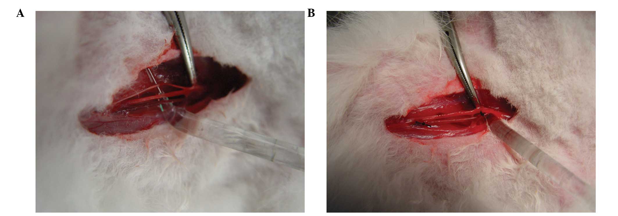
Modeling method
All surgical manipulations were conducted under
aseptic conditions, and the rabbits were anesthetized with 10%
chloral hydrate (Weihai Wego Medical Systems Co., Ltd, Weihai,
China), by means of an intramuscular injection (3 ml/kg, body
weight). Following anesthetization, the two sides of the femoral
vein (4 cm) of the rabbits were exposed and separated from the
surrounding tissue, taking care not to damage the femoral artery
and nerve. Each end of the femoral vein was then completely ligated
using 2-0 silk thread (Weihai Wego Medical Systems Co., Ltd;
Fig. 1).
Delivery method
A total of 30 rabbits were randomly divided into
three groups, each containing 10 rabbits: Control, model and
Salvia. The Salvia group were treated with a daily
intravenous injection of 2 g/kg S. miltiorrhiza nto the ear
vein prior to generation of the ligation model. The model and
control groups received equal amounts of sodium chloride. Following
one week of injections the rabbit ligation models were generated,
according to the protocol described above. Each group of rabbits
received the same treatment as mentioned until the end of the
experiment. The response to stimuli (sensitivity to pain stimuli),
activity levels and appetite in the rabbits were recorded. The
rabbits were sacrificed by aeroembolism seven days after generation
of the ligation model.
Specimen collection
Venous blood was collected in the normal and heparin
tubes (Weihai Wego Medical Systems Co., Ltd), in order to measure
SOD activities, MDA content, coagulation function (including
prothrombin, activated partial prothrombin, fibrinogen and thrombin
times) and blood rheological parameters (including whole blood
viscosity, plasma viscosity and erythrocyte aggregation).
Coagulation function was measured by CoaguChek® (Roche
Diagnostics, Xinqin, China) and blood rheological parameters were
measured using a Blood Rheology Analyzer (SA-9000; Lemon, Zhejing,
China). These measurements were made prior to generation of the
model, and three and seven days afterwards. At the end of the seven
days the ligated femoral veins were placed in 10% formaldehyde
solution, fixed for 48 h and paraffin-embedded. Serial sections (4
μm) were collected for hematoxylin and eosin staining. Images of
the venous wall and thrombosis were captured under a microscope
(BX41; Olympus Corp., Tokyo, Japan). Low magnification (x10) was
used to evaluate the vascular wall, while endothelial cells were
observed at a higher magnification (x40).
Statistical analysis
The data were analyzed by Student’s t-test and
one-way analysis of variance to compare the differences between the
groups, using SPSS version 13.0 software (SPSS Inc., Chicago, IL,
USA). P<0.05 was considered to indicate a statistically
significant difference.
Results
Toxicity and quality of life
The response to stimuli, activity levels and
appetite of the model and Salvia groups were similar to that
of the control group. Furthermore, no fatal PE occurred in any of
the rabbits.
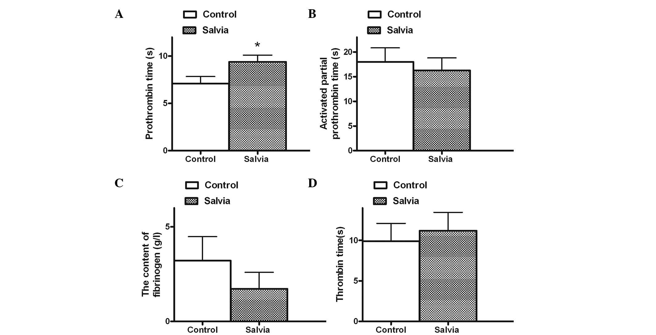
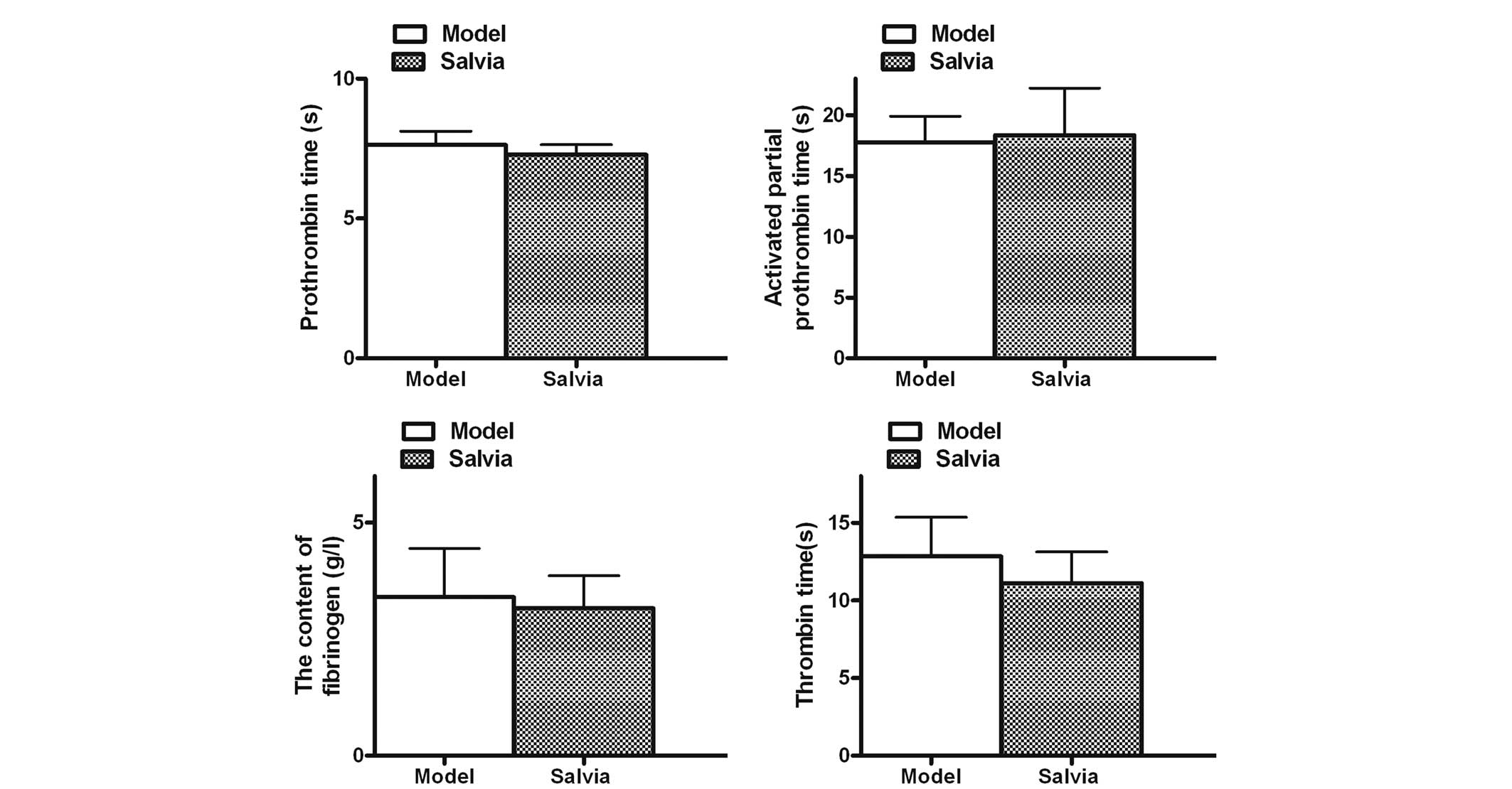
Detection of coagulation function prior
to generation of the ligation model
Following one week of S. miltiorrhiza
treatment, the coagulation function of the Salvia group was
not affected, except for prothrombin time (PT), which was
significantly increased, as compared with the control group
(P<0.05; Fig. 2).
Measurements of blood rheological
parameters and coagulation function, three days following
generation of the ligation model
Following generation of the model, blood rheological
parameters were measured, and whole blood viscosity (1/sec, 5/sec,
30/sec) and erythrocyte aggregation were shown to be significantly
increased in the model group, as compared with the control group
(P<0.05). These results suggest that ligation of the femoral
vein may result in a hypercoagulable state. There was also a
statistically significant increase in whole blood viscosity (1/sec,
5/sec, 30/sec) in the Salvia group, as compared with the
model group (P<0.05; Fig. 3).
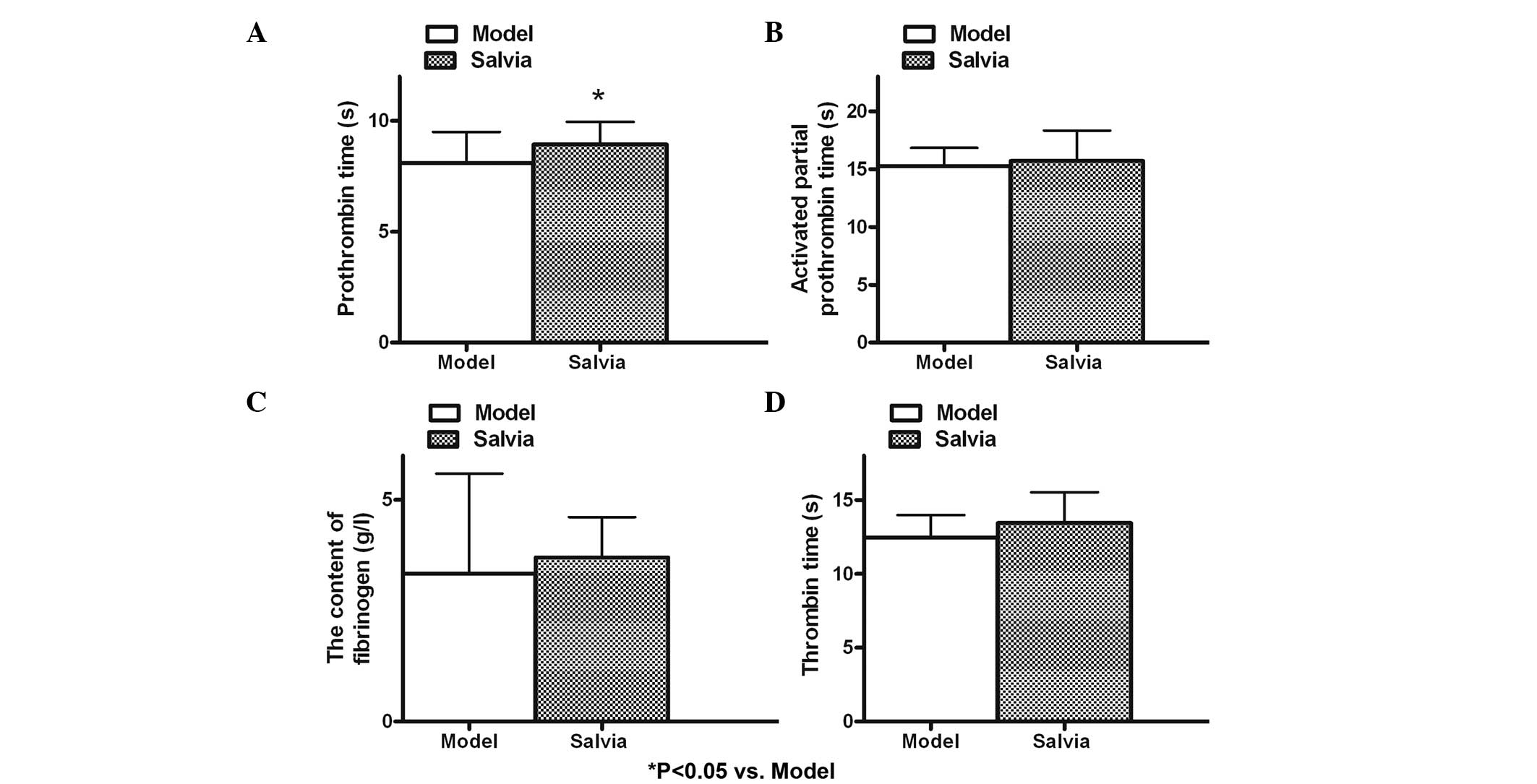
No significant differences were identified in the measurements of
coagulation function, except for PT, which was increased in the
Salvia group, as compared with the model group (P<0.05;
Fig. 4).
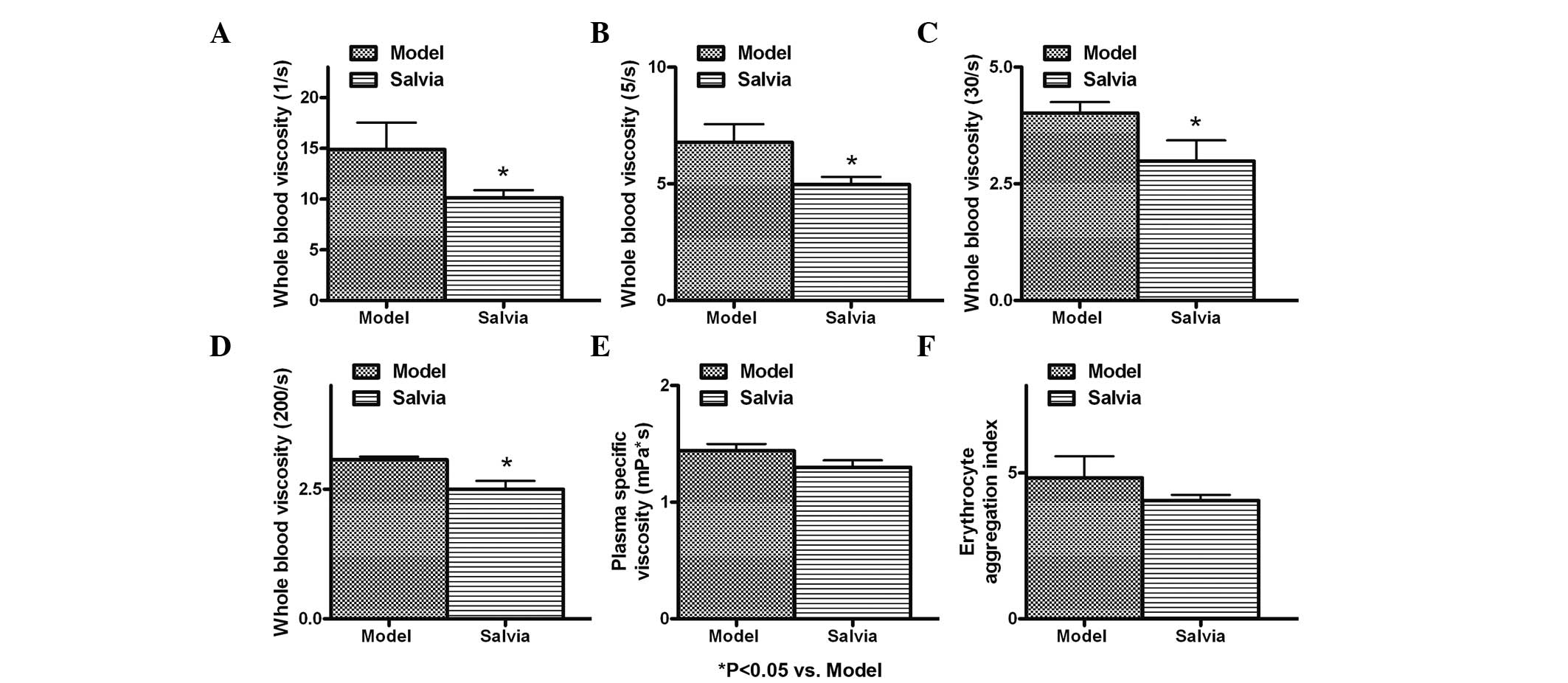
Measurements of blood rheological
parameters and coagulation function, seven days following
generation of the ligation model
Seven days after generation of the model, blood
rheological parameters were measured, and whole blood viscosity
(1/sec, 5/sec, 30/sec and 200/sec) was shown to be significantly
decreased in the Salvia group, as compared with the model
group (P<0.05; Fig. 5). There
were no significant differences in the coagulation function between
the Salvia and model groups (P>0.05; Fig. 6).
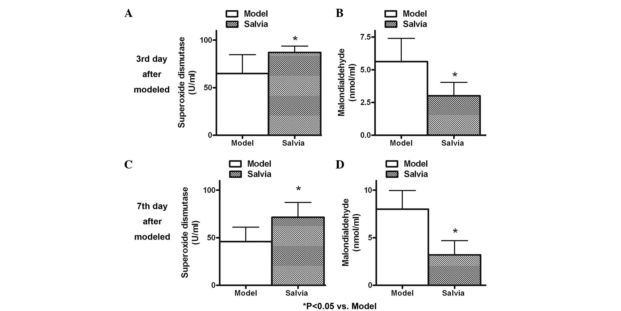
Antioxidative function
The present study evaluated the antioxidative
function of S. miltiorrhiza, the results of which are
presented in Fig. 7. The MDA
content was significantly decreased (P<0.05), and the activities
of SOD were notably increased (P<0.05) in the Salvia
group compared with the model group three days after generation of
the model. The MDA content was higher on the seventh day
(8.00±1.96) in the model group compared with the third day
(5.63±1.77). However, in response to treatment with S.
miltiorrhiza, MDA content was decreased (P<0.05), and SOD
activities were increased (P<0.05) compared with the model group
seven days after generation of the ligation model.
Histopathological observation
Following the sacrifice of the rabbits, the ligated
femoral veins were harvested and images of the veins were captured
under a microscope. The vascular wall was continuous in the control
group, endothelial integrity was maintained and no shedding of
endothelial cells was observed. The vascular wall of the model
group exhibited discontinuous change, intimal irregularities,
endothelial cell shedding, and thrombosis (mixed thrombus and red
thrombus), which was attached to the vascular wall. The vascular
wall of the Salvia group exhibited complete continuity,
regular intima, some endothelial cell shedding and a small amount
of intraluminal thrombosis, which was partly attached to the
vascular wall (Fig. 8).
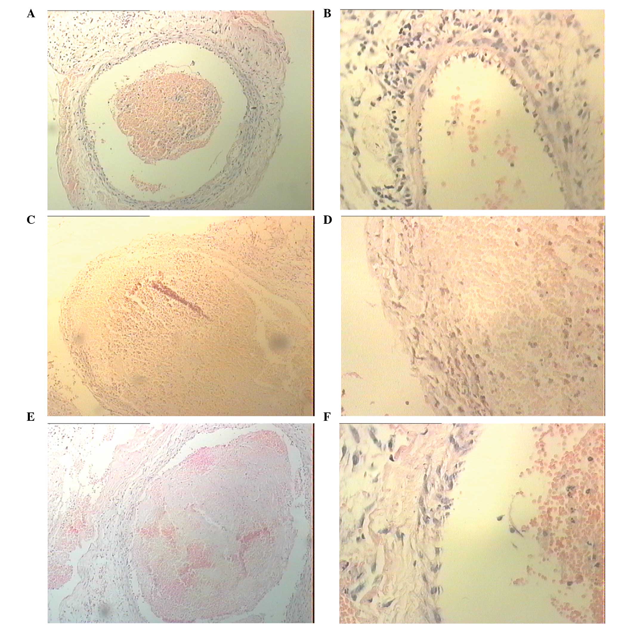 | Figure 8Images of the venous wall were
captured under a microscope. (A and B) In the control group the
vascular wall was continuous, endothelial integrity was maintained
and no shedding of endothelial cells was observed. (C and D) In the
model group the vascular wall showed discontinuous change, intimal
irregularities, endothelial cell shedding and thrombosis (mixed and
red thrombi), which was attached to the vein vascular wall. (E and
F) In the Salvia group the vascular wall exhibited complete
continuity, regular intima, some endothelial cell shedding and a
small amount of intraluminal thrombosis, which was partly attached
to the vein vascular wall. (A, C and E) Hematoxylin and eosin
(H&E); magnification, ×10. (B, D and F) H&E; magnification,
×40. |
Discussion
DVT is a well-known major public health problem,
which represents a significant clinical and economic disease
burden. Up to 21% of cases of DVT may lead to PE, which is a
potentially life-threatening complication (12). The conventional treatment for acute
DVT is immediate anticoagulation using low molecular-weight
heparin, followed by a period (3–6 months) of treatment with oral
anticoagulants (13,14). This treatment aims to prevent
thrombus propagation, and to reduce the risks of PE and DVT
recurrence. However, anticoagulation therapy does not possess
significant fibrinolytic activity, and patients with severe,
extensive and proximal DVTs remain at high risk (15). In DVT treated with anticoagulants
alone, it has been shown that early spontaneous clot lysis
frequently results in preservation of valvular function, which may
help to reduce post-thrombotic morbidity (16). The present study chose to
administer an intravenous injection of S. miltiorrhiza one
week prior to the generation of a ligation model in rabbits.
S. miltiorrhiza is a common traditional
Chinese medicine used for improving body function, which is capable
of promoting circulation and improving blood flow. In addition, it
has been used for the treatment of cardiovascular diseases,
including coronary heart disease, hyperlipidemia and
cerebrovascular disease (17,18).
S. miltiorrhiza has also been widely used in the United
States (10). When platelets are
challenged with outside stimuli to vascular endothelial cells they
increase the expression of the adhesion molecule CD31, which may
adhere to vascular endothelial cells, resulting in the formation of
a soft thrombus (19).
Furthermore, the formation of hard thrombi is initiated by the
enhanced adhesion of platelets, and the conversion of fibrinogen to
fibrin (19). Numerous studies
have been conducted regarding the effects of S. miltiorrhiza
on platelet aggregation. The inhibitory effects of S.
miltiorrhiza have been suggested to be associated with numerous
events, including the inhibition of Ca2+ influx in
platelets, an increase in the number of fibroblasts in the
G0/G1 phase and the attenuation of collagen
secretion (20). The present study
demonstrated that treatment with S. miltiorrhiza one week
after generation of a ligation model, did not affect the majority
of measurements of coagulation function, except for PT, which can
be used to evaluate the action of five different clotting factors
(I, II, V, VII, and X). Blood that takes a long time to clot in a
PT test has previously been shown to be an indicator of treatment
using warfarin (21). In the
present study the PT was significantly increased in the
Salvia group on the third day, but not on the seventh day,
after generation of the model.
Endothelium secretes factors that control vascular
relaxation and contraction, thrombogenesis and fibrinolysis, and
platelet activation and inhibition (22). Therefore, maintenance of the
functional integrity of endothelium is critical for preservation of
blood flow, and the prevention of thrombosis (23). A balance between growth and death
of endothelial cells is important for the integrity of the vascular
endothelium. An appropriate growth response of endothelial cells
helps maintain the integrity of the endothelium, and prevent the
development of atherosclerosis (24). The results of the present study
indicated that S. miltiorrhiza may protect vascular
endothelial cells in vivo. Previous studies have also
demonstrated the protective functions of S. miltiorrhiza on
human vascular endothelial cells in vitro (25–27).
Oxidative stress is the imbalance between the levels
of antioxidants and the production of oxygen-derived species
(28). In the present study S.
miltiorrhiza exhibited antioxidative functions in the vein
ligation model. Chan et al (29) investigated the effects of S.
miltiorrhiza on the pharmacodynamics and pharmacokinetics of
warfarin in rats. S. miltiorrhiza was shown to potentiate
the anticoagulant action of warfarin, by increasing the absorption
rate constant, the area under the plasma concentration time curve,
and the maximum concentration and half-life of warfarin. In
addition, S. miltiorrhiza decreased the clearance and the
apparent volume of distribution of warfarin. The present study did
not investigate the effects of warfarin, or the effects of a
combined treatment of S. miltiorrhiza with warfarin.
In conclusion, previous studies indicated that S.
miltiorrhiza promoted circulation and improved blood flow in
the treatment of cardiovascular diseases, while the present study
revealed that S. miltiorrhiza exhibited antioxidative and
protective effects on vascular endothelial cells. The results of
the present in vivo study demonstrated that S.
miltiorrhiza can decrease blood rheological parameters. S.
miltiorrhiza was also shown to exhibit antioxidative and
protective effects on vascular endothelial cells. These results
suggest that S. miltiorrhiza may have potential applications
for the treatment of DVT.
Acknowledgements
The present study was supported by the Hubei
Provincial Department of Education (grant no. Q20132108).
References
|
1
|
Anderson FA Jr, Wheeler HB, Goldberg RJ,
et al: A population-based perspective of the hospital incidence and
case-fatality rates of deep vein thrombosis and pulmonary embolism.
The Worcester DVT Study. Arch Intern Med. 151:933–938. 1991.
View Article : Google Scholar : PubMed/NCBI
|
|
2
|
Brown HL and Hiett AK: Deep vein
thrombosis and pulmonary embolism in pregnancy: diagnosis,
complications, and management. Clin Obstet Gynecol. 53:345–359.
2010. View Article : Google Scholar : PubMed/NCBI
|
|
3
|
Jaff MR, McMurtry MS, Archer SL, et al:
Management of massive and submassive pulmonary embolism,
iliofemoral deep vein thrombosis, and chronic thromboembolic
pulmonary hypertension: a scientific statement from the American
Heart Association. Circulation. 123:1788–1830. 2011. View Article : Google Scholar : PubMed/NCBI
|
|
4
|
Hardwick ME, Pulido PA and Colwell CW Jr:
A mobile compression device compared with low-molecular-weight
heparin for prevention of venous thromboembolism in total hip
arthroplasty. Orthop Nurs. 30:312–316. 2011. View Article : Google Scholar : PubMed/NCBI
|
|
5
|
Orken DN, Kenangil G, Ozkurt H, et al:
Prevention of deep venous thrombosis and pulmonary embolism in
patients with acute intracerebral hemorrhage. Neurologist.
15:329–331. 2009. View Article : Google Scholar : PubMed/NCBI
|
|
6
|
Lawless RA and Dangleben DA: Caval
agenesis with a hypoplastic left kidney in a patient with trauma on
warfarin for deep vein thrombosis. Vasc Endovascular Surg.
46:75–76. 2012. View Article : Google Scholar
|
|
7
|
Battistelli S, Genovese A and Gori T:
Heparin-induced thrombocytopenia in surgical patients. Am J Surg.
199:43–51. 2010. View Article : Google Scholar : PubMed/NCBI
|
|
8
|
Scarpa M, Pilon F, Pengo V, et al: Deep
venous thrombosis after surgery for inflammatory bowel disease: is
standard dose low molecular weight heparin prophylaxis enough?
World J Surg. 34:1629–1636. 2010. View Article : Google Scholar : PubMed/NCBI
|
|
9
|
Yu S, Zhong B, Zheng M, Xiao F, Dong Z and
Zhang H: The quality of randomized controlled trials on DanShen in
the treatment of ischemic vascular disease. J Altern Complement
Med. 15:557–565. 2009. View Article : Google Scholar : PubMed/NCBI
|
|
10
|
Cheng TO: Cardiovascular effects of
Danshen. Int J Cardiol. 121:9–22. 2007. View Article : Google Scholar : PubMed/NCBI
|
|
11
|
Ho JH and Hong CY: Salvianolic acids:
small compounds with multiple mechanisms for cardiovascular
protection. J Biomed Sci. 18:302011. View Article : Google Scholar : PubMed/NCBI
|
|
12
|
Iverson RE and Gomez JL: Deep venous
thrombosis: prevention and management. Clin Plast Surg. 40:389–398.
2013. View Article : Google Scholar : PubMed/NCBI
|
|
13
|
Wang YP, Zhang XQ, Yu WN, et al:
Endovascular treatment of acute proximal deep venous thrombosis
secondary to iliac vein compression syndrome: a novel technique for
thrombus removal. Chin Med J (Engl). 126:3184–3186. 2013.
|
|
14
|
Deep venous thrombosis and pulmonary
embolism. Part 1. Initial treatment: usually a low-molecular-weight
heparin. Prescrire Int. 22:99–101. 103–104. 2013.PubMed/NCBI
|
|
15
|
Cho ES, Chung JJ, Kim S, Kim JH, Yu JS and
Yoon CS: CT venography for deep vein thrombosis using a low tube
voltage (100 kVp) setting could increase venous enhancement and
reduce the amount of administered iodine. Korean J Radiol.
14:183–193. 2013. View Article : Google Scholar : PubMed/NCBI
|
|
16
|
Liew A and Douketis J: Initial and
long-term treatment of deep venous thrombosis: recent clinical
trials and their impact on patient management. Expert Opin
Pharmacother. 14:385–396. 2013. View Article : Google Scholar : PubMed/NCBI
|
|
17
|
Wen XD, Wang CZ, Yu C, et al: Salvia
miltiorrhiza (dan shen) significantly ameliorates colon
inflammation in dextran sulfate sodium induced colitis. Am J Chin
Med. 41:1097–1108. 2013. View Article : Google Scholar : PubMed/NCBI
|
|
18
|
Zhang JP, Zhang YY, Zhang Y, et al: Salvia
miltiorrhiza (Danshen) injection ameliorates iron overload-induced
cardiac damage in mice. Planta Med. 79:744–752. 2013. View Article : Google Scholar : PubMed/NCBI
|
|
19
|
Gurbel PA, Serebruany VL, Shustov AR, et
al: Increased baseline levels of platelet P-selectin, and
platelet-endothelial cell adhesion molecule-1 in patients with
acute myocardial infarction as predictors of unsuccessful
thrombolysis. Coron Artery Dis. 9:451–456. 1998. View Article : Google Scholar : PubMed/NCBI
|
|
20
|
Han JY, Fan JY, Horie Y, et al:
Ameliorating effects of compounds derived from Salvia miltiorrhiza
root extract on microcirculatory disturbance and target organ
injury by ischemia and reperfusion. Pharmacol Ther. 117:280–295.
2008. View Article : Google Scholar
|
|
21
|
Arbring K, Uppugunduri S and Lindahl TL:
Comparison of prothrombin time (INR) results and main
characteristics of patients on warfarin treatment in primary health
care centers and anticoagulation clinics. BMC Health Serv Res.
13:852013. View Article : Google Scholar : PubMed/NCBI
|
|
22
|
Axtell AL1, Gomari FA and Cooke JP:
Assessing endothelial vasodilator function with the Endo-PAT 2000.
J Vis Exp. 15:21672010.
|
|
23
|
Luscher TF and Barton M: Biology of the
endothelium. Clin Cardiol. 20:II-3–10. 1997.
|
|
24
|
Ling S, Nheu L, Dai A, Guo Z and
Komesaroff P: Effects of four medicinal herbs on human vascular
endothelial cells in culture. Int J Cardiol. 128:350–358. 2008.
View Article : Google Scholar
|
|
25
|
Zhou Z, Wang SQ, Liu Y and Miao AD:
Cryptotanshinone inhibits endothelin-1 expression and stimulates
nitric oxide production in human vascular endothelial cells.
Biochim Biophys Acta. 1760:1–9. 2006. View Article : Google Scholar
|
|
26
|
Chan P, Chen YC, Lin LJ, Cheng TH, Anzai
K, Chen YH, Liu ZM, Lin JG and Hong HJ: Tanshinone IIA attenuates
H2O2-induced injury in human umbilical vein
endothelial cells. Am J Chin Med. 40:1307–1319. 2012. View Article : Google Scholar
|
|
27
|
Zhou ZW, Xie XL, Zhou SF and Li CG:
Mechanism of reversal of high glucose-induced endothelial nitric
oxide synthase uncoupling by tanshinone IIA in human endothelial
cell line EA.hy926. Eur J Pharmacol. 697:97–105. 2012. View Article : Google Scholar : PubMed/NCBI
|
|
28
|
Møller P, Danielsen PH, Karottki DG,
Jantzen K, Roursgaard M, Klingberg H, Jensen DM, Christophersen DV,
Hemmingsen JG, Cao Y and Loft S: Oxidative stress and inflammation
generated DNA damage by exposure to air pollution particles. Mutat
Res Rev Mutat Res. 762C:133–166. 2014. View Article : Google Scholar
|
|
29
|
Chan K, Lo AC, Yeung JH and Woo KS: The
effects of Danshen (Salvia miltiorrhiza) on warfarin
pharmacodynamics and pharmacokinetics of warfarin enantiomers in
rats. J Pharm Pharmacol. 47:402–406. 1995. View Article : Google Scholar : PubMed/NCBI
|