Introduction
Nasal polyposis (NP) is a common
otorhinolaryngological disease, with prevalence rates in the
worldwide population in the range of 2–4%; however, the disease is
difficult to treat (1). Although
specific etiological factors for NP have been proposed, including
allergies, infections and inflammatory reactions, the pathogenesis
of the disease remains to be elucidated (1). Previous studies have been established
that predominantly eosinophilic inflammation is involved in NP
pathogenesis (2–6). In addition, several cell-selecting
and activating chemokines and adhesion molecules have been
demonstrated to be important in NP development (2,7,8),
although their impact and contribution to disease persistence and
recurrence remains unclear.
Tissue remodeling often occurs in response to
inflammatory conditions and can result in localized normal
reconstruction or pathology. Although a certain degree of
remodeling is expected to occur with all inflammatory diseases, its
regulation is mainly disease-specific (9–12).
Abnormal airway remodeling is considered to be associated with
asthma and has been demonstrated to present epithelial damage,
increased blood vessel cross-sectional area, airway smooth-muscle
hyperplasia and hypertrophy, mucous gland and goblet cell
hyperplasia and increased collagen deposition (13–16).
Although NP, asthma and allergic rhinitis frequently occur
concurrently (17), whether nasal
and bronchial epithelia are afflicted by a common pathology is
unclear.
The authors of the present study have previously
demonstrated that eosinophil infiltration is associated with the
expression and eosinophil binding of intercellular adhesion
molecule-1 (ICAM-1) (2). ICAM-1
expression appears to initiate mucosal remodeling of NP; however,
the upstream regulation of ICAM-1 remains to be elucidated. Several
cytokine (interleukin-5, IL-5) and chemokine (eosinophil cationic
protein, ECP; and eotaxin) candidates for eosinophil activation
have emerged (18).
The aims of the present study were to: i)
Investigate the morphological changes that occur during epithelial
remodeling in NP; ii) further assess the role of ICAM-1 in
localized tissue remodeling; and iii) assess the potential
association between ICAM-1 expression and markers of eosinophil
activation.
Materials and methods
Patients and tissue samples
Nasal mucosal tissue was obtained from 28 patients
(male, 18; female, 10) diagnosed with NP (NP group) and admitted
for functional endoscopic sinus surgery in the Department of
Otorinholaryngology-Head and Neck Surgery of China-Japan Union
Hospital at Jilin University (Changchun, China) between January
2008 and January 2009. The patients from which the NP specimens
were obtained had a mean age of 34 years (range, 15–64 years).
Tissue specimens were also obtained from 10 healthy volunteers
(male, 6; female, 4) undergoing septal surgery for partial middle
turbinate removal for a deviated septum (control group). The
patients from which the control specimens were obtained had a mean
age of 42 years (range, 14–57 years). All the healthy volunteers
were non-smokers and did not suffer from infectious nasal and sinus
diseases.
All the patients involved in the present study had
not undergone any prior nasal surgery and were not administered any
antihistamines, antibiotics, topical steroids or oral steroids
within the month preceding the surgery. The NP population was
limited to cases with eosinophilic inflammation. A mean measurement
from five observed fields in the subepithelial area identifying
≥350 eosinophils per microscopic field (magnification, ×400) was
considered to be indicative of eosinophilic inflammation. Patients
with a history of asthma and allergic rhinitis, which was confirmed
by a skin prick assessment and measurement of total and specific
immunoglobulin E expression, were excluded from this study. The
diagnosis of NP was based on the endoscopic findings, nasal sinus
CT scan, clinical history and symptoms and was confirmed by
post-surgical pathological examination. The interval between NP
diagnosis and surgical treatment was <1 month in all cases. All
the patients provided written consent prior to surgery allowing the
use of their biological specimens in biomedical research. The
procurement experiments were approved by the Ethics Committee of
China-Japan Union Hospital, Jilin University.
Histopathological stains
The surgically procured tissue specimens obtained
from the NP and control groups were formalin-fixed and
paraffin-embedded. Sections (3 μm) were stained with the following:
Hematoxylin and eosin (H&E; Beijing Biosynthesis Biotechnology
Co., Ltd., Beijing, China), used to quantify the density of
infiltrating eosinophils [number within the field of view (FOV) at
magnification of ×400] and the percentage (area-wise) of damaged
epithelium; alcian blue-periodic acid-Schiff (AB-PAS; Beijing
Leagene Biotech Co., Ltd, Beijing, China), used to calculate the
density of goblet cells in the epithelium (number per
mm2 of basement membrane in FOV at magnification of
×400) and the density of subepithelial glands (number per
mm2 of basement membrane); and Masson’s Trichrome (MT;
Beijing Leagene Biotech Co., Ltd), used to determine the thickness
of the basement membrane and subepithelium collagen. The extent of
epithelial damage and basement membrane thickening was categorized
using the staging system described by Ponikau et al
(3). A total of five random FOVs
were evaluated for each stain at a magnification of ×400 by two
experienced pathologists.
Immunohistochemical analysis
ECP and ICAM-1 were detected by the
streptavidin-peroxidase (SP) method using an SP (mouse) kit (Wuhan
Boster Biological Technology, Ltd., Wuhan, China), according to the
manufacturer’s instructions. Briefly, formalin-fixed,
paraffin-embedded tissue sections (3 μm) were deparaffinized,
rehydrated and pretreated with 5% normal rabbit serum (Beijing
Biosynthesis Biotechnology Co., Ltd.) prior to antigen retrieval in
citrate buffer (pH 6.0) at 95°C for 10 min. Next, the sections were
incubated with the following polyclonal rabbit anti-human primary
antibodies overnight at 4°C: ECP, diluted 1:100 in
phosphate-buffered saline (PBS; USCN Life Science Inc., Houston,
TX, USA); and ICAM-1, diluted 1:200 in PBS (Beijing Biosynthesis
Biotechnology Co., Ltd.). Signal detection was performed using an
SP kit. Negative controls were treated following the aforementioned
procedure, without addition of primary antibodies. In addition,
positive controls were treated following the aforementioned
procedure, using previously characterized tissue specimens known to
express the target antigen rather than the experimental specimens.
The number and percentage of ECP and ICAM-1 immunopositive cells
were assessed by two independent observers (magnification, ×400),
with a bright field microscope (CX31; Olympus Corp., Tokyo, Japan)
using a double-blind experimental procedure. Immunoreactivity
values are expressed as the calculated mean of 10 randomly selected
FOVs for each specimen. The means were calculated from the
incorporated values reported by the two independent reviewers.
Statistical analysis
Data are presented as the mean ± standard deviation.
The normal distribution was verified using the Kolmogorov-Smirnov
test (approximation method). Differences between the NP and control
group mean values were assessed using Student’s t-test.
Correlations between the infiltration and activation of
eosinophils, ICAM-1 and ECP expression and the indices of
epithelial damage and collagen deposition were assessed using
Pearson’s analysis. P<0.05 was considered to indicate a
statistically significant difference. All the statistical analyses
were performed using the SPSS 13.0 software (SPSS, Inc., Chicago,
IL, USA).
Results
Morphological characteristics of mucosa
remodeling in NP
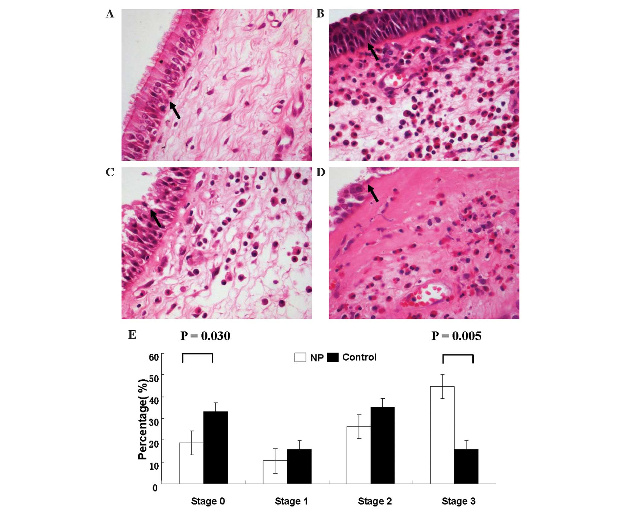
Using the staging criteria of Ponikau et al
(3), damage was detected in the
epithelial layers of the NP and control groups (refer to Fig. 1 for a description of the four
stages and representative micrographs). However, as determined from
the H&E-stained micrographs, the mean percentage of the nasal
mucosa that exhibited stage 3 epithelial damage was higher in the
NP specimens (44.58±7.50%) compared with the control specimens
(15.83±3.67%; P=0.005). By contrast, the control group specimens
exhibited greater stage 0 intact epithelia and mild epithelial
shedding (33.09±7.53%) compared with the NP group controls
(18.75±4.44%; P=0.030; Fig. 1).
However, the extent of stage 1 and 2 epithelial damage in NP
specimens (10.42±3.21% and 26.25±4.66%, respectively) was found to
be similar to that in control specimens (15.83±5.34% and
35.25±5.70%, respectively; P>0.05).
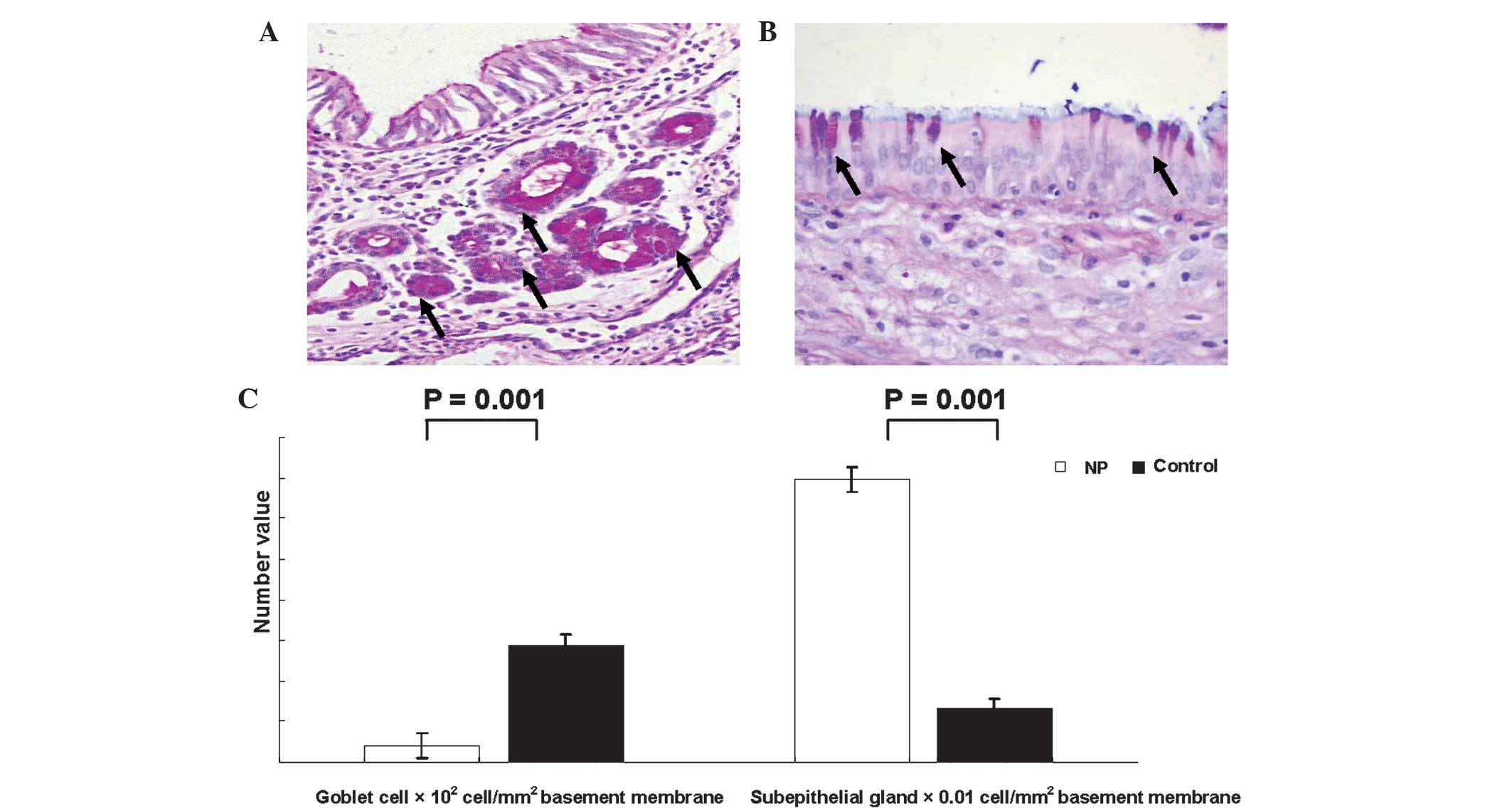
As shown in Fig. 2,
AB-PAS staining revealed that the NP specimens presented a markedly
reduced density of goblet cells (800.00±298.1 cells/mm2
basement membrane in FOV) when compared with control specimens
(5,750.00±462.91 cells/mm2 basement membrane in FOV). In
addition, NP specimens exhibited a greater density of epithelial
glands (1.39±0.33 glands/mm2 basement membrane in FOV)
compared with the control group specimens (0.26±0.09
glands/mm2 basement membrane in FOV; P=0.001). The nasal
mucosa of NP specimens exhibited an asymmetric clustering of
subepithelial hyperplasia glands, which was not observed in the
control specimens (Fig. 2).
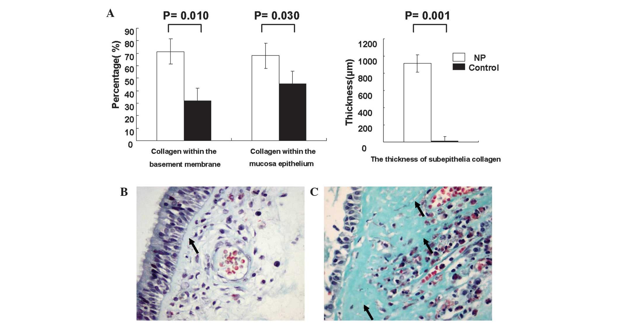
As indicated by MT-positive immunoreactivity, the
extent of collagen deposition within the mucosal epithelium
differed between the NP and control groups. The NP group specimens
had a higher percentage of collagen within the basement membrane
(71.25±20.31%) and throughout the mucosal epithelium
(68.13±16.89%), compared with the control specimens (31.88±7.99%,
P=0.010 and 45.63±7.76%, P=0.030, respectively; Fig. 3A). Furthermore, a morphological
investigation revealed a thickened subepithelial collagen layer
within the basement membrane in NP specimens compared with the
controls, which resembled a broad band (Fig. 3B and C). The average thickness of
the collagen band was significantly greater in the NP group (91.6
μm) compared with the controls (13.65 μm; P=0.001; Fig. 3A). Furthermore, the lamina propria
of the nasal mucosa in NP specimens was often destroyed by collagen
and, in certain cases, the thickened collagen extended beyond the
lamina propria. Collagen was detected in the subepithelium of the
specimens presenting the most severe epithelial damage (Fig. 3C).
Activated eosinophil distribution and
ICAM-1 expression
In NP specimens, the number of total (P=0.030),
activated (immunopositive for ECP, ECP+; P=0.006) and
ICAM-1 immunopositive (ICAM-1+; P=0.007) eosinophils was
significantly greater compared with the control turbinate tissue
specimens (Fig. 4). In addition,
the mean number of eosinophils in the NP group (39.00±4.24 per FOV)
was 3-fold higher compared with the control group (11.90±5.24 per
FOV), while the mean number of ECP+ eosinophils in the
NP group (33.58±8.32 per FOV) was almost 8-fold higher compared
with the control group (4.13±1.36 per FOV; Fig. 4D). A similar trend was observed for
the number of infiltrating eosinophils, with the mean number of
ICAM-1-expressing eosinophils in the NP specimens (33.57±10.937 per
FOV) being 4-fold higher compared with the control specimens
(7.625±0.92 per FOV). In the NP specimens, infiltrating eosinophils
were found to be located primarily around the vessels and glands
within the subepithelial layer (Fig.
4C). Eosinophil activation (ECP+ eosinophils/total
H&E-stained eosinophils) in the NP specimens (86.10%;
33.58/39.00) was elevated when compared with the control sections
(34.71%; 4.13/11.90; Fig. 4B.
Analysis of the serial sections confirmed that the majority of
ICAM-1+ cells exhibited eosinophil morphological
characteristics (Fig. 4A–C).
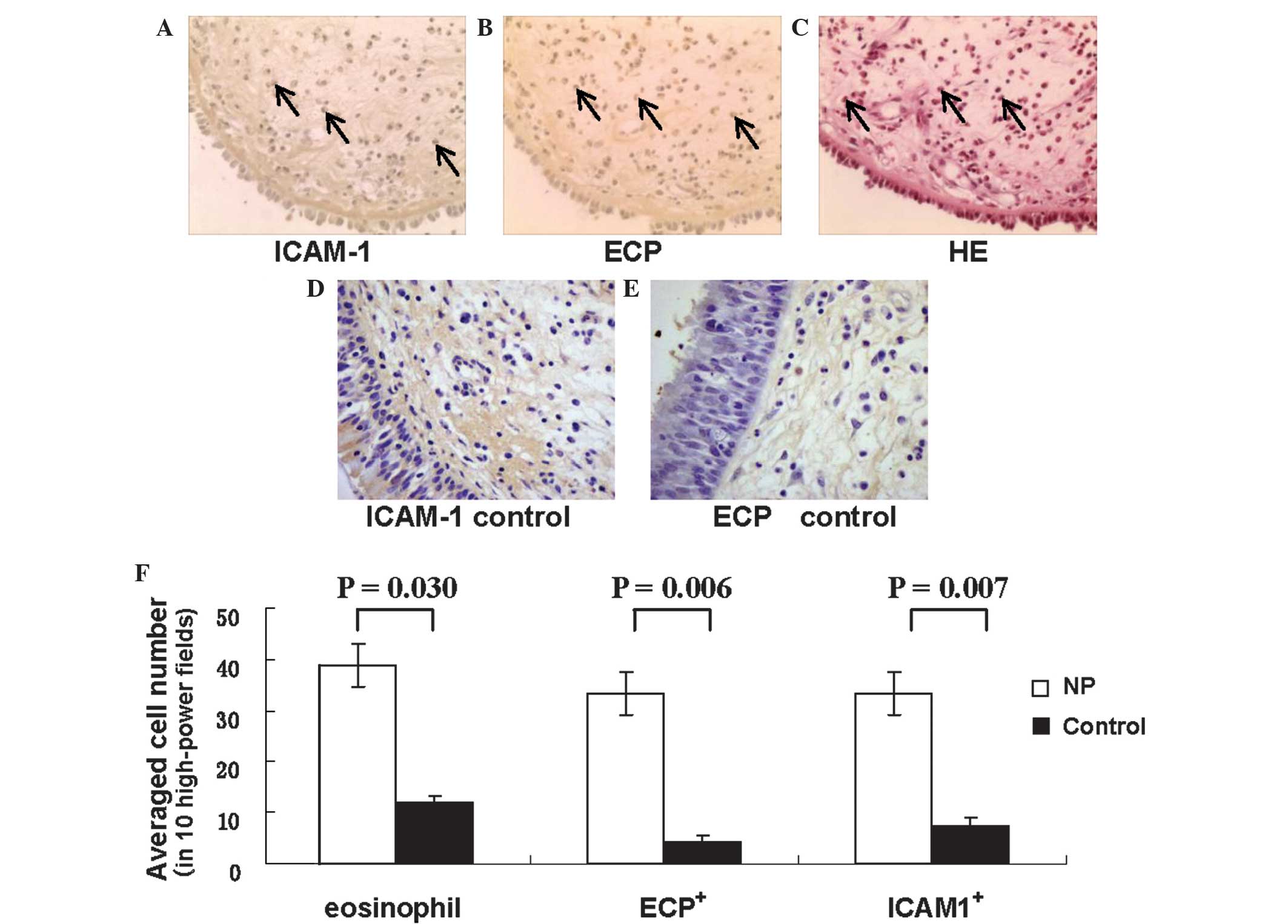 | Figure 4Distribution of total, infiltrating
and activated eosinophils in the NP and control nasal mucosa
specimens. Representative micrographs of (A) ICAM-1 and (B) ECP
immunohistochemistry (brown stain) in NP specimens (magnification,
×200), revealing a large number of ICAM-1+ and
ECP+ cells (arrows). (C) A representative micrograph of
hematoxylin and eosin-stained NP specimens depicting marked
eosinophil infiltration (arrows) of the nasal mucosa
(magnification, ×200). Representative micrographs of negative
control for (D) ICAM-1 and (E) ECP immunohistochemistry
(magnification, ×400). (F) Distribution of total, ECP+
and ICAM-1+ eosinophils in NP and control groups. NP
specimens exhibited higher cell counts for total eosinophils
(P=0.030), ECP+ cells (P=0.006) and ICAM-1+
cells (P=0.007) than did control specimens. NP, nasal polyposis;
ECP, eosinophil cationic protein; ICAM-1, intercellular adhesion
molecule-1. |
The extent of ECP+ cells in the nasal
mucosal epithelium of the NP group correlated moderately with
epithelial damage (r=0.424, P=0.038) and weakly with basement
membrane thickness (r=0.325, P=0.012). The density of
ICAM-1+ cells in the nasal mucosa of NP specimens
correlated markedly with ECP+ cell density (r=0.739,
P=0.010) and basement membrane thickness (r=0.603, P=0.010).
Discussion
Nasal polyposis is a chronic inflammatory disease of
the sino-nasal mucosa, characterized by edema, the infiltration of
inflammatory cells, hyperplasia of the fibrous tissue, and
vascularization(15,19,20).
In the present study, tissue sections of NP and normal nasal mucosa
were investigated for evidence of morphological changes and
remodeling. Epithelial damage, basement membrane thickening and
deposition of subepithelial collagen were increased in the NP
specimens when compared with the healthy control specimens.
Furthermore, positive correlations were observed between tissue
remodeling indicators and the expression levels of ICAM-1 and ECP
in NP nasal mucosa.
Mucus secreted by epithelial goblet cells is an
important component in the maintenance of nasal physiological
function and serves as a form of non-specific defense (21,22).
However, epithelial cells, which are located on the outer layer of
the nasal mucosa, are also capable of exhibiting an immune
response, including the release of arachic acid derivatives,
polypeptidases, matrix proteins, cytokines and other
immune-associated substances (4,14).
In the present study, severe damage was observed on the epithelium
of the NP specimens, which included the absence of cilia and
erosion of upper epithelial cell layers. These findings may be a
result of increased accumulation of eosinophils and other
inflammatory cells during nasal polyposis pathogenesis. Such
inflammatory cells are capable of generating cytotoxic proteins,
oxygen free radicals, proteinases and metalloproteinases, which may
affect ciliary motor function of epithelium mucosae, as well as
induce direct damage to the epithelium (13,23)
through sodium and chloride channel dysfunction and eventual
cellular edema (24). Epithelial
damage is associated with increased epithelial shedding as a result
of decreased adhesive power between the epithelium and basement
membrane (15,23).
Tos et al (21) observed that the median density of
goblet cells was 3,450 cells/mm2 in the anterior region
of the nose, compared with 6,050 cells/mm2 in
posteriorly located polyps. In addition, Kitapçi et al
(22) identified that the goblet
cell numbers were significantly higher in NP specimens compared
with control specimens. By contrast, Malekzadeh et al
(25) reported decreased
subepithelial gland areas in NP specimens. In the present study,
lower numbers of goblet cells and higher numbers of subepithelial
glands were detected in the NP specimens compared with the healthy
controls. Goblet cell number reduction has been previously
associated with metaplasia (26,27);
therefore, the presence of additional glandular tissue in the
current study is consistent with the possibility of metaplasia
occurring in the specimens. However, the presence and extent of
cell type conversion, as well as the mechanism of goblet cell
reduction, in NP remain to be elucidated. The increase in
subepithelial glands may be a compensatory mechanism in response to
reduced goblet cell counts and abnormal mucus secretion (13,15,24).
The coupled decrease in goblet cell density and increase in
supepithelial gland density reported in the present study may have
resulted in the formation of a defective mucus blanket, which may
in turn have led to ciliary structural damage and disrupted the
directional movement of the mucus blanket, resulting in cumulative
effects. In addition, exogenous physical, chemical and biological
stimulants may also have contributed to epithelial mucosa damage if
they were averted from the nasal tunical mucosa. This hypothesis is
plausible, considering the tissue’s nested anatomical position
within the ingress of the respiratory tract.
The extracellular matrix provides a crucial scaffold
for epithelial organization and its deposition serves as a keystone
during tissue remodeling. Deposition of the extracellular matrix by
fibroblast or myofibroblastic cells in NP tissue increases
epithelial mechanical strength, thereby averting tissue edema and
curtailing expansion of blood vessels and glands (3,15,20).
Extracellular matrix regulation in NP tissue has received scholarly
attention (28,29), particularly in relation to matrix
metalloproteinase 9 (MMP-9) activity. MMP-9 degrades the
extracellular matrix and is involved in tissue remodeling; MMP-9
gene polymorphisms have been demonstrated to affect susceptibility
to the development of chronic rhinosinusitis with NP in Chinese
patients (30). Several studies
have supported the hypothesis that MMP-9 is important in
eosinophil-selective migration into NP tissue (2,24,29);
however, Kahveci et al (28) did not identify a correlation
between MMP-9 and eosinophils. Therefore, NP severity and
recurrence may involve a mechanism other than MMP-9 (29,31).
ICAM-1, which is expressed widely in the ciliated
epithelium, basal cells, submucosa, vascular endothelium and
inflammatory cells, is important in the eosinophil-selective
migration out of blood vessels, local aggregation and eosinophil
infiltration (7). The expression
of ICAM-1 correlates with nuclear factor-κB (NF-κB) signaling
(32). Furthermore, upregulation
of MMP-9 expression through activation of the NF-κB signaling
pathway has been described in numerous diseases, including
bronchial epithelial cells in asthma (32,33)
To the best of our knowledge, no systematic and
anatomical investigations of collagen deposition in NP tissues
exist. The data presented in the current study indicated that
collagen, the principal component of the extracellular matrix,
accounts for 71.25% of the basement membrane and 68.13% of the
subepithelial area in NP tissues. Compared with healthy control
tissues (31.88% of basement membrane and 45.63% of subepithelial
area), the collagen content in NP tissues presented a ~2.2-fold and
1.5-fold increase, respectively.
Morphological examination under a light microscope
revealed band-like thickening of subepithelial collagen in NP
tissues, but not in control specimens. Therefore, the present study
hypothesized that the observed excessive collagen generation,
basement membrane thickening and subepithelial collagen banding
pattern are indicative of extracellular matrix deposition in NP. In
addition, the normal presentation of fibrin layers in reticular
arrangements, beneath the basement membrane and within the lamina
propria adjacent to the periosteum, was not observed in the NP
specimens, which in contrast exhibited a submucosal reticular
skeleton structure that was damaged or replaced by diffusely
distributed collagen fibers. In certain cases, thickening collagen
filled the lamina propria adjacent to the periosteum. A positive
correlation between the extent of the observed nasal epithelial
cell damage and the subcutaneous collagen thickness was also
identified in the present study. Therefore, direct exposure to
bacteria, viruses, external toxins and other types of super-antigen
following epithelial damage (6),
was hypothesized to result in stimulation of the basement membrane
and subsequent collagen hyperplasia in the present study. In
addition, submucosal mesh stent structural damage may be a
characteristic of tissue remodeling in NP.
Local eosinophil infiltration and activation are
classical hallmarks of nasal polyp formation and development
(5,24,34).
Although cases of NP without eosinophilic inflammation have been
described (35,36), the present study included only
cases with eosinophilic inflammation. Eosinophil activation has
been previously demonstrated to be associated with epithelial
damage and subepithelial collagen thickness (5,6);
however, the results of the present study revealed only moderate
correlations between markers of tissue remodeling and indicators of
eosinophil infiltration (such as ICAM-1) and activation (such as
ECP). Elucidation of tissue remodeling associations may be
confounded by the employment of multiple regulators and pathways.
Activated eosinophils release a variety of proteins and cytokines,
including ECP (37), transforming
growth factor-β1, fibroblast growth factor-2, MMP-9, tissue
inhibitor of metalloproteinases 1, interleukin (IL)-13 and IL-17,
which may contribute to the ultimate epithelial damage and collagen
deposition that occurs (19,29,30).
In addition, ICAM-1 has been revealed to be expressed in vascular
endothelial cells and be active in the migration of eosinophils
from blood vessels to tissues (2,7). In
the present study, morphological investigation under a light
microscope revealed the expression of ICAM-1 in activated
eosinophil cells, indicating that, in addition to its role in
eosinophil-selective transmembrane migration, ICAM-1 may also be
directly involved in eosinophil activation through mechanisms that
remain unclear. Therefore, eosinophil activation and ICAM-1
expression may contribute to, rather than govern, the initiation of
tissue remodeling in NP.
In conclusion, the present study observations
indicated that eosinophil-mediated tissue remodeling may play a
major role in NP pathogenesis. Although NP remodeling is often
irreversible, identification of key factors, including ICAM-1
expressing eosinophils, may lead to new preventive and therapeutic
strategies in combating NP recurrence. For instance, in the case
that ECP or ICAM-1 are involved in NP pathogenesis, patients with
NP may benefit from anti-ECP or anti-ICAM-1 therapies. Further
investigation is required to identify additional regulators and
elucidate the mechanisms of action in NP tissue remodeling,
including the potential role of the NF-κB-inducing molecule, ICAM-1
(8,34).
Acknowledgements
This study was supported by the Key Clinical Program
of the Ministry of Health (grant no. NSC 07090138) and the
Supporting Program of the ‘Eleventh Five-Year Plan’ for Science and
Technology Research of China (grant no. 2007CB516706).
References
|
1
|
Nemati S, Mojtahedi A, Naghavi SE, Banan R
and Zia F: Investigating Helicobacter pylori in nasal polyposis
using polymerase chain reaction, urease test and culture. Eur Arch
Otorhinolaryngol. 269:1457–1461. 2012. View Article : Google Scholar
|
|
2
|
Kong H, Dong Z, Guo Y, Yang Z and Bu G:
Intercellular adhesion molecule-1 and accumulation of eosinophils
in nasal polyp tissue. Chin Med J (Engl). 112:366–368. 1999.
|
|
3
|
Ponikau JU, Sherris DA, Kephart GM, et al:
Features of airway remodeling and eosinophilic inflammation in
chronic rhinosinusitis: is the histopathology similar to asthma? J
Allergy Clin Immunol. 112:877–882. 2003. View Article : Google Scholar : PubMed/NCBI
|
|
4
|
Fan GK, Wang H and Takenaka H: Eosinophil
infiltration and activation in nasal polyposis. Acta Otolaryngol.
127:521–526. 2007. View Article : Google Scholar : PubMed/NCBI
|
|
5
|
Eliashar R and Levi-Schaffer F: The role
of the eosinophil in nasal diseases. Curr Opin Otolaryngol Head
Neck Surg. 13:171–175. 2005. View Article : Google Scholar : PubMed/NCBI
|
|
6
|
Saitoh T, Kusunoki T, Yao T, et al:
Relationship between epithelial damage or basement membrane
thickness and eosinophilic infiltration in nasal polyps with
chronic rhinosinusitis. Rhinology. 47:275–279. 2009.PubMed/NCBI
|
|
7
|
Papon JF, Coste A, Gendron MC, et al:
HLA-DR and ICAM-1 expression and modulation in epithelial cells
from nasal polyps. Laryngoscope. 112:2067–2075. 2002. View Article : Google Scholar : PubMed/NCBI
|
|
8
|
Valera FC, Umezawa K, Brassesco MS, et al:
Suppression of inflammatory cytokine secretion by an NF-κB
inhibitor DHMEQ in nasal polyps fibroblasts. Cell Physiol Biochem.
30:13–22. 2012. View Article : Google Scholar
|
|
9
|
Grunig G, Marsh LM, Esmaeil N, Jackson K,
Gordon T, Reibman J, Kwapiszewska G and Park SH: Perspective:
ambient air pollution: inflammatory response and effects on the
lung’s vasculature. Pulm Circ. 4:25–35. 2014. View Article : Google Scholar : PubMed/NCBI
|
|
10
|
Koczy-Baron E and Kasperska-Zając A: The
role of vascular endothelial growth factor in inflammatory
processes. Postepy Hig Med Dosw (Online). 68:57–65. 2014.(In
Polish). View Article : Google Scholar
|
|
11
|
Bautista-Molano W, Romero-Sánchez C, De
Ávila J, Londoño J and Valle-Oñate R: Bone remodeling in
spondyloarthritis. Rev Med Chil. 141:1182–1189. 2013.(In Spanish).
View Article : Google Scholar
|
|
12
|
Berraies A, Hamzaoui K and Hamzaoui A:
Link between vitamin D and airway remodeling. J Asthma Allergy.
7:23–30. 2014.PubMed/NCBI
|
|
13
|
Hamid Q: Pathogenesis of small airways in
asthma. Respiration. 84:4–11. 2012. View Article : Google Scholar : PubMed/NCBI
|
|
14
|
Van Bruaene N and Bachert C: Tissue
remodeling in chronic rhinosinusitis. Curr Opin Allergy Clin
Immunol. 11:8–11. 2011. View Article : Google Scholar
|
|
15
|
Pawankar R and Nonaka M: Inflammatory
mechanisms and remodeling in chronic rhinosinusitis and nasal
polyps. Curr Allergy Asthma Rep. 7:202–208. 2007. View Article : Google Scholar : PubMed/NCBI
|
|
16
|
Royce SG, Cheng V, Samuel CS and Tang ML:
The regulation of fibrosis in airway remodeling in asthma. Mol Cell
Endocrinol. 351:167–175. 2012. View Article : Google Scholar : PubMed/NCBI
|
|
17
|
Rabago D, Guerard E and Bukstein D: Nasal
irrigation for chronic sinus symptoms in patients with allergic
rhinitis, asthma, and nasal polyposis: a hypothesis generating
study. WMJ. 107:69–75. 2008.PubMed/NCBI
|
|
18
|
Sun DI, Joo YH, Auo HJ and Kang JM:
Clinical significance of eosinophilic cationic protein levels in
nasal secretions of patients with nasal polyposis. Eur Arch
Otorhinolaryngol. 266:981–986. 2009. View Article : Google Scholar
|
|
19
|
Figueiredo CR, Silva ID and Weckx LL:
Inflammatory genes in nasal polyposis. Curr Opin Otolaryngol Head
Neck Surg. 16:18–21. 2008. View Article : Google Scholar : PubMed/NCBI
|
|
20
|
Pawliczak R, Lewandowska-Polak A and
Kowalski ML: Pathogenesis of nasal polyps: an update. Curr Allergy
Asthma. 5:463–471. 2005. View Article : Google Scholar
|
|
21
|
Tos M, Larsen PL and Moller K: Goblet cell
density in nasal polyps. Ann Otol Rhinol Laryngol. 99:310–315.
1990. View Article : Google Scholar : PubMed/NCBI
|
|
22
|
Kitapçi F, Muluk NB, Atasoy P and Koc C:
Role of mast and goblet cells in the pathogenesis of nasal polyps.
J Otolaryngol. 35:122–132. 2006. View Article : Google Scholar : PubMed/NCBI
|
|
23
|
Bernstein JM: Update on the molecular
biology of nasal polyposis. Otolaryngol Clin North Am.
38:1243–1255. 2005. View Article : Google Scholar : PubMed/NCBI
|
|
24
|
Pawankar R: Nasal polyposis: an update:
editorial review. Curr Opin Allergy Clin Immunol. 3:1–6. 2003.
View Article : Google Scholar : PubMed/NCBI
|
|
25
|
Malekzadeh S, Hamburger MD, Whelan PJ,
Biedlingmaier JF and Baraniuk JN: Density of middle turbinate
subepithelial mucous glands in patients with chronic
rhinosinusitis. Otolaryngol Head Neck Surg. 127:190–195. 2002.
View Article : Google Scholar : PubMed/NCBI
|
|
26
|
Boucherat O, Chakir J and Jeannotte L: The
loss of Hoxa5 function promotes Notch-dependent goblet cell
metaplasia in lung airways. Biol Open. 1:677–691. 2012. View Article : Google Scholar : PubMed/NCBI
|
|
27
|
Ren X, Shah TA, Ustiyan V, et al: FOXM1
promotes allergen-induced goblet cell metaplasia and pulmonary
inflammation. Mol Cell Biol. 33:371–386. 2013. View Article : Google Scholar :
|
|
28
|
Kahveci OK, Derekoy FS, Yilmaz M, Serteser
M and Altuntas A: The role of MMP-9 and TIMP-1 in nasal polyp
formation. Swiss Med Wkly. 138:684–688. 2008.PubMed/NCBI
|
|
29
|
Wang LF, Chien CY, Chiang FY, Chai CY and
Tai CF: Expression of matrix metalloproteinase-2 and matrix
metalloproteinase-9 in recurrent chronic rhinosinusitis with nasal
polyposis. Kaohsiung J Med Sci. 29:26–31. 2013. View Article : Google Scholar
|
|
30
|
Wang LF, Chien CY, Tai CF, Kuo WR, Hsi E
and Juo SH: Matrix metalloproteinase-9 gene polymorphisms in nasal
polyposis. BMC Med Genet. 11:852010. View Article : Google Scholar : PubMed/NCBI
|
|
31
|
Wang LF, Chien CY, Chiang FY, Chai CY and
Tai CF: Corelationship between matrix metalloproteinase 2 and 9
expression and severity of chronic rhinosinusitis with nasal
polyposis. Am J Rhinol Allergy. 26:e1–e4. 2012. View Article : Google Scholar : PubMed/NCBI
|
|
32
|
Jung J, Ko SH, Yoo do Y, et al:
5,7-Dihydroxy-3,4, 6-trimethoxyflavone inhibits intercellular
adhesion molecule 1 and vascular cell adhesion molecule 1 via the
Akt and nuclear factor-kappaB-dependent pathway, leading to
suppression of adhesion of monocytes and eosinophils to bronchial
epithelial cells. Immunology. 137:98–113. 2012. View Article : Google Scholar : PubMed/NCBI
|
|
33
|
Li J, Lau G, Chen L, et al: Interleukin 23
promotes hepatocellular carcinoma metastasis via NF-kappa B induced
matrix metalloproteinase 9 expression. PLoS One. 7:e462642012.
View Article : Google Scholar : PubMed/NCBI
|
|
34
|
Ishitoya J, Sakuma Y and Tsukuda M:
Eosinophilic chronic rhinosinusitis in Japan. Allergol Int.
59:239–245. 2010. View Article : Google Scholar : PubMed/NCBI
|
|
35
|
Derycke L, Zhang N, Holtappels G, Dutre T
and Bachert C: IL-17A as a regulator of neutrophil survival in
nasal polyp disease of patients with and without cystic fibrosis. J
Cyst Fibros. 11:193–200. 2012. View Article : Google Scholar
|
|
36
|
Wen W, Liu W, Zhang L, et al: Increased
neutrophilia in nasal polyps reduces the response to oral
corticosteroid therapy. J Allergy Clin Immunol. 129:1522–1528.
2012. View Article : Google Scholar : PubMed/NCBI
|
|
37
|
Nikolovski Z, Buzon V, Ribo M, et al:
Thermal unfolding of eosinophil cationic protein/ribonuclease 3: a
nonreversible process. Protein Sci. 15:2816–2827. 2006. View Article : Google Scholar : PubMed/NCBI
|