Introduction
Non-small cell lung cancer (NSCLC) remains the
leading cause of cancer-related mortality in China despite the fact
that great advances have been made in the therapeutic treatment
methods, including surgical resection, chemotherapy and
radiotherapy (1). NSCLC mainly
comprises two types: adenocarcinoma and squamous cell carcinoma.
Two-thirds of NSCLC patients are diagnosed at a late stage of the
disease when the tumors are not resectable. In addition, up to 60%
of all resected patients relapse and succumb to the disease
(2). Although radiotherapy has
been widely used to treat unresectable and metastatic tumors, radio
resistance usually leads to a low overall 5-year survival rate of
less than 15% (3). Therefore, the
development of new treatment options including gene therapy is an
urgent requirement.
Gene therapy is one of the innovative approaches
which have offered new hope for enhancing antitumor effects. The
retinoblastoma (RB94) tumor suppressor gene, which lacks the
NH2-terminal 112 amino acid residues of the full-length RB protein
(RB110), has been identified to have marked tumor suppressor
efficacy compared with wild-type RB110 (4,5). The
RB94 protein has been reported to remain in a hypo-phosphorylated
state in transfected tumor cells and demonstrate a significantly
longer half-life than the RB110 protein (4,5). In
addition, RB94 exhibits an increased antitumor efficacy in
RB-negative and RB-positive human tumors, including fibrosarcoma
and bladder, lung and prostate carcinoma, while the effects of
RB110 are limited to RB-negative tumors (4). Preclinical studies also demonstrate
that adenovirus-mediated (Ad)-RB94 gene transfer significantly
suppresses the growth of human head and neck cancer, bladder
carcinoma, pancreatic carcinoma and esophageal cancer in
vitro and in vivo (6–9).
Moreover, the combination of RB94 and radiation therapy (XRT)
results in synergistic tumor growth suppression of head and neck
squamous cell carcinoma (10).
Therefore, RB94 appears to be a promising candidate for gene
therapy in NSCLC.
In the present study, an adenoviral vector carrying
the RB94 gene (Ad-RB94) was constructed. Subsequently, we
investigated whether Ad-RB94 exerted therapeutic effects in NSCLC
as well as examining the underlying molecular mechanisms.
Materials and methods
Cell culture
The human NSCLC cell line A549 has been well
characterized and is known to express wild-type RB (11). The A549 cells were cultured in RPMI
1640 (Hyclone, Logan, UT, USA) with 10% fetal bovine serum at 37°C
in 5% CO2.
Construction of recombinant adenoviral
vectors
The construction of recombinant adenovirus vector
for the RB94 gene was carried out using Gateway™ clone technology
(GeneSil, Wuhan, China). Briefly, total RNA was extracted from a
human embryo and reversed transcribed to obtain object cDNA, then
the hRB94 gene fragment was amplified by polymerase chain reaction
(PCR). The attB-flanked PCR primers were designed and used to
amplify the hRB94 gene by PCR. An entry clone was performed by a BP
recombination reaction with attB-PCR products and donor vector
pDONRTM221. Then the entry clone and the target vector
Ad/CMV/V5-DEST with attR1 and attR2 sites was recombined in
vivo to create the expression clone (Ad-hRB94) using an
efficient LR recombination reaction. Next, the expression clone was
confirmed by PCR and sequencing. Ad-hRB94 was digested with Pac I
and transferred into 293A cells to be packaged into adenovirus
stock. Ad-hRB94 was amplified by infection of 293A cells and the
titer was measured. Ad-LacZ, a replication-defective control
adenovirus not carrying the RB94 gene, was obtained from Invitrogen
Life Technologies (Carlsbad, CA, USA). The viruses were amplified
and plaque-purified. Titers were determined by standard plaque
assays.
Western blot analysis
The cells were lysed by adding 100 μl
radioimmunoprecipitation assay lysis buffer [1% Triton X-100, 1%
deoxycholate, 0.1% sodium dodecyl sulfate (SDS), 1 mM
phenylmethylsulfonyl fluoride] purchased from Beyotime Institute of
Biotechnology (Shanghai, China) and then quantitated using a
bicinchoninic acid (BCA) assay kit with phosphate-buffered saline
(PBS) as a standard (Pierce, Rockford, IL, USA). Equal quantities
of protein (50 μg) from the different cells were separated by 10%
SDS-polyacrylamide gel electrophoresis. The separated proteins were
transferred onto nitrocellulose (GE Healthcare, Buckinghamshire,
UK). Non-specific binding sites were blocked using PBS-Tween-20 and
5% non-fat dried milk for 1 h at room temperature. Following
blocking, the samples were incubated overnight at 4°C with primary
antibody mouse-antihuman RB (BD Biosciences Pharmingen, San Diego,
CA, USA), which recognizes both full-length RB and
NH2-terminal-truncated RB94 proteins, at a concentration of 1:200
in Tris-buffered saline-Tween-20. Membranes were washed three times
for 10 min in a buffer containing PBS and 0.1% Tween-20, and then
incubated with an appropriate goat antimouse secondary antibody
conjugated to horseradish peroxidase (Amersham Pharmacia Biotech,
Piscataway, NJ, USA) applied for 1 h at room temperature. Reactive
bands were detected with the ECL chemiluminescence reagent
(Amersham Pharmacia Biotech, Freiburg, Germany). β-actin was
detected using monoclonal anti-β-actin (Sigma, St. Louis, MO,
USA).
Reverse transcription-quantitative PCR
(RT-qPCR)
Total mRNA was extracted from tumors in each of the
studied groups with an mRNA isolation kit (Takara, Dalian, China),
and 3 μg total RNA was used for cDNA synthesis by SuperScript II
reverse transcriptase (Invitrogen) according to the standard
instructions. qPCR was performed in a final volume of 20 μl
containing 1 μl each cDNA template, 2 μl each 10 nM primer (RB94 F:
5′-GAA TCT GCT TGT CCT CTT AAT CTT AAT CTT CC-3′ and R: 5′-GAA GAT
GGT GAT GGG ATT TC-3′; cyclinB1 F: 5′-CAG TCA GAC CAA AAT ACC TAC
TGG GT-3′ and R: 5′-ACA CAA ACC AGC TGC AGC ATC TTC TT-3′), and 10
μl SYBR-Green Master mix. Quantification was carried out using the
comparative cycle threshold (Ct) method, and water was used as the
negative control. Ct values were calculated by determining the
cycle number at which the fluorescence exceeded the threshold
limit. The average Ct values for the target gene were normalized to
an endogenous housekeeping gene encoding 18S rRNA.
Flow cytometric analysis
Cells were seeded at a density of 1×10⁶
cells/well in six-well tissue culture plates and allowed to adhere
overnight. Media were removed and cells were incubated with either
Ad-RB94 or Ad-LacZ at a multiplicity of infection of 10, or control
with PBS in 5 ml media for 2 h, after which 10 ml media was added.
After 24 h, harvested cells were trypsinized, washed with PBS,
fixed in suspension in 75% ethanol, and kept at −20°C until
analysis. Fixed cells were centrifuged and resuspended in PBS.
After adding RNase (1 mg/ml) to the cell suspension, the cells were
incubated for 30 min at 37°C, and finally stained with propidium
iodide (50 μg/ml) for 1 h at 4°C and then analyzed with FACScan.
Cell distribution was analyzed by WinMDI 2.8 software (Joseph
Trotter, San Diego, CA, USA).
Xenograft tumor in vivo
Eighteen six-week-old female BALB/c nude mice were
injected subcutaneously into the posterior flank region with
5×10⁶ A549 cells in 100 μl normal saline. After 10 days,
skin flaps were raised and tumors were exposed. Then mice were
divided into three groups randomly: a control group, an Ad-LacZ
group and an Ad-RB94 group (n=6 for each). The tumors were injected
intratumorally on day 0, 4, 7 and 14 following the establishment of
the groups. The mice were sacrificed and the tumors were measured
using calipers on day 21. The volumes were calculated according to
the formula: V (mm3) = larger diameter (mm) × smaller
diameter2 (mm2) × π/6. Institutional
guidelines with regard to animal experimentation and care were
followed for the nude mice during the experiment.
Apoptosis detection by Hoechst
staining
The apoptosis of tumors was evaluated by Hoechst
staining according to the manufacturer’s instructions. First,
paraffin sections were deparaffinized, washed once with PBS for 10
min and stained with Hoechst 33258 (Beyotime Institute of
Biotechnology) for 5 min. Then they were given neutral gummi and
observed by fluorescent Olympus microscopy. Four hundred cells were
counted in four different visual fields for every slide. The
percentage of apoptotic positive cells was calculated by counting
the positive cells and the total number of cells from every visual
field.
Statistical analysis
All the results are expressed as the means ±
standard deviation. Statistical significance was determined using
SPSS 17.0 for Windows (SPSS, Inc., Chicago, IL, USA). One-way
analysis of variance was performed for multiple comparisons
followed by Fisher least significant difference post hoc
comparisons. P<0.05 was considered to indicate a statistically
significant difference.
Results
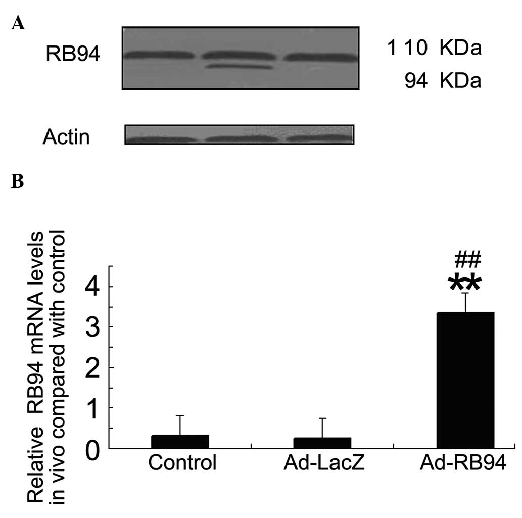
Expression of RB94 following Ad-RB94
transfection
Following Ad-RB94 transfection, western blot
analysis revealed two bands at 110 kDa (wild-type RB) and 94 kDa
(RB94) in the A549 cells (Fig. 1A)
with the RB antibody. However, the control and Ad-LacZ-transfected
A549 cells only expressed wild-type RB protein at 110 kDa. Next,
the expression of RB94 in vivo was determined by RT-qPCR. As
shown in Fig. 1B, the in
vivo RB94 mRNA level was higher in the Ad-RB94 group than in
the Ad-LacZ and control groups. There was no significant difference
between the control and the Ad-LacZ group.
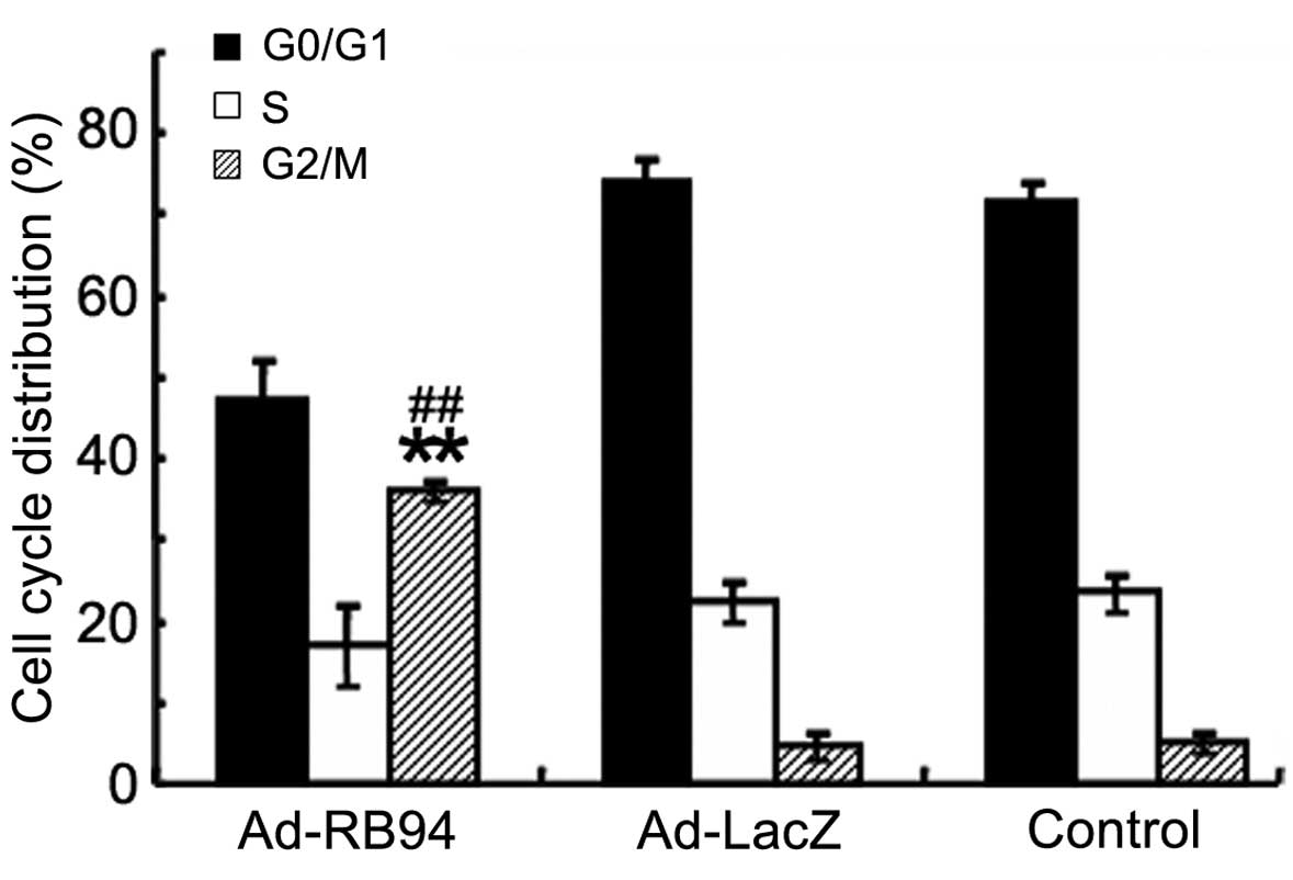
Ad-RB94 induces G2/M cell cycle
arrest
As shown in Fig. 2,
flow cytometry revealed that overexpression of Ad-RB94 increased
the proportion of A549 cells in the G2/M phase, whereas it
decreased the proportion of A549 cells in the G0/G1 phase and the S
phase, indicating that Ad-RB94 may induce G2/M cell cycle arrest in
A549 cells.
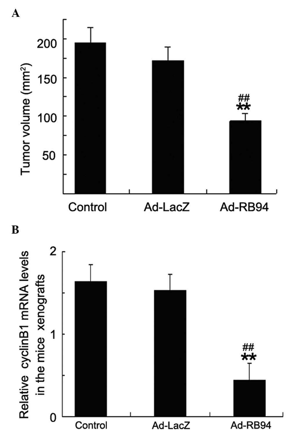
RB94 transfer inhibits tumors growth and
cyclinB1 mRNA levels in vivo
Following the 21 days of treatment, mice were
sacrificed and the volumes of tumors were examined. Treatment with
Ad-RB94 significantly inhibited the growth of tumors compared with
the control and Ad-LacZ. There was no significant difference
between the control and Ad-LacZ groups (P>0.05; Fig. 3A). The cyclinB1 mRNA level of the
tumors following Ad-RB94 treatment decreased significantly compared
with those of the Ad-LacZ and the control groups (Fig. 3B). There were no significant
differences in the levels of cyclinB1 mRNA between the Ad-LacZ and
the control groups.
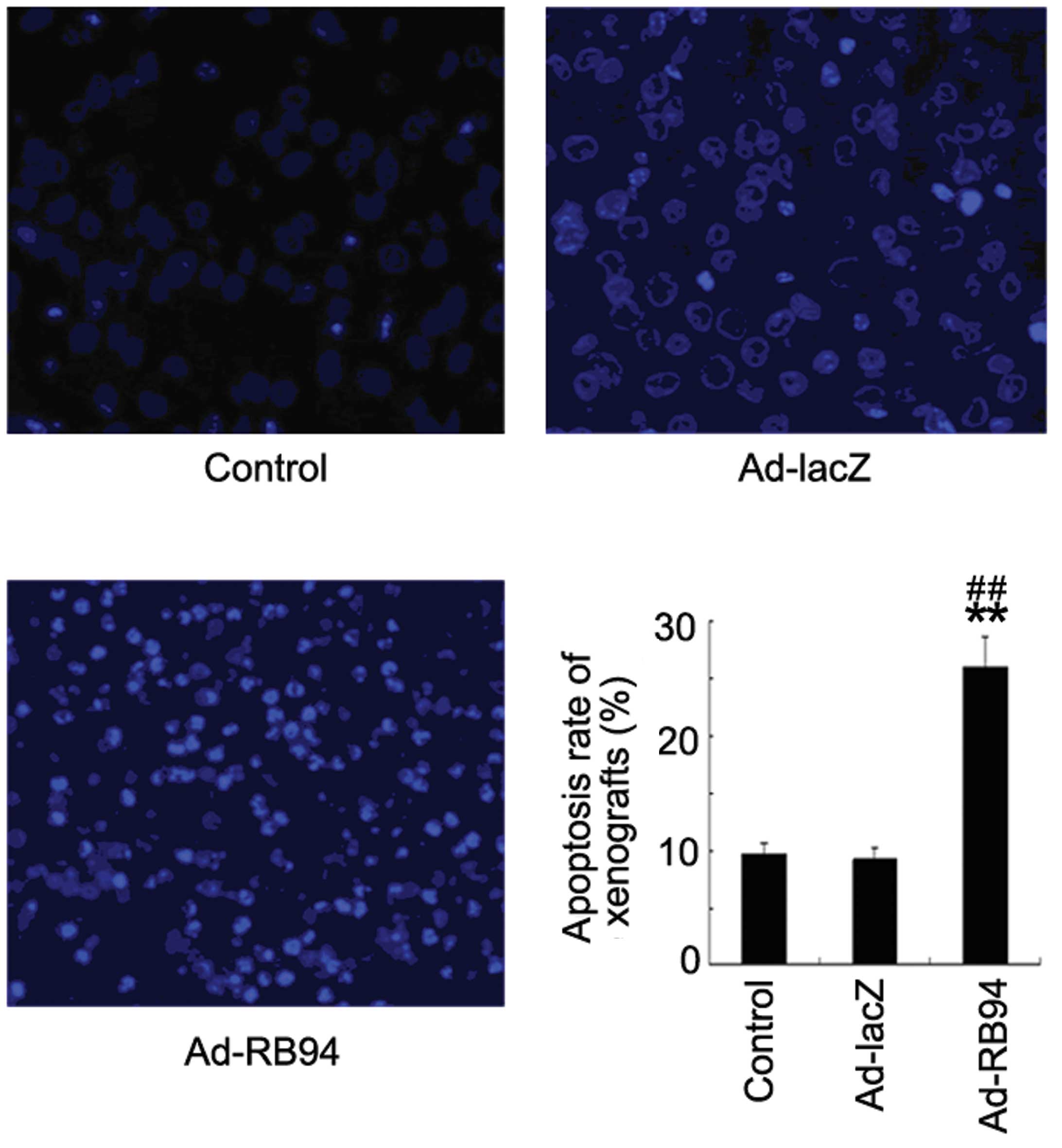
Apoptosis detection in vivo
To further investigate whether Ad-RB94 induced NSCLC
apoptosis in vivo, an apoptosis detection kit was used to
determine apoptosis-related molecular markers in tumor sections
(Fig. 4). The results demonstrated
that treatment with Ad-RB94 induced an apoptosis rate of 26%.
However, only a small number of positive cells were observed in the
control and the Ad-LacZ-treated mice.
Discussion
The results of the present study demonstrate that
RB94 overexpression induces the G2/M cell cycle arrest of NSCLC
in vitro. Overexpression of RB94 in vivo also induced
the apoptosis of NSCLC cells and decreased the tumor cyclinB1
levels, which may cause G2/M phase block (12,13),
and thereby inhibited the growth of the transplanted tumor.
It has been reported that RB94 is capable of
inducing apoptosis in human pancreatic tumors and in head and neck
cancer (6,8,10).
Ad-RB94 transfection also induces cell cycle arrest in the G2/M
phase and decreases telomerase activity in human head and neck
squamous cell carcinoma (6). In
the present study, the results demonstrated that Ad-RB94
transfection induced the G2/M phase block in the A549 lung cancer
cells. The in vivo investigation also suggested that Ad-RB94
transfection decreased the levels of cyclinB1. The Cdk1/cyclinB1
complex plays a significant role in the progression of the cell
cycle from the G2 to M phase, and downregulating cyclinB1 could
cause G2/M phase block (12,13).
Thus, these data indicated that the overexpression of Ad-RB94 may
result in G2/M phase block in lung cancer cells. In the tumor
xenograft study, the present study confirms that an adenoviral
vector efficiently delivers the RB94 gene into targeted cells and
suppresses the tumor cell growth. RB94 expression in the NSCLC
tumor cells leads to apoptotic tumor cell death in the xenograft
nude mouse model.
In conclusion, the present study provided evidence
that RB94 significantly inhibits the growth of NSCLC cells in
vitro and in vivo. These effects are elicited by
arresting the cells at the G2/M phase and triggering apoptosis of
the tumor cells. These results suggest that RB94 is a promising
candidate for adjuvant therapy with radiation or chemotherapy, as
tumor cells are most sensitive to radiation or cytotoxic drugs in
this cell cycle phase. Further research is required to better
elucidate the molecular pathways underlying these effects of
RB94.
References
|
1
|
Goya T, Asamura H, Yoshimura H, et al:
Prognosis of 6644 resected non-small cell lung cancers in Japan: a
Japanese lung cancer registry study. Lung Cancer. 50:227–234. 2005.
View Article : Google Scholar : PubMed/NCBI
|
|
2
|
Pisters KM, Evans WK, Azzoli CG, et al:
Cancer Care Ontario and American Society of Clinical Oncology
adjuvant chemotherapy and adjuvant radiation therapy for stages
I–IIIA resectable non small-cell lung cancer guideline. J Clin
Oncol. 25:5506–5518. 2007. View Article : Google Scholar : PubMed/NCBI
|
|
3
|
Erridge SC, Moller H, Price A and Brewster
D: International comparisons of survival from lung cancer: pitfalls
and warnings. Nat Clin Pract Oncol. 4:570–577. 2007. View Article : Google Scholar : PubMed/NCBI
|
|
4
|
Xu HJ, Zhou Y, Seigne J, et al: Enhanced
tumor suppressor gene therapy via replication-deficient adenovirus
vectors expressing an N-terminal truncated retinoblastoma protein.
Cancer Res. 56:2245–2249. 1996.PubMed/NCBI
|
|
5
|
Xu HJ, Xu K, Zhou Y, Li J, Benedict WF and
Hu SX: Enhanced tumor cell growth suppression by an N-terminal
truncated retinoblastoma protein. Proc Natl Acad Sci USA.
91:9837–9841. 1994. View Article : Google Scholar : PubMed/NCBI
|
|
6
|
Li D, Day KV, Yu S, et al: The role of
adenovirus-mediated retinoblastoma 94 in the treatment of head and
neck cancer. Cancer Res. 62:4637–4644. 2002.PubMed/NCBI
|
|
7
|
Zhang X, Multani AS, Zhou JH, et al:
Adenoviral-mediated retinoblastoma 94 produces rapid telomere
erosion, chromosomal crisis, and caspase-dependent apoptosis in
bladder cancer and immortalized human urothelial cells but not in
normal urothelial cells. Cancer Res. 63:760–765. 2003.PubMed/NCBI
|
|
8
|
Roig JM, Molina MA, Cascante A, et al:
Adenovirus-mediated retinoblastoma 94 gene transfer induces human
pancreatic tumor regression in a mouse xenograft model. Clin Cancer
Res. 10:1454–1462. 2004. View Article : Google Scholar : PubMed/NCBI
|
|
9
|
Zhang H, Li J, Wang YY, et al:
Retinoblastoma 94 enhances radiation treatment of esophageal
squamous cell carcinoma in vitro and in vivo. J Radiat Res.
53:117–124. 2012. View Article : Google Scholar : PubMed/NCBI
|
|
10
|
Araki K, Ahmad SM, Li G, et al:
Retinoblastoma RB94 enhances radiation treatment of head and neck
squamous cell carcinoma. Clin Cancer Res. 14:3514–3519. 2008.
View Article : Google Scholar : PubMed/NCBI
|
|
11
|
Edelman MJ, Quam H and Mullins B:
Interactions of gemcitabine, carboplatin and paclitaxel in
molecularly defined non-small-cell lung cancer cell lines. Cancer
Chemother Pharmacol. 48:141–144. 2001. View Article : Google Scholar : PubMed/NCBI
|
|
12
|
Vairapandi M, Balliet AG, Hoffman B and
Liebermann DA: GADD45b and GADD45g are cdc2/cyclinB1 kinase
inhibitors with a role in S and G2/M cell cycle checkpoints induced
by genotoxic stress. J Cell Physiol. 192:327–338. 2002. View Article : Google Scholar : PubMed/NCBI
|
|
13
|
Gomathinayagam R, Sowmyalakshmi S,
Mardhatillah F, Kumar R, Akbarsha MA and Damodaran C: Anticancer
mechanism of plumbagin, a natural compound, on non-small cell lung
cancer cells. Anticancer Res. 28:785–792. 2008.PubMed/NCBI
|