Introduction
Programmed cell death, or apoptosis, has a key role
in the regulation of cell division and a number of developmental
processes. The hallmarks of apoptosis include cell shrinkage,
nuclear condensation, membrane blebbing, DNA fragmentation and the
appearance of apoptotic bodies (1). It has been reported that numerous
antitumor agents act via the induction of apoptosis in order to
curb tumor promotion and progression. A family of proteases known
as caspases are activated in cells undergoing apoptosis, which
results in the onset of a number of molecular and structural
changes, including the condensation of nuclear heterochromatin,
cell shrinkage and the degradation of DNA repair enzymes
(polyribose polymerase; PARP), and DNA-dependent protein kinases.
The apoptotic bodies that form following apoptosis are then
phagocytocysed by neighboring cells or macrophages (2).
Osteosarcoma is the most prevalent type of malignant
bone tumor in children and teenagers as well as the eighth most
commonly occurring carcinoma in children, which accounts for ~2.4%
of all pediatric cancers and ~20% of all primary bone cancers.
Osteosarcoma have been reported to be more prevalent in males
compared with females, with the highest rates of occurrence during
adolescence (3,4).
In the past two decades, chemotherapy and surgery
have been the primary therapies used in the treatment of
osteosarcoma. However, there are various complications associated
with the intense doses of chemotherapeutic agents that patients
receive. A number of osteosarcoma patients do not show any response
to chemotherapy due to the development of multidrug resistance in
the cancer cells. Furthermore, there are numerous serious
side-effects associated with chemotherapy, including impaired renal
function, gonadal dysfunction and cardiac dysfunction (5–8).
Hence, there is an urgent requirement for novel, safer therapeutic
approaches for the treatment of osteosarcoma.
In the current study, the anticancer effects of
sclareol, a plant-derived diterpene, were investigated against
osteosarcoma cancer cells in vitro. The study examined the
effects of sclareol on MG63 human osteosarcoma cancer cells using a
3-(4,5-dimethyl-thiazol-2-yl)-2,5-diphenyltetrazolium bromide (MTT)
assay. Flow cytometry and fluorescence microscopy were used to
examine the effect of sclareol on the induction of apoptosis and
the loss of mitochondrial membrane potential (ΛΨm) in osteosarcoma
cells. To the best of our knowledge, no previous studies have
investigated the effect of this plant-derived diterpene on
osteosarcoma.
Materials and methods
Cell culture and treatment
MG63 human osteosarcoma cancer cells were procured
from the Shanghai Institute of Cell Biology (Shanghai, China). The
cancer cells were grown in Eagle’s minimal essential medium
(Gibco-BRL, Carlsbad, CA, USA) supplemented with 5% (v/v) fetal
bovine serum (FBS; Sigma, St. Louis, MO, USA), 2 mM L-glutamine,
100 U/ml penicillin and 100 μg/ml streptomycin (Gibco-BRL,
Carlsbad, CA, USA). The cells were allowed to attach for 2 h prior
to the addition of sclareol. The cells were maintained at 37°C in a
humidified atmosphere containing 5% (v/v) CO2. Sclareol
(Sigma) was dissolved in DMSO at 10 mg/ml as a stock solution and
diluted to the necessary concentration with fresh medium prior to
use. The final DMSO concentration in the cultures was <0.1%
(v/v), which did not influence cell growth when compared with that
of the vehicle-free controls. Cells were grown in the media
containing an equivalent amount of DMSO without sclareol to serve
as a negative control.
MTT assay for cell viability
An MTT assay (Sigma) was used to assess cell
viability. Briefly, cells were seeded in 96-well plates at a
density of 1×10⁶ cells/well. Following a 12-h
incubation, sclareol (10, 30, 50, 70 or 100 μM) was added to
the cells, while DMSO served as a negative control. The cells were
cultured for 12, 24 and 48 h, followed by incubation with MTT (0.5
mg/ml) for 3 h at 37°C. Water-insoluble formazan crystals formed
during incubation, which were dissolved by the addition of 100
μl/well DMSO. The optical densities (OD) at 570 nm were
measured using an immunosorbent assay plate reader (HR801; Shenzhen
Highcreation Technology Co., Ltd. Guangdong, China). Wells that
contained culture medium and MTT but no cells acted as blanks. The
percentage cell viability was calculated with the following
equation: Cell viability = (ODdrug −
ODblank/ODcontrol − ODblank) ×
100.
Lactate dehydrogenase (LDH) release
assay
An LDH assay was performed using a CytoTox
96® Non-Radioactive Cytotoxicity Assay kit from Promega
(Madison, WI, USA). Cells were seeded at a density of
2×10⁵ cells per well into 24-well plates 12 h prior to
the experiments. Following different treatments, media from each
well was collected to quantify the amount of released LDH, whereas
isolated wells exposed to lysis buffer (10% Triton X-100) and media
were collected to measure the total amount of cellular LDH.
Cellular LDH was measured using a Cytotoxicity Detection kit (Roche
Pharmaceutical Co., Basal, Switzerland) and a Colorimetric
microplate reader (Thermo Molecular Devices Co.). Lactate
dehydrogenase activity was determined by change in absorbance at
490 nm. For the purpose of calculating percent cytotoxicity values,
background LDH release from culture cells was considered as low
control and triton-X 100 (0.01%) treated cells as high control.
Leakage (%)=[A490 (sample)−A490 (low control)/A490 (high control) −
A490 (low control)]×100%
Detection of apoptosis
Cells grown in 12-well plates on cover-slips were
exposed to different concentrations (30, 50, 70 and 100 μM)
of sclareol for 48 h and incubated with Hoechst 33258 (Hoechst
Staining kit; Beyotime Biotechnology, Haimen, China) according to
the manufacturer’s instructions. Fluorescence microscopy was used
to monitor changes in cell shape; cells were washed once with PBS,
and then observed under a RX50-RFL Biological fluorescence
microscope (Nikon, Tokyo, Japan). The condensed DNA of apoptotic
cells was identified by intense local staining in the nucleus, in
contrast to diffused staining of DNA in normal cells. Cell
morphology was evaluated from six random visual fields and minimum
of 600 cells were counted; each experiment was performed in
triplicate.
Measurement of the effect of sclareol on
cell cycle phase distribution
MG63 osteosarcoma cells (1×10⁶) in a
60-mm dish were exposed to varying concentrations (50, 70 and 100
μM) of sclareol for 48 h. The cells were collected by
trypsinization and washed twice with phosphate-buffered saline
(PBS; Guangzhou Geneshun Biotech, Ltd, Guangzhou, China). Cells
were incubated in 60% ethanol at −20°C overnight and then treated
with 40 μg/ml RNase A (Guangzhou Geneshun Biotech, Ltd) and
stained with 20 μg/ml of propidium iodide (PI; Guangzhou
Geneshun Biotech, Ltd). The stained cells were analyzed using a
FACScan flow cytometer (BD Biosciences, San Jose, CA, USA) at a
wavelength of 488 nm.
Measurement of the effect of sclareol on
ΛΨm
The ΛΨm in MG63 cells was measured with rhodamine
123 (Rh-123; Guangzhou Geneshun Biotech Ltd) dye. MG63 osteosarcoma
cells (5×10⁵) were treated with different concentrations
(50, 70 and 100 μM) of sclareol and the ΛΨm was measured by
flow cytometry. Rh-123 (5 mM) was added 2 h prior to the
termination of experiment. The cells were then washed with PBS and
incubated with PI (20 μg/ml) for 30 min. The cells were
analyzed using a FACScan flow cytometer.
Statistical analysis
The data are expressed as the mean ± standard
deviation. Differences between two groups were analyzed using
Student’s t-test and P<0.05 was considered to indicate a
statistically significant difference.
Results
Sclareol has an antiproliferative effect
on MG63 osteosarcoma cells
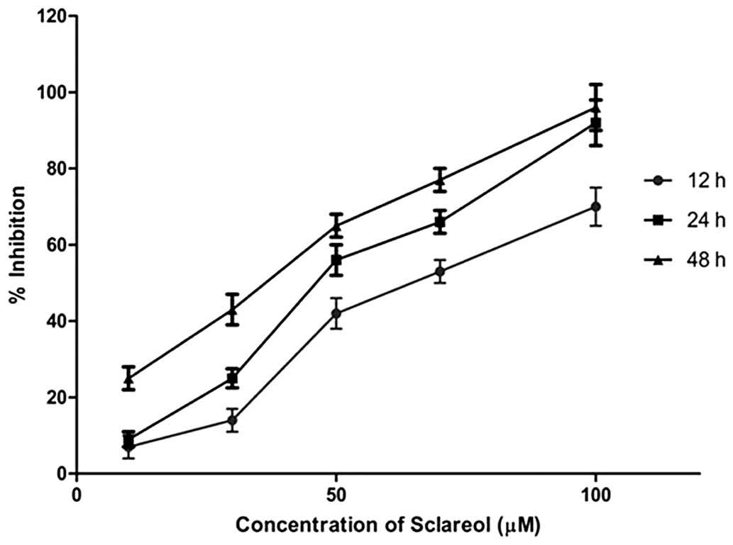
The antiproliferative activity of sclareol was
evaluated against cultured MG63 cells using an MTT assay following
12, 24 and 48 h incubation intervals. The osteosarcoma cancer cells
showed susceptibility to treatment with increasing doses of
sclareol. Sclareol displayed dose- and time-dependent inhibition of
cancer cell growth as presented in Fig. 1. The IC50 value of the
drug was found to be 65.2 μM at 12 h of incubation.
Following a 48-h incubation, growth inhibition was observed
compared with the growth at 12- and 24-h incubation intervals.
Evaluation of sclareol-induced cell death
with an LDH release assay
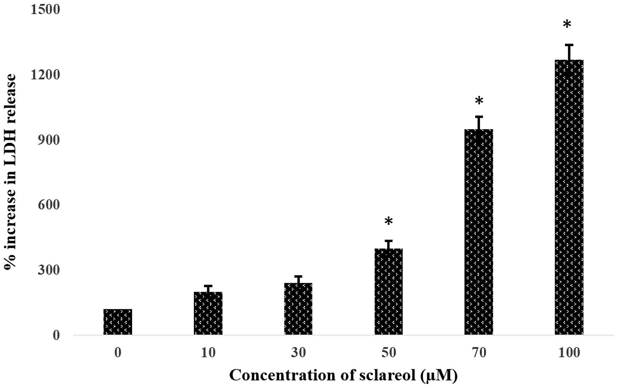
To examine whether sclareol induces cell death in
human MG63 osteosarcoma cancer cells, groups of cells were exposed
to different concentrations (10, 30, 50, 70 and 100 μM) of
sclareol. The extent of cell death was measured after a 48-h
incubation period using an LDH release assay. Since the amount of
LDH release from the dying cells into the culture medium is
proportional to the amount of cell death, this assay provides an
acceptable estimation of the cell death induced by sclareol.
Fig. 2 shows the significant
increase in the sclareol-induced LDH release in a dose-dependent
manner compared with that of the control (P<0.05).
Sclareol induces apoptosis in
osteosarcoma cells
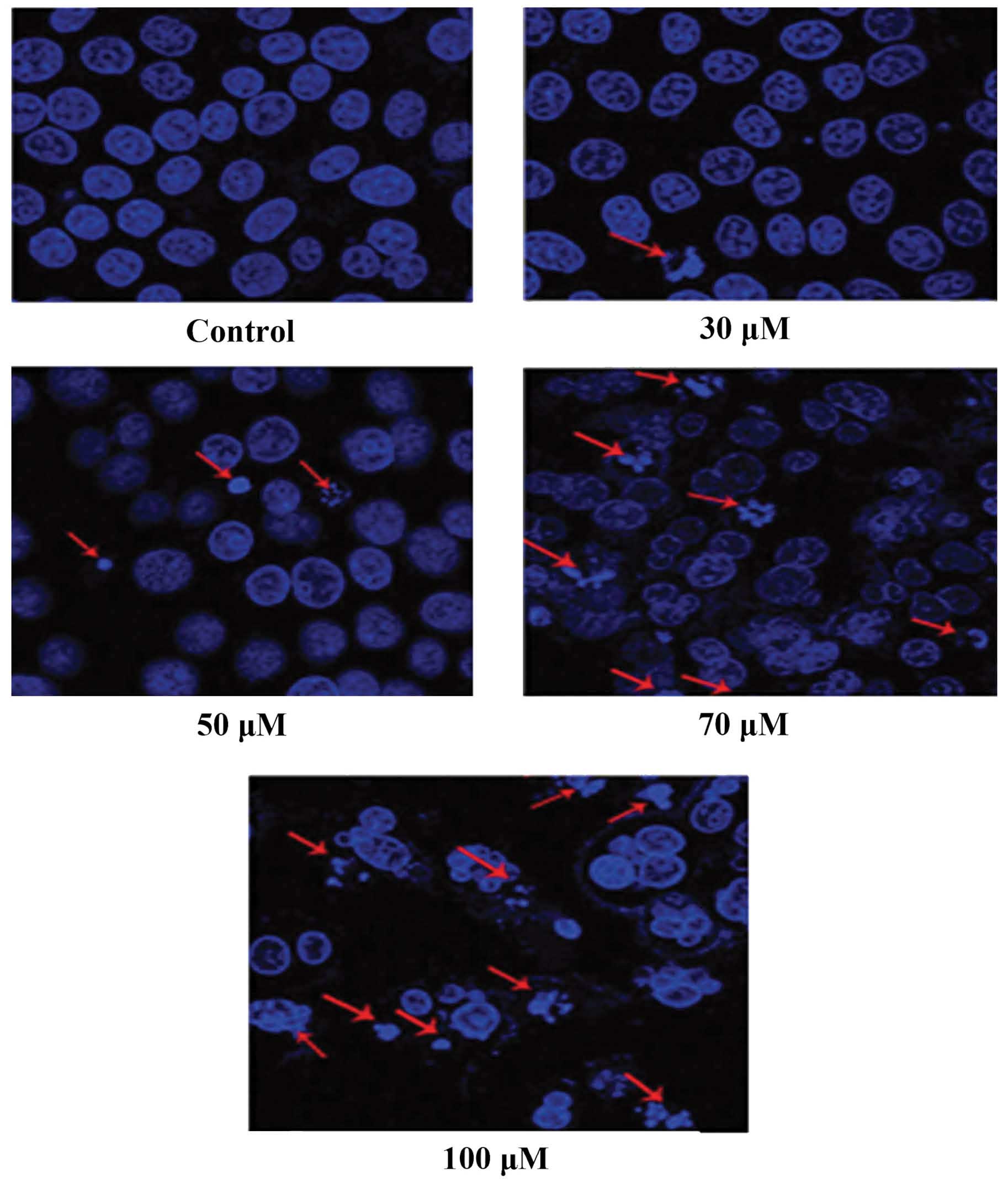
The apoptotic effects of sclareol were evaluated in
MG63 osteosarcoma cells exposed to different concentrations (30,
50, 70 and 100 μM) of sclareol for 48 h. The osteosarcoma
cells were stained and evaluated for nuclear morphological changes
using an inverted fluorescence microscope. The untreated cells
showed normal, evenly dispersed blue fluorescence within their
nuclei, whereas sclareol-treated cells displayed chromatin
condensation or dense staining fragmentation known as ‘apoptotic
bodies’, which correspond to an early apoptotic event (Fig. 3) (9). Notably, at higher concentrations of
sclareol, a more significant reduction in the total number of
cancer cells was observed compared with that in the control cells,
indicating that sclareol exerts potent apoptotic cell death effects
at higher doses.
Sclareol induces G1-phase
arrest in osteosarcoma cells
Apoptosis and cell cycle deregulation are closely
related biochemical events, and any disruption in cell cycle
progression may lead to apoptotic cell death. In order to have a
mechanistic overview of the growth inhibitory effect exerted by
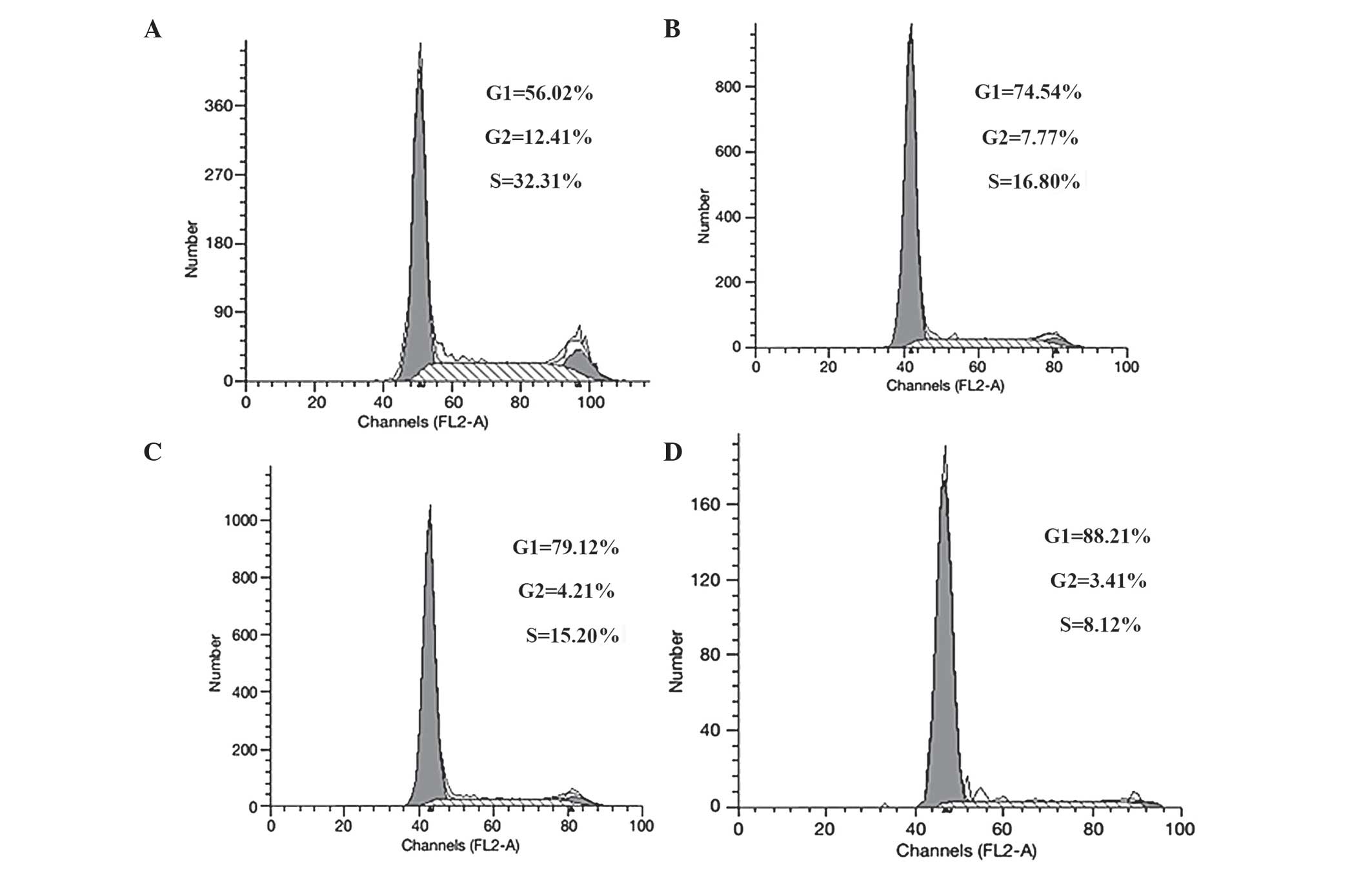
sclareol in osteosarcoma cancer cells, flow cytometric analysis was
performed to detect whether sclareol induces cell cycle arrest in
the MG63 cell line. The results indicated that treatment with
varying concentrations of sclareol for 48 h induced
G1-phase growth arrest in osteosarcoma cells (Fig. 4). Following a 48-h treatment with
50, 70 or 100 μM sclareol, the G1-phase cell
population was significantly increased from 56.02% in the DMSO
control cells to 74.54, 79.12 and 88.21% in the 50, 70 and 100
μM sclareol-treated cells, respectively. This increase in
the G1-phase cell population was accompanied by
corresponding decreases in the S and G2 phases of the
cell populations. In summary, the cancer growth inhibitory effects
of sclareol can be explained on the basis that it induces strong
G1-phase arrest in osteosarcoma cells.
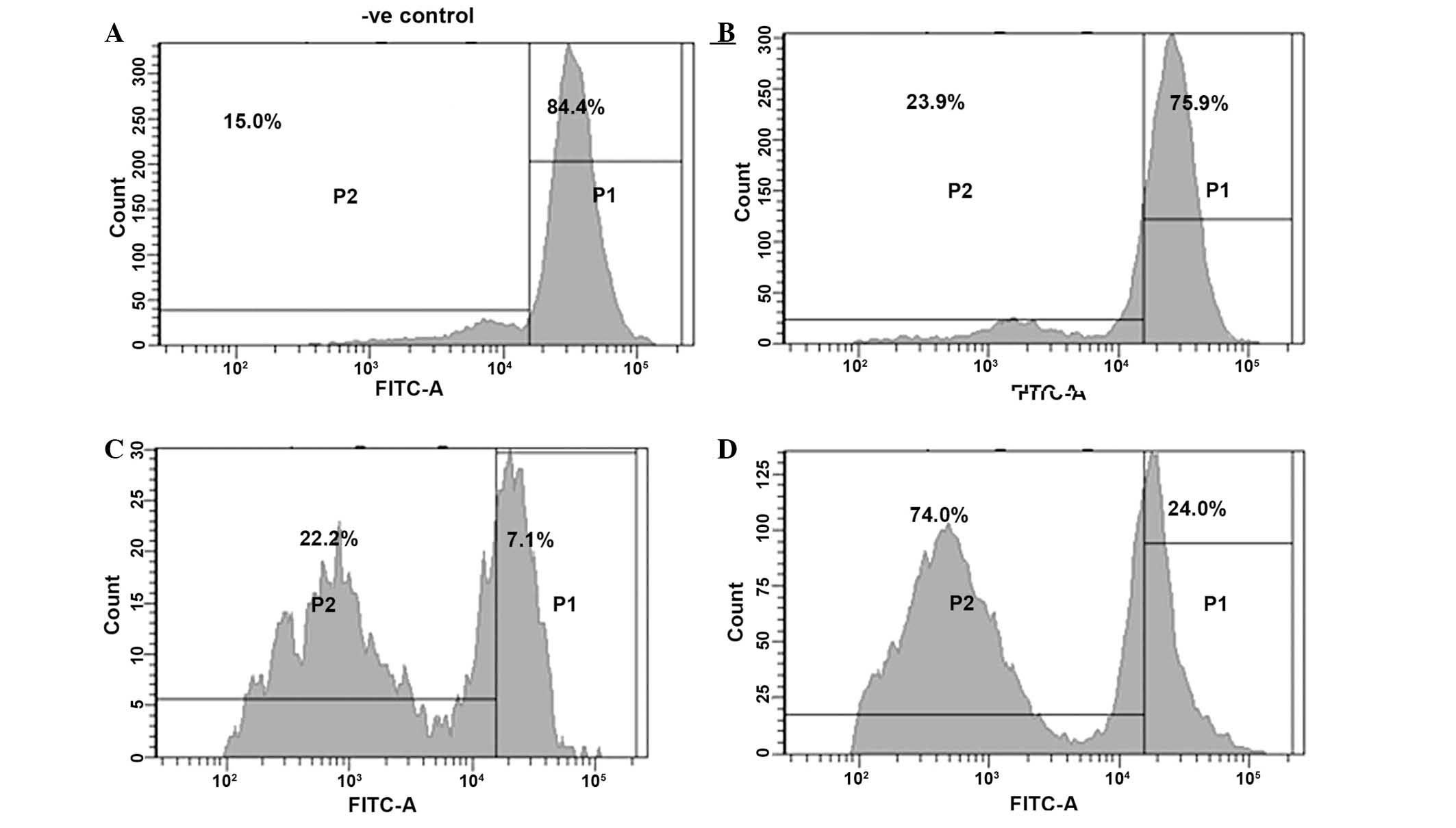
Sclareol induces significant ΛΨm loss in
MG63 osteosarcoma cancer cells
One important and indicative stage in the intrinsic
apoptosis pathway is the depolarization of the mitochondrial
membrane and the subsequent leakage of the outer membrane through
pore formation. This is accompanied by the release of pro-apoptotic
molecules and cytochrome c. The fluorescent dye Rh-123 is a
specific probe for the detection of alterations in the ΛΨm in
living cells (10). The results of
the current study revealed that sclareol induced a progressive
reduction in the number of cells with intact ΛΨm and increased the
number of cells with low ΛΨm after 48 h incubation. The loss of ΛΨm
in osteosarcoma cells increased in a dose-dependent manner
following treatment with 50, 70 and 100 μM of sclareol for
48 h (Fig. 5). The percentage of
cells with a reduced ΛΨm was 15.0% in the solvent-negative control
and 23.9, 22.2 and 74.0% at 50, 70 and 100 μM sclareol,
respectively. Thus, this experiment confirms that treatment of
osteosarcoma cancer cells with sclareol induces a loss in ΛΨm
significantly.
Discussion
Sclareol is a fragrant chemical compound found in
Salvia sclarea from which it derives its name. It is a
bicyclic diterpene alcohol with a sweet, balsamic scent. It is used
as a fragrance in cosmetics and perfumes and as flavoring in food.
From a pharmaceutical point of view, sclareol has been reported to
kill human leukemic and colon cancer cells by inducing apoptosis.
The addition of sclareol to cultures of HCT-116 human colon cancer
cells resulted in the inhibition of DNA synthesis, the arrest of
cells at the G1-phase of the cell cycle, activation of
caspases-8 and -9, PARP degradation and DNA fragmentation (11,12).
The anticancer activity of sclareol in vivo has been
assessed using human colon cancer xenograft/mouse models. It was
observed that sclareol arrested the growth of p53-deficient human
colon cancer cells and subsequently induced apoptosis through the
activation of caspases-8 and -9 in vivo (13). In another study, sclareol was
reported to exhibit an anticancer effect against MN1 and MDD2 human
breast cancer cell lines derived from the MCF-7 parental cell line.
The study used flow cytometry to demonstrate that sclareol was able
to inhibit DNA synthesis, induce cell cycle arrest at the
G0/G1 phase and induce apoptosis independent
of p53 status (14). An additional
study reported that liposome-incorporated sclareol shows cytotoxic
and antitumor activity against human colon cancer xenografts. The
liposomal and free sclareol were initially tested in vitro
for their activity against human cancer cells using the
sulphorhodamine B assay. Liposomal-incorporated sclareol showed a
reduction in the growth rate of human colon cancer cells in SCID
mice, with no significant side-effects (15).
The present study investigated the anticancer
activity of sclareol against MG63 osteosarcoma tumor cells, along
with its effects on cell cycle progression and ΛΨm, which is the
first such study to be published to the best of our knowledge. The
extent of cell death was measured with an LDH release assay, as the
amount of LDH released from dying cells into the culture medium is
proportional to the extent of cell death. Sclareol was shown to
induce dose- and time-dependent cell death in MG63 osteosarcoma
tumor cells. Furthermore, following exposure to different doses of
sclareol, morphological changes characteristic of apoptosis were
detected in these cells using fluorescence microscopy. Flow
cytometric analysis revealed that sclareol induces G1
cell cycle arrest in these cells. Following a 48-h treatment with
50, 70 and 100 μM doses of sclareol, the G1-phase
cell population was significantly increased from 56.02% in the DMSO
control cells to 74.54, 79.12 and 88.21%, respectively. Using a
Rh-123 fluorescent probe, it was revealed that sclareol caused a
significant reduction in the ΛΨm in these tumor cells.
In conclusion, sclareol significantly inhibits the
growth of osteosarcoma tumor cells by inducing apoptosis
accompanied by G1 phase cell cycle growth arrest and a
concomitant loss of ΛΨm. Furthermore, fluorescence microscopy
revealed that sclareol induces morphological changes characteristic
of apoptosis in these cells. Taking all of the results into
consideration, sclareol may be developed further as a possible
anticancer agent for the treatment of osteosarcoma.
References
|
1
|
Wyllie AH: Apoptosis: an overview. Br Med
Bull. 53:451–465. 1997. View Article : Google Scholar : PubMed/NCBI
|
|
2
|
Hengartner MO: The biochemistry of
apoptosis. Nature. 407:770–776. 2000. View
Article : Google Scholar : PubMed/NCBI
|
|
3
|
Enneking WF and Springfield DS:
Osteosarcoma. Orthop Clin North Am. 8:785–803. 1977.PubMed/NCBI
|
|
4
|
Ottaviani G and Jaffe N: The epidemiology
of osteosarcoma. Cancer Treat Res. 152:3–13. 2009. View Article : Google Scholar
|
|
5
|
Ferguson WS and Goorin AM: Current
treatment of osteosarcoma. Cancer Invest. 19:292–315. 2001.
View Article : Google Scholar : PubMed/NCBI
|
|
6
|
Bacci G and Lari S: Adjuvant and
neoadjuvant chemotherapy in osteosarcoma. Chir Organi Mov.
86:253–268. 2001.
|
|
7
|
Biermann JS and Baker LH: The future of
sarcoma treatment. Semin Oncol. 24:592–597. 1997.PubMed/NCBI
|
|
8
|
La Quaglia MP: Osteosarcoma. Specific
tumor management and results. Chest Surg Clin N Am. 8:77–95.
1998.PubMed/NCBI
|
|
9
|
Yang Y, Yang L, You QD, Nie FF, et al:
Differential apoptotic induction of gambogic acid, a novel
anticancer natural product, on hepatoma cells and normal
hepatocytes. Cancer Lett. 256:259–266. 2007. View Article : Google Scholar : PubMed/NCBI
|
|
10
|
Johnson LV, Walsh ML and Chen LB:
Localization of mitochondria in living cells with rhodamine 123.
Proc Natl Acad Sci USA. 77:990–994. 1980. View Article : Google Scholar : PubMed/NCBI
|
|
11
|
Dimas K, Kokkinopoulos D, Demetzos C, Vaos
B, et al: The effect of sclareol on growth and cell cycle
progression of human leukemic cell lines. Leuk Res. 23:217–234.
1999. View Article : Google Scholar : PubMed/NCBI
|
|
12
|
Dimas K, Hatziantoniou S, Tseleni S, Khan
H, et al: Sclareol induces apoptosis in human HCT116 colon cancer
cells in vitro and suppression of HCT116 tumor growth in
immunodeficient mice. Apoptosis. 12:685–694. 2007. View Article : Google Scholar : PubMed/NCBI
|
|
13
|
Mahaira LG, Tsimplouli C, Sakellaridis N,
Alevizopoulos K, et al: The labdane diterpene sclareol
(labd-14-ene-8, 13-diol) induces apoptosis in human tumor cell
lines and suppression of tumor growth in vivo via a p53-independent
mechanism of action. Eur J Pharmacol. 666:173–182. 2011. View Article : Google Scholar : PubMed/NCBI
|
|
14
|
Dimas K, Papadaki M, Tsimplouli C,
Hatziantoniou S, et al: Labd-14-ene-8,13-diol (sclareol) induces
cell cycle arrest and apoptosis in human breast cancer cells and
enhances the activity of anticancer drugs. Biomed Pharmacother.
60:127–133. 2006. View Article : Google Scholar : PubMed/NCBI
|
|
15
|
Hatziantoniou S, Dimas K, Georgopoulos A,
Sotiriadou N and Demetzos C: Cytotoxic and antitumor activity of
liposome-incorporated sclareol against cancer cell lines and human
colon cancer xenografts. Pharmacol Res. 53:80–87. 2006. View Article : Google Scholar
|