Introduction
Hepatoblastoma (HB) is a type of liver cancer, which
is common in children (1). The
primary challenges for the improvement of treatment efficacy
include HB metastasis and drug resistance (2,3).
Understanding the molecular mechanisms underlying HB metastasis may
be useful for the development of novel therapeutic gene targets and
the identification of diagnostic biomarkers for HB.
Epithelial-mesenchymal transition (EMT) is associated with the
progression of a number of epithelial tumors (4–6). EMT
consists of a coordinated series of stages, where epithelial cells
lose their cell polarity and their cell-cell adhesion, and develop
into mesenchymal cells, which results in tumor invasion and
metastasis. The process of EMT is essential for HB metastasis and
invasion to occur (7,8).
Thymosin β4 (Tβ4), a small peptide originally
isolated from calf thymus, is found in human tissues and blood
platelets. Tβ4 predominantly functions as a G-actin sequestering
factor, modulating the dynamic changes of the actin cytoskeleton.
In addition, Tβ4 may be involved in a number of physiological and
pathological processes, including the migration of fibroblasts
(9,10), endothelial cells and keratinocytes
(11,12), and wound healing (13,14).
In addition, the prospect of targeting Tβ4 in cancer therapy has
recently been proposed. A previous study suggested that Tβ4 was
overexpressed in osteosarcoma, colorectal carcinoma and esophageal
cancer cells (15). Wang et
al (16) demonstrated that Tβ4
overexpression suppressed epithelial-cadherin (E-cadherin)
expression and led to the increased motility of SW480 human colon
carcinoma cells. The present study therefore investigated the
involvement of Tβ4 in HB metastasis.
Materials and methods
Materials and specimen preparation
Fetal bovine serum (FBS) was purchased from Gibco
Life Technologies (Carlsbad, CA, USA). Tβ4 was obtained from
Prospec (Rehovot, Israel). Rabbit monoclonal anti-E-cadherin (cat.
no 3195), rabbit monocloncal anti-neural-cadherin (anti-N-cadherin;
cat. no 13116), rabbit monocloncal anti-β-catenin (cat. no. 9582)
and rabbit monocloncal anti-glyceraldehyde 3-phosphate
dehydrogenase (anti-GAPDH; cat. no. 9582) antibodies were obtained
from Cell Signaling Technology Inc. (Danvers, MA, USA). Rabbit
polycloncal anti-Tβ4 antibody (sc-67114, for western blot analysis)
was obtained from Santa Cruz Biotechnology, Inc. (Dallas, TX, USA).
Rabbit polyclonal anti-Tβ4 antibody (ab14335, for
immunohistochemical and immunofluorescence analysis) was purchased
from Abcam (Hong Kong, China). Mouse monoclonal anti-α-smooth
muscle actin (anti-α-SMA; A5228) was obtained from Sigma-Aldrich
(St. Louis, MO, USA). Lipfectamin RNAiMAX was purchased from
Invitrogen Life Technologies (Carlsbad, CA, USA). Liver samples
from HB and adjacent healthy tissue were obtained from 19 patients
with HB at Xinhua Hospital and First Affiliated Hospital of Bengbu
Medical College (Anhui, China). This study was performed according
to a protocol approved by the faculty of the Medicine Ethics
Committee of the First Affiliated Hospital of Bengbu Medical
College. Written informed consent (BYKF-D-2009-0914) was obtained
from the patients’ guardians prior to specimen collection.
Immunohistochemistry and
immunofluorescence analysis
Immunohistochemistry was performed using a
diaminobenzidine (DAB; Dako, Glostrup, Denmark) chromogen method as
described previously (17).
Specimens were initially incubated using xylol. Endogenous
peroxidases were then removed by incubating the samples with 0.3%
hydrogen peroxide. The primary antibodies (anti-E-cadherin, 1:400;
anti-Tβ4, 1:1,000; and anti-β-catenin, 1:100) were incubated
overnight in a chamber with water bottom at 4°C. The slides were
washed in phosphate-buffered saline (PBS) and incubated with the
horseradish peroxidase (HRP)-conjugated goat anti-rabbit
immunoglobulin (Ig)G secondary antibody (1:200; #7074; Cell
Signaling Technology, Inc.). Antibody binding was visualized using
a liquid DAB Substrate Chromogen System (Dako). In order to conduct
an immunofluorescence assay, the cells were fixed with 4%
paraformaldehyde. Subsequently, the cells were incubated with the
anti-Tβ4 antibody (1:200) and blocked using 3% bovine serum albumin
(MP Biomedicals, Auckland, New Zealand). Following three wash
stages with PBS, the secondary antibody, conjugated to fluorescein
isothiocyanate (1:200; #111-095-045; goat anti-rabbit; Jackson
ImmunoResearch, Inc., West Grove, PA, USA), was applied to the
cells. The nuclei were counter-stained using 4′,
6-diamidino-2-phenylindole (DAPI; Dojindo, Kumamoto, Japan). The
results were visualized using a fluorescence microscope (Eclipse;
Nikon, Tokyo, Japan). The nuclei staining and Tβ4 staining were
then merged (magnification, ×40).
Cell culture
The L02 human healthy liver and HepG2 hepatoblastoma
cell lines were cultured in Dulbecco’s modified Eagle’s medium
(DMEM; Gibco Life Technologies), supplemented with 10% fetal bovine
serum (FBS; Gibco Life Technologies) at 37°C in a humidified
atmosphere of 5% CO2. Tβ4-small interfering RNA
(Tβ4-siRNA) duplexes were synthesized by Genepharma Co., Ltd.
(Shanghai, China). The siRNA sequences were
5′-CUUCCAAAGAAACGAUUGATT-3′ 5′ - UCGAUAAGUCGAAACUGAATT-3′ and
5′-GAGGUUGGAUCAAGUUUAATT-3′. Tβ4-siRNA transfection was conducted
using Lipfectamin RNAiMAX (cat. no. 13778; Life Technologies,
Carlsbad, CA, USA) according to the manufacturer’s
instructions.
TGF-β1 EMT in vitro
HepG2 cells were stimulated for 72 h in a
conditioned medium containing TGF-β1 (10 ng/ml), supplemented with
0.5% FBS. Total protein was harvested in order to conduct western
blot analysis.
Western blotting
Protein concentrations of cell lysates were
determined using a bicinchoninic acid protein assay kit (Pierce,
Biotechnology, Inc., Rockford, IL, USA). Protein was separated
using electrophoresis on a Novex® 10% Tris-glycine gel
(Life Technologies) and transferred onto a nitrocellulose membrane
(Life Technologies). The membranes were incubated with the
following primary antibodies overnight at 4°C: Anti-E-cadherin
(1:1,000), anti-N-cadherin (1:1,000), anti-α-SMA (1:200),
anti-β-catenin (1:1,000), anti-Tβ4 (1:200) and anti-GAPDH
(1:1,000). The membranes were then incubated with a HRP-conjugated
anti-rabbit (1:2,000; cat. no. 7074) and anti-mouse (1:2,000; cat.
no. 7076) IgG secondary antibody (Cell Signaling Technology, Inc.)
after washing with PBS three times. The antibodies were detected
using an enhanced chemiluminescence detection kit (Thermo Fisher
Scientific, Waltham, MA, USA) and the Molecular Imager®
ChemiDoc™ XRS+ System (Bio-Rad Laboratories, Hercules, CA,
USA).
Wound healing
Confluent cells in 6-well plates were scratched
using 100-μl pipette tips. The cells were then incubated at 37°C to
allow cell migration into the wound. Following fixation, the number
of cells that had migrated into the wound were counted using a
microscope (Eclipse Ti; Nikon, Tokyo, Japan). Migration ratio (%)
was calculated as the wound width at 24 h/wound width at 0 h.
Transwell migration assay
Migration of HepG2 cells was determined using
24-well Transwell chambers (Corning Life Sciences, Tewksbury, MA,
USA), according to the manufacturer’s instructions. DMEM (500 μl)
was added to the lower chambers of the 24-well plate, containing
10% FBS. Cells (1×10⁴/well) were mixed with 100 μl of
DMEM without FBS, and the mixture was added to the upper chambers
of the 24-well plate. Transwell chambers were incubated at 37°C in
a 5% CO2 humidified atmosphere for 24 h. Cells that had
migrated to the lower surface of the polycarbonate membranes (12-mm
pore size) were fixed, stained with crystal violet (Amresco, Solon,
OH, USA) and quantified by counting five fields of view using a
microscope (Eclipse Ti; Nikon; magnification, ×40).
Statistical analysis
All data are expressed as the mean ± standard
deviation. Statistical significance for comparisons made between
two groups was determined using Student’s t-test analysis in SPSS
version 13 (SPSS Inc., Chicago, IL, USA).
Results
Tβ4 expression is associated with HB
metastasis
The clinico-pathological characteristics for the 19
patients with HB used in the present study are provided in Tables I and II. The expression of Tβ4 protein in HB
tumor tissue samples and adjacent healthy control tissues was
analyzed. Tβ4 expression was higher in HB samples compared with the
adjacent healthy samples (Fig. 1).
No significant associations were observed between Tβ4 expression
levels and the gender, age or tumor subtype of patients with HB
(Table I). Tβ4 expression was
significantly higher in metastatic HB samples compared with the
that of the non-metastatic HB samples (91% vs. 25%; P<0.01;
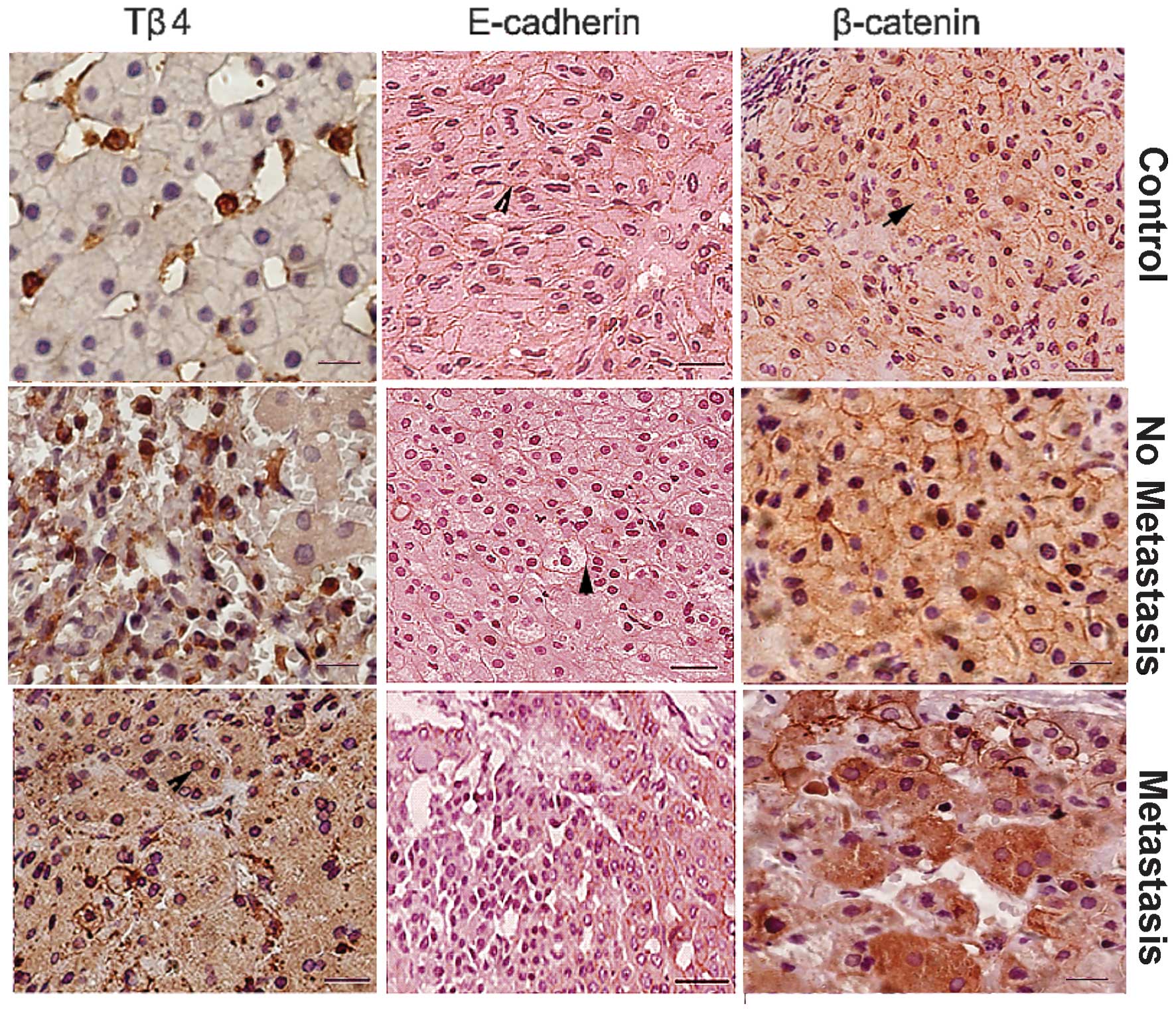
Table I). Expression of Tβ4 in
metastatic HB specimens was found to be increased, while E-cadherin
expression as well as the cytosolic accumulation of β-catenin were
reduced in these samples (Fig. 2
and Table II). Overall, these
results indicate that upregulation in the expression of Tβ4 may be
associated with HB metastasis.
 | Table ICharacteristics of patients with
hepatoblastoma. |
Table I
Characteristics of patients with
hepatoblastoma.
| Clinical
parameters | N | Tβ4 PR (%) | χ2 | P-value |
|---|
| Gender |
| M | 14 | 9 (64) | 0.13 | >0.05 |
| F | 5 | 3 (60) | | |
| Age |
| <3 years | 13 | 8 (61) | 0.00 | >0.05 |
| ≥3 years | 6 | 4 (67) | | |
| Tumor subtype |
| Fetal | 9 | 6 (67) | | |
| Embryonal | 5 | 3 (60) | 0.23 | >0.05 |
|
Undifferentiated | 2 | 2 (67) | | |
|
Epithelial/mesenchymal | 3 | 1 (50) | | |
| Lymph node
metastasis |
| Yes | 11 | 10 (91) | 8.65 | <0.01 |
| No | 8 | 2 (25) | | |
| Table IIClinicopathological features and
results of Tβ4, E-cadherin and β-catenin immunostaining in
hepatoblastoma samples. |
Table II
Clinicopathological features and
results of Tβ4, E-cadherin and β-catenin immunostaining in
hepatoblastoma samples.
| Gender | Age | Tumor subtype | LNM (Y/N) | Tβ4 | EC | Cβ-c |
|---|
| M | 8 Ye | Fetal | N | +/− | ++ | − |
| M | | | | | | |
| M | 7 Ye | Fetal | N | +/− | − | − |
| M | | | N | | | |
| M | 5 M | Fetal | N | +/− | +/− | − |
| M | 4 Ye | Embryonal | N | +/− | ++ | − |
| M | 13 M | Embryonal | N | +/− | +/− | +/− |
| M | 15 M |
Undifferentiated | N | +/− | ++ | − |
| M | 8 M |
Undifferentiated | N | ++ | − | ++ |
| F | 5 Ye |
Epithelial/mesenchymal | N | ++ | +/− | − |
| M | 11 M | Fetal | Y | ++ | − | +/− |
| F | 9 M | Fetal | Y | +++ | − | − |
| M | 11 M | Fetal | Y | +++ | − | +++ |
| M | 4 Ye | Fetal | Y | +/− | − | +++ |
| M | 6 M | Fetal | Y | +++ | − | +/− |
| F | 2 Ye | Fetal | Y | ++ | − | ++ |
| F | 4 M | Embryonal | Y | +++ | − | ++ |
| M | 5 Ye | Embryonal | Y | + | − | +++ |
| F | 13 M | Embryonal | Y | ++ | − | ++ |
| M | 9 M |
Epithelial/mesenchymal | Y | +++ | − | +/− |
| M | 14 M |
Epithelial/mesenchymal | Y | +++ | − | ++ |
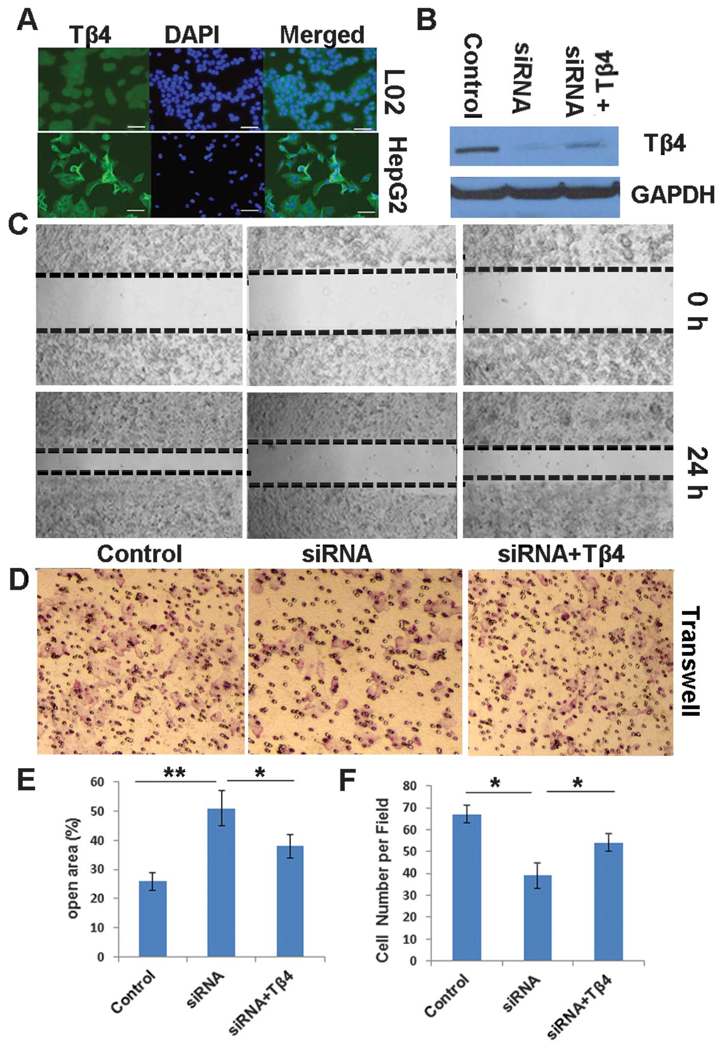
Expression of Tβ4 promotes HB cell
motility
In order to investigate the association between
endogenous Tβ4 levels and the migratory capability of HB cells,
siRNA was used to target Tβ4 mRNA and knockdown Tβ4 gene expression
in HepG2 cells. Silencing of Tβ4 expression in HepG2 cells (Tβ4
siRNA-transfected cells; Fig. 2A and
B) resulted in a significant reduction in cell migratory
capability compared with that of control HepG2 cells, according to
a wound healing assay (P<0.01; Fig.
2C and E). Following treatment with Tβ4 (siRNA + Tβ4 Fig. 2C and E) the migratory capability of
HepG2 cells was significantly greater than that of control HepG2
cells, and significantly lower than that of Tβ4 silenced (siRNA)
HepG2 cells (P<0.05 and 0.01, respectively). Similar results
were observed in the Transwell assays (P<0.05; Fig. 2D and F).
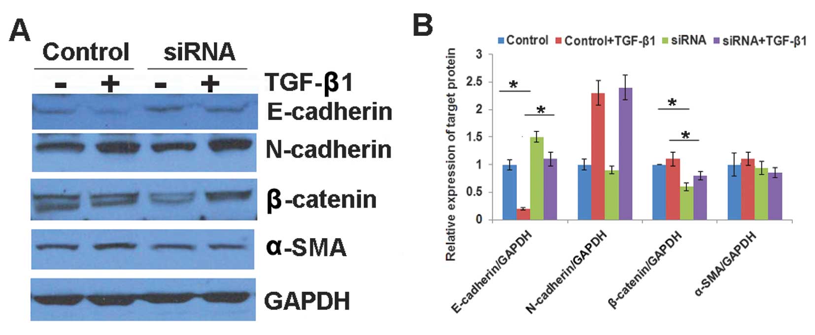
Tβ4 depletion inhibits EMT in HB
cells
The results of the present study suggested that
downregulated Tβ4 expression may be associated with reduced HepG2
cell migratory capability. Markers of EMT were examined in order to
investigate the mechanisms underlying these observations.
E-cadherin expression was higher in Tβ4-siRNA-transfected HepG2
cells compared with that in control cells, whereas the expression
levels of two mesenchymal markers (β-catenin and α-SMA) were lower
in Tβ4-siRNA-transfected HepG2 cells compared with those in control
cells (Fig. 3A and B). Since
TGF-β1 is involved in the induction of EMT (18), the association between Tβ4
expression and TGF-β1-induced EMT was examined. Following TGF-β1
treatment, the expression of E-cadherin in HepG2 cells was
significantly lower and the levels of N-cadherin and α-SMA were
significantly higher, compared with those in the control HepG2
cells. Following transfection with Tβ4-siRNA, the expression of
genes involved in TGF-β1-induced EMT was significantly reduced in
HepG2 cells compared with that in control HepG2 cells (Fig. 3A and B).
Discussion
Tβ4 is a cellular, actin-sequestering protein, which
is associated with angiogenesis induction and the metastatic
potential of tumor cells (19-24).
The results of the present study suggested that Tβ4 is involved in
HB metastasis. Tβ4 expression was significantly higher in HB tissue
cells compared with that in healthy adjacent cells (Fig. 1). Statistical analysis demonstrated
that Tβ4 expression in tumor tissues was significantly associated
with HB-derived lymph node metastasis (Table I and II). Tβ4 gene expression knockdown, using
siRNA transfection, resulted in a decrease in the migratory
capability of HepG2 cells compared with that in control cells
(Fig. 2). Furthermore, the
inhibition of Tβ4 expression suppressed the process of
TGF-β1-induced EMT in HepG2 cells (Fig. 3).
Tumor metastasis is a multistep process, in which
cancer cells disseminate from their primary sites and develop
secondary malignant growths at distant sites. The process involves
local invasion, intravasation, transportation, extravasation and
colonization (25). EMT involves a
series of steps, in which cell-cell and cell-extracellular matrix
interactions are altered in order to release epithelial cells from
the surrounding tissue (26). EMT
has been shown to be involved in promoting metastasis in
epithelium-derived carcinoma (25). A loss of E-cadherin has been
hypothesized to promote β-catenin expression, which binds with the
transcription factor, T-cell factor/lymphoid enhancer factor, and
modulates gene transcription (27). In the present study, it was found
that Tβ4 was upregulated in HB metastatic liver samples; by
contrast, E-cadherin expression and the cytosolic accumulation of
β-catenin were downregulated in these specimens (Table II and Fig. 1). As the principle binding partner
of β-catenin, E-cadherin is involved in the stabilization and
promotion of β-catenin expression. E-cadherin and β-catenin are
associated with the adhesion and the maintenance of epithelial cell
layers. The upregulation of Tβ4 expression may lead to the
downregulation of E-cadherin expression, and disrupt actin
filaments (28). This may
subsequently promote the release of β-catenin from the cell
membrane, thereby activating target genes and facilitating HB
metastasis. The TGF-β pathway appears to induce EMT (29). In the present study, HepG2 cells
were treated with TGF-β1 in order to induce EMT. The expression
E-cadherin was lower, and that of N-cadherin and β-catenin was
higher, in TGF-β1-treated cells, compared with expression of these
molecules in the control cells (Fig.
3). These results suggest that Tβ4 depletion may inhibit EMT in
HB cells. Adherent cell locomotion is a highly integrated process,
initiated by the forward extension of lamellipodia, followed by
repeated cycles of protrusion, adhesion and contraction (30,31).
Tβ4 is a candidate regulator of cell protrusion that is involved in
protrusion-associated processes, such as actin polymerization and
matrix metalloproteinase (MMP) expression. Wang et al
(16) demonstrated that an
increase in the invasiveness of SW480 colon carcinoma cells
over-expressing Tβ4, was associated with an increase in MMP-7
expression. The involvement of MMP expression in the processes
underlying the association between Tβ4 expression and HB metastasis
requires further investigation.
In conclusion, elevated Tβ4 expression in HB cells
may promote HB metastasis via the deregulation of EMT.
References
|
1
|
Czauderna P, Lopez-Terrada D, Hiyama E,
Häberle B, Malogolowkin MH and Meyers RL: Hepatoblastoma state of
the art: pathology, genetics, risk stratification, and
chemotherapy. Curr Opin Pediatr. 26:19–28. 2014. View Article : Google Scholar
|
|
2
|
Stocker JT: Hepatic tumors in children.
Clin Liver Dis. 5:259–281. 2001. View Article : Google Scholar : PubMed/NCBI
|
|
3
|
Green LK and Silva EG: Hepatoblastoma in
an adult with metastasis to the ovaries. Am J Clin Pathol.
92:110–115. 1989.PubMed/NCBI
|
|
4
|
Gupta SK, Oommen S, Aubry MC, Williams BP
and Vlahakis NE: Integrin α9β1 promotes malignant tumor growth and
metastasis by potentiating epithelial-mesenchymal transition.
Oncogene. 32:141–150. 2013. View Article : Google Scholar
|
|
5
|
Yadav A, Kumar B, Datta J, Teknos TN and
Kumar P: IL-6 promotes head and neck tumor metastasis by inducing
epithelial-mesenchymal transition via the JAK-STAT3-SNAIL signaling
pathway. Mol Cancer Res. 9:1658–1667. 2011. View Article : Google Scholar : PubMed/NCBI
|
|
6
|
Meng F, Han Y, Staloch D, Francis T,
Stokes A and Francis H: The H4 histamine receptor agonist,
clobenpropit, suppresses human cholangiocarcinoma progression by
disruption of epithelial mesenchymal transition and tumor
metastasis. Hepatology. 54:1718–1728. 2011. View Article : Google Scholar : PubMed/NCBI
|
|
7
|
Zucchini-Pascal N, Peyre L and Rahmani R:
Crosstalk between beta-catenin and snail in the induction of
epithelial to mesenchymal transition in hepatocarcinoma: role of
the ERK1/2 pathway. Int J Mol Sci. 14:20768–20792. 2013. View Article : Google Scholar : PubMed/NCBI
|
|
8
|
Cannito S, Novo E, Compagnone A, et al:
Redox mechanisms switch on hypoxia-dependent epithelial-mesenchymal
transition in cancer cells. Carcinogenesis. 29:2267–2278. 2008.
View Article : Google Scholar : PubMed/NCBI
|
|
9
|
Nagamalleswari K and Safer D: Sequestered
actin in chick embryo fibroblasts. Mol Cell Biochem. 209:63–67.
2000. View Article : Google Scholar : PubMed/NCBI
|
|
10
|
Reti R, Kwon E, Qiu P, Wheater M and Sosne
G: Thymosin beta4 is cytoprotective in human gingival fibroblasts.
Eur J Oral Sci. 116:424–430. 2008. View Article : Google Scholar : PubMed/NCBI
|
|
11
|
Roy P, Rajfur Z, Jones D, Marriott G, Loew
L and Jacobson K: Local photorelease of caged thymosin beta4 in
locomoting keratocytes causes cell turning. J Cell Biol.
153:1035–1048. 2001. View Article : Google Scholar : PubMed/NCBI
|
|
12
|
Mu H, Ohashi R, Yang H, et al: Thymosin
beta10 inhibits cell migration and capillary-like tube formation of
human coronary artery endothelial cells. Cell Motil Cytoskeleton.
63:222–230. 2006. View
Article : Google Scholar : PubMed/NCBI
|
|
13
|
Francis Godschalk M: Pressure ulcers: a
role for thymosin beta4. Ann N Y Acad Sci. 1112:413–417. 2007.
View Article : Google Scholar : PubMed/NCBI
|
|
14
|
Smart N, Rossdeutsch A and Riley PR:
Thymosin beta4 and angiogenesis: modes of action and therapeutic
potential. Angiogenesis. 10:229–241. 2007. View Article : Google Scholar : PubMed/NCBI
|
|
15
|
Jo JO, Kang YJ, Ock MS, Kleinman HK, Chang
HK and Cha HJ: Thymosin β4 expression in human tissues and in
tumors using tissue microarrays. Appl Immunohistochem Mol Morphol.
19:160–167. 2011. View Article : Google Scholar
|
|
16
|
Wang WS CP, Hsiao HL, Ju SY and Su Y:
Overexpression of the thymosin beta-4 gene is associated with
malignant progression of SW480 colon cancer cells. Oncogene.
22:3297–3306. 2003. View Article : Google Scholar : PubMed/NCBI
|
|
17
|
Bataille F, Rohrmeier C, Bates R, et al:
Evidence for a role of epithelial mesenchymal transition during
pathogenesis of fistulae in Crohn’s disease. Inflamm Bowel Dis.
14:1514–1527. 2008. View Article : Google Scholar : PubMed/NCBI
|
|
18
|
Flier SN, Tanjore H, Kokkotou EG, Sugimoto
H, Zeisberg M and Kalluri R: Identification of epithelial to
mesenchymal transition as a novel source of fibroblasts in
intestinal fibrosis. J Biol Chem. 285:20202–20212. 2010. View Article : Google Scholar : PubMed/NCBI
|
|
19
|
Kim NS, Kang YJ, Jo JO, et al: Elevated
expression of thymosin β4, vascular endothelial growth factor
(VEGF), and hypoxia inducible factor (HIF)-1α in early-stage
cervical cancers. Pathol Oncol Res. 17:493–502. 2011. View Article : Google Scholar : PubMed/NCBI
|
|
20
|
Liu JM, Kusinski M, Ilic V, et al:
Overexpression of the angiogenic tetrapeptide AcSDKP in human
malignant tumors. Anticancer Res. 28:2813–2817. 2008.PubMed/NCBI
|
|
21
|
Cha HJ, Jeong MJ and Kleinman HK: Role of
thymosin beta4 in tumor metastasis and angiogenesis. J Natl Cancer
Inst. 95:1674–1680. 2003. View Article : Google Scholar : PubMed/NCBI
|
|
22
|
Kim A, Son M, Kim KI, et al: Elevation of
intracellular cyclic AMP inhibits NF-kappaB-mediated thymosin beta4
expression in melanoma cells. Exp Cell Res. 315:3325–3335. 2009.
View Article : Google Scholar : PubMed/NCBI
|
|
23
|
Kielosto M, Nummela P, Järvinen K, Yin M
and Hölttä E: Identification of integrins alpha6 and beta7 as
c-Jun- and transformation-relevant genes in highly invasive
fibrosarcoma cells. Int J Cancer. 125:1065–1073. 2009. View Article : Google Scholar : PubMed/NCBI
|
|
24
|
Kobayashi T, Okada F, Fujii N, et al:
Thymosin-beta4 regulates motility and metastasis of malignant mouse
fibrosarcoma cells. Am J Pathol. 160:869–882. 2002. View Article : Google Scholar : PubMed/NCBI
|
|
25
|
Tsai JH and Yang J: Epithelial-mesenchymal
plasticity in carcinoma metastasis. Genes Dev. 27:2192–2206. 2013.
View Article : Google Scholar : PubMed/NCBI
|
|
26
|
Radisky DC: Epithelial-mesenchymal
transition. J Cell Sci. 118:4325–4326. 2005. View Article : Google Scholar : PubMed/NCBI
|
|
27
|
Semb H and Christofori G: The
tumor-suppressor function of E-cadherin. Am J Hum Genet.
63:1588–1593. 1998. View
Article : Google Scholar : PubMed/NCBI
|
|
28
|
Huang HC, Hu CH, Tang MC, Wang WS, Chen PM
and Su Y: Thymosin beta4 triggers an epithelial-mesenchymal
transition in colorectal carcinoma by upregulating integrin-linked
kinase. Oncogene. 26:2781–2790. 2007. View Article : Google Scholar
|
|
29
|
Katsuno Y, Lamouille S and Derynck R:
TGF-β signaling and epithelial-mesenchymal transition in cancer
progression. Curr Opin Oncol. 25:76–84. 2013. View Article : Google Scholar
|
|
30
|
Lauffenburger DA and Horwitz AF: Cell
migration: a physically integrated molecular process. Cell.
84:359–369. 1996. View Article : Google Scholar : PubMed/NCBI
|
|
31
|
Pollard TD and Borisy GG: Cellular
motility driven by assembly and disassembly of actin filaments.
Cell. 112:453–465. 2003. View Article : Google Scholar : PubMed/NCBI
|