Introduction
The klotho gene has been identified as an anti-aging
factor (1). Several phenotypes of
klotho mutant mice have been found to exhibit multiple disorders
resembling human aging syndromes, including shortened life span,
growth retardation, arteriosclerosis, skin and muscle atrophy and
osteoporosis (2). In addition,
overexpression of klotho extends the lifespan of mice by 20–30%
(3).
Klotho is predominantly expressed in the choroid
plexus of the brain and distal convoluted tubules of the kidney in
normal individuals. A significant decline in the gene and protein
expression levels of klotho have been reported in the kidneys of
diabetic rats (4,5). Klotho overexpression has been found
to modulate compensatory renal hypertrophy following nephrectomy by
suppressing the insulin-like growth factor 1 (IGF-1) signaling
pathway. This suppresses glomerulonephritis-induced renal fibrosis
and preserves renal function (6,7).
In vitro and in vivo experiments have demonstrated
that reduced renal expression of klotho enhanced the activity of
transforming growth factor-β1 (TGF-β1) and aggravated renal
fibrosis by unilateral ureteral obstruction in mice (8). By contrast, overexpression of klotho
has been shown to inhibit TGF-β1 signaling and suppress renal
fibrosis (9). In addition, a
previous study revealed that klotho suppressed renal fibrosis by
inhibiting Wnt signaling in a model of kidney obstruction (10). Despite these observations, the role
of klotho in diabetes-induced renal hypertrophy and fibrosis and
the precise molecular mechanism remain to be elucidated.
The RhoA/Rho-associated coiled-coil kinase (ROCK)
signaling pathway has been implicated in several diseases,
including diabetic nephropathy. In diabetic milieu, high levels of
glucose, advanced glycation endproducts, reactive oxygen species,
the hexosamine pathway and oxidized low-density lipoprotein can
activate the ROCK pathway in vascular and renal cells (11–17).
In addition, the Rho/ROCK signaling pathway can be activated to
mediate the effects of hormones, cytokines and mechanical stress
involved in diabetic renal pathophysiology, including angiotensin
II, aldosterone, vascular endothelial growth factor, TGF-β1 and
mechanical stress (18). The
increases in mesangial activity of RhoA and ROCK induced by high
glucose levels are associated with an increased production and
expression of collagen IV and the reorganization of fibronectin,
vascular endothelial growth factor and actin (19,20).
These effects are inhibited by the Y27632 or fasudil ROCK
inhibitors or by the transfection of mesangial cells with an
inactive RhoA mutant or RhoA-targeting small interfering RNA
(19,20). Furthermore, in a study by Peng
et al, ROCKI was demonstrated to prevent the high
glucose-induced activation of activator protein 1, which is a
transcription factor known to be involved in the upregulation of
TGF-β1 and fibronectin genes (20). Inhibition of ROCK activity, through
the administration of 30 mg/kg fasudil for 18 weeks, was found to
attenuate the development of proteinuria, glomerulosclerosis and
tubulointerstitial fibrosis and prevent the decrease in glomerular
filtration rate in uninephrectomized streptozotocin-induced
diabetic rats (21). In addition,
this ROCK activity inhibition ameliorated the increased renal
expression levels of TGF-β1, connective tissue growth factor and
extracellular matrix (ECM), which were induced by diabetes
(21). In non-diabetic models of
kidney disease, the epithelial-mesenchymal transition (EMT), which
is a process closely associated with the development of renal
fibrosis, was found to be mediated by the Rho/ROCK signaling
pathway (22,23). Furthermore, increased expression
levels of EMT markers, including fibroblast specific protein 1,
α-smooth muscle actin and vimentin (VIM), have been observed in
uninephrectomized diabetic rats. ETM marker expression was
attenuated by fasudil treatment, resulting in beneficial effects on
renal fibrosis and reduced expression levels of TGF-β1 and
connective tissue growth factor (21). In addition, Kikuchi et al
(13) reported the beneficial
effects of a high dose of fasudil (100 mg/kg) on proteinuria,
glomerulosclerosis and interstitial fibrosis in a rat model of type
2 diabetes (Otsuka Long-Evans Tokushima Fatty rats). The aim of the
present study was to examine whether klotho regulates
diabetic-induced renal hypertrophy and fibrosis by suppressing the
RhoA/ROCK pathway.
Materials and methods
Construction of a recombinant
adeno-associated virus (AAV) vector containing the mouse klotho
gene
A recombinant AAV vector carrying mouse klotho (mKL)
full-length cDNA (rAAV.mKL) was prepared. Fresh mouse kidneys were
obtained from the Experimental Animal Center of Chongqing Medical
University (Chongqing, China). Kidneys were obtained within 5 min
following sacrifice of a Kunming mouse (aged 5 weeks) through
cervical dislocation following anesthetic with 10% chloral hydrate
(HeChang Chemical Co., Ltd, Wuhan, China). The kidney tissue was
then frozen in liquid nitrogen (Chongqing Jiangbei Manulife Gas
Co., Ltd, Chongqing, China) and stored at −80°C overnight. The
coding sequence of the mouse klotho mRNA was obtained from the
GenBank database (NM_013823.2) and specific primers were designed
(forward, 5′-CCGGAATTCATGCTAGCCCG-3′, and reverse,
5′-GCCGCTCGAGTTACTTATAACTTCTC-3′) to amplify the mouse klotho
coding sequence. Following reverse transcription-polymerase chain
reaction (RT-PCR), the PCR product was inserted into a pMD19-T
vector (Takara Biotechnology Co., Ltd.) and verified by DNA
sequencing using the Applied Biosystems 3730 DNA Sequencing system
(Applied Biosystems Life Technologies, Foster City, CA, USA).
Subsequently, the mouse klotho coding sequence was cut from the
pMD19-T.mKL vector using EcoRI and XhoI and was
subcloned into a pAAV-internal ribosome entry site (IRES)-humanized
Renilla (Hr) green fluorescent protein (GFP) backbone
between the EcoRI and XhoI sites. Following sequence
verification, mKL expression was driven by a human cytomegalovirus
intermediate-early promoter, followed by the IRESs, HrGFP and human
growth hormone poly(A). The expression cassette was flanked by rAAV
inverted terminal repeats. Large-scale vector production and
purification was performed by the Vector Gene Technology Company,
Ltd. (Shanghai, China), using rAAV.GFP as the control vector, prior
to use in the present study. The vector batch titer was
3×10⁸ vector genomes (vg)/ml for AAV.mKL and
1×10⁸ vg/ml for rAAV.GFP.
Animals
A total of 28 Male Sprague-Dawley (SD) rats
(weighing between 200 and 250 g; aged 7 weeks) were purchased from
the Experimental Animal Center of Chongqing Medical University
(Chongqing, China) and housed under identical conditions with free
access to food and water throughout the experiment. All the animal
handling procedures were in accordance with the Regulations for The
Administration of Affairs Concerning Experimental Animals. The
study was approved by the Ethics Committee of the First Affiliated
Hospital of Chongqing Medical University. All the surgical
procedures were performed under anesthesia (10% chloral hydrate;
300 mg/kg; HeChang Chemical Co., Ltd.) and all efforts were made to
minimize animal suffering. Diabetes was induced in the rats by a
administering 60 mg/kg streptozotocin (Sigma-Aldrich, St. Louis,
MO, USA) in 0.1 mol/l citrate buffer (pH 4.5) via a single tail
vein injection (24,25). Age-matched rats were administered
with an equivalent volume of citrate buffer via the same procedure
and were used as the control group (SD rats).
Streptozotocin-treated rats with blood glucose levels >16.7
mmol/l at one week after injection were considered diabetic
(24).
Different treatment regimens were used in the
present study, as follows: The diabetic rats, referred to as DM
rats, were randomly divided into three groups (n=7 in each group).
Prior to gene delivery, the body weight and fasting plasma glucose
of the rats were measured, blood was obtained from the tail vein
and serum was separated by centrifugation at 2,500 × g for 15 min
for further analysis. The three groups of DM rats received a tail
vein intravenous injection of rAAV.mKL (4×10⁸
particles/rat; 0.5 ml), rAAV.GFP (4×10⁸ particles/rat;
0.5 ml) or phosphate-buffered saline (PBS; 0.5 ml). The SD group
(n=7) received PBS (0.5 ml) and served as the control. Body weights
and fasting plasma glucose were measured every two weeks following
gene delivery. At 12 weeks post-rAAV delivery, all the rats were
sacrificed by exsanguination under anesthesia using 10% chloral
hydrate (300 mg/kg). Fasting blood samples were obtained from the
heart of the rats and the serum was separated by centrifugation at
2,500 × g for 15 min for further analysis. The kidneys were
dissected and analyzed, as described in a subsequent section.
ELISA
The plasma level of klotho was determined using an
ELISA kit (R&D Systems, Minneapolis, MN, USA), according to the
manufacturer’s instructions. In addition, the level of fasting
plasma glucose was measured using a LifeScan One Touch UltraEasy
Blood Glucose meter (LifeScan, High Wycombe, UK), according to the
manufacturer’s instructions.
Tissue preparation
The kidneys were washed with saline and weighed. The
kidney weight and body weight ratio (g/g) ×103 were
calculated for each rat in order to determine the kidney
hypertrophy index. A coronal section through the kidney midline at
the level of the renal pelvis was fixed in 4% paraformaldehyde at
4°C for 24 h. Paraffin-embedded sections (4 μm) were
prepared for staining, using the Periodic acid-Schiff (PAS)
staining and Masson’s trichrome staining kits, which were purchased
from BoPei Biological Technology Co., Ltd (Chongqing, China) and
immunohistochemical analysis, using Immunohistochemical (SP-9001)
and Diaminobenzidine kits obtained from Zhongshan Golden Bridge
Biotechnology Co., Ltd (Beijing, China). The remainder kidney
sample was stored at −80°C for further analysis.
Fluorescence microscopy
Unfixed kidney samples were frozen at −80°C and cut
into 20-μm sections. The expression of GFP in the kidneys
was visualized with a fluorescein isothiocyanate filter using a
Leica fluorescence microscope (DMIL4000, Leica Microsystems, Inc.,
Buffalo Grove, IL, USA) to determine the localization of the
transgene.
Immunohistochemical analysis
For immunohistochemical analysis, paraffin-embedded
tissue sections (4 μm) were incubated with
peroxidase-blocking solution (Beijing Zhongshan Golden Bridge
Biotechnology Co., Ltd) for 10 min, followed by incubation with
protein blocker (Beijing Zhongshan Golden Bridge Biotechnology Co.,
Ltd) for 15 min. The sections were then incubated overnight at 4°C
with the following primary antibodies: Polyclonal rabbit
anti-rat/mouse klotho antibody(1:150; Abcam, Cambridge, MA, USA),
polyclonal rabbit anti-rat fibronectin (FN) antibody (1:200;
Bioworld Technology Inc., St Louis Park, MN, USA) and polyclonal
rabbit anti-rat VIM antibody (1:100; Proteintech Group, Inc.,
Chicago, IL, USA). Next, the samples were incubated with a goat
anti-rabbit immunoglobulin (Ig)G secondary antibody (1:6,000;
Zhongshan Golden Bridge Biotechnology Co, Ltd) for 30 min at
37°C.
Histological examination
For histological examination, the PAS-stained kidney
tissue sections were analyzed under a microscope at x400 and the
glomerular volumes were evaluated using an automatic inverted
microscope (DMI4000, Leica Microsystems, Inc.). Glomerular tuft
volumes were calculated from the midsection areas using the maximal
planar area method (24). The
glomerular volumes and total glomerular cell numbers of ≥25
randomly selected glomeruli from the renal cortex of each animal
were measured (in total, 350–450 glomeruli from 5–7 rats per
group). Masson’s trichrome staining was used to detect the
accumulation of interstitial collagen fiber as a marker of renal
fibrosis. The percentage of interstitial fibrosis of the total
kidney area in a particular visual field or the collagen volume
fraction was quantified. The samples were visualized and images
were captured using a DMIL4000 Leica microscope. The images were
then analyzed using the Image-Pro Plus 6.0 image analysis software
(Media Cybernetics, Inc., Rockville, MD, USA) and the integrated
optical density values of the metachromatic granules were
recorded.
Semi-quantitative RT-PCR
The expression levels of mKL, ROCKI and β-actin in
the kidney tissues were examined using semi-quantitative RT-PCR.
Total RNA was extracted from the freshly-frozen kidney samples
using an RNAiso Plus kit (Takara Biotechnology Co., Ltd.) and cDNA
was synthesized using a PrimeScript® RT reagent kit
(Takara Biotechnology Co., Ltd.), according to the manufacturer’s
instructions. Subsequently, mKL, ROCKI and β-actin were amplified
using a Multiplex PCR kit (Takara Biotechnology Co., Ltd.). The PCR
(25 μl) conditions were as follows: initial denaturation at
94°C for 5 min; 35 cycles of denaturation at 94°C for 30 sec,
annealing at 60°C for 30 sec and elongation at 72°C for 35 sec; and
final extension at 72°C for 5 min. The specific primers were
designed based on published rat sequences from the GenBank database
(Table I).
 | Table IPrimer sequences used in reverse
transcription-quantitative polymerase chain reaction. |
Table I
Primer sequences used in reverse
transcription-quantitative polymerase chain reaction.
| Primer target | Primer | Primer
sequence | Product length
(bp) |
|---|
| mKL | Sense |
5′-GGGTCACTGGGTCAATCT-3′ | 710 |
| Antisense |
5′-GCAAAGTAGCCACAAAGG-3′ | |
| ROCKI | Sense |
5′-ATCCACCAGGAAGGTTTATGC-3′ | 226 |
| Antisense |
5′-AGGCACATCGTAGTTGCTCAT-3′ | |
| β-actin | Sense |
5′-ACTGTGCCCATCTACGAGG-3′ | 678 |
| Antisense |
5′-GAAAGGGTGTAACGCAACTA-3′ | |
Western blotting
The kidney tissues were homogenized in
radioimmunoprecipitation assay lysis buffer (Beyotime Institute of
Biotechnology, Jiangsu, China) containing 100 mg/ml
phenylmethylsulfonyl fluoride and 1 mg/ml aprotinin and the lysate
was centrifuged at 12,000 × g for 5 min. The protein concentration
of the supernatant was quantified using a Bicinchoninic acid
Protein Assay kit (Beyotime Institute of Biotechnology). Equal
quantities of protein from each sample were separated by 6% sodium
dodecyl sulfate-polyacrylamide gel electrophoresis and then
electrotransferred onto polyvinylidene difluoride membranes
(Beyotime Institute of Biotechnology). After blocking with 5%
bovine serum albumin (Beyotime Institute of Biotechnology), the
membranes were incubated with polyclonal rabbit anti-rat
phosphorylated myosin phosphatase target subunit 1 (p-MYPT1;
Thr696/Thr853) antibody (1:800; Cell Signaling Technology, Inc.,
Danvers, MA, USA), followed by incubation with horseradish
peroxidase-conjugated affinipure goat anti-rabbit IgG secondary
antibody (1:6,000; Zhongshan Golden Bridge Biotechnology Co., Ltd).
Subsequently, the protein samples were visualized by enhanced
chemiluminescence (Beyotime Institute of Biotechnology), exposed to
an X-ray film (Kodak, Rochester, NY, USA) and developed with an
X-ray processor (Chremi DOC XRS Volber Lourmat; Bio-Rad,
Laboratories, Inc., Hercules, CA, USA). The protein band
intensities were quantified using the Quantity One analysis
software 4.6.2 (Bio-Rad Laboratories, Inc.) and the protein
expression was normalized against the expression of β-actin.
Statistical analyses
The data are presented as the mean ± standard error
of the mean. Differences among the groups were assessed by one-way
analysis of variance, followed by Tukey’s post-hoc multiple
comparison test, using the SPSS software package for Windows
(Version 17.0; SPSS, Inc., Chicago, IL, USA). P<0.05 was
considered to indicate a statistically significant difference.
Results
Klotho gene delivery does not alter the
blood glucose levels or body weight of DM rats
To investigate the effect of klotho gene delivery on
DM rats, blood glucose levels were measured. The three DM rat
groups received intravenous injection of rAAV.mKL, rAAV.GFP or PBS,
wheareas the SD group received PBS (control group). The SD rats
exhibited stable blood glucose levels (3.3–5.6 mmol/l) throughout
the experimental period. However, the fasting blood glucose levels
of the DM rats (18.10–25.70 mmol/l) were significantly higher
compared with the SD rats, and this persistent hyperglycemia was
accompanied by weight loss. In addition, the DM rats exhibited
symptoms generally associated with diabetes, including dull fur,
hair loss, lack of grooming, inactivity, polydipsia, polyuria and
polyphagia. The results of the present study revealed that klotho
gene delivery did not significantly alter the levels of blood
glucose or the body weight of the DM rats (Table II).
| Table IIEffect of klotho gene delivery on
fasting plasma glucose, body weight and kidney weight. |
Table II
Effect of klotho gene delivery on
fasting plasma glucose, body weight and kidney weight.
| Group | n | FPG (mmol/l) | BW (g) | KW (g) | KHI (g/g ×
1,000) |
|---|
| DM-PBS | 7 | 21.86±1.94a |
495.58±39.66a | 3.25±0.34a | 6.56±0.33a |
| DM-GFP | 7 | 20.88±1.94a |
492.92±74.02a | 3.21±0.48a | 6.52±0.29a |
| DM-mKL | 7 | 20.44±1.59a |
497.57±64.69a | 3.04±0.61a | 6.06±0.52b |
| SD-PBS | 7 | 4.63±0.42 | 586.50±64.18 | 2.03±0.21 | 3.47±0.19 |
Klotho gene delivery reduces renal
hypertrophy in DM rats
To understand how klotho gene delivery affects renal
hypertrophy, histological examination of the kidney tissues was
performed. The results demonstrated that the kidney hypertrophy
index was significantly increased in the DM rats compared with the
SD rats (P<0.05); however, the index was found to be markedly
reduced by klotho gene delivery (DM-mKL; P<0.05; Table II). In addition, the rat kidney
sections were stained with PAS for morphological analysis (Fig. 1A–D). An increase in the average
glomerular volume and total glomerular cell number were observed in
the DM rats compared with the SD rats (P<0.05), and this change
was attenuated by klotho gene delivery (P<0.05; Fig. 1E and F). The kidney sections were
also stained by Masson’s trichrome for tubulointerstitial collagen,
which revealed that the degree of collagen deposition in the DM
rats was more evident and fibrosis was enhanced compared with the
SD rats. However, the degree of tubulointerstitial fibrosis in the
rAAV.mKL-treated rats was markedly suppressed compared with the
PBS-treated DM rats (Fig. 2A–D).
Quantification of the tubulointerstitial collagen demonstrated that
the collagen volume fraction was found to be significantly
increased in the DM rats compared with the SD rats (P<0.05) and
significantly decreased following klotho gene delivery (P<0.05;
Fig. 2E). These data suggested
that klotho gene delivery reduced renal hypertrophy in the DM
rats.
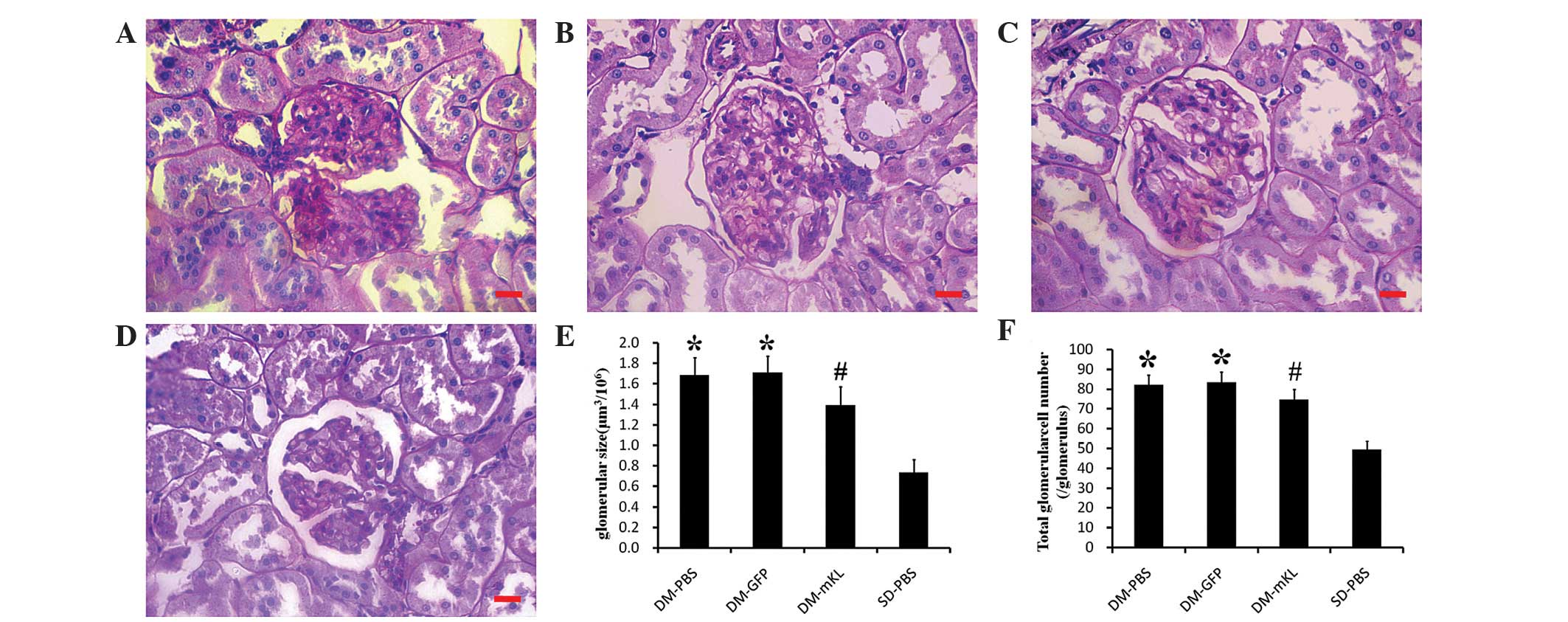 | Figure 1Representative microscopic images of
the kidney tissues stained with periodic acid-Schiff
(magnification, x400). The kidneys were obtained from DM rats
treated with (A) PBS (DM-PBS group), (B) GFP (DM-GFP) and (C) mKL
(DM-mKL). (D) SD rats were treated with PBS (SD-PBS; control).
Scale bar=25 μm. (E) Glomerular size and the (F) total
glomerular cell number were determined in each group, by examining
350–450 glomeruli per group. The data are expressed as the mean ±
standard error of the mean. *P<0.05, vs. SD-PBS
group; and #P<0.05, vs. DM-PBS group. n=7 in each
group. DM, diabetic; PBS, phosphate-buffered saline; GFP, green
fluorescent protein; mKL, mouse klotho; SD, Sprague-Dawley. |
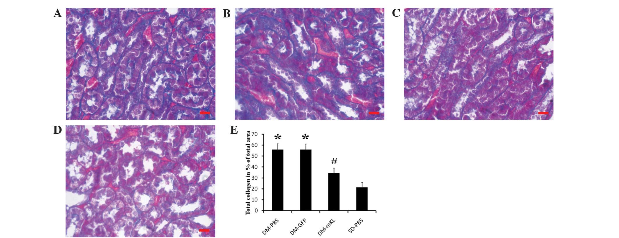 | Figure 2Representative microscopic images
(magnification, x400) of kidney tissues stained with Masson’s
trichrome, obtained from DM rats treated with (A) PBS (DM-PBS
group), (B) GFP (DM-GFP group) and (C) mKL (DM-mKL group). (D) SD
rats were treated with PBS (SD-PBS group). Scale bar=25 μm.
(E) Calculated collagen volume fraction. The data are expressed as
the mean ± standard error of the mean. *P<0.05, vs.
SD-PBS group; and #P<0.05, vs. DM-PBS group. n=7 in
each group. DM, diabetic; PBS, phosphate-buffered saline; GFP,
green fluorescent protein; mKL, mouse klotho; SD,
Sprague-Dawley. |
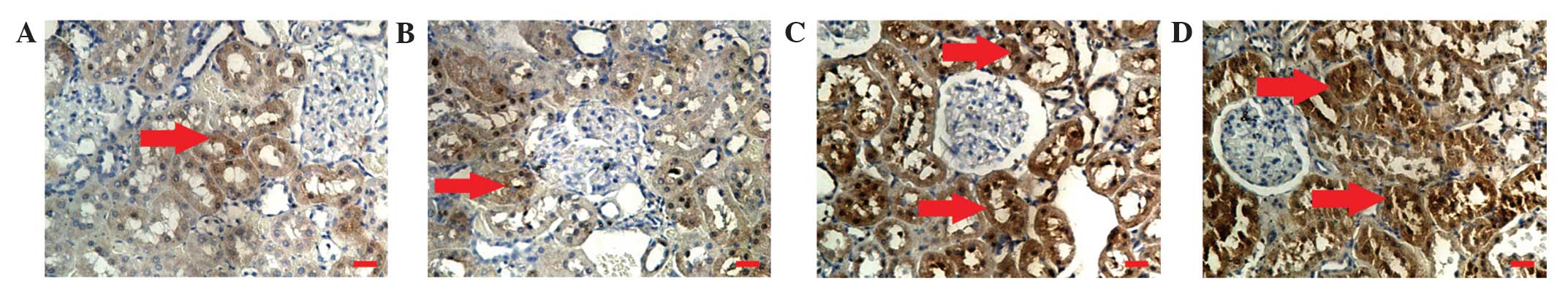
Klotho gene delivery increases the
protein expression of klotho, but decreases the protein expression
levels of FN and VIM in the kidneys of DM rats
In order to investigate the effect of klotho gene
delivery on the protein expression levels of klotho, FN and VIM,
immunohistochemical analysis was performed. The results revealed
that the protein expression of klotho (brown staining) was
localized in the renal tubule epithelial cells (Fig. 3A–D). In addition, the renal
expression of klotho was lower in the DM rats compared with the SD
rats. However, klotho gene delivery increased the protein
expression of klotho (dark brown staining) in the renal tubule
epithelial cells of the DM rats (Fig.
3C). In addition, immunohistochemical analysis indicated that
the protein expression of FN (brown staining) was localized in the
extracellular membrane of the renal tubule epithelial cells
(Fig. 4A–D). The renal expression
of FN was found to be higher in the DM rats compared with the SD
rats. However, the expression of FN in the DM rats treated with
rAAV.mKL was lower compared with the DM rats treated with PBS or
rAAV.GFP (Fig. 4E). Similar to FN,
the protein expression of VIM (brown staining) was also localized
in the extracellular membrane of the renal tubule epithelial cells
(Fig. 5A–D). The renal expression
of VIM was found to be higher in the DM rats compared with the SD
rats. However, the expression of VIM in the DM rats treated with
rAAV.mKL was lower compared with the DM rats treated with PBS or
rAAV.GFP (Fig. 5E). These results
indicated that klotho gene delivery increased the protein
expression of klotho, but decreased the protein expression levels
of FN and VIM in the kidneys of DM rats.
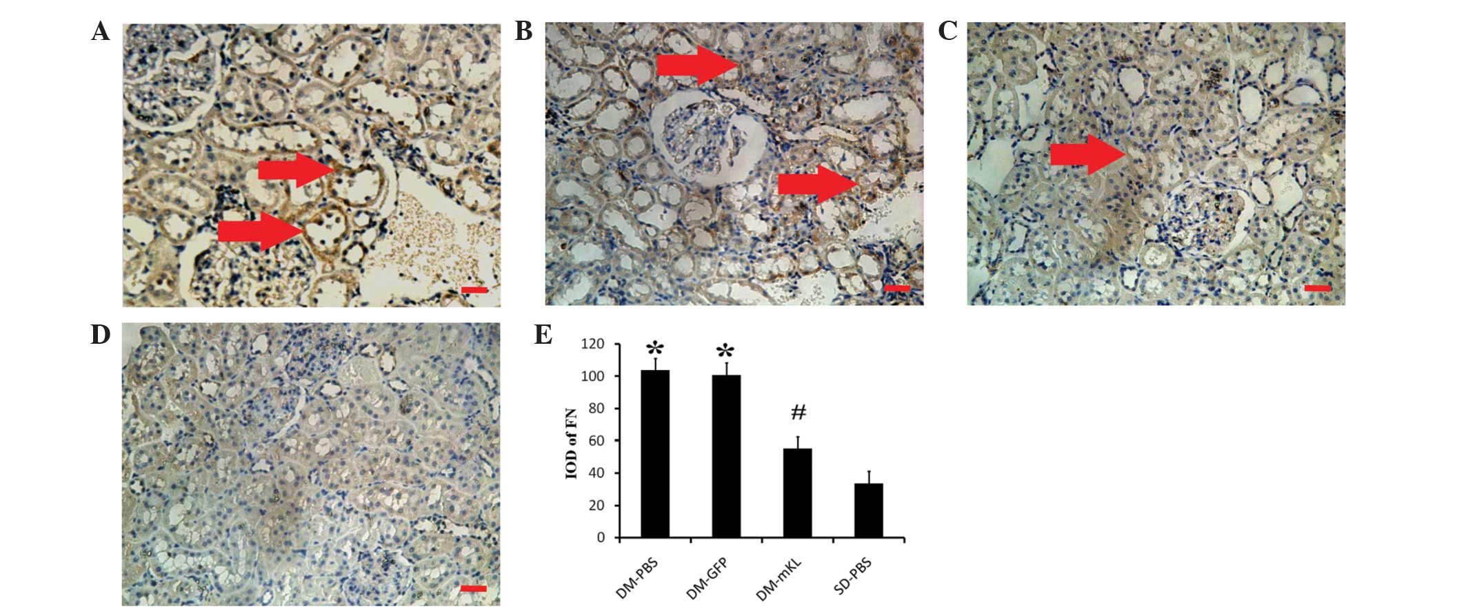 | Figure 4Immunohistochemical analysis
(magnification, x200) of the expression of FN in kidney tissues
obtained from DM rats treated with (A) PBS (DM-PBS group), (B) GFP
(DM-GFP group) and (C) mKL (DM-mKL group), as well as (D) SD rats
treated with PBS (SD-PBS group). Scale bar=25 μm. The arrows
indicate the protein expression of FN in the extracellular membrane
of the renal tubule epithelial cells (brown staining). (E)
Quantitative analysis of the protein expression of FN. The data are
expressed as the mean ± standard error of the mean.
*P<0.05, vs. SD-PBS group; and #P<0.05,
vs. DM-PBS group. n=7 in each group. FN, fibronectin; DM, diabetic;
PBS, phosphate-buffered saline; GFP, green fluorescent protein;
mKL, mouse klotho; SD, Sprague-Dawley. |
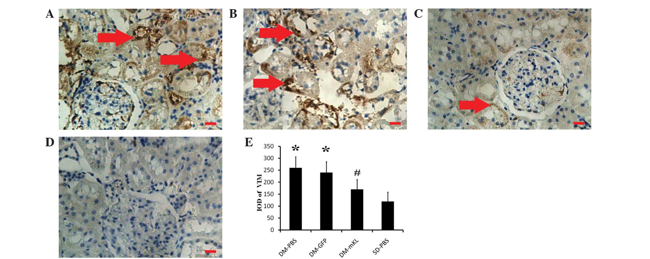 | Figure 5Immunohistochemical analysis
(magnification, x200) of the expression of VIM in kidney tissues
obtained from DM rats treated with (A) PBS (DM-PBS group), (B) GFP
(DM-GFP group) and (C) mKL (DM-mKL group), as well as (D) SD rats
treated with PBS (SD-PBS group). Scale bar=25 μm. The arrows
indicate the protein expression of VIM in the extracellular
membrane of the renal tubule epithelial cells (brown staining). (E)
Quantitative analysis of the protein expression of VIM. The data
are expressed as the mean ± standard error of the mean.
*P<0.05, vs. SD-PBS group; and #P<0.05,
vs. DM-PBS group. n=7 for each group. VIM, vimentin; DM, diabetic;
PBS, phosphate-buffered saline; GFP, green fluorescent protein;
mKL, mouse klotho; SD, Sprague-Dawley. |
Klotho gene delivery suppresses ROCK
activation in DM rats
In order to investigate the activation of ROCK and
the protein expression of p-MYPT1 (Thr696/Thr853), RT-qPCR and
western blotting were performed. The mRNA expression level of ROCKI
was analyzed in the kidney tissues of each experimental group and
was found to be significantly higher in the DM rats compared with
the SD rats (P<0.05). However, following treatment with
rAAV.mKL, the mRNA expression of ROCKI was significantly suppressed
compared with the PBS- or rAAV.GFP-treated DM rats (P<0.05;
Fig. 6A and B). Consistent with
the semi-quantitative RT-PCR results, western blotting revealed
that p-MYPT1 (Thr696/853) was significantly elevated in the DM rats
(Fig. 6C) and was suppressed
following treatment with rAAV.mKL (P<0.05; Fig. 6D–E). These results indicated that
klotho gene delivery suppressed the mRNA expression of ROCKI, and
thus, the activation of the ROCK signaling pathway in the DM
rats.
 | Figure 6mRNA expression of ROCKI in kidney
tissues (A) determined by semi-quantitative reverse
transcription-polymerase chain reaction and (B) obtained by
quantitative analysis. Protein expression levels of p-MYPT1, (C)
determined by western blotting, for (D) p-MYPT1 (Thr696) and (E)
p-MYPT1 (Thr853). The data are expressed as the mean ± standard
error of the mean. *P<0.05, vs. SD-PBS group; and
#P<0.05, vs. DM-PBS group. n=7 for each group.
Groups: M, DL 2000 DNA Marker; 1, DM-PBS; 2, DM-GFP; 3, DM-mKL; and
4, SD-PBS. ROCKI, Rho-associated coiled-coil kinase 1; p-MYPT1,
phosphorylated myosin phosphatase target subunit 1; DM, diabetic;
PBS, phosphate-buffered saline; GFP, green fluorescent protein;
mKL, mouse klotho; SD, Sprague-Dawley. |
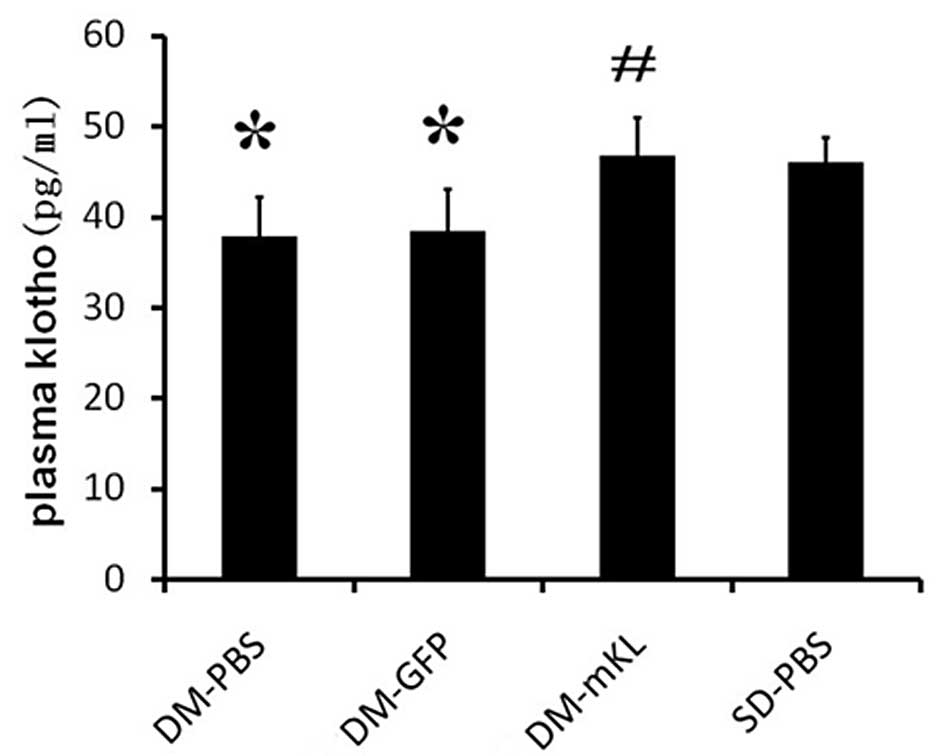
Klotho gene delivery upregulates plasma
levels of the klotho protein in DM rats
In order to determine the plasma levels of klotho
protein, ELISA was performed. The data indicated that the levels of
plasma klotho were lower in the DM rats compared with the SD rats.
Klotho gene delivery increased the plasma klotho levels in the
rAAV.mKL-treated rats compared with the DM rats treated with PBS
and rAAV.GFP (Fig. 7). Therefore,
klotho gene delivery was found to upregulate the levels of klotho
protein in the plasma of DM rats.
rAAV results in long-term mRNA expression
of GFP and mKL in the rat kidneys
To visualize the mRNA expression levels of GFP and
mKL in the rat kidneys, fluorescence microscopy was used. In the
DM-GFP and DM-mKL groups, GFP expression was detected (Fig. 8B and C), indicating that GFP was
expressed in the rats 12 weeks after the delivery of rAAV.GFP and
rAAV.mKL. By contrast, no GFP expression was detected in the
kidneys of the DM-PBS or SD-PBS groups. In addition, mouse klotho
was highly expressed in the kidney of the rAAV.mKL-treated rats 12
weeks after gene delivery (Fig.
8E), indicating the successful delivery of the mouse klotho
gene. However, no mouse klotho mRNA was observed in any other
group. These results demonstrated that rAAV resulted in long-term
transgene expression.
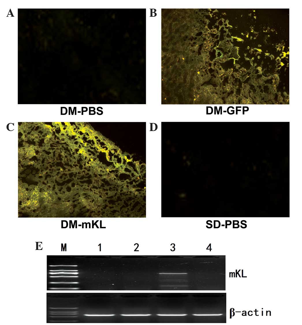 | Figure 8mRNA expression of GFP and mKL in
kidney tissues. Fluorescent photomicrographs of GFP expression in
the kidneys of DM rats treated with (A) PBS, (B) rAAV.GFP or (C)
rAAV.mKL and (D) SD rats treated with PBS. (E) Semi-quantitative
reverse transcription-polymerase chain reaction detection of the
mRNA expression of mouse klotho in the kidneys of rats treated with
rAAV.mKL using a mouse klotho-specific primer. Groups: M, DL 2000
DNA Marker; 1, DM-PBS; 2, DM-GFP; 3, DM-mKL; and 4, SD-PBS. DM,
diabetic; PBS, phosphate-buffered saline; GFP, green fluorescent
protein; mKL, mouse klotho; SD, Sprague-Dawley; AAV,
adeno-associated virus. |
Discussion
The main characteristic pathological features of
diabetic kidney damage are early glomerular hypertrophy, glomerular
capillary basement membrane thickening and gradual accumulation of
ECM in the mesangial and renal tubular interstitial area (26). The inherent structure of the kidney
is then damaged and eventually results in glomerular sclerosis and
interstitial fibrosis that leads to kidney failure. In the present
study, DM rats were transfected with exogenous rAAV.mKL and the
klotho gene was found to have no effect on body weight or fasting
glucose level. However, the klotho gene was able to inhibit kidney
hypertrophy and fibrosis, delay the pathological changes of
diabetic kidney, including glomerular hypertrophy, glomerular
capillary basement membrane thickening, mesangial and renal tubular
interstitial area collagen fibrosis, and reduce the kidney
hypertrophy index of the DM rats. Therefore, increased klotho gene
expression was shown to have a protective effect on the kidney of
DM rats.
Renal hypertrophy is an early characteristic
pathological change occurring in diabetic nephropathy and is
closely associated with late renal fibrosis (27). The mammalian target of rapamycin
(mTOR) pathway is the most important downstream signaling pathway
of compensatory renal hypertrophy following nephrectomy. Rapamycin
is an mTOR inhibitor, which inhibits compensatory tubular cell
hypertrophy. Furthermore, compensatory renal hypertrophy is
suppressed in ribosomal protein S6 knock-out mice, which do not
exhibit compensatory renal growth or tubular cell proliferation
following nephrectomy (28,29).
Therefore, mTOR is essential in mediating increased RNA and protein
synthesis in compensatory renal hypertrophy (29). The increase of reactive oxygen
species caused by NAD(P)H oxidase activation is important in renal
glomerular hypertrophy signal transduction and cell proliferation
following unilateral nephrectomy (30). Nagasu et al (6) investigated the compensatory renal
hypertrophy induced by unilateral nephrectomy in a klotho
transgenic mouse model. The results revealed that overexpression of
the klotho gene restrained the activation of mTOR and the increase
in reactive oxygen species resulting from NAD(P) H oxidase
activation and, thus, reduced renal hypertrophy (6). These effects are achieved by the
inhibition of the insulin-like growth factor-1 (IGF-1) signaling
pathway by the klotho gene. However, whether the overexpression of
the klotho gene inhibits diabetes-induced renal hypertrophy through
inhibition of the IGF-1 signaling pathway remains to be
elucidated.
Renal fibrosis, including glomerular sclerosis and
renal interstitial fibrosis, is the final common pathway in the
majority of cases of end-stage renal disease. FN, an adhesion
molecule, is an important component of ECM and its expression can
be increased with a prolonged duration of DM. In the glomerular
mesangial and renal tubular interstitial areas, excessive ECM
deposition results in glomerular sclerosis and renal interstitial
fibrosis (31,32). The present study demonstrated that
exogenous rAAV.mKL transfection inhibited the expression of FN and
reduced the generation and accumulation of ECM in the kidneys of
the DM rats.
EMT is a process by which fully differentiated
epithelial cells undergo transition to a fibroblast phenotype
(33). The migration of epithelial
cells from the tubular structure into the interstitium, where
matrix is produced (including collagen types I and IV and FN),
contributes to the formation of renal fibrosis (34). Previous studies have demonstrated
that, in addition to renal tubular epithelial cells, sertoli and
endothelial cells also undergo phenotypic transformations, which
are termed podocyte- and endothelial-mesenchymal transitions,
respectively (35,36). VIM is an intermediate filament
constituent of the cytoskeleton of mesenchymal cells. The
expression of VIM is associated with the cell phenotype, including
shape, motility and adhesion. VIM is an EMT-specific marker and its
expression is closely associated with the degree of EMT (37). Previous studies have revealed that
a reduced expression level of klotho in the kidneys aggravates
renal interstitial fibrosis in mice induced by unilateral ureteral
obstruction (8). In addition, the
secreted klotho suppresses the TGF-β1-induced EMT response in
cultured cells, including decreased expression levels of epithelial
markers, increased expression levels of mesenchymal markers and/or
increased cell migration. Furthermore, secreted klotho inhibits Wnt
and IGF-1 signaling, which promotes EMT (9). The present study demonstrated that
exogenous rAAV.mKL transfection in DM rats decreased the protein
expression of VIM and reduced the pathological changes in the
kidney and collagen fiber formation. Therefore, as discussed
earlier, the klotho gene may reduce the progression of
diabetes-induced renal fibrosis by inhibiting ECM and EMT.
Previous in vivo and in vitro
experiments have demonstrated that the activation of ROCK is
involved in the occurrence and development of diabetic nephropathy
(18). Kolavennu et al and
Peng et al identified that the expression of FN in mesangial
cells cultured in a high glucose environment was increased upon
activation of the ROCKI signaling pathway (19,20).
In addition, Komers et al observed that activation of the
ROCK signaling pathway in kidneys with diabetic nephropathy
increased the expression of VIM and aggravated renal fibrosis;
however, ROCK-specific inhibitors reduced the process of renal
fibrosis (21). Furthermore, a
previous study demonstrated that administering fasudil in rats
exhibiting diabetic cardiomyopathy inhibited the mRNA transcription
and protein activity of ROCKI and thus, reduced diabetic myocardial
fibrosis, oxidative stress and the apoptosis of myocardial cells
(38). The results of the present
study demonstrated that increased expression of klotho gene in the
kidneys of DM rats inhibited the protein expression of FN and VIM,
which may be associated with the inhibition of the mRNA expression
and protein activity of ROCKI.
A previous study revealed that RhoA/ROCK signaling
pathway activation is closely associated with the expression of
klotho (39). However, the
mechanism underlying the regulation of the mRNA transcription and
protein activity ROCKI by klotho remains unclear. The increased
mRNA transcription and protein activity of ROCK have been
associated with the remnant lipoprotein and inflammatory factors,
including angiotensin II and interleukin-1, which stimulate the
protein kinase C/nuclear factor (NF)-κB pathway (40,41).
In addition, Zhao et al identified that increased exogenous
klotho protein levels in db/db diabetic mice inhibited NF-κB
activity (42). Therefore, the
present study hypothesized that: i) Klotho may inhibit the mRNA
transcription and protein activity of ROCKI, possibly by inhibiting
NF-κB activity; ii) klotho may produce two proteins, including the
membrane and secretion klotho proteins. Membrane klotho protein
functions as an obligatory co-receptor for fibroblast growth factor
23, whereas the function of secretion klotho protein can vary with
the circulation to affect various signal transduction pathways
(43–45). However, further studies are
required to elucidate whether the membrane and secretion klotho
proteins have associated sites for direct binding with the small
GTPase protein, RhoA, which indirectly affects the activity of
ROCK.
Several studies have demonstrated that the gene and
protein expression levels of klotho are serially declined in
chronic renal failure patients and diabetic rats (4,5,46).
The present study demonstrated that transfection with exogenous
rAAV.mKL stabilized the expression of klotho for at least 12 weeks
and upregulated the protein expression of klotho in the serum of DM
rats. These results provide a basis for understanding the mechanism
by which klotho functions in diabetes and associated diseases and
may be helpful in the development of a novel long-term treatment
for age-related diseases, including diabetes.
Klotho protein is unique since it inhibits four
signaling pathways simultaneously, offering a major advantage over
numerous individual inhibitors in clinical and preclinical
development, including IGF-1 receptor antibodies, tyrosine kinase
(47) and Wnt signaling inhibitors
(48), TGF-β1 neutralizing
antibodies, soluble TGF-βR2, TGF-β receptor kinase inhibitors
(49,50) and ROCK signaling inhibitors. In
addition, klotho therapy is considered to be safe since
overexpression of klotho extends the lifespan of mice (3). Therefore, klotho protein may be a
novel therapeutic agent for the treatment of tissue hypertrophy and
fibrosis with a unique mechanism of action.
In conclusion, the present study demonstrated that
exogenous rAAV.mKL transfection in DM rats upregulated the
expression of klotho and delayed the progression of renal
hypertrophy and fibrosis induced by diabetes. To a certain extent,
these results may be due to klotho suppressing the ROCK signaling
pathway. However, further studies are required to understand how
klotho affects the mRNA transcription and protein activity of
ROCKI.
Acknowledgments
This study was supported by grants from the National
Natural Science Foundation of China (no. 30672212), the Medical
Science Research Program of Chongqing Municipal Health Bureau (no.
2010-2-079), the Science Research Project of Creative Foundation of
Chongqing Medical University (no. CX0507) and the National Key
Clinical Specialties Construction Program of China [no. (2013)544].
This study was conducted at the Chongqing Key Laboratory of
Ophthalmology and Experimental Research Center of The First
Affiliated Hospital of Chongqing Medical University (Chongqing,
China).
References
|
1
|
Kuro-o M, Matsumura Y, Aizawa H, et al:
Mutation of the mouse klotho gene leads to a syndrome resembling
ageing. Nature. 390:45–51. 1997. View
Article : Google Scholar : PubMed/NCBI
|
|
2
|
Masuda H, Chikuda H, Suga T, Kawaguchi H
and Kuro-o M: Regulation of multiple ageing-like phenotypes by
inducible klotho gene expression in klotho mutant mice. Mech Ageing
Dev. 126:1274–1283. 2005. View Article : Google Scholar : PubMed/NCBI
|
|
3
|
Kurosu H, Yamamoto M, Clark JD, et al:
Suppression of aging in mice by the hormone Klotho. Science.
309:1829–1833. 2005. View Article : Google Scholar : PubMed/NCBI
|
|
4
|
Ishizaka N, Matsuzaki G, Saito K, Furuta
K, Mori I and Nagai R: Downregulation of klotho gene expression in
streptozotocin-induced diabetic rats. Geriatr Gerontol Int.
7:285–292. 2007. View Article : Google Scholar
|
|
5
|
Cheng MF, Chen LJ and Cheng JT: Decrease
of Klotho in the kidney of streptozotocin-induced diabetic rats. J
Biomed Biotechnol. 2010:5138532012.
|
|
6
|
Nagasu H, Satoh M, Kuwabara A, et al:
Overexpression of klotho protein modulates uninephrectomy-induced
compensatory renal hypertrophy by suppressing IGF-I signals.
Biochem Biophys Res Commun. 407:39–43. 2011. View Article : Google Scholar : PubMed/NCBI
|
|
7
|
Haruna Y, Kashihara N, Satoh M, et al:
Amelioration of progressive renal injury by genetic manipulation of
Klotho gene. Proc Natl Acad Sci USA. 104:2331–2336. 2007.
View Article : Google Scholar : PubMed/NCBI
|
|
8
|
Sugiura H, Yoshida T, Shiohira S, et al:
Reduced klotho expression level in kidney aggravates renal
interstitial fibrosis. Am J Physiol Renal Physiol. 302:1252–1264.
2012. View Article : Google Scholar
|
|
9
|
Doi S, Zou Y, Togao O, et al: Klotho
inhibits transforming growth factor-β1 (TGF-β1) signaling and
suppresses renal fibrosis and cancer metastasis in mice. J Biol
Chem. 286:8655–8665. 2011. View Article : Google Scholar : PubMed/NCBI
|
|
10
|
Satoh M, Nagasu H, Morita Y, Yamaguchi TP,
Kanwar YS and Kashihara N: Klotho protects against mouse renal
fibrosis by inhibiting Wnt signaling. Am J Physiol Renal Physiol.
303:F1641–F1651. 2012. View Article : Google Scholar : PubMed/NCBI
|
|
11
|
Shimokawa H and Takeshita A: Rho-kinase is
an important therapeutic target in cardiovascular medicine.
Arterioscler Thromb Vasc Biol. 25:1767–1775. 2005. View Article : Google Scholar : PubMed/NCBI
|
|
12
|
Uehata M, Ishizaki T, Satoh H, et al:
Calcium sensitization of smooth muscle mediated by a Rho-associated
protein kinase in hypertension. Nature. 389:990–994. 1997.
View Article : Google Scholar : PubMed/NCBI
|
|
13
|
Kikuchi Y, Yamada M, Imakiire T, et al: A
Rho-kinase inhibitor, fasudil, prevents development of diabetes and
nephropathy in insulin-resistant diabetic rats. J Endocrinol.
192:595–603. 2007. View Article : Google Scholar : PubMed/NCBI
|
|
14
|
Kawamura H, Yokote K, Asaumi S, et al:
High glucose-induced upregulation of osteopontin is mediated via
Rho/Rho kinase pathway in cultured rat aortic smooth muscle cells.
Arterioscler Thromb Vasc Biol. 24:276–281. 2004. View Article : Google Scholar
|
|
15
|
Reiniger N, Lau K, McCalla D, et al:
Deletion of the receptor for advanced glycation end products
reduces glomerulosclerosis and preserves renal function in the
diabetic OVE26 mouse. Diabetes. 59:2043–2054. 2010. View Article : Google Scholar : PubMed/NCBI
|
|
16
|
Zhang Y, Peng F, Gao B, et al: Mechanical
strain-induced RhoA activation requires NADPH oxidase-mediated ROS
generation in caveolae. Antioxid Redox Signal. 13:959–973. 2010.
View Article : Google Scholar : PubMed/NCBI
|
|
17
|
Seibold S, Schürle D, Heinloth A, et al:
Oxidized LDL induces proliferation and hypertrophy in human
umbilical vein endothelial cells via regulation of p27Kip1
expression: role of RhoA. J Am Soc Nephrol. 15:3026–3034. 2004.
View Article : Google Scholar : PubMed/NCBI
|
|
18
|
Komers R: Rho kinase inhibition in
diabetic nephropathy. Curr Opin Nephrol Hypertens. 20:77–83. 2011.
View Article : Google Scholar
|
|
19
|
Kolavennu V, Zeng L, Peng H, Wang Y and
Danesh FR: Targeting of RhoA/ROCK signaling ameliorates progression
of diabetic nephropathy independent of glucose control. Diabetes.
57:714–723. 2008. View Article : Google Scholar
|
|
20
|
Peng F, Wu D, Gao B, Ingram AJ, Zhang B,
Chorneyko K, McKenzie R and Krepinsky JC: RhoA/Rho-kinase
contribute to the pathogenesis of diabetic renal disease. Diabetes.
57:1683–1692. 2008. View Article : Google Scholar : PubMed/NCBI
|
|
21
|
Komers R, Oyama TT, Beard DR, Tikellis C,
Xu B, Lotspeich DF and Anderson S: Rho kinase inhibition protects
kidneys from diabetic nephropathy without reducing blood pressure.
Kidney Int. 79:432–442. 2011. View Article : Google Scholar
|
|
22
|
Patel S, Takagi KI, Suzuki J, Imaizumi A,
Kimura T, Mason RM, Kamimura T and Zhang Z: RhoGTPase activation is
a key step in renal epithelial mesenchymal transdifferentiation. J
Am Soc Nephrol. 16:1977–1984. 2005. View Article : Google Scholar : PubMed/NCBI
|
|
23
|
Rodrigues-Díez R, Carvajal-González G,
Sánchez-López E, Rodríguez-Vita J, Rodrigues Díez R, Selgas R,
Ortiz A, Egido J, Mezzano S and Ruiz-Ortega M: Pharmacological
modulation of epithelial mesenchymal transition caused by
angiotensin II. Role of ROCK and MAPK pathways. Pharm Res.
25:2447–2461. 2008. View Article : Google Scholar : PubMed/NCBI
|
|
24
|
Lin G, Craig GP, Zhang L, Yuen VG, Allard
M, McNeill JH and MacLeod KM: Acute inhibition of Rho-kinase
improves cardiac contractile function in streptozotocin-diabetic
rats. Cardiovasc Res. 75:51–58. 2007. View Article : Google Scholar : PubMed/NCBI
|
|
25
|
Sharma V, Parsons H, Allard MF and McNeill
JH: Metoprolol increases the expression of β3-adrenoceptors in the
diabetic heart: effects on nitric oxide signaling and forkhead
transcription factor-3. Eur J Pharmacol. 595:44–51. 2008.
View Article : Google Scholar : PubMed/NCBI
|
|
26
|
Ziyadeh EF: The extracellular matrix in
diabetic nephropathy. Am J Kidney Dis. 22:736–744. 1993. View Article : Google Scholar : PubMed/NCBI
|
|
27
|
Wolf G and Ziyadeh FN: Molecular
mechanisms of diabetic renal hypertrophy. Kidney Int. 56:393–405.
1999. View Article : Google Scholar : PubMed/NCBI
|
|
28
|
Chen JK, Chen J, Neilson EG and Harris RC:
Role of mammalian target of rapamycin signaling in compensatory
renal hypertrophy. J Am Soc Nephrol. 16:1384–1391. 2005. View Article : Google Scholar : PubMed/NCBI
|
|
29
|
Chen JK, Chen J, Thomas G, Kozma SC and
Harris RC: S6 kinase 1 knockout inhibits uninephrectomy- or
diabetes-induced renal hypertrophy. Am J Physiol Renal Physiol.
297:585–593. 2009. View Article : Google Scholar
|
|
30
|
Ozeki M, Nagasu H, Satoh M, Namikoshi T,
Haruna Y, Tomita N, Sasaki T and Kashihara N: Reactive oxygen
species mediate compensatory glomerular hypertrophy in rat
uninephrectomized kidney. J Physiol Sci. 59:397–373. 2009.
View Article : Google Scholar : PubMed/NCBI
|
|
31
|
Mauer SM, Lane P, Zhu D, Fioretto P and
Steffes MW: Renal structure and function in insulin-dependent
diabetes mellitus in man. J Hypertens Suppl. 10:S17–S20. 1992.
View Article : Google Scholar : PubMed/NCBI
|
|
32
|
Van Vliet A, Baelde HJ, Vleming LJ, de
Heer E and Bruijn JA: Distribution of fibronectin isoforms in human
renal disease. J Pathol. 193:256–262. 2001. View Article : Google Scholar : PubMed/NCBI
|
|
33
|
Carew RW, Wang B and Kantharidis P: The
role of EMT in renal fibrosis. Cell Tissue Res. 347:103–116. 2012.
View Article : Google Scholar
|
|
34
|
Kalluri R and Neilson EG:
Epithelial-mesenchymal transition and its implications for
fibrosis. J Clin Invest. 112:1776–1784. 2003. View Article : Google Scholar : PubMed/NCBI
|
|
35
|
Moeller MJ, Soofi A, Hartmann I, Le Hir M,
Wiggins R, Kriz W and Holzman LB: Podocytes populate cellular
crescents in a murine model of inflammatory glomerulonephritis. J
Am Soc Nephrol. 15:61–67. 2004. View Article : Google Scholar
|
|
36
|
Zeisberg EM, Potenta SE, Sugimoto H,
Zeisberg M and Kalluri R: Fibroblasts in kidney fibrosis emerge via
endothelial-to-mesenchymal transition. J Am Soc Nephrol.
19:2282–2287. 2008. View Article : Google Scholar : PubMed/NCBI
|
|
37
|
Galichon P and Hertig A: Epithelial to
mesenchymal transition as a biomarker in renal fibrosis: are we
ready for the bedside? Fibrogenesis Tissue Repair. 4:112011.
View Article : Google Scholar : PubMed/NCBI
|
|
38
|
Guan SJ, Ma ZH, Wu YL, Zhang JP, Liang F,
Weiss JW, Guo QY, Wang JY, Ji ES and Chu L: Long-term
administration of fasudil improves cardiomyopathy in
streptozotocin-induced diabetic rats. Food Chem Toxicol.
50:1874–1882. 2012. View Article : Google Scholar : PubMed/NCBI
|
|
39
|
Narumiya H, Sasaki S, Kuwahara N, et al:
HMG-CoA reductase inhibitors up-regulate anti-aging klotho mRNA via
RhoA inactivation in IMCD3 cells. Cardiovasc Res. 64:331–336. 2004.
View Article : Google Scholar : PubMed/NCBI
|
|
40
|
Oi K, Shimokawa H, Hiroki J, et al:
Remnant lipoproteins from patients with sudden cardiac death
enhance coronary vasospastic activity through upregulation of
Rho-kinase. Arterioscler Thromb Vasc Biol. 24:918–922. 2004.
View Article : Google Scholar : PubMed/NCBI
|
|
41
|
Hiroki J, Shimokawa H, Higashi M, et al:
Inflammatory stimuli upregulate Rho-kinase in human coronary
vascular smooth muscle cells. J Mol Cell Cardiol. 37:537–546. 2004.
View Article : Google Scholar : PubMed/NCBI
|
|
42
|
Zhao Y, Banerjee S, Dey N, et al: Klotho
depletion contributes to increased inflammation in kidney of the
db/db mouse model of diabetes via RelA (serine) 536
phosphorylation. Diabetes. 60:1907–1916. 2011. View Article : Google Scholar : PubMed/NCBI
|
|
43
|
Urakawa I, Yamazaki Y, Shimada T, et al:
Klotho converts canonical FGF receptor into a specific receptor for
FGF23. Nature. 444:770–774. 2006. View Article : Google Scholar : PubMed/NCBI
|
|
44
|
Wang Y and Sun Z: Current understanding of
klotho. Ageing Res Rev. 8:43–51. 2009. View Article : Google Scholar :
|
|
45
|
Hu MC, Kuro-o M and Moe OW: Renal and
extrarenal actions of Klotho. Semin Nephrol. 33:118–129. 2013.
View Article : Google Scholar : PubMed/NCBI
|
|
46
|
Koh N, Fujimori T, Nishiguchi S, Tamori A
and Shiomi S: Severely reduced production of klotho in human
chronic renal failure kidney. Biochem Biophys Res Commun.
280:1015–1020. 2001. View Article : Google Scholar : PubMed/NCBI
|
|
47
|
Weroha SJ and Haluska P: IGF-1 Receptor
Inhibitors in Clinical Trials-Early Lessons. J Mammary Gland Biol
Neoplasia. 13:471–483. 2008. View Article : Google Scholar : PubMed/NCBI
|
|
48
|
Huang SM, Mishina YM, Liu S, et al:
Tankyrase inhibition stabilizes axin and antagonizes Wnt
signalling. Nature. 461:614–620. 2009. View Article : Google Scholar : PubMed/NCBI
|
|
49
|
Yingling JM, Blanchard KL and Sawyer JS:
Development of TGF-β signalling inhibitors for cancer therapy. Nat
Rev Drug Discov. 3:1011–1022. 2004. View Article : Google Scholar : PubMed/NCBI
|
|
50
|
Prud’homme GJ: Pathobiology of
transforming growth factor β in cancer, fibrosis and immunologic
disease, and therapeutic considerations. Lab Invest. 7:1077–1091.
2007. View Article : Google Scholar
|