Introduction
Colorectal cancer is the third most common type of
cancer and remains one of the leading causes of cancer-associated
mortality worldwide (1). Despite
advances in the treatment of cancer, treatment failure and tumor
recurrence remain a problem. Previously, it was suggested that all
neoplastic cells within a tumor are potentially tumorigenic.
However, according to subsequent studies, a small subset of cancer
initiating cells, termed cancer stem cells (CSCs) have been
suggested as the factor involved in drug resistance and
tumorigenesis (2,3). The presence of CSCs, their stem-like
role and their implications in the treatment of cancer have been
well established in several types of cancer (4–8).
Previous studies have reported that there are two different methods
to isolate CSCs, based on the expression of CD133 and the Hoechst
33342 dye exclusion technique. In the latter, a small population of
cancer cells can be been identified at the side of the overal
population in fluorescence-activated cell sorting (FACS) analysis,
which exclude Hoechst 33342 dye and are, therefore, termed side
population (SP) cells. These SP cells share the characteristic
features of CSCs, including the expression of stem cells surface
markers, high level of tumorigenicity and differentiation potential
(9–12). Furthermore, the SP cells have high
expression levels of adenosine triphosphate-binding cassette (ABC)
transporters, enhanced DNA-repair capacity and resistance to
apoptosis, which leads to chemoresistance and tumor relapse
(13). Therefore, the isolation
and characterization of SP cells can assist in identifying and
targeting CSCs and, as a result, increase long-term disease-free
survival rates.
It was previously reported, based on the expression
of CD133, that isolated colon CSCs are highly resistant to
apoptosis, associated with the autocrine production of interleukin
(IL)-4 (14). Previous studies on
several types of cancer have demonstrated that IL-4 is involved in
resistance to apoptosis and increased expression of antiapoptotic
proteins (15). Therefore, the
present study was designed to identify colon CSCs, based on the
Hoecst 33342 dye exclusion technique. Furthermore, the sorted colon
CSCs were analyzed for CD133 positivity, drug resistance and the
expression of IL-4.
Materials and methods
Tissue collection and cell culture
Colon adenocarcinoma tissue samples were collected
from patients during colon resection (n=10, 20–60 years old, 5 male
and 5 female), according to standard ethical standards. The study
was approved by the ethics committee of The Second Affiliated
Hospital of Nanchang University (Nanchang, China) and consent was
obtained from all patients. The diagnosis of colon cancer was made
based on clinical, pathological and laboratory tests performed by
qualified physicians. Following isolation, the tissues were
diagnosed, based on their histological type and grade. The cancer
tissues were washed thoroughly in phosphate-buffered saline (PBS;
Sigma-Aldrich, St. Louis, MO, USA) solution, containing antibiotics
and were incubated overnight in Dulbecco's modified Eagle's medium
(DMEM)/F12 (Gibco Life Technologies, Carslbad, CA, USA) containing
penicillin (500 U/ml), streptomycin (500 g/ml), and amphotericin B
(1.25 03bcg/ml) (Gibco Life Technologies) at room
temperature. Enzymatic digestion was performed using collagenase
(1.5 mg/ml) (Gibco Life Technologies) and hyaluronidase (20
03bcg/ml; Sigma-Aldrich) in PBS for 1 hr at room
temperature. The cells were cultured in DMEM with 10% fetal bovine
serum (FBS; Sigma-Aldrich), supplemented with antibiotics, and
maintained in T-75 flasks at 37°C in a humidified 5% CO2
and 95% air atmosphere. At 90% confluence, the cells were removed
from the culture flask using trypsin-EDTA (0.25%: 53 mM EDTA;
Sigma-Aldrich) washed in distilled water, and the cells were
suspended in 10% DMEM. The number of cells were counted using a
hemocytometer (Z359629; Bright-Line; Sigma-Aldrich).
FACS analysis
The following experimental groups were included in
the present study: The control group, containing cells + Hoechst
33342 dye (n=5); and the drug-treated group, containing cells +
verapamil (Sigma-Aldrich) + Hoechst 33342 dye (n=5). The techniques
for the Hoechst 33342 dye labeling and immunofluorescence were
obtained from Dr Wanshan Li of the Department of Oral and
Maxillofacial Surgery, Chongqing Medical University (Chongqing,
China). The cells (~106 cells/ml of 10% DMEM) were
labeled with Hoechst 33342 stock (Sigma-Aldrich)-bis-benzimide (5
03bcl/ml), with either the dye alone, or in combination with
verapamil (0.8 03bcl/ml). The cells were resuspended in 500
03bcl Hank's balanced salt solution (HBBS; Sigma-Aldrich),
containing 10 mM HEPES for FACS analysis. Finally, the cells were
counterstained with 2 03bcg/ml propidium iodide (PI;
Sigma-Aldrich). The cells were sorted into SP and non-SP cells
using a flow cytom eter (Attune NxT; Life Technologies, Grand
Island, NY, USA), and the sorted cells were then cultured and
maintained in DMEM/F-12, supplemented with 10% FBS at room
temperature.
Immunocytochemistry
The sorted SP cells and non-SP cells were seeded in
35 mm culture plates (~100 03bcl) with 1 ml 10% DMEM.
Following incubation overnight, the cells were rinsed with PBS and
fixed in 4% paraformaldehyde (Sigma-Aldrich). Subsequently, the
cells were blocked with 1% bovine serum albumin (BSA;
Sigma-Aldrich)-Tris-buffered saline (TBS; Sigma-Aldrich) with RNase
(10 03bcl/1,000 03bcl 3% BSA-TBS). After 1 h
incubation at room temperature, the cells were rinsed with PBS and
were incubated with primary antibody against CD133 (cat no.
orb99113; polyclonal, rabbit; Biorbyt, San Francisco, CA, USA) in
1% BSA-TBS (1:100; 2 03bcl/200 03bcl), incubated
overnight at 40°C. The cells were then washed with 1X PBS, followed
by incubation with fluorescein isothiocyanate (FITC)-conjugated
secondary antibody (1:100 in 1% BSA-TBS; goat anti-rabbit
immunoglobulin G with alkaline phosphatase markers; cat. no.
sc-2043; Santa Cruz Biotechnology, Inc., Dallas, TX, USA), at room
temperature for 1 h. The cells were then washed again with PBS, and
PI was added (1 03bcl/200 03bcl PBS). The tumor
spheres formed were counterstained with Hoechst 33342 staining (200
03bcl for 15 min; Life Technologies) to visualize the
nuclei. The cells were then viewed under a confocal laser scanning
microscope (Leica TCS; Leica Microsystems, Inc., Buffalo Grove, IL,
USA). Image analysis and figures were prepared using Adobe
Photoshop CS6 (Adobe Systems, Inc., San Jose, CA, USA).
Cell resistance assay
The SP and non-SP cells were cultured in 96-well
plates at a concentration of 1×10³ cells/plate at 37°C.
After 24 h, 5-fluorouracil (5-FU; Sigma-Aldrich) was added to the
cultures to a final concentration of 10 03bcg/ml. The cells
were also treated with cisplatin (20 03bcmol/l;
Sigma-Aldrich), paclitaxel (2 03bcmol/l; Sigma-Aldrich) and
oxaliplatin (100 mM; Sigma-Aldrich). The plates were placed in a
hatch box for 48 h, following which, each well was supplemented
with 10 03bcl Cell Counting kit-8 (CCK-8) solution, and the
plates were incubated for 3 h at 37°C. The mean optical density
(OD) at 450nm was determined using a UV/Vis spectrophotometer
(UV-5800; Metash, Shanghai, China), and was represented as a graph.
The resistance of the cells in the two groups was calculated using
the following formula: Cell resistance rate (%) = (experimental
group OD450/control group OD450) × 100, as described previously
(16). Furthermore, the analysis
of cell death of colon cancer spheres was evaluated using orange
acridine/ethidium bromide staining (200 03bcl for 15 min;
Sigma-Aldrich), which was viewed under confocal microscopy (LSM
510; Carl Zeiss GmbH, Jena, Germany). Images were captured and
processed using Adobe Photoshop CS6 (Adobe Systems, Inc.).
Sphere formation assay
The sorted SP cells and non-SP cells were plated at
a density of 1,000 cells/ml, resuspended in tumor sphere medium,
containing a serum-free 1:1 mixture of Ham's F-12/DMEM
(Sigma-Aldrich), N2 supplement, 10 ng/ml human recombinant bFGF and
10 ng/ml epidermal growth factor (Sigma-Aldrich). The cells were
then cultured in ultra-low attachment plates for ~2 weeks ay 37°C.
The sorted SP and non-SP cells were seeded at a low density of 20
cells/L, and the number of generated spheres, measuring >100 lm,
were counted following 7 days of culture.
Biochemistry
The expression levels of proteins were analyzed
using western blot analysis. The proteins were extracted from the
SP and non-SP cells. Cell pellets were resuspended in ice-cold
NP-40 lysis buffer (50 mM Tris-HCl, pH 7.5, 150 mM NaCl, 1 mM EGTA,
1% NP-40) containing protease inhibitors., and the protein
concentrations were determined using a Bradford assay (Bio-Rad
Laboratories, Inc., Hercules, CA, USA). Following 10% sodium
dodecyl sulfate-poly acrylamide gel electrophoresis and transfer
onto nitrocellulose membranes (Bio-Rad Laboratories, Inc.), the
gels were incubated with the following primary antibodies: Rabbit
anti-human polyclonal ABC sub-family G member 2 (ABCG2; 1:500; cat.
no. AV43649; Sigma-Aldrich), rabbit polyclonal Bcl-2 (1:500; cat.
no. sc-492, Santa Cruz Biotechnology, Inc.) and rabbit polyclonal
IL-4 (1:500, cat. no. sc-7919, Santa Cruz Biotechnology, Inc.),
secondary antibody (goat anti-rabbit immunoglobulin G with alkaline
phosphatase markers; 1:1,000, cat. no. sc-2043; Santa Cruz
Biotechnology, Inc.) and chemiluminescence reagent. The blots were
detected and scanned using a densitometer (Bio-Rad GS-710; Bio-Rad
Laboratories, Inc.).
Statistical analysis
One-way analysis of variance and Student's t-test
were performed to determine significant differences between the
treatment and control groups using SAS statistical software (SAS
Institute Inc., Cary, NC, USA). P<0.05 was considered to
indicate a statistically significant difference.
Results
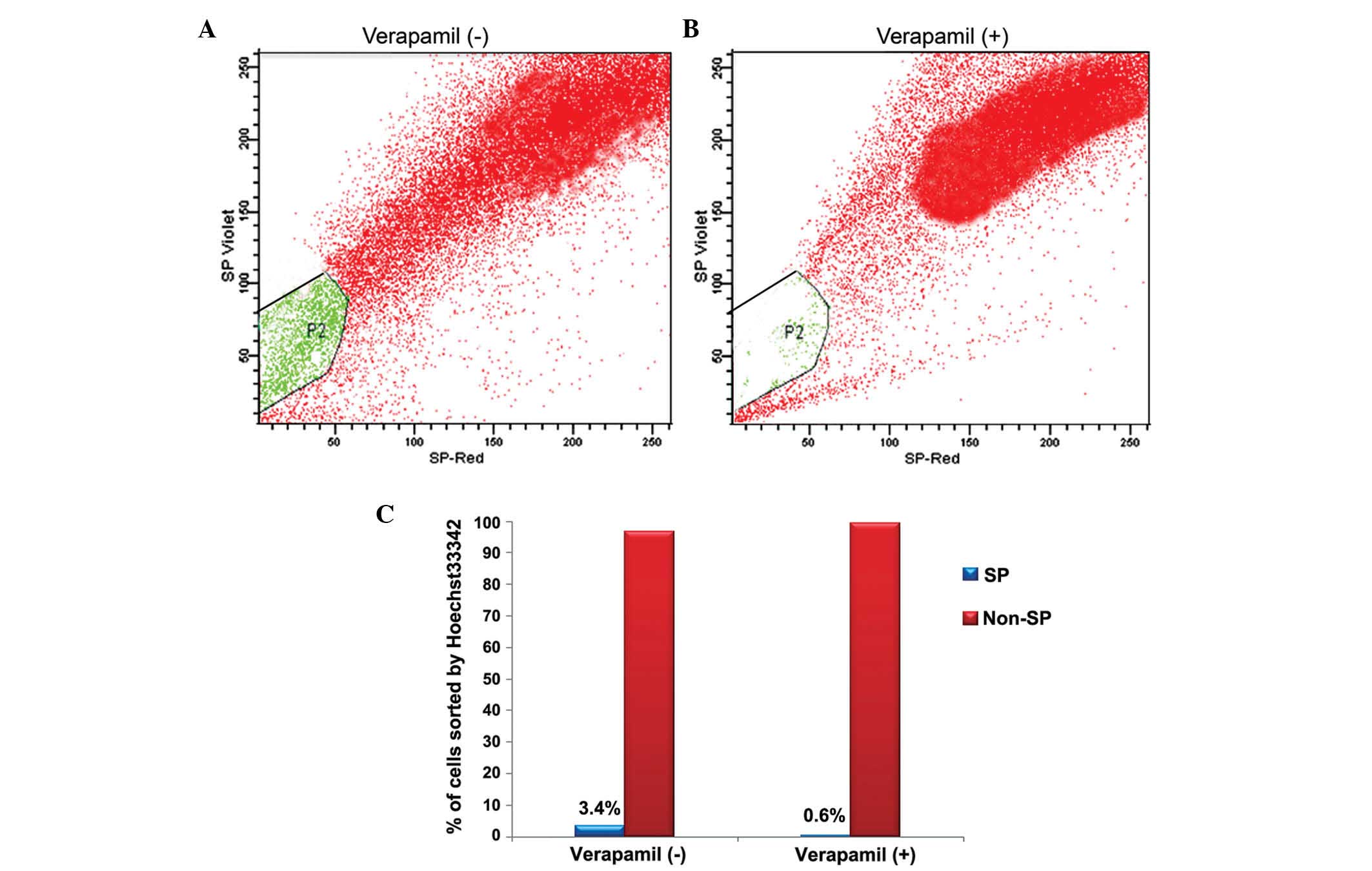
FACS analysis of SP cells containing
multidrug resistance transporter 1 (MDR1) using Hoechst 33342
The present study analyzed the colon cancer cells
for the presence of cancer stem-like SP cells using a Hoechst 33342
dye exclusion method. During FACS analysis, the live cells were
selected using PI staining, which was excluded by the dead cells
(P1-gated region of Fig. 1A and
B). The FACS analysis identified ~3.4% of the SP cells from the
colon cancer, which expelled Hoechst 33342 dye and occurred as a
distinct population (P2 region of Fig.
1A). Hoechst 33342 efflux by SP cells is an active process by
MDR1, an ABC transporter transmembrane protein. Therefore, the
cells were subsequently treated with verapamil, an MDR1 inhibitor,
which inhibits drug efflux by the cells. Following treatment with
verapamil, the presence of SP cells was reduced between 3.4% and
0.6% (P2-gated region of Fig. 1B).
Therefore, these data suggested that the presence of SP cells and
the expression of ABC transporters in colon cancer may be
responsible for chemotherapeutic drug efflux and subsequent
chemoresistance.
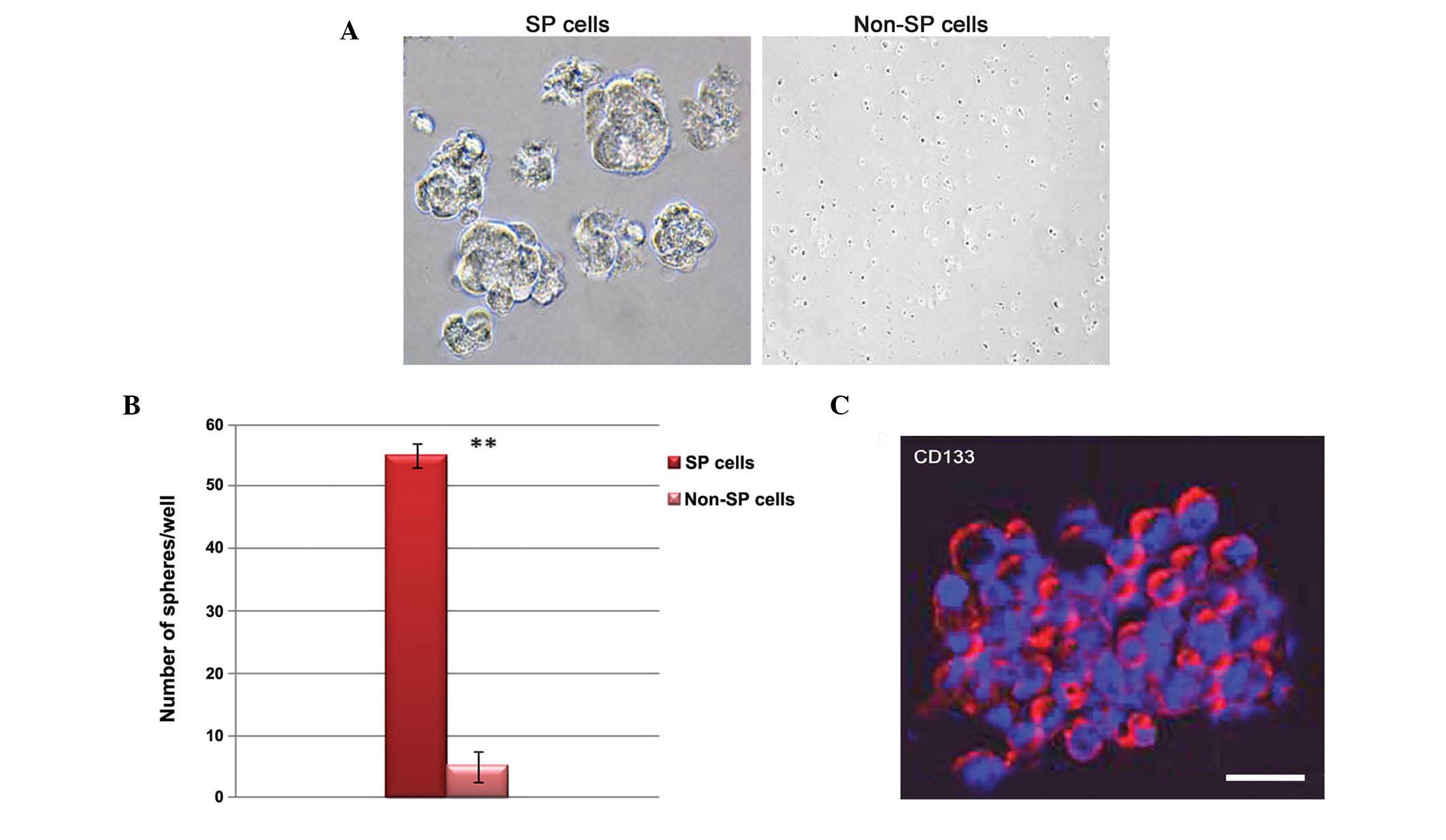
Characterization of tumor spheres formed
by colon cancer SP cells
The present study subsequently aimed to characterize
the FACS-sorted SP cells by analyzing the ability of the SP cells
to form tumor spheres and by determining their positivity to CD133
under standard adherent conditions. The sphere formation assay
revealed that the SP cells readily formed tumor spheres and formed
a cluster of sphere-like cells compared with the non-SP cells
(Fig. 2A). These SP cells
exhibited faster growth rates and spheres were observed on day 5,
unlike in the non-SP cells. The total numbers of tumor spheres
formed by the SP and non-SP cells following 7 days of culture were
also examined. As shown in Fig.
2B, the number of tumor spheres formed by the SP cells in
serum-free medium was significantly higher than the number formed
by the non-SP cells. Subsequently, the tumor spheres were analyzed
for CD133 positivity by immunofluorescence. The tumor spheres
generated by the SP cells exhibited positive expression of CD133
(Fig. 2C). These findings
indicated that the cancer stem-like SP cells were markedly
tumorigenic.
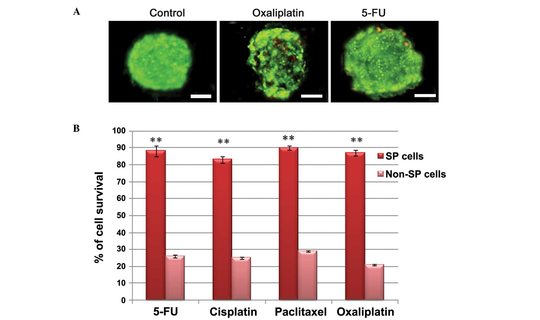
Multidrug resistance and resistance to
apoptosis in colon cancer SP cells
For characterization, the sorted colon cancer SP and
non-SP cells were subjected to a drug-resistance assay in order to
determine the rate of cell survival following treatment with
several chemotherapeutic drugs. Immunofluorescence analysis
revealed that the tumor spheres contained more viable cells upon
treatment with drugs, including 5-FU and oxaliplatin (Fig. 3A). The percentage of cell survival
was also quantified for the SP and non-SP cells following treatment
with cisplatin, paclitaxe, 5-FU and oxaliplatin. Following
treatment with these drugs, the SP cells exhibited significantly
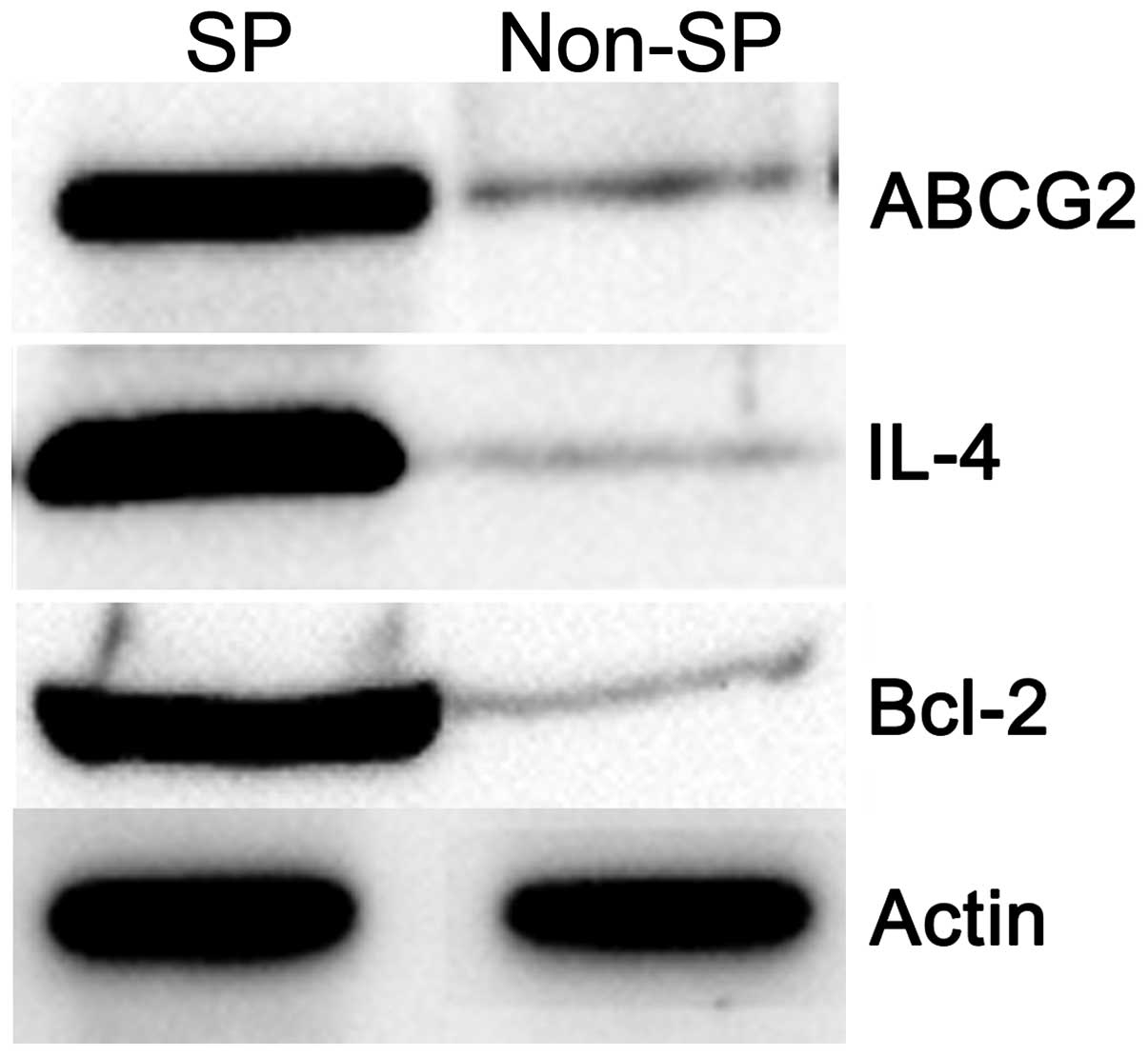
higher survival rates compared with the non-SP cells (Fig. 3B). Furthermore, the biochemical
data revealed that the SP cells exhibited elevated expression
levels of the ABCG2 MDR1 transporter protein, the antiapoptotic
factor, Bcl-2, and auto-crine secretion of IL-4, whereas the levels
of these proteins were comparatively lower in the non-SP cells
(Fig. 4). These results suggested
that the colon cancer SP cells were highly multidrug resistant with
a reduced rate of apoptosis, which contributed to enhanced survival
following treatment with multiple drugs.
Discussion
Cancer is a disease, which contains heterogenous
populations of cells exhibiting multiple differentiation due to
randomly occurring successive mutations. It has been suggested that
cancerous growth, metastasis and tumor recurrence are orchestrated
by a small population of cells, termed CSCs (17). According to the CSC theory, the
traditional therapeutic approaches, used to eradicate the majority
of tumor cells and induce repression of tumor lesions, leave the
CSCs unaffected, which is responsible for treatment failure and
tumor recurrence. Thus traditional treatment fails to prevent
disease relapse and tumor metastasis (3). Therefore, it is essential to develop
a novel therapeutic drug, which can efficiently target the CSCs and
CSC-mediated pathways. However, these hypotheses remain to be
confirmed. Several reports have demonstrated that CSCs can be
isolated and characterized based on either the use of a Hoechst
33342 dye exclusion technique or cell surface markers, including
CD133 (7). Cells, which exclude
Hoechst 33342 dye are referred to as SP cells, and share
characteristic features of CSCs, including tumor initiating
capacity, expression of stem-like genes and resistance to
chemotherapeutic drugs (18). The
results of the present study demonstrated that 3.4% of the cells in
colon cancer were cancer stem-like SP cells, and their prevalence
is reduced upon treatment with verapamil, an MDR1 inhibitor.
It was previously reported that, in colon cancer, a
small population of CD133 positive cells are able to form spheres
and are highly tumorigenic (19).
Similar observations have been made for other types of solid tumor,
including those in melanoma, and brain, ovarian, prostate and
breast carcinoma (4–8). Furthermore, Dean et al
reported that CSCs have the ability to resist death-inducing
signals, as well as enhance the expression level of antiapoptotic
proteins and high levels of drug transporters (13,20).
In accordance, the present study demonstrated high expression
levels of ABC transporter proteins and the Bcl-2 anti-apoptotic
factor in SP cells by western blot analysis. In addition, the tumor
spheres generated by the SP cells were CD133 positive and
effectively resisted chemotherapeutic drugs, including 5-FU,
cisplatin, oxaliplatin and paclitaxel. This confirmed that CSCs
exhibit chemotherapy resistance and are highly tumorigenic.
Considerable data has suggested that the secretion
of certain interleukins, including IL-3 and IL-4, promotes and
increases the survival of cancer cells by upregulating
anti-apoptotic factors (21–23).
In addition, neutralization of these IL secretions effectively
increase the number of death receptors (14). The present study demonstrated that
colon cancer SP cells also exhibited increased expression levels of
IL-4, together with upregulated protein levels of Bcl-2. These
results confirmed that IL-4-dependent protection is involved in
cell death resistance in colon CSCs. Similar to these findings, it
has been previously demonstrated that the secretion of IL-4 and
IL-10 alters the sensitivity of cancer cells to chemotherapeutic
drug-induced cell death (24).
Therefore, the chemoresistant CSCs remaining following standard
conventional treatment strategies can effectively accelerate the
tumor growth and results in treatment failure and relapse.
Therefore, elevated expression levels of ABCG2, IL-4 and Bcl-2
effectively function together and contribute to chemotherapy
resistance and tumor relapse. However, the functional interactions
between IL-4, Bcl-2 and ABCG2, and their signaling pathways in SP
cells require further investigation. Together with others reports,
the findings of the present study may assist in further
characterizing CSCs for designing effective and novel anticancer
drugs.
Acknowledgments
The authors would like to thank Dr Wanshan Li,
Department of Oral and Maxillofacial Surgery, Chongqing Medical
University for sharing the FACS protocol by personal
communication.
References
|
1
|
Greenlee RT, Hill-Harmon MB, Murray T and
Thun M: Cancer statistics, 2001. CA Cancer J Clin. 51:15–36. 2001.
View Article : Google Scholar : PubMed/NCBI
|
|
2
|
Presnell SC, Petersen B and Heidaran M:
Stem cells in adult tissues. Semin Cell Dev Biol. 13:369–376. 2002.
View Article : Google Scholar : PubMed/NCBI
|
|
3
|
Reya T, Morrison SJ, Clarke MF and
Weissman LL: Stem cells, cancer, and cancer stem cells. Nature.
414:105–111. 2001. View
Article : Google Scholar : PubMed/NCBI
|
|
4
|
Bonnet D and Dick JE: Human acutemyeloid
leukemia is organized as a hierarchy that originates from a
primitive hematopoietic cell. Nat Med. 3:730–737. 1997. View Article : Google Scholar : PubMed/NCBI
|
|
5
|
Collins AT, Berry PA, Hyde C, Stower MJ
and Maitland NJ: Prospective identification of tumorigenic prostate
cancer stem cells. Cancer Res. 65:10946–10951. 2005. View Article : Google Scholar : PubMed/NCBI
|
|
6
|
Dontu G, Abdalla WM, Fole JM, et al: In
vitro propagation and transcriptional profiling of human mammary
stem/progenitor cells. Genes Dev. 17:1253–1270. 2003. View Article : Google Scholar : PubMed/NCBI
|
|
7
|
Bapat SA, Mali AM, Koppikar CB and Kurrey
NK: Stem and progenitor-like cells contribute to the aggressive
behavior of human epithelial ovarian cancer. Cancer Res.
65:3025–3029. 2005.PubMed/NCBI
|
|
8
|
Singh SK, Hawkins C, Clarke ID, et al:
Identification of human brain tumor initiating cells. Nature.
432:396–401. 2004. View Article : Google Scholar : PubMed/NCBI
|
|
9
|
Horst D, Kriegl L, Engel J, Kirchner T and
Jung A: CD133 expression is an independent prognostic marker for
low survival in colorectal cancer. Brit J Cancer. 99:1285–1289.
2008. View Article : Google Scholar : PubMed/NCBI
|
|
10
|
Shapiro HM: Microbial analysis at the
single-cell level: Tasks and techniques. J Microbiol Methods.
42:3–16. 2000. View Article : Google Scholar : PubMed/NCBI
|
|
11
|
Kruger J, Singh K, O'Neill A, Jackson C,
Morrison A and O'Brien P: Development of a microfluidic device for
fluorescence activated cell sorting. J Micromech Microeng.
12:486–494. 2002. View Article : Google Scholar
|
|
12
|
Preffer FI, Dombkowski D, Sykes M, Scadden
D and Yang YG: Lineage-negative side-population (SP) cells with
restricted hematopoietic capacity circulate in normal human adult
blood: Immunophenotypic and functional characterization. Stem
Cells. 20:417–427. 2002. View Article : Google Scholar : PubMed/NCBI
|
|
13
|
Dean M, Fojo T and Bates S: Tumor stem
cells and drug resistance. Nat Rev Cancer. 5:275–284. 2005.
View Article : Google Scholar : PubMed/NCBI
|
|
14
|
Todaro M, Zerilli M, Ricci-Vitiani L, et
al: Autocrine production of interleukin-4 and interleukin-10 is
required for survival and growth of thyroid cancer cells. Cancer
Res. 66:1491–1499. 2006. View Article : Google Scholar : PubMed/NCBI
|
|
15
|
Conticello C, Pedini F, Zeuner A, et al:
IL-4 protects tumor cells from anti-CD95 and chemotherapeutic
agents via up-regulation of antiapoptotic proteins. J Immunol.
172:5467–5477. 2004. View Article : Google Scholar : PubMed/NCBI
|
|
16
|
Qz H, Xz L, Wang K, et al: Isolation and
characterization of cancer stem cells from high grade serous
ovarian carcinomas. Cell Physiol Biochem. 33:173–184. 2014.
View Article : Google Scholar
|
|
17
|
Yoo MH and Hatfield DL: The cancer stem
cell theory: Is it correct? Mol Cells. 26:514–516. 2008.PubMed/NCBI
|
|
18
|
Hirschmann-Jax C, Foster AE, Wulf GG, et
al: A distinct 'side population' of cells with high drug efflux
capacity in human tumor cells. Proc Natl Acad USA. 101:14228–14233.
2004. View Article : Google Scholar
|
|
19
|
Ricci-Vitiani L, Lombardi DG, Pilozzi E,
et al: Identification and expansion of human
colon-cancer-initiating cells. Nature. 445:111–115. 2007.
View Article : Google Scholar
|
|
20
|
Eramo A, Ricci-Vitiani L, Zeuner A, et al:
Chemotherapy resistance of glioblastoma stem cells. Cell Death
Differ. 13:1238–1241. 2006. View Article : Google Scholar : PubMed/NCBI
|
|
21
|
Dancescu M, Rubio-Trujillo M, Biron G,
Bron D, Delespesse G and Sarfati M: Interleukin 4 protects chronic
lymphocytic leukemic B cells from death by apoptosis and
upregulates Bcl-2 expression. J Exp Med. 176:1319–1326. 1992.
View Article : Google Scholar : PubMed/NCBI
|
|
22
|
Kieslinger M, Woldman I, Moriggl R, et al:
Antiapoptotic activity of Stat5 required during terminal stages of
myeloid differentiation. Genes Dev. 14:232–244. 2000.PubMed/NCBI
|
|
23
|
Prokopchuk O, Liu Y, Henne-Bruns D and
Kornmann M: Interleukin-4 enhances proliferation of human
pancreatic cancer cells: Evidence for autocrine and paracrine
actions. Br J Cancer. 92:921–928. 2005. View Article : Google Scholar : PubMed/NCBI
|
|
24
|
Todaro M, Perez AM, Di BA, et al: Colon
Cancer Stem Cells Dictate Tumor Growth and Resist Cell Death by
Production of Interleukin-4. Cell Stem Cell. 1:389–402. 2007.
View Article : Google Scholar
|