Introduction
MicroRNAs (miRs) are small, 19–25 nucleotide long
non-coding RNAs that function as critical regulators of gene
expression. They bind to the 3′-untranslated region (UTR) of target
genes and, along with other accessory proteins, form an RNA-induced
silencing complex that is responsible for promoting mRNA
degradation and inhibiting mRNA translation (1). Increasing evidence indicates that
miRNAs are essential in a number of biological processes, including
development, apoptosis, cell proliferation, differentiation,
disease survival and cell death (2–4).
Aberrantly expressed miRNAs are also associated with several
neurological diseases, such as Parkinson's disease, dementia and
glioma (5–8). Current research has revealed that
ectopic overexpression of microRNA-34a (miR-34a) can induce cell
cycle arrest, apoptosis and senescence to inhibit cancer
recurrence, migration and metastasis (9,10).
Multiple studies have also indicated that miR-34a regulates a
variety of target mRNAs, including cyclin-dependent kinase 4/6
(CDK4/6), E2F transcription factor3 (E2F3), Cyclin E2, B-cell
lymphoma 2 (Bcl-2) and NAD-dependent deacetylase sirtuin-1 (SIRT1)
(11,12). TP53 is one of the most common
apoptosis-related genes in mammalian cells, and its gene product
p53, activates the transcription of a set of miRNAs, including
members of the miR-34 family (13). Bcl-2 is another apoptosis-related
gene. Bcl-2 family members were originally characterized with
respect to their roles in regulating apoptosis through complex
interactions that dictate the integrity of the outer mitochondrial
membrane. As an NAD-induced deacetylase, SIRT1 is a transcriptional
regulator and can inhibit the expression of pro-apoptotic proteins
(14). SIRT1 regulates
p53-dependent apoptosis by deacetylating and destabilizing p53. It
has been confirmed that SIRT1 mediates miR-34a-induced apoptosis by
regulating p53 activity. A positive feedback loop has been
identified, in which p53 induces expression of miR-34a, suppressing
SIRT1 and increasing p53 activity (15). PC12 cells are derived from rat
adrenal medulla pheochromocytoma, and are widely utilized in in
vitro studies of neurological diseases. However, thus far,
there have been no experimental studies of the effect of miR-34a in
PC12 cells.
It was hypothesized that Bcl-2 and SIRT1 may be
critical downstream targets of miR-34a that participate in cellular
apoptosis. In the present study, miR-34a mimics or inhibitors were
transfected into PC12 cells, and the apoptosis and proliferation
rates were measured. The aim of the present study was to establish
whether miR-34a-induced PC12 cell apoptosis occurs via suppression
of SIRT1 and Bcl-2.
Materials and methods
Cell culture
PC12 cells (obtained from the Biomedical Laboratory
of Xinjiang Medical University, Ürümqi, China) were cultured in
RPMI medium (GE Healthcare, Logan, UT, USA) containing 10% horse
serum (Hangzhou Sijiqing Biological Engineering Materials Co.,
Hangzhou, China) and 5% fetal bovine serum (Hangzhou Sijiqing
Biological Engineering Materials Co.,) in a CO2
humidified incubator at 37°C. Transfection was performed using
Lipofectamine 2000 (Invitrogen Life Technologies, Carlsbad, CA,
USA) kit according to the manufacturer's instructions. The cells
were divided into the following groups: Negative control group
(control group), 100 nM miR-34a mimic (miR-34a mimic group) and 100
nM miR-34a inhibitor (miR-34a inhibitor group). The miR-34a mimic
and inhibitor were obtained from Shanghai Genechem Co., Ltd.
(Shanghai, China).
3-(4,5-dimethylthiazol-2-yl)-2-5
diphenyltetrazolium bromide (MTT) assay
PC12 cells were seeded into 96-well plates at a
density of 4×10³ cells/well. The effect of miR-34a on
cell growth and viability was determined by an MTT assay. After
transfection (24, 48 or 72 h) with either miR-34a mimic or miR-34a
inhibitor, cells were incubated with MTT (5 mg/ml) in
phosphate-buffered saline (PBS) for 4 h, and then lysed with 50%
N,N dimethylformamide and 10% SDS for an additional 3 h at 37°C.
The absorbance was measured at 570 nm using an ELISA reader
(DG-3022; Nanjing Huangdong Electronic Information & Technology
Co., Ltd, Nanjing, China). Samples were plated in triplicate, and
the average value for each group was calculated.
Senescence-associated β-galactosidase
staining
After transfection with miR-34a mimics or
inhibitors, PC12 cells were stained for SA-β-gal activity analysis.
Cells were fixed with 4% formaldehyde for 15 min at room
temperature, washed three times with PBS, and incubated with 1 ml
X-gal solution (Hangzhou Sijiqing Biological Engineering Materials
Co.) for 12 h at 37°C, avoiding exposure to CO2.
Following incubation, a blue color developed in senescent cells,
observed under a microscope (IX71; Olympus Corporation, Tokyo,
Japan) and the proportions of senescent cells were observed by
digital imaging (Motic Images Plus 2.0; Motic China Group Co.,
Ltd., Xiamen, China).
Apoptosis analysis by
fluorescent-activated cell sorting
After transfection with miR-34a mimics or
inhibitors, PC12 cells were harvested, washed with ice-cold PBS,
resuspended in 500 µl binding buffer, and incubated with 5
µl propidium iodide (PI; Beyotime Institute of
Biotechnology, Jiangsu, China) and 5 µl Annexin
V-fluorescein isothiocyanate (FITC; Beyotime Institute of
Biotechnology) for 10 min in the dark. The cells were then washed
and resuspended in 500 µl PBS, and cell apoptosis was
analyzed by flow cytometry (using an Epics XL-MCL flow cytometer;
Beckman Coulter, Inc., Brea, CA, USA).
Western blot analysis
PC12 cell lysates were extracted using a BCA Protein
Assay Reagent kit (Beyotime Institute of Biotechnology, Shanghai,
China), following mimic or inhibitor treatment. Protein
concentration was determined using the bicinchoninic acid protein
assay reagent (Beyotime Institute of Biotechnology). Equal
quantities of protein from each sample were loaded and
electrophoresed on 10% SDS-PAGE gels (Beyotime Institute of
Biotechnology), and transferred to nitrocellulose membranes [Sangon
Biotech (Shanghai) Co., Ltd., Shanghai, China]. After blocking with
non-fat dried milk, membranes were probed with polyclonal rabbit
anti-human acetyl-p53 [dilution, 1:1,000 (cat. no. CS2525); Cell
Signaling Technology, Inc., Danvers, MA, USA], polyclonal rabbit
anti-human Bcl-2 [dilution, 1:1,000 (cat. no. BS4023); Bioworld
Technology, Inc., St. Louis Park, MN, USA] or polyclonal rabbit
anti-human SIRT1 [dilution, 1:1,000 (cat. no. CS2327); Cell
Signaling Technology, Inc.] antibodies. Antibody signals were
visualized using a Chemiluminescent Detection kit, according to the
manufacturer's instructions (Beyotime Institute of Biotechnology).
Experiments with blank and negative controls were conducted in
parallel. The relative band intensities of the blots were
quantified with Adobe Photoshop software (Adobe Systems, Inc., San
Jose, CA, USA)..
Statistical analysis
All experiments were repeated at least three times.
All values are expressed as the mean ± standard deviation. The
difference between means was analyzed by Student's unpaired t-test.
P<0.05 was considered to indicate a statistically significant
difference. All statistical analyses were conducted using SPSS 19.0
(SPSS, Inc., Armonk, NY, USA).
Results
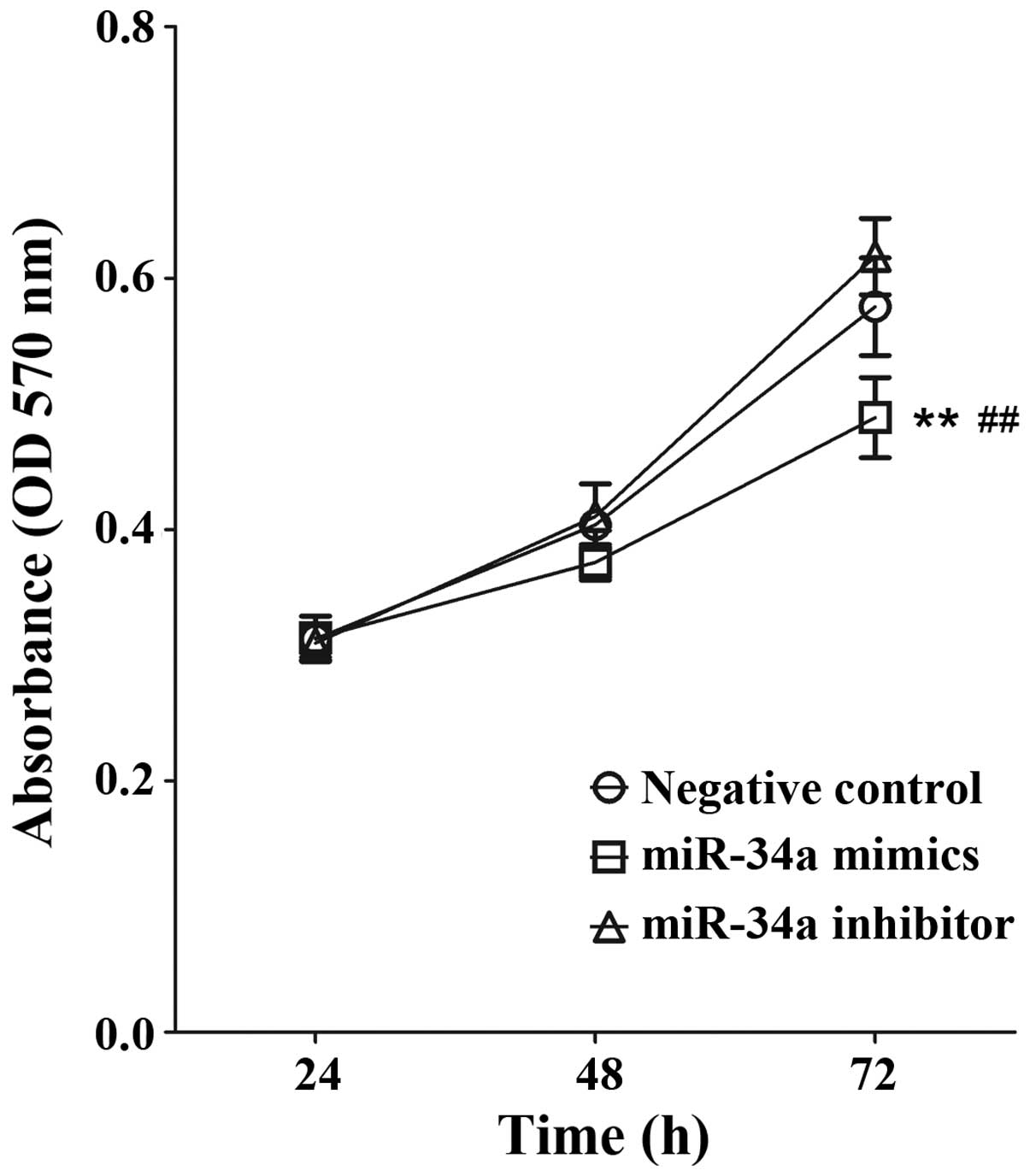
miR-34a mimics inhibit the proliferation
of PC12 cells
To assess the biological role of miR-34a during the
proliferation of PC12 cells, cells were transiently transfected
with miR-34a mimics or inhibitors, and the proliferation was
measured 24, 48 and 72 h following transfection using an MTT assay.
As shown in Fig. 1, following
transfection with miR-34a mimics, the proliferation of PC12 cells
was significantly decreased compared with that of the negative
control group (P<0.01). The proliferation of the miR-34a
inhibitor group was marginally higher than that of the negative
control group; however, no significant difference was identified
(P>0.05).
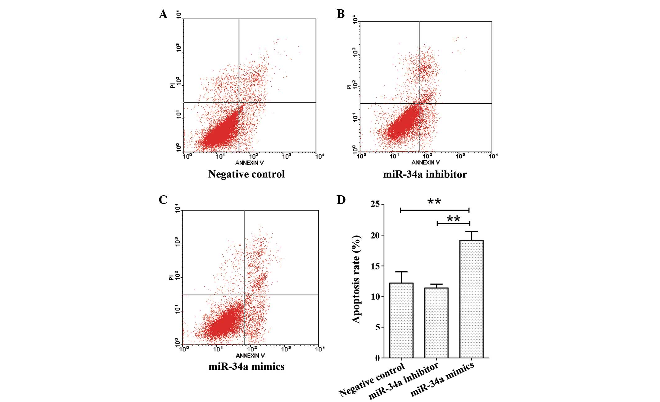
miR-34a induces PC12 cell apoptosis
It is well known that miR-34a is an important
component of the p53 tumor suppressor protein transcriptional
network, which regulates cell proliferation and cell cycle
progression. In order to investigate the biological effects of
miR-34a in nerve cells, PC12 cells were transiently transfected
with miR-34a mimics or miR-34a inhibitors, and the proportions of
apoptotic cells were quantified using an Annexin V-FITC/PI dual
staining assay. As shown in Fig.
2, after transfection, the total proportion of apoptotic cells
in the miR-34a mimic group was significantly increased compared
with that of the control group (P<0.01). The apoptotic rate of
miR-34a inhibitor group was marginally lower than that of the
control group, however, no significant difference was identified
(P>0.05). The total apoptotic rates for control, miR-34a
inhibitor and miR-34a mimic transfection groups were 12.2±1.06,
11.4±0.34 and 19.2±0.84%, respectively.
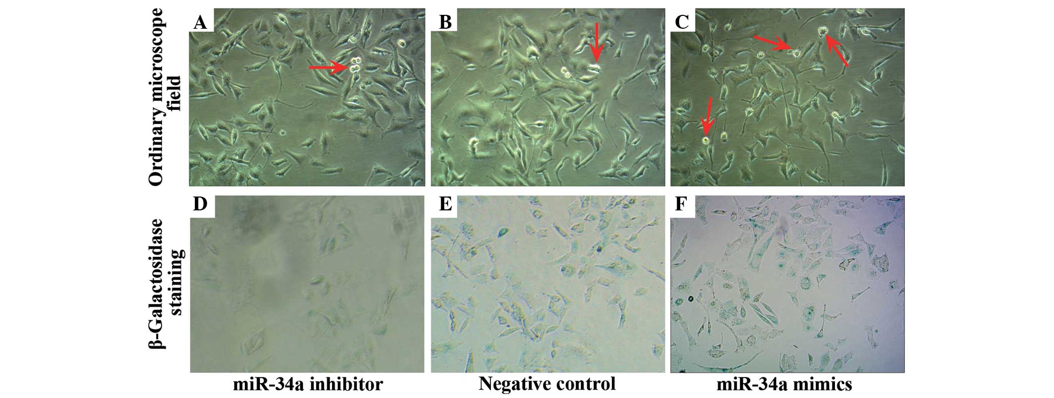
miR-34a induces PC12 cell senescence
The influence of miR-34a on cell senescence was then
evaluated using the β-galactosidase staining assay. The number of
non-adherent cells in the miR-34a mimic group was increasing
compared with the miR-34a inhibitor group and the control group,
however, no significant difference was identified between the
miR-34a inhibitor group and the control group (Fig. 3A–C). SA-β-gal staining analysis
showed that the miR-34a mimics greatly increased SA-β-gal activity
(Fig. 3D–F). These results
demonstrate that miR-34a increases PC12 cell apoptosis and
senescence.
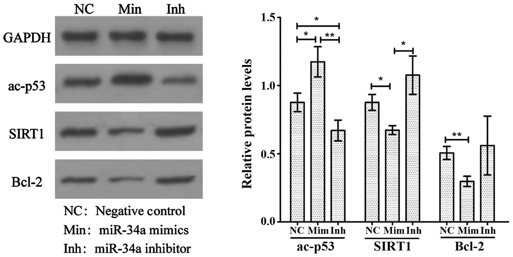
miR-34a reduces the expression of Bcl-2
and SIRT1
To determine whether miR-34a expression levels
correlate with the ac-p53, Bcl-2 and SIRT1 levels in PC12 cells,
the expression of each protein was quantified by western blot
analysis after transfection with miR-34a mimics or inhibitors. As
Fig. 4 shows, compared with the
control group, the expression levels of Bcl-2 (P<0.01) and SIRT1
(P<0.05) in the miR-34a mimic group were significantly reduced,
while the levels of ac-p53 (P<0.05) were elevated in this group.
In addition, the ac-p53 in miR-34a inhibitor group was reduced
compared with the control group (P<0.05). The levels of SIRT1 in
the miR-34a inhibitor group were significantly elevated compared
with the miR-34a mimics group (P<0.05), however, the SIRT1
levels were not significantly different from the control group. The
levels of Bcl-2 in the miR-34a inhibitor group were marginally
elevated compared with the miR-34a mimic and the control groups,
however the difference was not significant.
Discussion
PC12 is a cell line derived from a pheochromocytoma
of the rat adrenal medulla, which contains a mixture of
neuroblastic and eosinophilic cells (16). PC12 cells stop dividing and
terminally differentiate when treated with nerve growth factor or
dexamethasone, rendering PC12 cells a good in vitro model
for investigating neuronal differentiation and neurosecretion
(17,18). Additionally, due to their
widespread availability and transfectable features, PC12 cells are
one of the most commonly used models for investigating the
physiology, pathology and pharmacology of neural cell
differentiation. In recent years, researchers have developed
various neurodegenerative disease cell models, including
Alzheimer's and Parkinson's disease models, using PC12 cells
(19,20). As small, endogenously expressed
non-coding RNAs, miRNAs regulate gene expression by promoting the
degradation of target mRNA and inhibiting translation. miRNAs are
frequently involved in the regulation of cellular differentiation,
proliferation, metabolism and apoptosis. As a member of the miR-34
family, miR-34a has been widely investigated in recent years.
Several studies have indicated that upregulation of miR-34a
expression can induce apoptosis, senescence, differentiation, cell
cycle arrest and growth suppression (21,22).
Overexpression of miR-34a increases the proportion of postmitotic
neurons of mouse neural stem cells (23). SIRT1 is a nicotinamide adenine
dinucleotide (NAD)-dependent histone deacetylase that has been
implicated in inflammation, circadian rhythms, hypoxic responses,
cell survival, life longevity and metabolic processes (24,25).
SIRT1 also exhibits a protective role in certain neurodegenerative
disease models (26). It has been
reported that SIRT1 inhibits lipopolysaccharide-mediated
proinflammatory cytokine release in microglia and circumvents
dopaminergic neuronal injury induced by activated
microglial-derived factors via p53-caspase-3-dependent apoptosis,
which indicates that upregulation of SIRT1 may provide a promising
target for therapeutic intervention in neuroinflammatory diseases
(27).
p53 is a sensor of chronic and acute alterations in
cellular physiology and interacts with DNA to aid in regulating
chromosomal integrity (28).
miR-34a enhances p53 activity by reducing p53 deacetylation, which
in turn results in a decrease in SIRT1 expression (15). This decrease is achieved at the
post-transcriptional level, involving miRNA binding to the 3′-UTR
of SIRT1. In addition, the inhibition of SIRT1 activates
p53-dependent apoptosis through deacetylation and stabilization of
p53. As an important anti-apoptosis gene, Bcl-2 cooperates with
apoptosis activating factors and forms the Bcl-2-Apaf-1-caspase 9
complex, which inhibits the activation of caspases 9 and 3, and
prevents the initiation of mitochondrial apoptosis access. A study
showed that miR-34a inhibits the function and activity of Bcl-2 and
promotes cellular apoptosis (29).
As demonstrated in the present study, transfection
of PC12 cells with miR-34a mimics resulted in insignificant
alterations in cell viability compared with the control group.
Apoptosis analysis by flow cytometry revealed that the apoptosis
rate of the miR-34a mimic group was significantly higher compared
with the control group. Consistently, compared with the control
group, the expression of Bcl-2 and SIRT1 in the miR-34a mimic group
was decreased, while the ac-p53 was increased. Cell senescence is a
form of stagnation of cell growth and, the results obtained using
the SA-β-gal staining assay showed that the miR-34a mimic greatly
increased the SA-β-gal activity, suggesting that the miR34a mimic
could increase the senescence of PC12 cells. miR-34a binds to the
3′-UTR sequences of SIRT1 mRNA, immediately inhibiting the
translation of SIRT1. The decrease in SIRT1 expression leads to an
increase in the acetylation of p53, enhancing p53 transcriptional
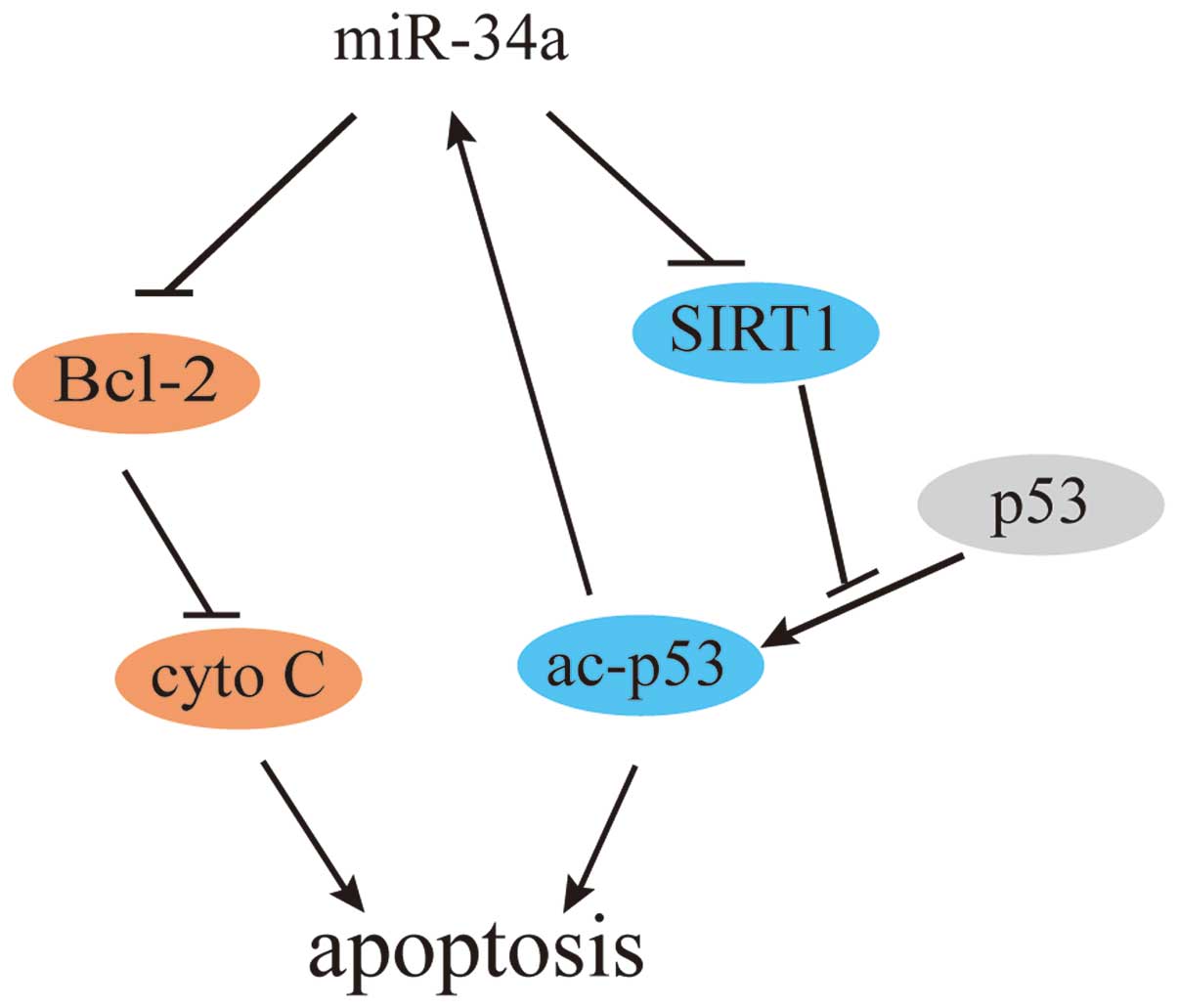
activity, through the positive feedback loop as shown in Fig. 5. As a highly-conserved
NAD+ induced deacetylase enzyme, SIRT1 was first
identified in yeast, where, through regulation of downstream
targets, such as p53, Foxo and Ku79, it is involved in reducing
oxidative stress and apoptosis, and regulating gene silencing and
cell cycle progression (30–32).
SIRT1 promotes the deacetylation of c-terminal lysine 382 of the
p53 protein, regulating the transcriptional activity of p53 and
inhibiting p53-induced apoptosis (33). It is also reported that p53 can
inhibit autophagy by reducing expression of SestrinZ and Draln, and
accelerating cellular senescence (34,35).
Studies have revealed that the deposition of β-amyloid protein (Aβ)
is a critical initiating factor in Alzheimer's disease, and Aβ is a
direct contributor to the neurofibrillary tangles in the brain and
results in the loss of neurons (36). In models of Alzheimer's disease,
high expression of SIRT1 in rat brains was found to strengthen the
catabolic pathway of the amyloid protein, and reduce Aβ deposition
(37). The present study showed
that transfection of miR-34a mimics can effectively reduce the
expression of SIRT1 in PC12 cells, while transfection of miR-34a
inhibitors can increase the expression of SIRT1. Therefore, it was
demonstrated that miR-34a activates SIRT1, which, due to the roles
of SIRT1 in Alzheimer's disease, indicates that miR-34a may aid in
preventing and curing Alzheimer's disease.
In conclusion, the present data indicates that
miR-34a induces PC12 cellular apoptosis, which may be associated
with the inhibition of SIRT1 and Bcl-2. Furthermore, this study
highlights the importance of the positive feedback loop formed by
miR-34a-SIRT1-p53 in cellular apoptosis. These results reveal that
miR-34a is a key regulator of cellular apoptosis and a potential
therapeutic target in neuronal diseases.
Acknowledgments
This study has been supported by the National
Science Foundation of China (grant no. 81372108).
References
|
1
|
Benfey PN: Molecular biology: Microrna is
here to stay. Nature. 425:244–245. 2003. View Article : Google Scholar : PubMed/NCBI
|
|
2
|
Chen LH, Tsai KL, Chen YW, Yu CC, Chang
KW, Chiou SH, Ku HH, Chu PY, Tseng LM and Huang PI: Microrna as a
novel modulator in head and neck squamous carcinoma. J Oncol.
2010:1356322010. View Article : Google Scholar : PubMed/NCBI
|
|
3
|
Ozata DM, Caramuta S, Velazquez-Fernandez
D, Akcakaya P, Xie H, Hoog A, Zedenius J, Backdahl M, Larsson C and
Lui WO: The role of microrna deregulation in the pathogenesis of
adrenocortical carcinoma. Endocr Relat Cancer. 18:643–655. 2011.
View Article : Google Scholar : PubMed/NCBI
|
|
4
|
Guo Z, Chi F, Song Y, Wang C, Yu R, Wei T,
Gui J and Zhu X: Transcriptome analysis of murine thymic epithelial
cells reveals ageassociated changes in microrna expression. Int J
Mol Med. 32:835–842. 2013.PubMed/NCBI
|
|
5
|
Santosh PS, Arora N, Sarma P, Pal-Bhadra M
and Bhadra U: Interaction map and selection of microrna targets in
parkinson's disease-related genes. J Biomed Biotechnol.
3631452009.
|
|
6
|
Sheinerman KS and Umansky SR: Circulating
cell-free microrna as biomarkers for screening, diagnosis and
monitoring of neurodegenerative diseases and other neurologic
pathologies. Front Cell Neurosci. 7:1502013. View Article : Google Scholar : PubMed/NCBI
|
|
7
|
Ai J, Sun LH, Che H, Zhang R, Zhang TZ, Wu
WC, Su XL, Chen X, Yang G, Li K, et al: Microrna-195 protects
against dementia induced by chronic brain hypoperfusion via its
anti-amyloidogenic effect in rats. J Neurosci. 33:3989–4001. 2013.
View Article : Google Scholar : PubMed/NCBI
|
|
8
|
Men D, Liang Y and Chen L: Decreased
expression of microrna-200b is an independent unfavorable
prognostic factor for glioma patients. Cancer Epidemiol.
38:152–156. 2014. View Article : Google Scholar : PubMed/NCBI
|
|
9
|
Liu C, Zhou C, Gao F, Cai S, Zhang C, Zhao
L, Zhao F, Cao F, Lin J, Yang Y, et al: Mir-34a in age and tissue
related radio-sensitivity and serum mir-34a as a novel indicator of
radiation injury. Int J Biol Sci. 7:221–233. 2011. View Article : Google Scholar : PubMed/NCBI
|
|
10
|
Boon RA, Iekushi K, Lechner S, Seeger T,
Fischer A, Heydt S, Kaluza D, Treguer K, Carmona G, Bonauer A, et
al: Microrna-34a regulates cardiac ageing and function. Nature.
495:107–110. 2013. View Article : Google Scholar : PubMed/NCBI
|
|
11
|
Li L, Yuan L, Luo J, Gao J, Guo J and Xie
X: Mir-34a inhibits proliferation and migration of breast cancer
through down-regulation of bcl-2 and sirt1. Clin Exp Med.
13:109–117. 2013. View Article : Google Scholar
|
|
12
|
Tomosugi M, Sowa Y, Yasuda S, Tanaka R, Te
RH, Ikawa H, Koyama M and Sakai T: Retinoblastoma gene-independent
g1 phase arrest by flavone, phosphatidylinositol 3-kinase inhibitor
and histone deacetylase inhibitor. Cancer Sci. 103:2139–2143. 2012.
View Article : Google Scholar : PubMed/NCBI
|
|
13
|
Bommer GT, Gerin I, Feng Y, Kaczorowski
AJ, Kuick R, Love RE, Zhai Y, Giordano TJ, Qin ZS, Moore BB, et al:
P53-mediated activation of mirna34 candidate tumor-suppressor
genes. Curr Biol. 17:1298–1307. 2007. View Article : Google Scholar : PubMed/NCBI
|
|
14
|
Matsushita N, Takami Y, Kimura M, Tachiiri
S, Ishiai M, Nakayama T and Takata M: Role of nad-dependent
deacetylases sirt1 and sirt2 in radiation and cisplatin-induced
cell death in vertebrate cells. Genes Cells. 10:321–332. 2005.
View Article : Google Scholar : PubMed/NCBI
|
|
15
|
Castro RE, Ferreira DM, Afonso MB,
Borralho PM, Machado MV, Cortez-Pinto H and Rodrigues CM:
Mir-34a/sirt1/p53 is suppressed by ursodeoxycholic acid in the rat
liver and activated by disease severity in human non-alcoholic
fatty liver disease. J Hepatol. 58:119–125. 2013. View Article : Google Scholar
|
|
16
|
Attiah DG, Kopher RA and Desai TA:
Characterization of pc12 cell proliferation and
differentiation-stimulated by ecm adhesion proteins and
neurotrophic factors. J Mater Sci Mater Med. 14:1005–1009. 2003.
View Article : Google Scholar
|
|
17
|
Leoni C, Menegon A, Benfenati F, Toniolo
D, Pennuto M and Valtorta F: Neurite extension occurs in the
absence of regulated exocytosis in pc12 subclones. Mol Biol Cell.
10:2919–2931. 1999. View Article : Google Scholar : PubMed/NCBI
|
|
18
|
Taupenot L: Analysis of regulated
secretion using pc12 cells. Curr Protoc Cell Biol. 15:12–15.
2007.
|
|
19
|
Martin D, Salinas M, Lopez-Valdaliso R,
Serrano E, Recuero M and Cuadrado A: Effect of the alzheimer
amyloid fragment abeta (25–35) on akt/pkb kinase and survival of
pc12 cells. J Neurochem. 78:1000–1008. 2001. View Article : Google Scholar
|
|
20
|
Zhang ZT, Cao XB, Xiong N, Wang HC, Huang
JS, Sun SG and Wang T: Morin exerts neuroprotective actions in
parkinson disease models in vitro and in vivo. Acta Pharmacol Sin.
31:900–906. 2010. View Article : Google Scholar : PubMed/NCBI
|
|
21
|
Hermeking H: The mir-34 family in cancer
and apoptosis. Cell Death Differ. 17:193–199. 2010. View Article : Google Scholar
|
|
22
|
Zhang C, Mo R, Yin B, Zhou L, Liu Y and
Fan J: Tumor suppressor microrna-34a inhibits cell proliferation by
targeting notch1 in renal cell carcinoma. Oncol Lett. 7:1689–1694.
2014.PubMed/NCBI
|
|
23
|
Aranha MM, Santos DM, Xavier JM, Low WC,
Steer CJ, Sola S and Rodrigues CM: Apoptosis-associated micrornas
are modulated in mouse, rat and human neural differentiation. BMC
Genomics. 11:5142010. View Article : Google Scholar : PubMed/NCBI
|
|
24
|
Yamakuchi M: Microrna regulation of sirt1.
Front Physiol. 3:682012. View Article : Google Scholar : PubMed/NCBI
|
|
25
|
Nogueiras R, Habegger KM, Chaudhary N,
Finan B, Banks AS, Dietrich MO, Horvath TL, Sinclair DA, Pfluger PT
and Tschop MH: Sirtuin 1 and sirtuin 3: Physiological modulators of
metabolism. Physiol Rev. 92:1479–1514. 2012. View Article : Google Scholar : PubMed/NCBI
|
|
26
|
Kim D, Nguyen MD, Dobbin MM, Fischer A,
Sananbenesi F, Rodgers JT, Delalle I, Baur JA, Sui G, Armour SM, et
al: SIRT1 deacetylase protects against neurodegeneration in models
for Alzheimer's disease and amyotrophic lateral sclerosis. EMBO J.
26:3169–3179. 2007. View Article : Google Scholar : PubMed/NCBI
|
|
27
|
Ye J, Liu Z, Wei J, Lu L, Huang Y, Luo L
and Xie H: Protective effect of sirt1 on toxicity of
microglial-derived factors induced by lps to pc12 cells via the
p53-caspase-3-dependent apoptotic pathway. Neurosci Lett.
553:72–77. 2013. View Article : Google Scholar : PubMed/NCBI
|
|
28
|
Liu TX, Howlett NG, Deng M, Langenau DM,
Hsu K, Rhodes J, Kanki JP, D'Andrea AD and Look AT: Knockdown of
zebrafish fancd2 causes developmental abnormalities via
p53-dependent apoptosis. Dev Cell. 5:903–914. 2003. View Article : Google Scholar : PubMed/NCBI
|
|
29
|
Potts MB, Vaughn AE, McDonough H,
Patterson C and Deshmukh M: Reduced apaf-1 levels in cardiomyocytes
engage strict regulation of apoptosis by endogenous xiap. J Cell
Biol. 171:925–930. 2005. View Article : Google Scholar : PubMed/NCBI
|
|
30
|
Bitterman KJ, Anderson RM, Cohen HY,
Latorre-Esteves M and Sinclair DA: Inhibition of silencing and
accelerated aging by nicotinamide, a putative negative regulator of
yeast sir2 and human sirt1. J Biol Chem. 277:45099–45107. 2002.
View Article : Google Scholar : PubMed/NCBI
|
|
31
|
Hori YS, Kuno A, Hosoda R and Horio Y:
Regulation of foxos and p53 by sirt1 modulators under oxidative
stress. PLoS One. 8:e738752013. View Article : Google Scholar : PubMed/NCBI
|
|
32
|
Sun MF, Chang TT, Chang KW, Huang HJ, Chen
HY, Tsai FJ, Lin JG and Chen CY: Blocking the dna repair system by
traditional chinese medicine? J Biomol Struct Dyn. 28:895–906.
2011. View Article : Google Scholar : PubMed/NCBI
|
|
33
|
Solomon JM, Pasupuleti R, Xu L, McDonagh
T, Curtis R, DiStefano PS and Huber LJ: Inhibition of sirt1
catalytic activity increases p53 acetylation but does not alter
cell survival following dna damage. Mol Cell Biol. 26:28–38. 2006.
View Article : Google Scholar :
|
|
34
|
Tavernarakis N, Pasparaki A, Tasdemir E,
Maiuri MC and Kroemer G: The effects of p53 on whole organism
longevity are mediated by autophagy. Autophagy. 4:870–873. 2008.
View Article : Google Scholar : PubMed/NCBI
|
|
35
|
Maiuri MC, Malik SA, Morselli E, Kepp O,
Criollo A, Mouchel PL, Carnuccio R and Kroemer G: Stimulation of
autophagy by the p53 target gene sestrin2. Cell Cycle. 8:1571–1576.
2009. View Article : Google Scholar : PubMed/NCBI
|
|
36
|
Bloom GS: Amyloid-beta and tau: the
trigger and bullet in alzheimer disease pathogenesis. JAMA Neurol.
71:505–508. 2014. View Article : Google Scholar : PubMed/NCBI
|
|
37
|
Sun Q, Jia N, Wang W, Jin H, Xu J and Hu
H: Activation of sirt1 by curcumin blocks the neurotoxicity of
amyloid-beta25-35 in rat cortical neurons. Biochem Biophys Res
Commun. 448:89–94. 2014. View Article : Google Scholar : PubMed/NCBI
|