Introduction
Aortic aneurysm is a leading cause of cardiovascular
mortality and has the dismal prognosis of any major type of
cardiovascular condition (1–3).
Aortic aneurysm, determined by a pathological aortic expansion by
>50% of the normal vascular diameter, is divided into true
aortic aneurysm and aortic pseudo-aneurysm (4). Although considerable progress has
been made in improving cardiovascular survival rates over the past
few decades, the five-year survival rate has remained <35%
(5). The poor prognosis of
cardiovascular conditions is in part due to the emergence of drug
resistance (6). Consequently,
there is an urgent requirement to explore the underlying molecular
mechanisms of aortic aneurysm.
As is commonly known, mRNA is a type of
single-stranded RNA, which carries genetic information that guides
protein expression. Gene transcription occurs via the expression of
mRNA molecules in each cell (7).
However, abnormal RNA splicing (trans-splicing) may occur, leading
to the covalent attachment of a single RNA molecule to its other
end and resulting in the formation of cyclic RNA (circRNA); this is
usually a rare biochemical reaction. Initially, circRNAs were
identified in plants, in which they are capable of encoding
sub-viral factors (8). In
unicellular organisms, circRNAs occur mostly due to the
self-splicing intron of ribosomal RNA, but may also be produced by
evolutionarily conserved protein-coding genes (9,10).
In animal cells, circRNAs are formed in the splicing process by
joining the 5′ and 3′ end of the downstream exon (10,11).
By far the most well-known circRNA is that formed by anti-sense
transcription of the mRNA of the sex determining region Y gene,
which was shown to be highly expressed in the testes (12). Analysis of gene expression in
archaea and mammals showed that circRNAs are more abundant than
expected; however, the biological functions of circRNAs in animals
and plants have remained elusive. CircRNAs have been frequently
found by RNA seqencing, and it has been attempted to identify all
circRNAs present in animal cell systems. It has been revealed that
certain circRNAs act as molecular 'sponges' by combining and
encapsulating microRNAs (miRNAs), thereby performing their
regulatory effect on gene expression (13,14).
In addition, it was speculated that circRNAs have numerous other
functions, pointing out that the functions of RNA go beyond that of
a messenger between DNA and the encoded proteins (15). CircRNAs have not been discovered at
any earlier stages as traditional methods to isolate RNA from cells
discarded of these cyclic molecules. It has been wrongly assumed
that only linear RNAs are the products of gene transcription;
however, circRNA are the predominant transcript form of hundreds of
genes (16). CircRNAs have also
been widely ignored by classical RNA sequencing studies due to
their lack of a tail end (17).
CircRNAs are more abundant than expected and may be more important
than previously thought; however, their expression and function in
numerous conditions, including aortic aneurysm, have remained
elusive.
The circRNA labeled hsa-circ-000595 was first
identified by Memczak et al (18). It is located on chromosome 14, and
the gene coding starts from position 93753822 and ends on position
93762503 of the chromosome. In addition, it has been demonstrated
that hsa-circ-000595 has a connection with miR-19a and miR-19b
(18). miR-19a is a member of the
miR-17-92 cluster, which is located on chromosome 13q31.3 and is
involved in the pathogenesis of numerous types of tumor. It has
previously been reproted that miR-19a is upregulated in various
types of tumors (19). The present
study investigated the role of circRNAs in aortic aneurysm using an
epigenetic PCR array. As hypoxia is implicated in aortic aneurysm
(20), the effect of
CoCl2-induced hypoxia on the expression of
hsa-circ-000595, a circRNA that was found to be differentially
expressed in aortic aneurism compared with normal aortic tissues,
was examined. Furthermore, RNA interference was employed to
knockdown circRNA expression, and its effect on hypoxia-associated
apoptosis of a human aortic smooth muscle cell line was assessed.
The present study preliminarily explored the underlying mechanisms
of aortic aneurysm from the perspective of epigenetics.
Materials and methods
Cell line
Human aortic smooth muscle cells (V-SMC-6110) were
obtained from the American Type Culture Collection (Manassas, VA,
USA). Cells were cultured in low-glucose Dulbecco's modified
Eagle's Medium (DMEM; Invitrogen Life Technologies, Carlsbad, CA,
USA) containing 10% fetal bovine serum (FBS; Invitrogen Life
Technologies), 100 units/ml penicillin sodium and 100 µg/ml
streptomycin sulfate (Invitrogen Life Technologies) at 37°C in a
humidified chamber supplemented with 5% CO2.
Patient samples
Aortic tissues from patients with aortic aneurysm
were obtained from Zhejiang Second Hospital (Huangzhou, China)
between January 2005 and December 2013. The patients consisted of
28 males and 7 females aged between 35 and 84 years old with a mean
age of 54 years. The Bioethics Committee of the Zhejiang Second
Hospital approved all experiments, which were in accordance with
the National Institutes of Health Guide (Bethesda, MD, USA).
Small interfering (si)RNA
transfection
Prior to transfection, V-SMC were seeded at a
density of 3.0×10⁵, and 2 ml serum-free DMEM was added
into six-well plates. siRNA (Funeng Biological Technology Co.,
Ltd., Shanghai, China) (forward, 5′-GGCCUAAGAUAAGAAAUAUUU-3′, and
reverse, 5′-AUAUUUCUUAUCUUAGGCCUU-3′) was added to a final
concentration of 50 nM. siRNA targeted to circRNA hsa-circ-000595
was transfected into V-SMC cells with Lipofectamine 2000
(Invitrogen Life Technologies) following the manufacturer's
instructions. Knockdown was then confirmed by assessing the
expression of circRNA and miRNA by reverse transcription
quantitative polymerase chain reaction (RT-qPCR).
RT-qPCR
Total RNA was extracted from cultured cells using
TRIzol (Invitrogen Life Technologies) and determined its
concentration was determined. Total RNA (0.5 µg) was used as
a template to prepare cDNA (Reverse Transcription System, Promega
Corporation, Madison, WI, USA; cat. no. A3500). The mRNA expression
of target genes (hsa-circ-000595 and miR-19a) was quantified using
SYBR Premix EX Taq (Takara Bio, Inc., Shanghai, China) on the ABI
7500 squence detection system (Advanced Biosystem, Thermo Fisher
Scientific, Waltham, MA, USA). PCR was performed with the following
thermocycling conditions: An initial 5 min at 95°C, followed by 40
cycles of 95°C for 30 sec, 55°C for 30 sec and 72°C for 30 sec. The
thermocycler used in the present study was the StepOnePlus™
Real-Time PCR system (Applied Biosystems Life Technologies, Foster
City, CA, USA) The primers were obtained from Funengbio Co.
(Shanghai, China), and the sequences were as follows:
hsa-circ-000595, forward 5′-ACGCGGCCTAAATATGAGCA-3′, reverse
5′-GAAGCTTCCAGACTGAGCCAC-3′; m i R-19a, forward
5′-ACGTGTGCAAATCTATGCAAAAC-3′ and reverse 5′-GTGCAGGGTCCGAGGT-3′;
β-actin, forward 5′-CCTCGCCTTTGCCGATCC-3′ and reverse
5′-GGATCTTCATGAGGTAGTCAGTC-3′. Housekeeping gene β-actin was used
as an internal reference to normalize the results. All experiments
were performed in triplicate. Finally, the 2−ΔΔCt method
was performed to calculate the relative expression (21).
Western blot analysis
Western blotting was performed according to standard
protocols. Briefly, the total protein from tissue or cell was
extracted using radioimmunoprecipitation lysis buffer containing 1
mM phenylmethanesulfonylfluo-ride and the protein concentration was
determined using the Bradford method (Beyotime Institute of
Biotechnology, Nantong, China) according to the manufacturer's
instructions. 20 µg total protein sample was separated by
10% SDS-PAGE (Fdbio Science, Hangzhou, China) and transferred into
a nitrocellulose membrane (EMD Millipore, Billerica, MA, USA). The
membrane was blocked in phosphate-buffered saline containing Tween
20 (PBST) with 5% non-fat milk for 1 h at 4°C. The membrane was
then incubated with the following primary antibodies for 12 h at
4°C: Anti-caspase 8 (mouse anti-human monoclonal antibody; 1:1,000;
cat. no. sc-56070), anti-caspase 3 (mouse anti-human monoclonal
antibody; 1:800; cat. no. sc-65496) and anti-B-cell lymphoma
(Bcl)-2 (rabbit anti-human polyclonal antibody; 1:1,000; cat. no.
sc-492) (all Santa Cruz Biotechnology, Inc., Dallas, TX, USA),
followed by three washes with PBST and incubation for 30 min at 4°C
with secondary antibody labeled with horseradish peroxidase.
Finally, the membrane was washed three times with PBST and the
protein bands were visualized with Super Signal. Antibody binding
was detected using an enhanced chemiluminescence kit (EMD
Millipore). The blots were then incubated in a commercial stripping
solution (Pierce Biotechnology, Inc. Rockford, IL, USA) for 10 min.
The membranes were re-probed with a rabbit anti-human anti-β-actin
polyclonal antibody (1:1,000; cat. no. A2668; Sigma-Aldrich, St.
Louis, MO, USA) as a loading control.
Hematoxylin and eosin (HE) staining
The aortic tissue samples from patients with aortic
aneurysm were fixed in 4% paraformaldehyde (Aladdin, Shanghai,
China), dehydrated with graded ethanol, cleared in dimethylbenzene
(Aladdin) and embedded in paraffin (Aladdin). Paraffin sections (5
µm) were prepared using a Leica histotome (Leica
Microsystems, Wetzlar, Germany) and deparaffinized with immersion
in dimethylbenzene prior to rehydration. HE staining was
subsequently performed according to standard procedures (22).
Apoptosis assays
The V-SMC cells were pre-treated with
CoCl2, or without CoCl2 if they were to serve
as a control. The apoptosis levels of the V-SMC cells treated with
scramble or hsa-circ-000595 siRNA were subsequently analyzed using
an Annexin V-fluorescein isothiocyanate (FITC) Apoptosis Detection
kit (cat. no. C1063; Beyotime Institute of Biotechnology) according
to the manufacturer's instructions. The cells were seeded in 6-well
plates at a density of 1×10⁵ cells/well in DMEM medium
for 24 h. The cells were then digested with 0.25% trypsin
(Invitrogen Life Technologies) and resuspended in 300 µl
binding buffer (Beyotime Institute of Biotechnology) containing 5
µl Annexin V-FITC and 5 µl propidium iodide solution,
and incubated at room temperature in the dark for 20 min. The
stained cells were analyzed by flow cytometry (FACScan; BD
Biosciences, Franklin Lakes, NJ, USA).
TUNEL assay
The V-SMC cells were pre-treated with
CoCl2 or without CoCl2 if they were to serve
as a control. The cells were then treated with scramble siRNA or
hsa-circ-000595 siRNA for 24 h. The adherent cells were
subsequently detached using 0.1% trypsin/0.04% EDTA (Invitrogen
Life Technologies) and the cell apoptosis levels were assessed
using a DeadEnd™ Fluorometric TUNEL assay kit (cat. no. C1088;
Beyotime Institute of Biotechnology). Data were acquired using an
Infinite 200 PRO Multimode reader (Tecan Group, Ltd., Maennedorf,
Switzerland) and were analyzed using i-control 1.9 software (Tecan
Group, Ltd.).
Bioinformatics analysis
The bioinformatics data presented in the present
study may be obtained from GEO (http://www.ncbi.nlm.nih.gov/geo) with accession number
GSE24194.
Statistical analysis
Values are expressed as the mean ± standard
deviation. Differences between groups were analyzed by 1- or 2-way
analysis of variance. Differences in the rates of tumor inhibition
were validated by the χ2 test. P<0.05 was considered
to indicate a significant difference between groups. The analytical
software is SPSS 13.0 (SPSS Inc, Chicago, IL, USA) was used for
statistical analyses.
Results
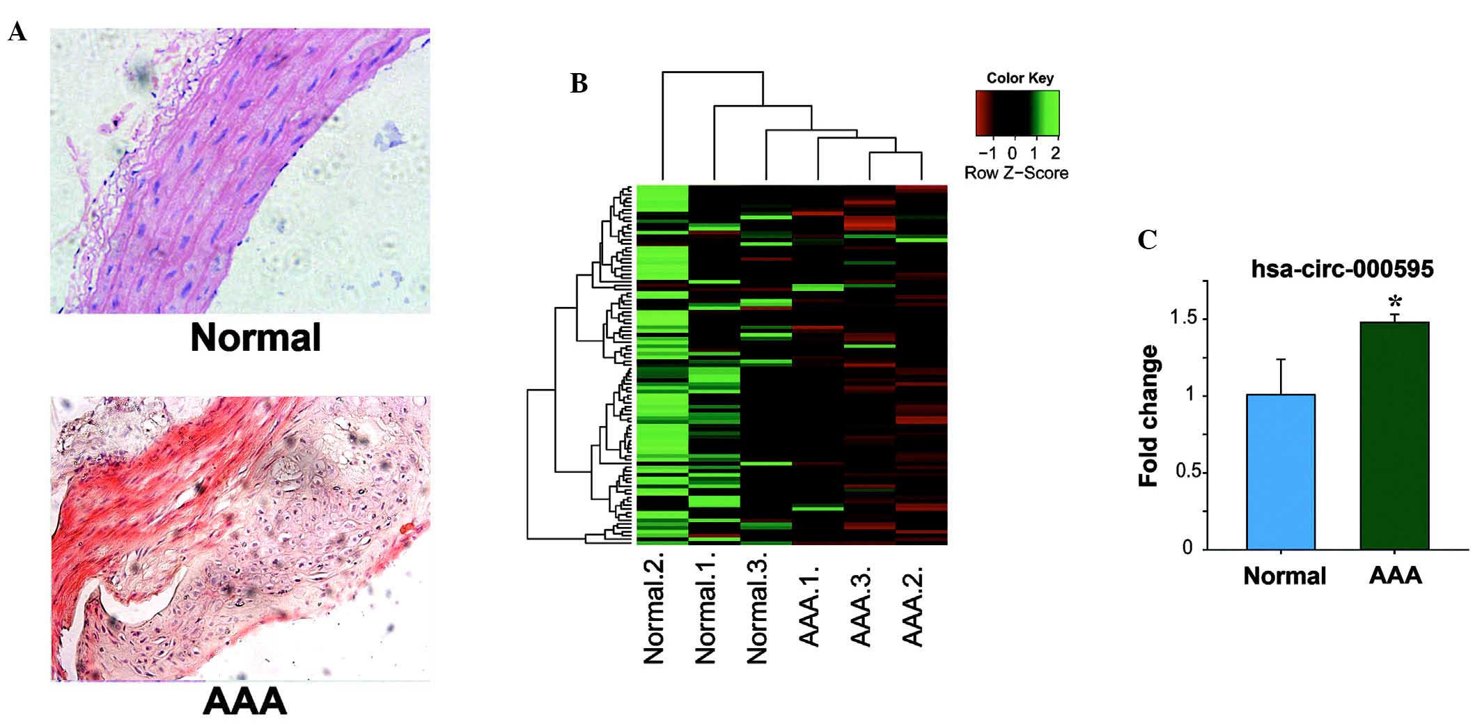
Expression of circRNA in aortic tissues
from patients with aortic aneurysm
Aortic tissues from patients with aortic aneurysm
were assessed by histochemical analysis and RT-qPCR. H&E
staining of aortic aneurysm and normal tissues was performed
(Fig. 1A). In the PCR array
assays, 94 circRNAs were assessed and the results revealed that the
expression of 12 circRNAs was increased by at least 1.5-fold of
that in normal tissues (Fig. 1B).
In aortic tissues from patients with aortic aneurysm,
hsa-circ-000595 was most significantly increased by 1.5-fold
compared with that in normal tissues (P<0.05) (Fig. 1C).
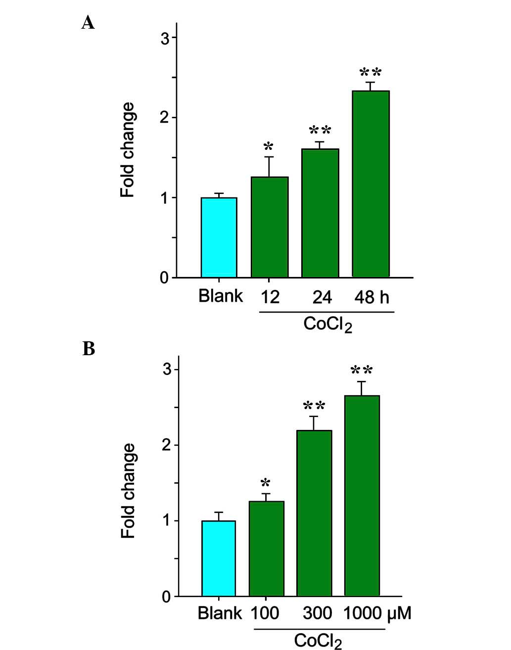
Hypoxia upregulates the expression of
hsa-circ-000595 in v-SMCs
As hypoxia may be one of the causes of aortic
aneurysm, the present study determined the expression of
hsa-circ-000595 during hypoxia, which was simulated by incubation
with CoCl2. The expression levels of hsa-circ-000595 in
v-SMCs treated with/without CoCl2 were assessed by
RT-qPCR. As shown in Fig. 2A,
V-SMC expressed circRNAs normally under normoxic conditions, while
treatment with CoCl2 (300 µM) led to a modest
upregulation of the expression of hsa-circ-000595 in V-SMCs in a
time-dependent manner (Fig. 2A;
P<0.05). Of note, high doses of CoCl2 significantly
increased the expression of hsa-circ-000595 (Fig. 2B; P<0.05). These results
indicated that hsa-circ-000595 expression was increased under
hypoxic conditions, further suggesting its implication in aortic
aneurysm.
Knockdown of hsa-circ-000595 attenuates
hypoxia-induced apoptosis of V-SMCs
To further explore how hsa-circ-000595 influences
the viability of V-SMCs, hsa-circ-000595 knockdown was performed in
V-SMCs. RT-qPCR confirmed that the expression of hsa-circ-000595
was obviously downregulated in V-SMCs transfected with two
different siRNAs directed towards hsa-circ-000595 for 48 h compared
with the expression in cells transfected with scrambled control
vector (P<0.05) (Fig. 3A). In
order to clarify the role of hsa-circ-000595 in apoptosis of
V-SMCs, the apoptotic rate was assessed by flow cytometry and
terminal deoxynucleotidyl transferase dUTP nick end labeling
(TUNEL) assays. As shown in Fig.
3B, trans-fection with either of the two siRNAs of
hsa-circ-000595 did not affect the apoptotic rate of V-SMC cells in
the absence of CoCl2; however, in the presence of
CoCl2, the apoptotic rate was decreased in
hsa-circ-000595-knockdown cells compared with that in cells
transfected with scrambled control. These results were validated by
TUNEL assays (Fig. 3C). Under
hypoxic conditions only, hsa-circ-000595 knockdown can also reduced
the protein levels of caspase 3, caspase 8 and Bcl-2, which are
apoptosis-associated proteins (Fig.
3D). Therefore, knockdown of hsa-circ-000595 attenuated
hypoxia-induced apoptosis of aortic cells, which may potentially
reduce the occurrence of aortic aneurysm.
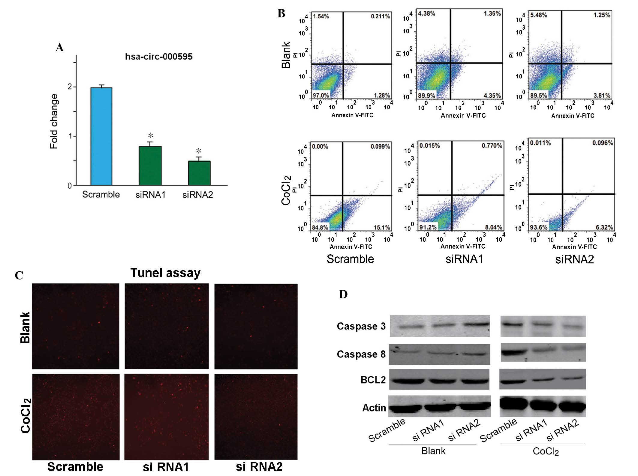 | Figure 3Effect of has-circ-000595 knockdown on
hypoxia-induced damage of V-SMCs. (A) Knockdown efficiency of two
siRNAs targeted to has-circ-000595 in V-SMCs. The data are
expressed as the mean ± standard deviation. *P<0.05
vs. scrabled control. (B) Flow cytometric detection of the
apoptotic rate of V-SMCs transfected with siRNAs. The two siRNAs
targeted to hsa-circ-000595 did not affect the apoptotic rate of
V-SMCs in the absence of CoCl2; however, under
CoCl2-induced hypoxia, the hsa-circ-000595 siRNAs
decreased the apoptotic rate of V-SMCs compared with that of
CoCl2-treated controls. (C) Tunel analysis of the
apoptotic rate of V-SMCs transfected with siRNAs in the absence or
presence of CoCl2. The amount of red fluorescence is
indicative of the apoptotic rate (magnification, ×20). (D)
Expression levels of caspases 3, caspase 8 and Bcl-2 in scramble-,
siRNA1- and siRNA2-transfected cells with or without
CoCl2 were detected by western blot analysis, and GAPDH
was used as a loading control. siRNA, small interfering RNA; V-SMC,
human aortic smooth muscle cell; circRNA, cyclic RNA; hsa, Homo
sapiens; Bcl-2, B-cell lymphoma 2; Tunel, terminal
deoxynucleotidyl transferase dUTP nick end labeling; PI, propidium
iodide; FITC, fluorescein isothiocyanate. |
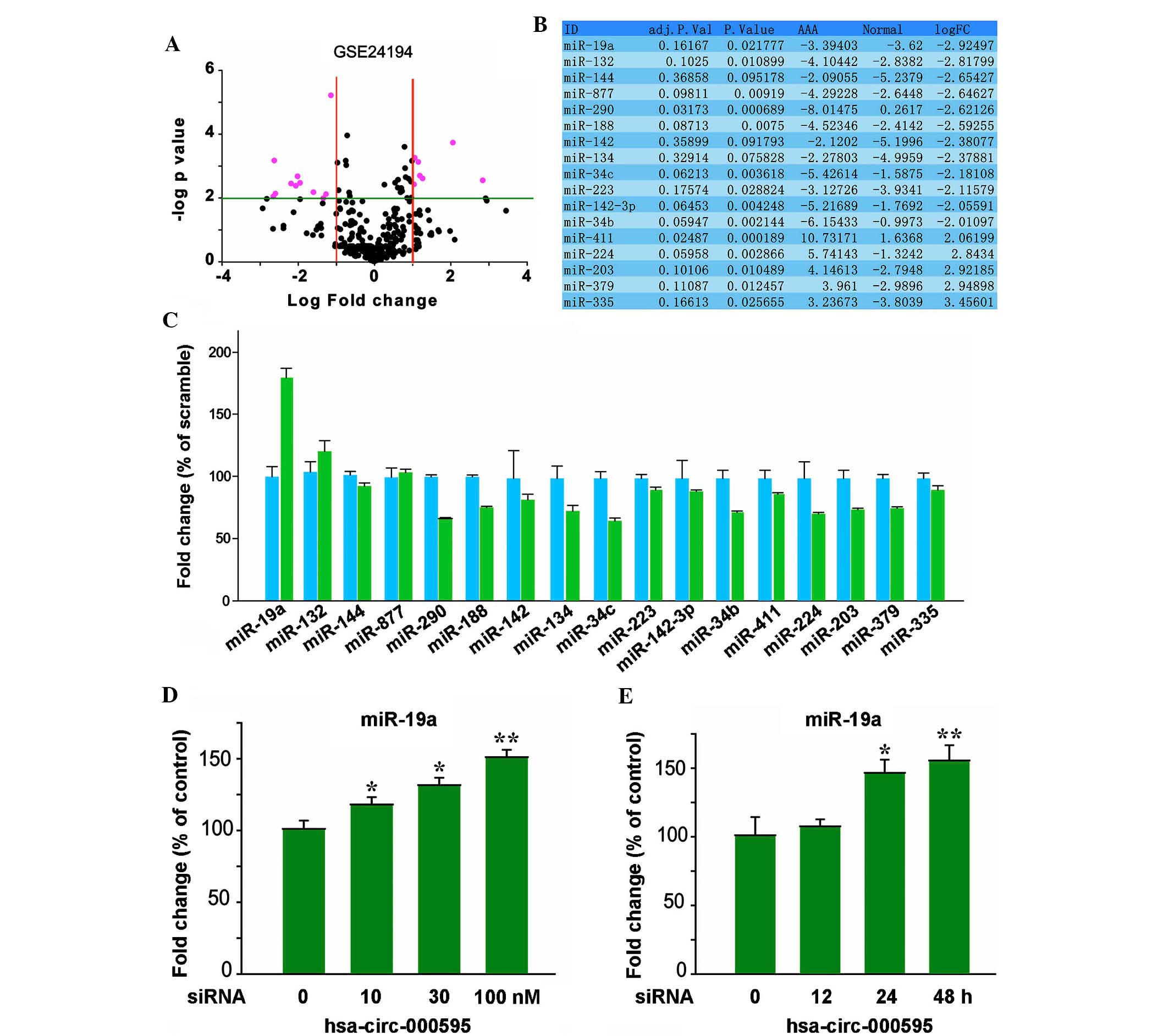
Hsa-circ-000595 regulates miR-19a
expression
Growing evidence indicated that hsa-circ-000595 is
strongly associated with apoptosis (23). CircRNAs were reported to be
silencers of miRNAs. By using bioinformatics tools, the present
study demonstrated that expression of 17 miRNAs was affected by
hsa-circ-000595 (Fig. 4A–C), and
that there was a statistically significant difference between the
expression levels of miR-19a and Hsa-circ-000595. These results
were obtained from the GSE24194 database. RT-PCR was then performed
to determine the expression levels of miR-19a in V-SMCs treated
with CoCl2. As shown in Fig. 4D and E, prior to CoCl2
treatment of V-SMCs, the cells were transfected with
hsa-circ-000595 siRNA, and subsequently the expression of miR-19a
was significantly increased in hsa-circ-000595-knockdown-treated
V-SMCs in a dose- and time-dependent manner, as compared with that
in native V-SMCs. These results suggest that knockdown of
hsa-circ-000595 is able to increase the expression of miR-19a.
Discussion
Evidence has shown that a combination of
hsa-circ-000595 knockdown with miR-19a was more effective in
preventing V-CMC cell apoptosis than either agent (24). In the present study, the expression
levels of hsa-circ-000595 were higher in tissue from patients with
aortic aneurysm, as compared with in normal tissue. In addition,
there was a negative correlation between hsa-circ-000595 and
miR-19a; the expression of miR-19a was enhanced after silencing of
hsa-circ-000595 expression. However, the therapeutical value of
this finding has not been exploited for any clinical applications
due to the low bioavailability of hsa-circ-000595 (25,26).
In order to overcome its low bioavailability, numerous analogues of
hsa-circ-000595 have been created over the past two decades, but
the outcomes have been disappointing (27). The present study performed a
preliminary analysis in order to elucidate the synergistic
mechanism of hsa-circ-000595 and miR-19a from an epigenetics
perspective aimed to lay a theoretical foundation for improving the
sensitivity to miRNA treatment in cardiovascular conditions.
Previous studies indicated that sensitivity to miRNA
was increased by upregulation of miR200 and miR21, which blocked
hsa-circ-000595 (28). The present
study showed that hsa-circ-000595-knockdown upregulated miR-19a
expression in hypoxic V-SMC cells. Previous studies reported that
cyclo-oxygenase (COX)-2 and nuclar factor (NF)-κB, are strongly
associated with cardiovascular diseases, are the target genes of
hsa-circ-000595 (18,29).
The present study generated a hypoxic cell model
using a cardiovascular V-SMC line. In these V-SMCs,
hypoxia-inducible factor (HIF)1α is strongly linked with
hypoxia-induced apoptosis and is aberrantly highly expressed at the
mRNA and protein level compared with that in normal cells.
According to previous studies, this is in part due to the
inactivation/activation of NF-κB, COX-2 and their downstream target
molecules (15). However, previous
studies found that miR-19a was markedly downregulated in V-SMCs
treated with a combination of circRNAs and hsa-circ-000595 compared
with circRNAs alone or not (30,31).
This suggested that circRNAs may increase the sensitivity to miRNA
treatment not only through modulation of miR21 and miR200
expression, but also through modulation of miR-19a expression by
hsa-circ-000595. In addition, hsa-circ-000595-knockdown was
associated with high miR-19a expression levels, as indicated by the
results of V-SMC cells treated with a combination of circRNAs and
hsa-circ-000595 compared with circRNAs alone or not. Therefore, it
is indicated that the levels of miR-19a may be altered in V-SMCs
treated with a combination of miR-19a and hsa-circ-000595. All of
these results suggested that aortic aneurysm can be induced through
upregulation of miR-19a expression by hsa-circ-000595 (32–34).
In conclusion, the present study showed that
hsa-circ-000595 was highly expressed aortic tissues of patients
with aortic aneurysm, and that hsa-circ-000595 can inhibit the
expression of miR-19a. These results laid a theoretical foundation
for exploring the mechanisms of the involvement of circRNAs in
aortic aneurysm and their roles in the regulation of miRNAs, which
may be harnessed in order to tune the sensitivity of aortic
aneurysm to miRNA treatments.
References
|
1
|
Quickley HA and Vitale S: Models of
open-angle glaucoma prevalence and incidence in the United States.
Invest Ophthalmol Vis Sci. 38:83–91. 1997.
|
|
2
|
Flammer J, Orgül S, Costa VP, Orzalesi N,
Krieglstein GK, Serra LM, Renard JP and Stefánsson E: The impact of
ocular blood flow in glaucoma. Prog Retin Eye Res. 21:359–393.
2002. View Article : Google Scholar : PubMed/NCBI
|
|
3
|
Kniestedt C and Kanngiesser HE: Dynamic
contour tonometry. Ophthalmologe. 103:713–721. 2006.In German.
View Article : Google Scholar
|
|
4
|
Romppainen T, Kniestedt C, Bachmann LM and
Stürmer J: Ocular pulse amplitude: A new biometrical parameter for
diagnose of glaucoma. Ophthalmologe. 104:230–235. 2007.In German.
View Article : Google Scholar : PubMed/NCBI
|
|
5
|
He G, Qing H, Tong Y, Cai F, Ishiura S and
Song W: Degradation of nicastrin involves both proteasome and
lysosome. J Neurochem. 101:982–992. 2007. View Article : Google Scholar : PubMed/NCBI
|
|
6
|
Sanger HL, Klotz G, Riesner D, Gross HJ
and Kleinschmidt AK: Viroids are single-stranded covalently closed
circular RNAmolecules existing as highly base-paired rod-like
structures. Proc Natl Acad Sci U S A. 73:3852–3856. 1976.
View Article : Google Scholar : PubMed/NCBI
|
|
7
|
Grabowski PJ, Zaug AJ and Cech TR: The
intervening sequence of the ribosomal RNA precursor is converted to
a circular RNA in isolated nuclei of Tetrahymena. Cell. 23:467–476.
1981. View Article : Google Scholar : PubMed/NCBI
|
|
8
|
Danan M, Schwartz S, Edelheit S and Sorek
R: Transcriptome-wide discovery of circular RNAs in Archaea.
Nucleic Acids Res. 40:3131–3142. 2012. View Article : Google Scholar :
|
|
9
|
Nigro JM, Cho KR, Fearon ER, Kern SE,
Ruppert JM, Oliner JD, Kinzler KW and Vogelstein B: Scrambled
exons. Cell. 64:607–613. 1991. View Article : Google Scholar : PubMed/NCBI
|
|
10
|
Cocquerelle C, Mascrez B, Hétuin D and
Bailleul B: Mis-splicing yields circular RNA molecules. FASEB J.
7:155–160. 1993.PubMed/NCBI
|
|
11
|
Capel B, Swain A, Nicolis S, Hacker A,
Walter M, Koopman P, Goodfellow P and Lovell-Badge R: Circular
transcripts of the testis-determining gene Sry in adult mouse
testis. Cell. 73:1019–1030. 1993. View Article : Google Scholar : PubMed/NCBI
|
|
12
|
Chao CW, Chan DC, Kuo A and Leder P: The
mouse formin (Fmn) gene: Abundant circular RNA transcripts and
gene-targeted deletion analysis. Mol Med. 4:614–628.
1998.PubMed/NCBI
|
|
13
|
Burd CE, Jeck WR, Liu Y, Sanoff HK, Wang Z
and Sharpless NE: Expression of linear and novel circular forms of
an INK4/ARF-associated non-coding RNA correlates with
atherosclerosis risk. PLoS Genet. 6:e10012332010. View Article : Google Scholar : PubMed/NCBI
|
|
14
|
Hansen TB, Wiklund ED, Bramsen JB,
Villadsen SB, Statham AL, Clark SJ and Kjems J: miRNA-dependent
gene silencing involving Ago2-mediated cleavage of a circular
antisense RNA. EMBO J. 30:4414–4422. 2011. View Article : Google Scholar : PubMed/NCBI
|
|
15
|
Hansen TB, Jensen TI, Clausen BH, Bramsen
JB, Finsen B, Damgaard CK and Kjems J: Natural RNA circles function
as efficient microRNA sponges. Nature. 495:384–388. 2013.
View Article : Google Scholar : PubMed/NCBI
|
|
16
|
Salzman J, Gawad C, Wang PL, Lacayo N and
Brown PO: Circular RNAs are the predominant transcript isoform from
hundreds of human genes in diverse cell types. PLoS One.
7:e307332012. View Article : Google Scholar : PubMed/NCBI
|
|
17
|
Bartel DP: MicroRNAs: Genomics,
biogenesis, mechanism and function. Cell. 116:281–297. 2004.
View Article : Google Scholar : PubMed/NCBI
|
|
18
|
Memczak S, Jens M, Elefsinioti A, Torti F,
Krueger J, Rybak A, Maier L, Mackowiak SD, Gregersen LH, Munschauer
M, et al: Circular RNAs are a large class of animal RNAs with
regulatory potency. Nature. 495:333–338. 2013. View Article : Google Scholar : PubMed/NCBI
|
|
19
|
Chen L, Li C, Zhang R, Gao X, Qu X, Zhao
M, Qiao C, Xu J and Li J: miR-17-92 cluster microRNAs confers
tumorigenicity in mutiple myeloma. Cancer Lett. 309:62–70. 2011.
View Article : Google Scholar : PubMed/NCBI
|
|
20
|
Xu Q, Ji YS and Schmedtje JF Jr: Sp1
increases expression of cyclo-oxygenase-2 in hypoxic vascular
endothelium Implications for the mechanisms of aortic aneurysm and
heart failure. J Biol Chem. 275:24583–24589. 2000. View Article : Google Scholar : PubMed/NCBI
|
|
21
|
Schmittgen TD and Livak KJ: Analyzing
real-time PCR data by the comparative C(T) method. Nat Protoc.
3:1101–1108. 2008. View Article : Google Scholar : PubMed/NCBI
|
|
22
|
Zhang M, Sun L, Wang X, Chen S, Kong Y,
Liu N, Chen Y, Jia Q and Zhang L and Zhang L: Activin B promotes
BMSC-mediated cutaneous wound healing by regulating cell migration
via the JNK-ERK signaling pathway. Cell Transplant. 23:1061–1073.
2014. View Article : Google Scholar : PubMed/NCBI
|
|
23
|
Brown JR and Sanseau P: A computational
view of microRNAs and their targets. Drug Discov Today. 10:595–601.
2005. View Article : Google Scholar : PubMed/NCBI
|
|
24
|
Ebert MS, Neilson JR and Sharp PA:
MicroRNA sponges: Competitive inhibitors of small RNAs in mammalian
cells. Nat Methods. 4:721–726. 2007. View Article : Google Scholar : PubMed/NCBI
|
|
25
|
Franco-Zorrilla JM, Valli A, Todesco M,
Mateos I, Puga MI, Rubio-Somoza I, Leyva A, Weigel D, García JA and
Paz-Ares J: Target mimicry provides a new mechanism for regulation
of microRNA activity. Nat Genet. 39:1033–1037. 2007. View Article : Google Scholar : PubMed/NCBI
|
|
26
|
Poliseno L, Salmena L, Zhang J, Carver B,
Haveman WJ and Pandolfi PP: A coding- independent function of gene
and pseu-dogene mRNAs regulates tumour biology. Nature.
465:1033–1038. 2010. View Article : Google Scholar : PubMed/NCBI
|
|
27
|
Tay Y, Kats L, Salmena L, Weiss D, Tan SM,
Ala U, Karreth F, Poliseno L, Provero P and Di Cunto F:
Coding-independent regulation of the tumor suppressor PTEN by
competing endogenous mRNAs. Cell. 147:344–357. 2011. View Article : Google Scholar : PubMed/NCBI
|
|
28
|
Cesana M, Cacchiarelli D, Legnini I,
Santini T, Sthandier O, Chinappi M, Tramontano A and Bozzoni I: A
long noncoding RNA controls muscle differentiation by functioning
as a competing endogenous RNA. Cell. 147:358–369. 2011. View Article : Google Scholar : PubMed/NCBI
|
|
29
|
Ebert MS and Sharp PA: Emerging roles for
natural microRNA sponges. Curr Biol. 20:R858–R861. 2010. View Article : Google Scholar : PubMed/NCBI
|
|
30
|
Salzman J, Gawad C, Wang PL, Lacayo N and
Brown PO: Circular RNAs are the predominant transcript isoform from
hundreds of human genes in diverse cell types. PLoS One.
7:e307332012. View Article : Google Scholar : PubMed/NCBI
|
|
31
|
Wiertz E, Hill A, Tortorella D and Ploegh
H: Cytomegaloviruses use multiple mechanisms to elude the host
immune response. Immunol Lett. 57:213–216. 1997. View Article : Google Scholar : PubMed/NCBI
|
|
32
|
Sullivan CS: New roles for large and small
viral RNAs in evading host defences. Nat Rev Genet. 9:503–507.
2008. View
Article : Google Scholar : PubMed/NCBI
|
|
33
|
Gottwein E and Cullen BR: Viral and
cellular microRNAs as determinants of viral pathogenesis and
immunity. Cell Host Microbe. 3:375–387. 2008. View Article : Google Scholar : PubMed/NCBI
|
|
34
|
Keck K, Volper EM, Spengler RM, Long DD,
Chan CY, Ding Y and McCaffrey AP: Rational design leads to more
potent RNA interference against hepatitis B virus: Factors
effecting silencing efficiency. Mol Ther. 17:538–547. 2009.
View Article : Google Scholar :
|