Introduction
Bone is a highly specialized tissue comprising a
network of extracellular matrix components (1,2).
Bone exhibits a poor capacity for self-repair; therefore, even
small defects may require surgery (3). Tissue engineering approaches using
autologous cells and various scaffolds are becoming a promising
alternative to conventional surgery. A combinatorial approach for
treating large bone defects includes seeding cells, growth factors
and biomaterials used in bone tissue engineering, which can enhance
the success of cell therapy (4–7).
Several scaffolds fabricated from natural or synthetic biomaterials
have been suggested for use in bone tissue engineering. Selection
of an appropriate biomaterial is important, as the behaviors and
fates of cells are markedly affected by the structure and
components of the biomaterial (8).
Growth factors have been shown to be critical in the
induction and maintenance of osteoblast cell phenotype, and are
active in the regulation of cell proliferation and differentiation
in bone tissue engineering (9).
Transforming growth factor-β1 (TGF-β) is the most abundant cytokine
in bone and, due to its abundance (200 mg/kg), it may be central in
bone turnover (10). TGF-β1 is
critical in bone formation, mineral storage and hematopoietic cell
generation. Of the TGF-β superfamily, TGF-β1 exhibits the most
marked chemotactic effect towards human osteoblasts, and
application of this cytokine in a dog model was observed to enhance
mechanical fixation, bone ingrowth and gap bone formation in the
presence of unloaded implants surrounded by a gap (11); an effect, which was pronounced with
low concentrations of TGF-β1, but not with higher concentrations
(12). TGF-β1 has been widely used
to promote the osteogenesis of bone marrow stromal cells in a
variety of in vitro culture systems, by supplying it in the
medium continuously for >4 weeks (13–15).
Arious technologies have been engineered with the aim of local
delivery and controlled release of an appropriate concentration of
these growth factors in vitro (16–22).
Therefore, the controlled administration of TGF-β1 may represent an
emerging tissue engineering technology, which may modulate cellular
responses to encourage bone regeneration of skeletal defects
(23,24).
Osteoblasts are mononucleated cells, which are
responsible for bone formation. They arise from osteoblastic
precursors located in the deeper layer of periosteum and the bone
marrow, and produce a matrix of osteoid, which is composed
predominantly of type I collagen (25). TGF-β1 has a variety of widely
recognized roles in bone formation. For example, TGF-β1 enhances
osteoblast proliferation (26),
and it also enhances the production of extracellular bone matrix
protein by osteoblasts in the early stages of osteoblast
differentiation (27).
The silk fibroin-chitosan (SF-CS) scaffold has been
recognized as a suitable material for applications in orthopedics
and maxillofacial surgery due to it being biodegradable,
biocompatible and exhibiting osteoconductive properties (28–30).
In our previous study, SF and CS were combined into a three
dimensional (3D) scaffold to provide unique chemical, structural
and mechanical properties, for utilization in bone tissue
engineering and regenerative applications. The investigation found
that the most appropriate proportion of CS and SF was 5:5 for bone
tissue engineering (31).
In the present study, TGF-β1 was introduced into the
SF-CS scaffold to reconstruct a 3D scaffold for the first time, to
the best of our knowledge. The subsequent aim was to examine the
application and biocompatibility of the TGF-β1-SF-CS 3D scaffolds
in meeting the requirements of bone tissue engineering scaffolds.
The effect of the TGF-β1-SF-CS composite scaffolds on hFOB1.19
osteoblast cell morphology, differentiation and function remain to
be elucidated, and have not been investigated previously. TGF-β1
was used in the present study to evaluate the suitability of the
SF-CS scaffolds as matrices, using hFOB1.19 osteoblast cells. The
cellular activity, viability, and biochemistry were analyzed for
bone tissue engineering. The critical effects of the materials in
morphogenesis were analyzed from the in vitro response to
identify the clinical relevance of the SF-CS scaffold.
Materials and methods
Materials
Raw silk was purchased from Silk Co., Ltd.
(Nanchong, Sichuan, China). Chitosan was purchased from Tongxing
Company (Jiangsu, China). TGF-β1 (100 ng/bottle) was purchased from
ProSpec-Tany TechnoGene, Ltd. (Rehovot, Israel). hFOB1.19
osteoblast cells were purchased from BioHermes Co., Ltd. (Shanghai,
China). NaHCO3, CaCl2 and ethanol were
purchased from Nanjing Jiancheng Bioengineering Institute,
(Nanjing, China). All other chemicals and reagents were of
analytical grade, unless specified otherwise.
Preparation of the TGF-β1-SF-CS and SF-CS
scaffolds
The Bombyx mori silk fibers were treated
twice with 0.5% (w/w) NaHCO3 solution at 100°C for 30
min, and were then rinsed with 70°C distilled water to remove the
sericin for 30 min, followed by drying at 37°C. The degummed silk
was dissolved in a solvent mixture of CaCl2 (44.4
g)/CH3CH2OH (46 ml)/H2O (57.5 ml)
with a molar ratio of 1:2:8, at 70°C for 6 h, and was filtered to
obtain the SF solution. Following dialysis in cellulose dialysis
tubing (molecular weight cut-off; MWCO=50,000; Nanjing Jiancheng
Biotechnology Institute, Nanjing, China) against distilled water
for 3 days, with the water replaced every 12 h. The final
concentration of SF used was 8%.
The CS solutions were prepared by dissolving high
molecular-weight CS (82.7% deacetylation; Tongxing Company) at
3.66% (w/v) in 2% acetic acid (Nanjing Jiancheng Biotechnology
Institute). The final concentration of CS used was 2%.
TGF-β1 solution was prepared by dissolving 100 ng
TGF-β1 in 1 ml deionized sterile water (100 ng/ml).
The SF/CS blend solution (100 ml) with a SF/CS
weight ratio of 5:5, was prepared in the same solvent at 10 wt%
(combined weight of CS and SF). Following dialysis in cellulose
dialysis tubing (MWCO=50,000) against distilled water for 3 days,
with water replaced every 12 h, the CS and SF solution was
obtained. Subsequently,1 ml TGF-β1 solution was added and stirred
in a magnetic stirrer (JB-2A; Bante Instruments Limited, Shanghai,
China) for 50 min. The mixture of CS, SF and TGF-β1 was then added
into a 24-hole teflon culture plate. The CS, SF and TGF-β1 solution
was then lyophilized (VFD-2000; Boyikang Laboratory Instruments
Co., Ltd., Beijing, China) to obtain the TGF-β1-SF-CS scaffold. The
SF-CS scaffolds were prepared using the same method.
To improve water stability, the dry TGF-β1-SF-CS and
SF-CS scaffolds were treated in methanol solution (concentration
<10%) for 2 h to crystallize the SF content and neutralize the
CS content. The TGF-β1-SF-CS and SF-CS scaffolds were then
lyophilized to remove the excess methanol.
hFOB1.19 osteoblast cell culture
The hFOB1.19 osteoblast cells, supplied by
BioHermes, were cultured in DMEM/F12 medium supplemented with 10%
fetal bovine serum (ProSpec-Tany TechnoGene, Ltd.), 200 mM/l
glutamine, 2 mg/ml sodium bicarbonate and 100 mg/ml
penicillin/streptomycin (ProSpec-Tany TechnoGene, Ltd.). The cells
were cultured in 25 cm2 flasks at 37°C in a humidified
atmosphere of 5% CO2. Confluent monolayers were split by
treatment with phosphate-buffered saline (PBS) and 0.05%
trypsin/EDTA solution (Nanjing Jiancheng Biotechnology Institute),
and the culture medium was replaced every 2 days.
The samples were sterilized with 75% alcohol under
ultraviolet light overnight, and were then rinsed extensively three
times with sterile PBS. Prior to cell culture, the scaffolds were
pre-soaked by immersion in DMEM for 12 h in the 37°C incubator.
The hFOB1.19 osteoblast cells were cultured onto the
TGF-β1-SF-CS scaffolds, SF-CS scaffolds and 24-well tissue culture
plate wells as a control group (n=3; diameter 15 mm; height 13 mm)
for 3 and 6 days at 37°C under an atmosphere of 5% CO2
and 95% air, with an original cell culture density of 4,000
cells/well.
Cell morphology
The morphological features of the osteoblast cells
on the TGF-β1-SF-CS scaffold and SF-CS scaffold were observed on
days 1, 3 and 7 of culture using a scanning electron microscope
(SEM; TM3000; Hitachi, Ltd., Tokyo, Japan). The samples were washed
with PBS and fixed with 3.0% glutaraldehyde (Nanjing Jiancheng
Biotechnology Institute) at 4°C for 4 h. Subsequently, the samples
were dehydrated through a series of graded ethanol solutions (30,
50, 70, 80, 90, 95 and 100%), air-dried overnight, and sputtered
with gold for SEM observation. Adherent cell morphology, patterns
of cellular adherence with respect to structural features of the
TGF-β1-SF-CS scaffold and SF-CS scaffold, cell-cell interactions,
and adherent cell density were assessed.
Adhesion of cells
The cells, which had adhered to the TGF-β1-SF-CS
scaffold, SF-CS scaffold and control group were harvested, and the
concentration was adjusted to 1×10⁴/ml. The cells were
placed in cell culture plates, which were pre-coated with the
TGF-β1-SF-CS scaffold and SF-CS scaffold (1 ml per well). Cells
cultured in culture plates with no scaffold served as controls. A
total of six parallel wells were used for each group. The cells
were cultured in an incubator at 37°C with 5% CO2. The
numbers of non-adhered cells were quantified at 1, 2, 4, 8 and 24
h.
The cell adhesion rate was calculated according to
the following formula: Adhesion rate (%) = (number of seeded cells
- non-adhered cells) / number of seeded cells × 100.
Cell Counting Kit-8 (CCK-8) assay
In order to examine the survival of the grafted
cells, the cell viability and proliferation rates were determined
using a CCK-8 assay (Nanjing Jiancheng Biotechnology Institute).
Briefly, following culture of the samples for 1, 3, 5 and 7 days,
the culture medium was replaced with serum-free culture medium
containing CCK-8 (0.5 mg/ml). Following culture for 4 h, the
samples were transferred onto a 96-well plate. The absorbance was
measured at 450 nm using an ELISA reader (Bio-Rad 550 Bio-Rad
Laboratories, Inc., Hercules, CA, USA).
Alkaline phosphatase (ALP) activity
The activities of intracellular ALP in the scaffolds
and control group were measured using an ALP assay kit (Nanjing
Jiancheng Bioengineering Institute). Briefly, the samples were
cultured 1, 4, 7 and 10 days, and then washed with PBS three times.
The cells in the samples and control were then immersed in 500
µl of cell lysis solution containing 0.1% Triton X-100
(Nanjing Jiancheng Biotechnology Institute) and 5 mM
MgCl2 (Nanjing Jiancheng Biotechnology Institute) at
room temperature overnight. The whole solution was transferred into
a tube and centrifuged at 28,340 × g for 10 min at 4°C. ALP
activity was measured by mixing 50 µl of supernatant with 50
µl p-nitrophenyl phosphate (5 mM) in 150 mM
2-amino-2-methyl-1-propanol buffer solution at room temperature for
30 min in the dark. Following the incubation period, the reaction
was terminated by adding 50 µl of 0.2 N NaOH to denature the
ALP. The optical density (OD) was then measured at 520 nm using an
ELISA reader (Bio-Rad Model 550; Bio-Rad Laboratories, Inc.).
Statistical analysis
All quantitative data are expressed as the mean ±
standard deviation. Statistical analyses were performed using the
statistical program, SPSS 17 (SPSS, Inc., Chicago, IL, USA). The
results were analyzed using one-way analysis of variance (ANOVA)
with Student's t-test. P<0.05 were considered to indicate
a statistically significant difference. Prior to results being
analyzed using one-way ANOVA, all quantitative data were confirmed
as normally distributed.
Results
Cell morphology
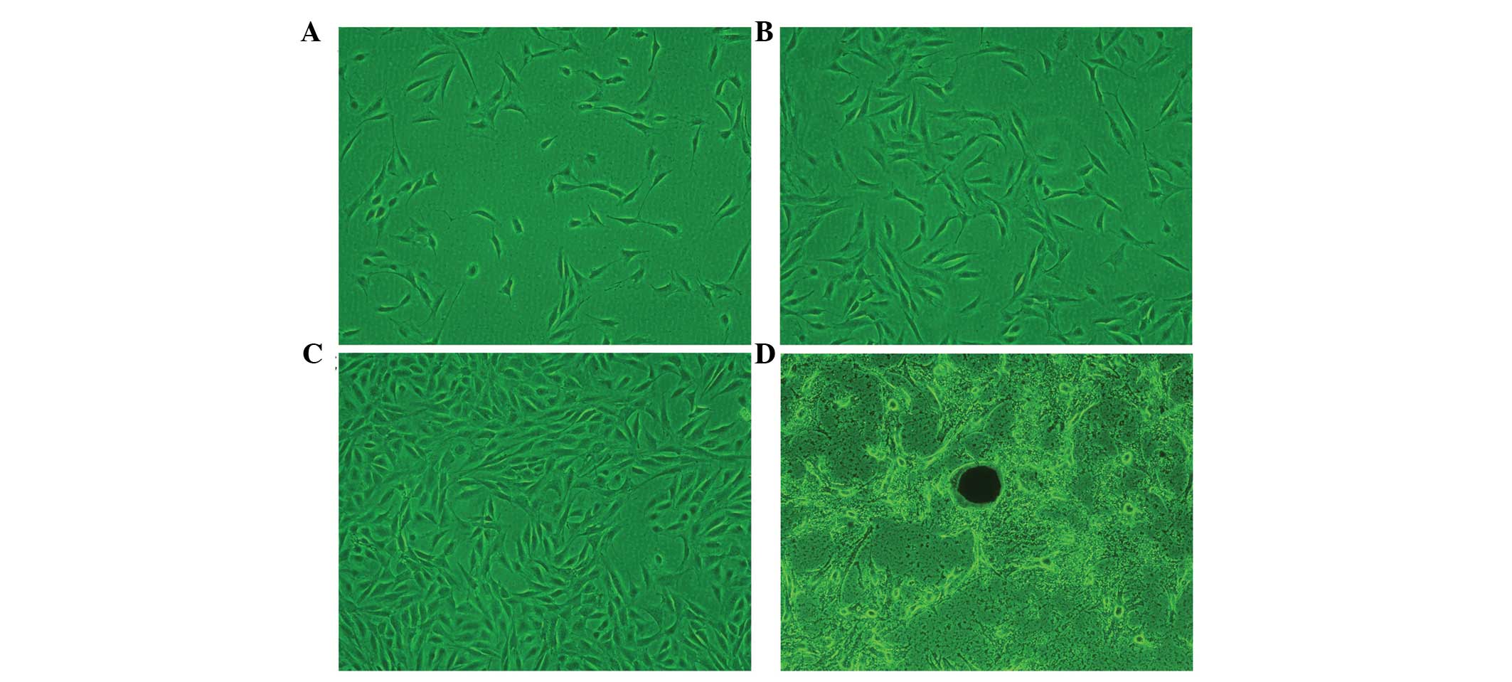
Cell morphology was examined using a light-inverted
microscope (CKX41; Olympus Corporation, Tokyo, Japan) (Fig. 1), in which the hFOB1.19 osteoblast
cells demonstrated a high rate of survival and rapid
proliferation.
In the primary cells, the hFOB1.19 osteoblast cells
exhibited a round morphology. After 12 h, the hFOB1.19 cells were
sparsely attached to the culture flasks, and exhibited a
fibroblast-like, spindle-shaped morphology. As the cell number
increased and the cells continued to grow, a substantial number of
cells on day 3 had adhered to the surface of the culture flask, and
the majority of the cells had become large and flattened, with a
polygonal or triangular shape. After 6 days of culture, the cells
showed adequate growth and exhibited active proliferation. A higher
density was observed in the primary and transformed cells, which
did not change throughout the different passages. The osteoblast
cells had become well-formed mineral nodules at day 18 (Fig. 1).
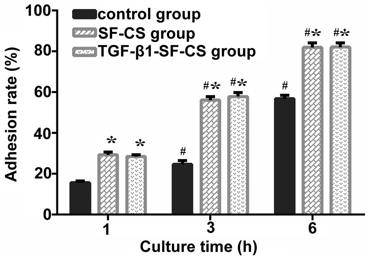
Adhesion rate of the hFOB1.19 osteoblast
cells is not altered by TGF-β1
Following culture for 1, 3 and 6 h, cell adhesion
rate increased with increasing duration in the three groups. The
adhesion rates in the TGF-β1-SF-CS group and SF-CS group were
higher, compared with that in the control group (P<0.01;
Fig. 2). After 1 h, the adhesion
rate of the major populations of cells in the SF-CS scaffolds were
almost the same as those in the TGF-β1-SF-CS scaffolds, with no
significant differences between the scaffold groups (P>0.05).
The rapid time-course of osteoblast cell adhesion to the SF-CS and
TGF-β1-SF-CS scaffolds was significant, exhibiting 29% adhesion by
60 min post-seeding. After 3 and 6 h, there were significant
increases in the numbers of adherent cells, compared with the
numbers after 1 h (Fig. 2). The
adhesion rates in the TGF-β1-SF-CS scaffold and SF-CS scaffold
groups were significantly higher, compared with that in the control
group (P<0.01), however, there remained no significant
differences between the two scaffold groups (P>0.05). These
results confirmed that the SF-CS scaffold increased the adhesion of
the osteoblasts cells. The results also revealed that TGF-β1 did
not promote hFOB1.19 osteoblast cell adhesion in the SF-CS
scaffolds.
SEM images of hFOB1.19 cells cultured
with the TGF-β-SF-CS scaffolds indicate biocompaitibilty
The SEM images showed randomly distributed cells on
the surface and inside the TGF-β1-SF-CS scaffold and SF-CS
scaffold.
On examining the growth of cells on the scaffold at
1 day, scattered round cells had adhered to the scaffold and a
large number of particles were observed on the cell surface. Cell
microfilaments were connected with the scaffold material (Fig. 3A).
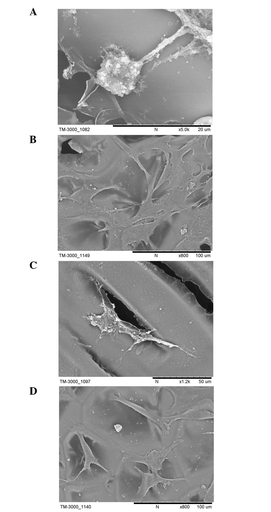 | Figure 3Scanning electron microscopy images of
the hFOB1.19 cells cultured with the transforming growth
factor-β1-silk fibroin-chitosan scaffold for (A) 1 day, scattered
round cells had adhered to the scaffold and a large number of
particles were observed on the cell surface, cell microfilaments
were connected with the scaffold material; (B) 3 days, single round
cells had become polygonal or triangular in shape, granular and
filiform substances were observed around the cells and cell
microfilaments and pseudopodia were tightly connected to the
scaffold; and (C) 7 days, the cells exhibited a typical osteoblast
cell morphology, and adhered tightly on the scaffold surface or
inside via the formation of lamellipodia and filopodia, indicative
of cell spreading. A number of cells present were observed to
extend towards other cells in the culture. (D) hFOB1.19 cells
cultured with the SF-CS scaffold at 7 days, fewer and more sparse
attachment of cells was observed compared with the TGF-β1-SF-CS
scaffolds. TGF-β1, transforming growth factor-β1; SF, silk fibroin;
CS, chitosan. |
After 3 days, a large number of the cells had
adhered to the surface and pores of the scaffold. The cell
aggregates had become enlarged and actively proliferating. The SEM
results showed that the single round cells had become polygonal or
triangular in shape. Granular and filiform substances were observed
around the cells, and cell microfilaments and pseudopodia were
tightly connected to the scaffold (Fig. 3B).
After 7 days, an increased number of cells had
adhered to the surface and pores of the scaffold, compared with the
number at 3 days, and the cells had grown and proliferated well.
The cells exhibited a typical osteoblast cell morphology, and
adhered tightly on the scaffold surface or inside via the formation
of lamellipodia and filopodia, indicative of cell spreading. A
number of cells present were observe to extend towards other cells
in the culture. The cells were well attached to the TGF-β1-SF-CS
scaffolds and appeared to have spread. The majority of the cells
had collected together to form cellular aggregations, which is
crucial for the viability and function of hFOB1.19 cells in
vitro. The SF-CS scaffolds showed fewer or more sparse
attachment of cells, compared with the TGF-β1-SF-CS scaffolds
(Fig. 3C).
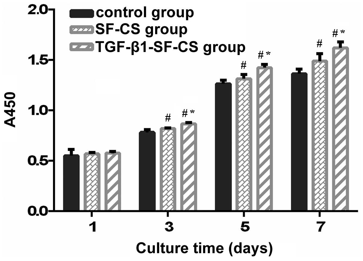
TGF-β1 promotes the proliferation of
hFOB1.19 cells
The proliferation rats of the hFOB1.19 cells on the
VEGF-SF-CS scaffolds, SF-CS scaffolds and control group cultured
for 1, 3, 5 and 7 days were compared using a CCK-8 assay. The
absorbance values for the TGF-β1-SF-CS scaffolds and SF-CS
scaffolds gradually increased, indicating significant cell growth
within the two scaffolds (Fig. 4).
The hFOB1.19 cells cultured without scaffolds showed similar
results. The number of cells increased with culture duration in all
three groups. hFOB1.19 cell proliferation was observed in the
TGF-β1-SF-CS scaffolds and SF-CS scaffolds, and there were no
significant differences between these groups and the cells cultured
in the control group after 1 day (P>0.05). However, after 3
days, the numbers of hFOB1.19 cells cultured in the TGF-β1-SF-CS
scaffolds were higher than thoses cultured in the SF-CS scaffolds,
and the numbers of hFOB1.19 cells cultured in the two scaffold
groups were significantly higher, compared with the number of cells
cultured without scaffolds (P<0.05).
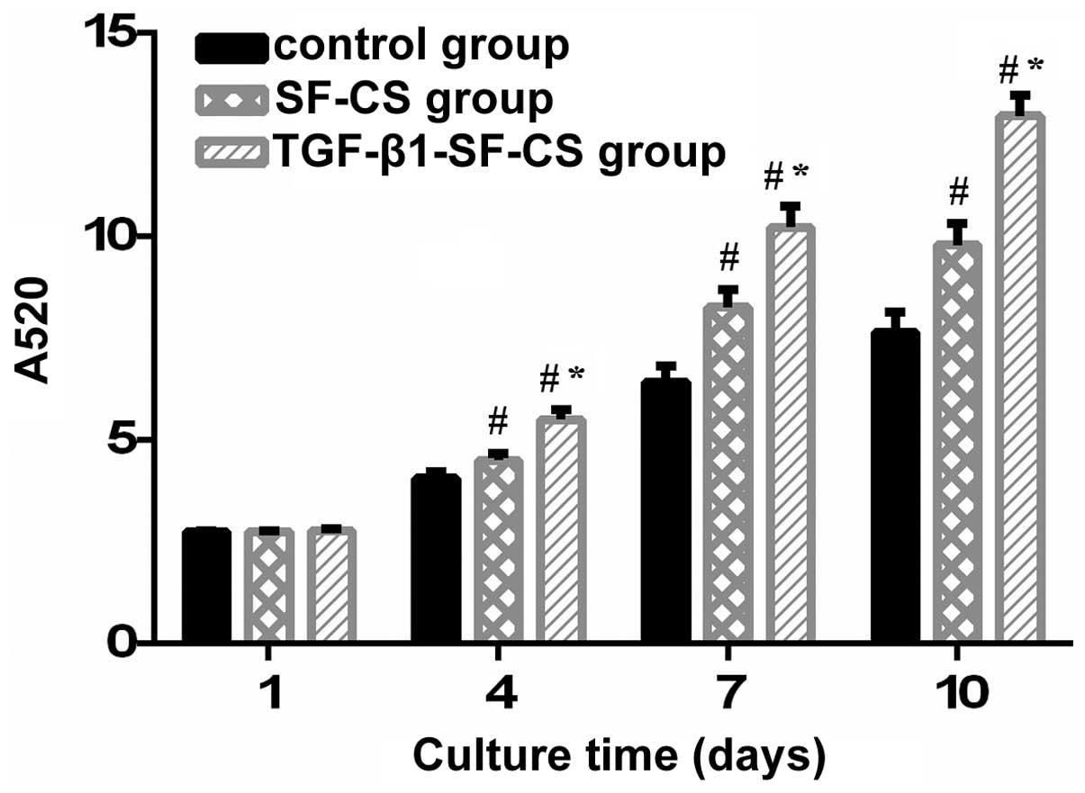
TGF-β1 promotes the activity of ALP in
hFOB1.19 cells
The ALP activity of the hFOB1.19 cells cultured in
the TGF-β1-SF-CS scaffolds, SF-CS scaffolds and control group are
shown in Fig. 5. In the
measurements of total ALP activity during the culture period
between days 1 and 4, no significant difference in OD values were
identified between the TGF-β1-SF-CS, SF-CS and control groups
(P>0.05). However, between days 4 and 10, significant
differences in the OD values were found between the scaffold groups
and the control group (P<0.01). The OD values in the
TGF-β1-SF-CS groups were the highest. This suggested that TGF-β1
had an effect on the activity of the hFOB1.19 cells. As the
duration of culture increased between days 4 and 10, the growth
trend of the OD values in three groups increased rapidly, and the
OD values peaked after 10 days.
Discussion
The development of biomimetic scaffolds to enhance
bone-formation has largely focused on altering the chemical
composition of scaffolds. Therefore, the present study combined
SF-CS scaffolds with the TGF-β1 growth factor, in order to promote
bone-formation. The results of the present study demonstrated that
the SF-CS scaffolds provided a suitable environment for osteoblast
cell proliferation and differentiation in the presence of TGF-β1
in vitro.
The response of cells to TGF-β1 varies depending on
the dose, length of exposure and target tissue (32). A prolonged exposure and high dose
of TGF-β1 may result in pathological conditions, including
extensive fibrosis, hypertrophic scarring and the formation of
osteophytes (32,33). An engineered system capable of
inducing cell-mediated TGF-β1 activation can begin to address these
problems through bone tissue engineering. The novel strategy
investigated in the present study aimed to incorporate TGF-β1 onto
an SF-CS scaffold and allow osteoblast cells to sense and activate
TGF-β1, as required.
When fabricating the TGF-β1-SF-CS scaffold for bone
tissue engineering, it is critical to maintain the bioactivity of
TGF-β1 as, during the course of manufacture, TGF-β1 bioactivity is
most likely to be destroyed in lyophilization and methanol
crosslinking. In lyophilization, the use of a cryoprotector may
reduce or even prevent the effects of stress on the activity of the
protein (34). Silk protein and
chitosan can be used as cryoprotectors to maintain the activity of
TGF-β1 (35,36). Klibanov (37) reported that the conformation of
proteins treated with low concentration methanol remain unchanged,
and the covalent structure of the protein molecule is not
destroyed. In the present study, the following measures were taking
in constructing the TGF-β1-SF-CS scaffold to prevent the TGF-β1
bioactivity from being destroyed: (i) Lyophilization temperature
was set at −40°C, and silk and chitosan were used as cryoprotectors
to further maintain TGF-β1 activity; (ii) methanol concentration
was <10%. Through ALP and CCK-8 assays, it was evident that no
significant degradation of TGF-β1 bioactivity had occurred.
In the present study, the SF-CS scaffold was
manufactured in aqueous solution to incorporate the active TGF-β1
molecule without damaging TGF-β1 activity. Mandal and Kundu
(38) utilized lyophilization to
prepare a 3D silk fibroin scaffold in bovine serum albumin, and
then examined its in vitro release and performance. The
results of the ALP and CCK-8 assays in the present study result
revealed that, in the TGF-β1-SF-CS scaffold, TGF-β1 was evenly and
continuously released within a certain period.
In the present study, osteoblast cell morphology was
investigated using SEM to further evaluate the biocompatibility of
the SF-CS and TGF-β1-SF-CS scaffolds. The seeding of hFOB1.19 on
TGF-β1-SF-CS scaffolds up to 7 days resulted in polygonal or
triangular cell morphologies, similar to what is typically observed
in standard cell culture flasks. These observations confirmed cell
survival and proliferation on the TGF-β1-SF-CS scaffolds, assuring
its biocompatibility. The evidence of cell-cell interactions and
cell spreading are considered to be signs of healthy cells and
indicative of a non-cytotoxic response of the cells on TGF-β1-SF-CS
scaffolds (39,40).
The time course of the adhesion of osteoblast cells
to the SF-CS and TGF-β1-SF-CS scaffolds clearly demonstrated that
adhesion of the hFOB1.19 to the SF-CS and TGF-β1-SF-CS scaffolds
occurred within minutes. The rapid time-course of osteoblast cell
adhesion to the SF-CS and TGF-β1-SF-CS scaffolds was significant,
exhibiting 29% adhesion by 60 min post-seeding. The adhesion rate
of the hFOB1.19 cells in the TGF-β1-SF-CS group and SF-CS group
significantly exceeded those in the control group (P<0.05),
although no statistically significant differences (P>0.05) were
observed between the SF-CS and TGF-β1-SF-CS scaffold groups.
Previous studies have indicated that SF-CS scaffolds increase the
adhesion and growth of osteoblast cells. The observations in the
present study achieved the primary objective in determining whether
SF-CS scaffolds have the potential to establish an adhesive
interface with osteoblast cells in vitro. The results also
confirmed that TGF-β1 was unable to promote the adhesion of
hFOB1.19 osteoblast cells to the SF-CS scaffolds.
The levels of cell growth and proliferation on the
scaffolds and control group were assessed in the present study
using CCK-8 and ALP assays in vitro. It was observed that
the absor-bance index of the assessed groups increased with the
increase in culture duration. After 1 day, no statistically
significant differences (P>0.05) were observed in cell activity
among the three groups. The activities of the hFOB1.19 cells
cultured on the TGF-β1-SF-CS and SF-CS scaffolds were higher than
that of the cells cultured in the control group after 3 days in
culture, which suggested that the scaffolds were beneficial to cell
development. The number of osteoblast cells on the TGF-β1-SF-CS
scaffolds were significantly higher, compared with those on the
SF-CS scaffolds. These results indicated that TGF-β1 significantly
promoted the attachment and proliferation of the osteoblast cells.
The results of the present study also support evidence from in
vitro experiments that TGF-β1 may regulate bone formation
through a direct effect on osteoblasts. It has been reported that
human TGF-β1 binds to osteoblasts in culture, and is capable of
inducing migration and alkaline phosphatase activity (41). The results of the present study
indicated differences in the activities of the osteoblast cells in
the different scaffold groups. The reason for the differences in
the production of extracellular matrices in the different scaffolds
may be due to the differences in the chemical compositions of the
matrices.
The enhancement of osteoblast cell proliferation and
activity by TGF-β1 was time-dependent. TGF-β1 did not significantly
promote the activity or proliferation of osteoblast cells during
the first 3 days of culture, however, it had a marked effect when
cultured between days 3 and 10, demonstrating that the effect of
TGF-β1, which promotes the activity and proliferation of osteoblast
cells, was time-dependent. These results indicated that the TGF-β1
delivery strategies have a marked affect on osteogenic
differentiation and require consideration when designing such a
delivery system.
Previous results have indicated that this 3D
environment, coupled with growth factor application, may be key in
recreating an in vitro tissue-specific microenvironment for
improving bone repair strategies. The results of the present study
support the suggested that TGF-β1 is important for promoting
hFOB1.19 osteoblast cell extracellular matrix production. Based on
the observations in the present study following 10 days of culture,
future bone tissue engineering investigations can be performed to
focus on how TGF-β1 combines with the SF-CS scaffold, whether the
sustained release of TGF-β1 is maintained and enhanced over longer
durations, and whether the culture of different type of cells on
the SF-CS-SF-CS scaffolds yield similar outcomes.
In conclusion, the results of the present study
confirmed that the direct effects of TGF-β1 on osteoblast cells
were an increase in the proliferation, migration, differentiation
and survival of the cells. The TGF-β1-SF-CS scaffolds were
biocompatible and had no negative effects on the hFOB1.19
osteoblast cells in vitro. The SF-CS composite material may
offer potential as an ideal scaffold material for bone tissue
engineering.
Acknowledgments
The current study was supported by the Science and
Technology Plan Project of Liaoning Province (grant. no. 20
12-B-00002012225082).
References
|
1
|
Vacanti JP and Langer R: Tissue
engineering: The design and fabrication of living replacement
devices for surgical reconstruction and transplantation. Lancet.
354(Supp 1): SI32–SI34. 1999. View Article : Google Scholar : PubMed/NCBI
|
|
2
|
Scheller EL, Krebsbach PH and Kohn DH:
Tissue engineering: State of the art in oral rehabilitation. J Oral
Rehabil. 36:368–389. 2009. View Article : Google Scholar : PubMed/NCBI
|
|
3
|
Hankenson KD, Dishowitz M, Gray C and
Schenker M: Angiogenesis in bone regeneration. Injury. 42:556–561.
2011. View Article : Google Scholar : PubMed/NCBI
|
|
4
|
Marsell R and Einhorn TA: The biology of
fracture healing. Injury. 42:551–555. 2011. View Article : Google Scholar : PubMed/NCBI
|
|
5
|
Muschler GF, Nakamoto C and Griffith LG:
Engineering principles of clinical cell-based tissue engineering. J
Bone Joint Surg Am. 86-A:1541–1558. 2004.PubMed/NCBI
|
|
6
|
Grayson WL, Chao PH, Marolt D, Kaplan DL
and Vunjak-Novakovic G: Engineering custom-designed osteochondral
tissue grafts. Trends Biotechnol. 26:181–189. 2008. View Article : Google Scholar : PubMed/NCBI
|
|
7
|
Vunjak-Novakovic G, Meinel L, Altman G and
Kaplan D: Bioreactor cultivation of osteochondral grafts. Orthod
Craniofac Res. 8:209–218. 2005. View Article : Google Scholar : PubMed/NCBI
|
|
8
|
Adams JC and Watt FM: Regulation of
development and differentiation by the extracellular matrix.
Development. 117:1183–1198. 1993.PubMed/NCBI
|
|
9
|
Zhang LH and Zhang QQ: Study on the
osteoblast and the growth factors of bone tissue engineering.
Zhongguo Yi Xue Ke Xue Yuan Xue Bao. 23:631–637. 2001.In
Chinese.
|
|
10
|
Bonewald LF and Dallas SL: Role of active
and latent transforming growth factor beta in bone formation. J
Cell Biochem. 55:350–357. 1994. View Article : Google Scholar : PubMed/NCBI
|
|
11
|
Saitta B, Passarini J, Sareen D, Ornelas
L, Sahabian A, Argade S, Krakow D, Cohn DH, Svendsen CN and Rimoin
DL: Patient-derived skeletal dysplasia induced pluripotent stem
cells display abnormal chondrogenic marker expression and
regulation by BMP2 and TGF-β1. Stem Cells Dev. 23:1464–1478. 2014.
View Article : Google Scholar : PubMed/NCBI
|
|
12
|
Lind M: Growth factor stimulation of bone
healing. Effects on osteoblasts, osteomies and implants fixation.
Acta Orthop Scand Suppl. 283:2–37. 1998.PubMed/NCBI
|
|
13
|
Connelly JT, Wilson CG and Levenston ME:
Characterization of proteoglycan production and processing by
chondrocytes and BMSCs in tissue engineered constructs.
Osteoarthritis Cartilage. 16:1092–1100. 2008. View Article : Google Scholar : PubMed/NCBI
|
|
14
|
Mouw JK, Connelly JT, Wilson CG, Michael
KE and Levenston ME: Dynamic compression regulates the expression
and synthesis of chondrocyte-specific matrix molecules in bone
marrow stromal cells. Stem Cells. 25:655–663. 2007. View Article : Google Scholar
|
|
15
|
Johnstone B, Hering TM, Caplan AI,
Goldberg VM and Yoo JU: In vitro chondrogenesis of bone
marrow-derived mesenchymal progenitor cells. Exp Cell Res.
238:265–272. 1998. View Article : Google Scholar : PubMed/NCBI
|
|
16
|
Vehof JW, Haus MT, de Ruijter AE, Spauwen
PH and Jansen JA: Bone formation in transforming growth factor
beta-I-loaded titanium fiber mesh implants. Clin Oral Implants Res.
13:94–102. 2002. View Article : Google Scholar : PubMed/NCBI
|
|
17
|
Holland TA, Tessmar JK, Tabata Y and Mikos
AG: Transforming growth factor-beta 1 release from oligo
(poly(ethylene glycol) fumarate) hydrogels in conditions that model
the cartilage wound healing environment. J Control Release.
94:101–104. 2004. View Article : Google Scholar
|
|
18
|
Motoyama M, Deie M, Kanaya A, Nishimori M,
Miyamoto A, Yanada S, Adachi N and Ochi M: In vitro cartilage
formation using TGF-beta-immobilized magnetic beads and mesenchymal
stem cell-magnetic bead complexes under magnetic field conditions.
J Biomed Mater Res A. 92:196–204. 2010. View Article : Google Scholar
|
|
19
|
Park H, Temenoff JS, Tabata Y, Caplan AI,
Raphael RM, Jansen JA and Mikos AG: Effect of dual growth factor
delivery on chondrogenic differentiation of rabbit marrow
mesenchymal stem cells encapsulated in injectable hydrogel
composites. J Biomed Mater Res A. 88:889–897. 2009. View Article : Google Scholar
|
|
20
|
Fischer U, Hempel U, Becker D, Bierbaum S,
Scharnweber D, Worch H and Wenzel KW: Transforming growth factor
beta1 immobilized adsorptively on Ti6Al4V and collagen type I
coated Ti6Al4 V maintains its biological activity. Biomaterials.
24:2631–2641. 2003. View Article : Google Scholar : PubMed/NCBI
|
|
21
|
Dickhut A, Dexheimer V, Martin K, Lauinger
R, Heisel C and Richter W: Chondrogenesis of human mesenchymal stem
cells by local TGF-beta delivery in a biphasic resorbable carrier.
Tissue Eng Part A. 16:453–464. 2010. View Article : Google Scholar
|
|
22
|
Park JS, Yang HJ, Woo DG, Yang HN, Na K
and Park KH: Chondrogenic differentiation of mesenchymal stem cells
embedded in a scaffold by long-term release of TGF-beta3 complexed
with chondroitin sulfate. J Biomed Mater Res A. 92:806–816.
2010.
|
|
23
|
Murphy CM, O'Brien FJ, Little DG and
Schindeler A: Cell-scaffold interactions in the bone tissue
engineering triad. Eur Cell Mater. 26:120–132. 2013.PubMed/NCBI
|
|
24
|
Roberts AB and Sporn MB: Physiological
actions and clinical applications of transforming growth
factor-beta (TGF-beta). Growth Factors. 8:1–9. 1993. View Article : Google Scholar : PubMed/NCBI
|
|
25
|
D'Ippolito G, Schiller PC, Ricordi C, Roos
BA and Howard GA: Age-related osteogenic potential of mesenchymal
stromal stem cells from human vertebral bone marrow. J Bone Miner
Res. 14:1115–1122. 1999. View Article : Google Scholar : PubMed/NCBI
|
|
26
|
Kassem M, Kveiborg M and Eriksen EF:
Production and action of transforming growth factor-beta in human
osteoblast cultures: Dependence on cell differentiation and
modulation by calcitriol. Eur J Clin Invest. 30:429–437. 2000.
View Article : Google Scholar : PubMed/NCBI
|
|
27
|
Alliston T, Choy L, Ducy P, Karsenty G and
Derynck R: TGF-beta-induced repression of CBFA1 by Smad3 decreases
cbfa1 and osteocalcin expression and inhibits osteoblast
differentiation. EMBO J. 20:2254–2272. 2001. View Article : Google Scholar : PubMed/NCBI
|
|
28
|
Park SJ, Lee KY, Ha WS and Park SY:
Structural changes and their effect on mechanical properties of
silk fibroin/chitosan blends. Journal of Applied Polymer Science.
74:2571–2575. 1999. View Article : Google Scholar
|
|
29
|
El Sayed K, Marzahn U, John T, Hoyer M,
Zreiqat H, Witthuhn A, Kohl B, Haisch A and Schulze-Tanzil G:
PGA-associated heterotopic chondrocyte cocultures: Implications of
nasoseptal and auricular chondrocytes in articular cartilage
repair. J Tissue Eng Regen Med. 7:61–72. 2013. View Article : Google Scholar
|
|
30
|
De Santis R, Gloria A, Russo T, et al: A
basic approach toward the development of nanocomposite magnetic
scaffolds for advanced bone tissue engineering. Journal of Applied
Polymer Science. 122:3599–3605. 2011. View Article : Google Scholar
|
|
31
|
Gobin AS, Froude VE and Mathur AB:
Structural and mechanical characteristics of silk fibroin and
chitosan blend scaffolds for tissue regeneration. J Biomed Mater
Res A. 74:465–473. 2005. View Article : Google Scholar : PubMed/NCBI
|
|
32
|
Saharinen J, Hyytiäinen M, Taipale J and
Keski-Oja J: Latent transforming growth factor-beta binding
proteins (LTBPs)-structural extracellular matrix proteins for
targeting TGF-beta action. Cytokine Growth Factor Rev. 10:99–117.
1999. View Article : Google Scholar
|
|
33
|
Mierisch CM, Cohen SB, Jordan LC,
Robertson PG, Balian G and Diduch DR: Transforming growth
factor-beta in calcium alginate beads for the treatment of
articular cartilage defects in the rabbit. Arthroscopy. 18:892–900.
2002. View Article : Google Scholar : PubMed/NCBI
|
|
34
|
Haines AH: Non-equivalence of D- and
L-trehalose in stabilising alkaline phosphatase against
freeze-drying and thermal stress. Is chiral recognition involved?
Org Biomol Chem. 4:702–706. 2006. View
Article : Google Scholar : PubMed/NCBI
|
|
35
|
Kreilgaard L, Jones LS, Randolph TW,
Frokjaer S, Flink JM, Manning MC and Carpenter JF: Effect of Tween
20 on freeze-thawing- and agitation-induced aggregation of
recombinant human factor XIII. J Pharm Sci. 87:1597–1603. 1998.
|
|
36
|
Pikal-Cleland KA and Carpenter JF:
Lyophilization-induced protein denaturation in phosphate buffer
systems: Monomeric and tetrameric beta-galactosidase. J Pharm Sci.
90:1255–1268. 2001. View
Article : Google Scholar : PubMed/NCBI
|
|
37
|
Klibanov AM: Enzymatic catalysis in
anhydrous organic solvents. Trends Biochem Sci. 14:141–144. 1989.
View Article : Google Scholar : PubMed/NCBI
|
|
38
|
Mandal BB and Kundu SC: Calcium alginate
beads embedded in silk fibroin as 3D dual drug releasing scaffolds.
Biomaterials. 30:5170–5177. 2009. View Article : Google Scholar : PubMed/NCBI
|
|
39
|
Nwe N, Furuike T and Tamura H: The
mechanical and biological properties of chitosan scaffolds for
tissue regeneration templates are significantly enhanced by
chitosan from gongronella butleri. Materials. 2:374–398. 2009.
View Article : Google Scholar
|
|
40
|
Neamnark A, Sanchavanakit N, Pavasant P,
Bunaprasert T, Supaphol P and Rujiravanit R: In vitro
biocompatibility evaluations of hexanoyl chitosan film.
Carbohydrate Polymers. 68:166–172. 2007. View Article : Google Scholar
|
|
41
|
Bonewald LF and Mundy GR: Role of
transforming growth factor beta in bone remodeling: A review.
Connect Tissue Res. 23:201–208. 1989. View Article : Google Scholar : PubMed/NCBI
|