Introduction
Periodontitis is a common inflammatory disease,
which induces destruction of the tooth-supporting apparatus and may
lead to tooth loss. However, current or emerging treatment options
for periodontitis have limited and variable outcomes, or have yet
to be developed for clinical use (1). The use of tissue engineering may be a
possible therapeutic strategy for periodontal regeneration
(2).
Periodontal ligament stem cells (PDLSCs) are
effective and readily available cells for periodontal regeneration
(3). However, due to the
limitations in acquiring these cells, their application in clinical
therapy is limited. The gingiva, which is an important component of
periodontal tissue, exhibits promising regenerative ability and is
able to rapidly repair injury. In addition, almost no scars remain
after healing (4), it is available
in large quantities and is easy to acquire. Previous studies have
identified the presence of mesenchymal stem cells (MSCs) in human
gingival connective tissue, and have reported on their
morphological and functional characteristics (4–6).
Therefore, gingival-derived MSCs (GMSCs) have recently received
increased attention (5,7,8).
Cell sheet technology refers to a scaffold-free
tissue reconstruction technique. Based on a unique temperature
responsive culture dish, cultured cells can be non-invasively
harvested as intact contiguous sheets along with their deposited
extracellular matrix (9,10). Cell sheets may be used to avoid
unforeseeable complications, including biomaterial degradation,
inflammation and limited cell-seeding efficiency (9,10).
It has the natural reaggregation potential of monodispersed cells
and the advantage of an affluent natural matrix (11–13).
It is well-known that tooth generation depends on
the interaction between dental epithelium and MSCs (14); this interaction is also important
in root and periodontal tissue formation (15–17).
Therefore, it has being suggested that the interaction between
dental epithelium and MSCs may be important in periodontal
regeneration. During tooth development, the periodontium is formed
by the apical mesenchyme of the tooth germ, which is associated
with the expression of several signaling molecules, including
transforming growth factor-β, Notch-1, fibroblast growth factors or
bone morphogenetic proteins (18–22).
The present study used apical tooth germ-conditioned
medium (APTG-CM) to induce differentiation of GMSCs and PDLSCs;
subsequently, transplant complexes containing dentin slices,
ceramic bovine bone (CBB), and GMSC and PDLSC sheets were
generated. The complex was subcutaneously transplanted into athymic
mice for 8 weeks to observe periodontal regeneration.
Materials and methods
Isolation and characterization of PDLSCs
and GMSCs
Gingival connective tissue samples were obtained
from 10 healthy male donors (age, 15–25 years) at the Stomatology
Hospital Affiliated to Tongji University (Shanghai, China). Under
local anesthetic a ~2×2×1 mm sample of keratinized gingival tissue
was obtained from the maxillary tuberosity. In addition, human
periodontal ligaments were obtained from healthy premolars
extracted from 10 male donors (age, 13–25 years) undergoing
orthodontic treatment at the Stomatology Hospital Affiliated to
Tongji University (Shanghai, China). Sample collection conformed to
a protocol approved by the ethical authorities at the School of
Stomatology, Tongji University (Shanghai, China) and all donors
provided informed consent.
Human GMSCs and PDLSCs were isolated as previously
described (3,6). Using the limiting dilution technique,
single-colony derived human GMSCs and PDLSCs were isolated and
expanded to obtain homogeneous populations. The cells were
confirmed by fluorescence-activated cell sorting analysis using
antibodies against CD45 (cat. no. FAB1430A), CD105 (cat. no.
FAB10971P) and STRO-1 (cat. no. MAB1038; all obtained from R&D
Systems, Minneapolis, MN, USA), according to the manufacturer's
protocol. The two types of stem cell were stimulated with
osteogenic conditioned medium [100 nM dexamethasone, 5 mM
β-glycerophosphate, 50 mg/ml ascorbate phosphate and 5% fetal
bovine serum (FBS; Gibco; Thermo Fisher Scientific, Inc., Waltham,
MA, USA)] and adipogenic conditioned medium [1 mM dexamethasone, 1
mg/ml insulin, 0.5 mM 3-isobutyl-1-methylxanthine (Sigma-Aldrich;
Merck Millipore, Darmstadt, Germany) and 5% FBS]. The conditions
used for the induction of calcium accumulation and adipogenesis
were the same as previously reported (12). Normal medium was used in the blank
control group. Calcium accumulation was detected using 2% Alizarin
Red (pH 4.2) staining (Sigma-Aldrich; Merck Millipore). Adipogenic
cultures were stained with fresh Oil Red O solution (Sigma-Aldrich;
Merck Millipore). Staining was observed by light microscopy (DX51;
Olympus Corporation, Tokyo, Japan).
APTG-CM and co-culture system
All experiments involving the use of animals were
reviewed and approved by the Animal Care Committee of Tongji
University. Five-day postnatal Sprague-Dawley rats (n=30; male and
female; weight, 10±2 g; Shanghai SLAC Laboratory Animal Co., Ltd.,
Shanghai, China) were bred in plastic cages (5 mice/cage) under
standard laboratory conditions with a 12 h light/dark cycle, at
20°C with 48% humidity. Food and water were provided. The rats were
sacrificed by decapitation under isoflurane anesthesia. Developing
mandibular first molar tooth germs were then dissected from the
jaws. The apical portion of the tooth germ was separated as
previously reported (14), and was
cultured in alpha-minimum essential medium (alpha-MEM; Gibco;
Thermo Fisher Scientific, Inc.) supplemented with 10% FBS. The
primary APTG culture medium was changed every 48 h until 100%
confluence for supernatant collection was reached. The cells were
then centrifuged at 2,000 × g for 15 min at 4°C. The
supernatants were mixed with an equal volume of fresh alpha-MEM
supplemented with 10% FBS and 50 µg/ml ascorbic phosphate
(Sigma-Aldrich; Merck Millipore), which was used as APTG-CM for
PDLSCs and GMSCs co-culture. Alpha-MEM supplemented with 10% FBS
and 50 µg/ml ascorbic phosphate was used as a control
medium.
Cell cycle analysis
Following 10 days of co-culture at 37°C with
APTG-CM, single-cell suspensions of GMSCs and PDLSCs were harvested
and were washed twice with phosphate-buffered saline (PBS).
Subsequently, the cells were fixed in cold 70% dehydrated alcohol
at 4°C for 24 h. After further washing, cell suspensions were
treated with RNase A and then stained with propidium iodide (PI;
Sigma-Aldrich; Merck Millipore) at 4°C for 30 min. The content of
DNA was assayed by flow cytometry to confirm the cell cycle
analysis and subjected to cell cycle analysis using MultiCycle
software (Beckman Coulter, Inc., Brea, CA, USA. One million cells
were counted per sample.
Alkaline phosphatase (ALP) activity
assay
An ALP activity assay was performed to determine the
influence of APTG-CM co-culture. Single-cell suspensions of GMSCs
and PDLSCs were seeded at a density of 1×10³ cells/well
in 96-well plates. After 3, 6, 9, 12 and 15 days of co-culture with
APTG-CM, the ALP activity of GMSCs and PDLSCs was detected using an
ALP assay kit (Nanjing Jiancheng Bioengineering Institute, Nanjing,
China.). The results were measured at 405 nm in a spectrophotometer
using a multiplate reader (BioTek Instruments, Inc., Winooski, VT,
USA).
Reverse transcription-quantitative
polymerase chain reaction (RT-qPCR) analysis
The co-cultured GMSCs and PDLSCs, and the control
cells, were harvested and RNA was extracted using
TRIzol® reagent (Invitrogen; Thermo Fisher Scientific,
Inc.) according to the manufacturer's protocol. The total RNA
concentration was assessed using a NanoDrop 2000 spectrophotometer
(Thermo Fisher Scientific, Inc.). RNA (1.0 mg) was reversed
transcribed into cDNA using PrimeScript First Strand cDNA Synthesis
kit (Takara Biotechnology Co., Ltd., Dalian, China) according to
the manufacturer's protocols. Subsequently cDNA synthesis was
conducted using a Superscript II First-strand cDNA synthesis kit
(Invitrogen; Thermo Fisher Scientific, Inc.), and RT-qPCR was
performed using a MyiQ2 Real-Time PCR Detection system (Bio-Rad
Laboratories, Inc., Hercules, CA, USA). The thermocycling
conditions were as follows: 95°C for 5 min; 30 cycles of 95°C for
30 sec, 56°C for 30 sec and extension at 72°C for 1 min. Relative
quantities of mRNA were calculated using the 2−ΔΔCq
method (23) and normalized to
housekeeping gene β-actin. The experiment was repeated ≥3 times.
Sequences for the sense and anti-sense primers (Sangon Biotech Co.,
Ltd., Shanghai, China) used in the present study are listed in
Table I.
 | Table IOligonucleotide primer sequences used
in polymerase chain reaction. |
Table I
Oligonucleotide primer sequences used
in polymerase chain reaction.
| Gene | Primer | Sequence |
|---|
| Osteocalcin | Forward |
5′-GGGTCAGGAGGAGAATCGT-3′ |
| Reverse |
5′-GTGTCTTAGCAGGCAGGGA-3′ |
| Bone
sialoprotein | Forward |
5′-CTGCTTCCTCACTCCAGGAC-3′ |
| Reverse |
5′-ATTGAGAAAGCACAGGCCAT-3′ |
| Alkaline
phosphatase | Forward |
5′-GCCAAGAAAGCAGGAAAGTC-3′ |
| Reverse |
5′-GAGTACCAGTTGCGGTTCAC-3′ |
| Type I collagen | Forward |
5′-CTGACCTTCCTGCGCCTGATGTCC-3′ |
| Reverse |
5′-GTCTGGGGCACCAACGTCCAAGGG-3′ |
| Cementum-derived
protein 23 | Forward |
5′-ATGGGCACATCAAGCACTGA-3′ |
| Reverse |
5′-CCCCATTAGTGTCATCCTGC-3′ |
| β-actin | Forward |
5′-CAGGCTGTGCTATCCCTGTA-3′ |
| Reverse |
5′-CATACCCCTCGTAGATGGGC-3′ |
GMSC and PDLSC sheets for the
regeneration of cementum/periodontal ligament (PDL)-like complexes
in mice
Single colony-derived GMSCs and PDLSCs wee cultured
for 2 weeks to a density to form cell sheets, which were then
detached from the bottom of the culture plates using a cell
scraper. Cell sheets were used to wrap dentin slices (from root of
tooth; 2×3×0.5 mm) and CBB (Research and Development Center for
Tissue Engineering, Fourth Military Medical University, Xi'an,
China) was placed on either side. Surgical sutures were used to
fasten the complex, and transplanted into the subcutaneous tissue
of the upper backs of six-week-old immunodeficient mice (n=30;
weight, 20±2 g; Shanghai SLAC Laboratory Animal Co., Ltd.). A total
of 30 complexes (10×3 groups) were implanted. Each immunodeficient
mouse received one of three different complexes (APTG-CM GMSC sheet
+ dentin + CBB; GMSC sheet + dentin + CBB; and APTG-CM PDLSC sheet
+ dentin + CBB). The implants were recovered 8 weeks
post-transplantation following decapitation under isoflurane
anesthesia, were fixed in 4% paraformaldehyde for 2 days and were
then decalcified for a further 28 days in 10% EDTA (pH 8.0), prior
to embedding in paraffin. For histological analysis, 5-µm
implant sections were prepared and stained with hematoxylin and
eosin (H&E; Sigma-Aldrich; Merck Millipore), according to the
manufacturer's protocol (24).
Staining was evaluated by light microscopy (DX51; Olympus
Corporation).
Statistical analyses
Data are presented as the mean ± standard deviation.
Experiments were repeated three times. Statistical significance was
analyzed using SPSS 17.0 software (SPSS, Inc., Chicago, IL, USA).
One-way analysis of variance followed by Tukey's test was used to
determine the significant differences among the groups. P<0.05
was considered to indicate a statistically significant difference.
All procedures were performed blinded.
Results
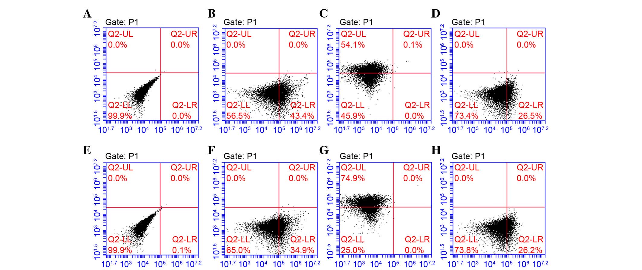
Characterization of PDLSCs and GMSCs
Using the limiting dilution technique, putative
clonogenic GMSCs and PDLSCs were isolated and purified. They were
identified by the positive expression of STRO-1 and CD105, and
negative expression of CD45 (Fig.
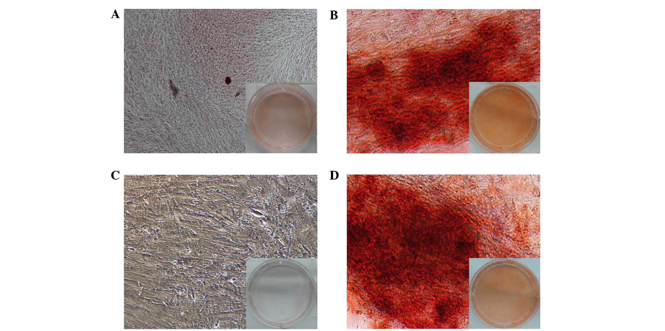
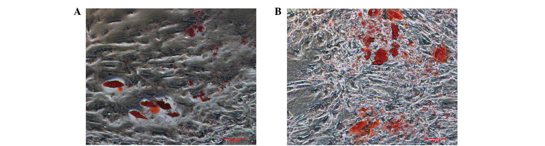
1). To investigate the differentiation potential of GMSCs and
PDLSCs, the osteogenic and adipogenic potential of the cells was
investigated. Osteogenesis was confirmed by the presence of
red-stained mineral nodules (Fig.
2), and adipogenesis was confirmed by intracellular lipid
droplet accumulation (Fig. 3).
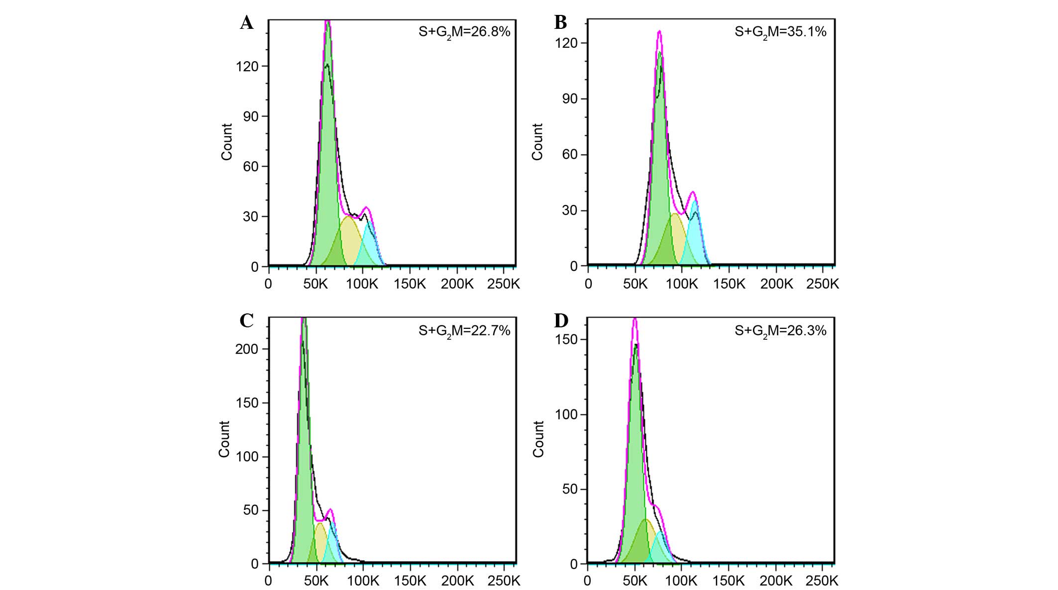
Cell cycle analysis of GMSCs and PDLSCs
co-cultured with APTG-CM
Cell cycle analysis was performed to investigate
whether APTG-CM was able to affect cell proliferation in
vitro. In GMSCs and PDLSCs co-cultured with APTG-CM a markedly
higher percentage of cells were present in S and G2/M
phases (GMSCs, 35.1%; PDLSCs, 26.3%) whereas a lower percentage of
cells were present in G0/G1 phase (Fig. 4) compared with in the untreated
cells. These results suggest that APTG-CM may exert a stimulating
effect on GMSCs and PDLSCs proliferation.
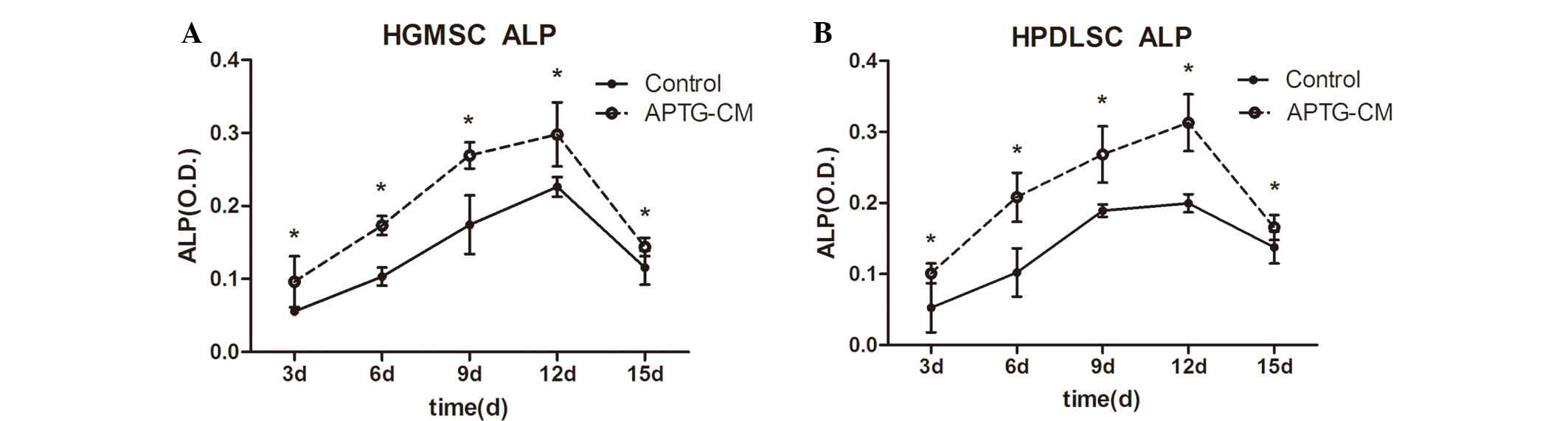
ALP activity assay
ALP is an early marker of dental MSC differentiation
towards the cementoblastic/osteoblastic phenotype (25). In the present study, ALP activity
of GMSCs and PDLSCs cultured with or without APTG-CM was measured.
ALP activity was markedly higher in the co-culture groups. The
effects of APTG-CM on GMSCs and PDLSCs increased in a
time-dependent manner, up to day 12, during the 15-day culture
period. As presented in Fig. 5
(P<0.05), ALP activity had steadily increased in the co-culture
group by day 3, and continued to increase until it reached a peak
at day 12, thus indicating that co-cultured cells continually
differentiate into hard tissue-forming cells. After day 12, ALP
activity gradually decreased until day 15, which may be correlated
with increasing mineral deposition.
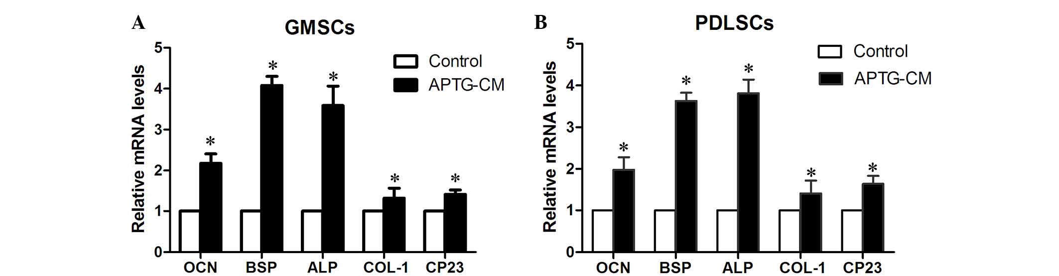
Gene expression in GMSCs and PDLSCs
co-cultured with APTG-CM
RT-PCR analysis was conducted following co-culture
of the two cell types with APTG-CM. The expression levels of bone-
and extracellular matrix-associated genes (Table I) were upregulated in GMSCs
(Fig. 6A) and PDLSCs (Fig. 6B) co-cultured with APTG-CM compared
with cells in the control group (P<0.05). The mRNA expression
patterns of bone-related genes suggest that GMSCs and PDLSCs
co-cultured with APTG-CM exhibit some molecular properties of
cementoblast-like cells.
Differentiation of GMSCs and PDLSCs
following heterotopic transplantation
A total of 30 complexes were harvested after 8 weeks
of transplantation and evaluated by light microscopy following
H&E staining (Fig. 7). In the
experimental group (APTG-CM GMSC sheet + dentin + CBB), a
cementum/PDL-like structure was regenerated on dentin surfaces, and
the collagen content and orientation of PDL-like tissues were
markedly obvious (Fig. 7B and C);
similar characteristics were observed in the positive control group
(APTG-CM PDLSC sheet + dentin + CBB; Fig. 7D and E). Furthermore, distinctive
dental cementum-like matrices were regenerated and clearly observed
on dentin after transplantation for 8 weeks. Conversely,
non-co-cultured GMSC sheet transplants (GMSC sheet + dentin + CBB)
formed small amounts of PDL-like tissue but no cementum-like
deposits (Fig. 7A). The images
were captured on a compound microscope.
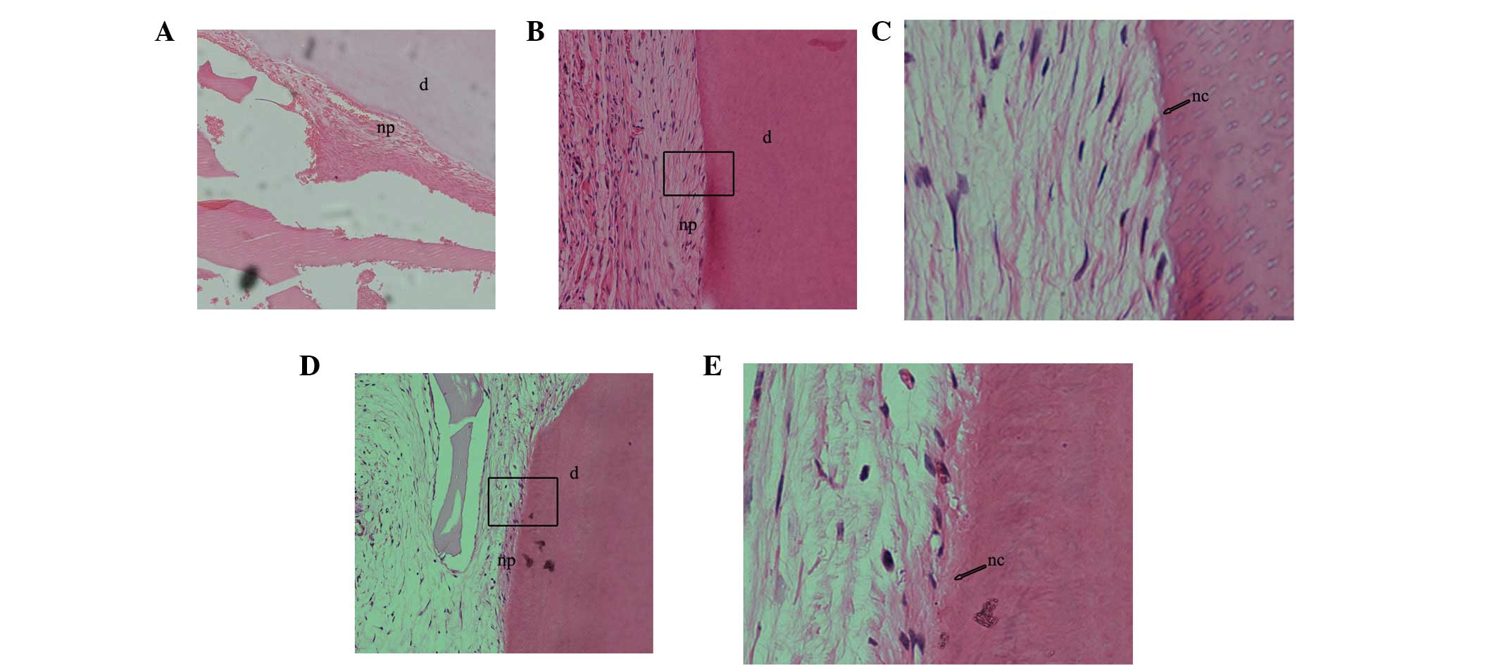 | Figure 7Following subcutaneous transplantation
for 8 weeks, the differentiation potential of gingival-derived
mesenchymal stem cells (GMSCs) and periodontal ligament stem cells
(PDLSCs) co-cultured with apical tooth germ-conditioned medium
(APTG-CM) were determined in vivo after hematoxylin and
eosin staining. (A) Non-co-cultured GMSCs formed a small amount of
periodontal ligament (PDL)-like tissue but no cementum-like
deposits; magnification, ×100. (B) GMSCs co-cultured with APTG-CM
exhibited tissue-regenerative capacity, were able to produce
cementum-like mineralized deposits on the surface of dentin slices,
and PDL-like collagen fibers connected with the newly formed
cementum-like tissues; magnification, ×100x. (C) Magnification of
the rectangular area in (B) (magnification, ×400). (D) PDLSCs
co-cultured with APTG-CM group also formed cementum and PDL-like
structures; magnification, ×100. (E) Magnification of the
rectangular area in (D) (magnification, ×400). np, new PDL-like
collagen fibers; nc: new cementum-like tissues; d, dentin. |
Discussion
Ideal periodontal reconstruction would involve the
development of Sharpey's fibers, which consist of collagen fibers.
Therefore, generation of a suitable regenerative periodontal
environment is of great importance. Furthermore, a mineralized
matrix is essential for periodontal regeneration. Certain
osteoinductive technologies, including the use of conditioned
medium from developing apical tooth germ cells, periapical follicle
stem cells and Hertwig's epithelial root sheath cells, as well as
osteoinductive medium, have previously been applied to create a
regenerative microenvironment (4,26–29).
The present study developed a periodontal complex using
APTG-CM-induced GMSC sheets, dentin slices and CBB for periodontal
regeneration. As hypothesized, transplantation of immunodeficient
mice with this periodontal complex resulted in the generation of a
dental cementum/PDL-like complex. These results indicated that the
development of this periodontal complex may provide an alternative
clinical approach for tooth reconstruction in future therapeutic
strategies.
In the present study, the APTG-CM used may contain
several molecular signals and growth factors that are necessary for
GMSC and PDLSC proliferation and differentiation, thus inducing
differentiation of GMSCs and PDLSCs towards a cementoblast
phenotype. As expected, the induced GMSCs exhibited several crucial
characteristics of cementoblast-like cells. Firstly, flow
cytometric cell cycle analysis demonstrated that cells co-cultured
with APTG-CM presented a higher percentage of cells in S and
G2/M phases. These results suggested that DNA synthesis
was enhanced and APTG-CM may provide an appropriate
microenvironment, which is necessary for the proliferation and
differentiation of GMSCs and PDLSCs. Secondly, ALP activity of
GMSCs and PDLSCs co-cultured with APTG-CM was increased. It is
well-known that when odontogenic mesenchymal cells are
differentiated towards cementum-like and osteogenic phenotypes, ALP
activity is an early marker (30).
The increase in ALP activity indicated that the mineralization
ability of GMSCs and PDLSCs co-cultured with APTG-CM was enhanced.
ALP is considered to be a prerequisite, which has a major role in
the formation of mineral tissue. Furthermore, several important
bone-associated genes, including osteocalcin, bone sialoprotein,
ALP, type I collagen and cementum-derived protein 23 were
upregulated in GMSCs and PDLSCs co-cultured with APG-CM, which may
enhance PDL-like tissue regeneration. These bone-associated genes
were also important markers associated with mineral extracellular
matrix (31). These changes may be
considered the mechanistic basis for altering the fates of GMSCs
and PDLSCs, and may contribute to the regeneration of periodontal
tissue. The results of an in vivo study detected similar
changes in GMSCs and PDLSCs induced by APTG-CM.
The results of the in vivo heterotopic
transplantation were consistent with the results of the in
vitro study. In the experimental and positive control groups,
PDL-like structures were regenerated on the dentin surfaces and
novel cementum matrix generation was detected. These findings were
the most important in the present study; to the best of our
knowledge, there are no reports regarding the use of GMSCs to
replace PDLSCs in periodontal regeneration, and the subsequent
regeneration of a PDL-like structure. However, it is worth noting
that the formation of PDL-like structures was continuous, this may
be due to the stability and compactness between cell sheets and
dentin slices due to the surgical sutures used, and may be
associated with novel cementum matrix generation, it is well-known
that acellular cementum regeneration is the gold standard of
periodontal regeneration (32).
Still, it was related to the environmental factors where the
complex located. In the present study, only heterotopic
transplantation was used to simulate periodontal regeneration. A
previous study demonstrated that the alveolar bone environment
differs from that of other areas (33) and is suitable for periodontal
regeneration (34). If the
complexes were orthotopic transplants into jaws, the
microenvironment would be better suited to periodontal
regeneration, this could be a target of future investigation.
Angiogenesis may also be important. In the present study
regeneration of the PDL-like structure was accompanied by
angiogenesis. Restoration of vascular supply was considered to be a
critical factor in successful tissue engineering (35,36).
Additional studies are required to confirm the differences between
periodontal tissue formed by GMSCs and PDLSCs.
In conclusion, the results of the present study
suggested that GMSCs induced by APTG-CM have the potential for
periodontal tissue regeneration. This may have general implications
for periodontal engineering. Future research is required to confirm
the differences between periodontal tissue formed by GMSCs and
PDLSCs.
Acknowledgments
The present study was supported by the National
Natural Science Foundation of China (grant no. 81271152).
References
|
1
|
Chen FM, Zhang J, Zhang M, An Y, Chen F
and Wu ZF: A review on endogenous regenerative technology in
periodontal regenerative medicine. Biomaterials. 31:7892–7927.
2010. View Article : Google Scholar : PubMed/NCBI
|
|
2
|
Chen FM and Jin Y: Periodontal tissue
engineering and regeneration: Current approaches and expanding
opportunities. Tissue Eng Part B Rev. 16:219–255. 2010. View Article : Google Scholar
|
|
3
|
Seo BM, Miura M, Gronthos S, Bartold PM,
Batouli S, Brahim J, Young M, Robey PG, Wang CY and Shi S:
Investigation of multi-potent postnatal stem cells from human
periodontal ligament. Lancet. 364:149–155. 2004. View Article : Google Scholar : PubMed/NCBI
|
|
4
|
Naveau A, Reinald N, Fournier B, Durand E,
Lafont A, Coulomb B and Gogly B: Gingival fibroblasts inhibit MMP-1
and MMP-3 activities in an ex-vivo artery model. Connect Tissue
Res. 48:300–308. 2007. View Article : Google Scholar : PubMed/NCBI
|
|
5
|
Zhang Q, Shi S, Liu Y, Uyanne J, Shi Y,
Shi S and Le AD: Mesenchymal stem cells derived from human gingiva
are capable of immunomodulatory functions and ameliorate
inflammation-related tissue destruction in experimental colitis. J
Immunol. 183:7787–7798. 2009. View Article : Google Scholar : PubMed/NCBI
|
|
6
|
Mitrano TI, Grob MS, Carrión F,
Nova-Lamperti E, Luz PA, Fierro FS, Quintero A, Chaparro A and Sanz
A: Culture and Characterization of mesenchymal stem cells from
human gingival tissue. J Periodontol. 81:917–925. 2010. View Article : Google Scholar : PubMed/NCBI
|
|
7
|
Wang F, Yu M, Yan X, Wen Y, Zeng Q, Yue W,
Yang P and Pei X: Gingival-derived mesenchymal stem cell-mediated
therapeutic for bone tissue regeneration. Stem Cells Dev.
20:2093–2102. 2011. View Article : Google Scholar : PubMed/NCBI
|
|
8
|
Zhang QZ, Su WR, Shi SH, Wilder-Smith P,
Xiang AP, Wong A, Nguyen AL, Kwon CW and Le AD: Human
gingiva-derived mesenchymal stem cells elicit polarization of M2
macrophages and enhance cutaneous wound healing. Stem Cells.
28:1856–1868. 2010. View
Article : Google Scholar : PubMed/NCBI
|
|
9
|
Kushida A, Yamato M, Konno C, Kikuchi A,
Sakurai Y and Okano T: Decrease in culture temperature releases
monolayer endothelial cell sheets together with deposited
fibronectin matrix from temperature-responsive culture surfaces. J
Biomed Mater Res. 45:355–362. 1999. View Article : Google Scholar : PubMed/NCBI
|
|
10
|
Yang J, Yamato M, Kohno C, Nishimoto A,
Sekine H, Fukai F and Okano T: Cell sheet engineering: Recreating
tissues without biodegradable scaffolds. Biomaterials.
26:6415–6422. 2005. View Article : Google Scholar : PubMed/NCBI
|
|
11
|
Hasegawa M, Yamato M, Kikuchi A, Okano T
and Ishikawa I: Human periodontal ligament cell sheets can
regenerate periodontal ligament tissue in an athymic rat model.
Tissue Eng. 11:469–478. 2005. View Article : Google Scholar : PubMed/NCBI
|
|
12
|
Yang Z, Jin F, Zhang X, Ma D, Han C, Huo
N, Wang Y, Zhang Y, Lin Z and Jin Y: Tissue engineering of
cementum/periodontal-ligament complex using a novel
three-dimensional pellet cultivation system for human periodontal
ligament stem cells. Tissue Eng Part C Methods. 15:571–581. 2009.
View Article : Google Scholar : PubMed/NCBI
|
|
13
|
Iwata T, Yamato M, Tsuchioka H, Takagi R,
Mukobata S, Washio K, Okano T and Ishikawa I: Periodontal
regeneration with multi-layered periodontal ligament derived cell
sheets in a canine model. Biomaterials. 30:2716–2723. 2009.
View Article : Google Scholar : PubMed/NCBI
|
|
14
|
Koyanagi M, Brandes RP, Haendeler J,
Zeiher AM and Dimmeler S: Cell-to-cell connection of endothelial
progenitor cells with cardiac myocytes by nanotubes: a novel
mechanism for cell fate changes? Circ Res. 96:1039–1041. 2005.
View Article : Google Scholar : PubMed/NCBI
|
|
15
|
Kikuchi H, Suzuki K, Sakai N and Yamada S:
Odontoblasts induced from mesenchymal cells of murine dental
papillae in three-dimensional cell culture. Cell Tissue Res.
317:173–185. 2004. View Article : Google Scholar : PubMed/NCBI
|
|
16
|
Smith AJ and Lesot H: Induction and
regulation of crown dentinogenesis: Embryonic events as a template
for dental tissue repair? Crit Rev Oral Biol Med. 12:425–437. 2001.
View Article : Google Scholar
|
|
17
|
Yu JH, Shi JN, Deng ZH, Zhuang H, Nie X,
Wang RN and Jin Y: Cell pellets from dental papillae can reexhibit
dental morphogenesis and dentinogenesis. Biochem Biophys Res
Commun. 346:116–124. 2006. View Article : Google Scholar : PubMed/NCBI
|
|
18
|
Harada H, Toyono T, Toyoshima K, Yamasaki
M, Itoh N, Kato S, Sekine K and Ohuchi H: FGF10 maintains stem cell
compartment in developing mouse incisors. Development.
129:1533–1541. 2002.PubMed/NCBI
|
|
19
|
Tummers M and Thesleff I: Root or crown: A
developmental choice orchestrated by the differential regulation of
the epithelial stem cell niche in the tooth of two rodent species.
Development. 130:1049–1057. 2003. View Article : Google Scholar : PubMed/NCBI
|
|
20
|
Harada H and Ohshima H: New perspectives
on tooth development and the dental stem cell niche. Arch Histol
Cytol. 67:1–11. 2004. View Article : Google Scholar : PubMed/NCBI
|
|
21
|
Nakashima M: Bone morphogenetic proteins
in dentin regeneration for potential use in endodontic therapy.
Cytokine Growth Factor Rev. 16:369–376. 2005. View Article : Google Scholar : PubMed/NCBI
|
|
22
|
Ohshima H, Nakasone N, Hashimoto E, Sakai
H, Nakakura-Ohshima K and Harada H: The eternal tooth germ is
formed at the apical end of continuously growing teeth. Arch Oral
Biol. 50:153–157. 2005. View Article : Google Scholar : PubMed/NCBI
|
|
23
|
Livak KJ and Schmittgen TD: Analysis of
relative gene expression data using real-time quantitative PCR and
the 2(-Delta Delta C(T)) method. Methods. 25:402–408. 2001.
View Article : Google Scholar
|
|
24
|
Wittekind D: Traditional staining for
routine diagnostic pathology including the role of tannic acid. 1.
Value and limitations of the hematoxylin-eosin stain. Biotech
Histochem. 78:261–270. 2003. View Article : Google Scholar
|
|
25
|
Thomas HF and Kollar EJ: Differentiation
of odontoblasts in grafted recombinants of murine epithelial root
sheath and dental mesenchyme. Arch Oral Biol. 34:27–35. 1989.
View Article : Google Scholar : PubMed/NCBI
|
|
26
|
Han C, Yang Z, Zhou W, Jin F, Song Y, Wang
Y, Huo N, Chen L, Qian H, Hou R, et al: Periapical follicle stem
cell: A promising candidate for cementum/periodontal ligament
regeneration and bio-root engineering. Stem Cells Dev.
19:1405–1415. 2009. View Article : Google Scholar : PubMed/NCBI
|
|
27
|
Flores MG, Hasegawa M, Yamato M, Takagi R,
Okano T and Ishikawa I: Cementum-periodontal ligament complex
regeneration using the cell sheet technique. J Periodontal Res.
43:364–371. 2008. View Article : Google Scholar : PubMed/NCBI
|
|
28
|
Flores MG, Yashiro R, Washio K, Yamato M,
Okano T and Ishikawa I: Periodontal ligament cell sheet promotes
periodontal regeneration in athymic rats. J Clin Periodontol.
35:1066–1072. 2008. View Article : Google Scholar : PubMed/NCBI
|
|
29
|
Bai Y, Bai Y, Matsuzaka K, Hashimoto S,
Fukuyama T, Wu L, Miwa T, Liu X, Wang X and Inoue T: Cementum- and
periodontal ligament-like tissue formation by dental follicle cell
sheets co-cultured with Hertwig's epithelial root sheath cells.
Bone. 48:1417–1426. 2011. View Article : Google Scholar : PubMed/NCBI
|
|
30
|
Sumita Y, Honda MJ, Ohara T, Tsuchiya S,
Sagara H, Kagami H and Ueda M: Performance of collagen sponge as a
3-D scaffold for tooth-tissue engineering. Biomaterials.
27:3238–3248. 2006. View Article : Google Scholar : PubMed/NCBI
|
|
31
|
Kumar S, Mahendra G and Ponnazhagan S:
Determination of osteoprogenitor-specific promoter activity in
mouse mesenchymal stem cells by recombinant adeno-associated virus
transduction. Biochim Biophys Acta. 1731:95–103. 2005. View Article : Google Scholar : PubMed/NCBI
|
|
32
|
Grzesik WJ and Narayanan AS: Cementum and
periodontal wound healing and regeneration. Crit Rev Oral Biol Med.
13:474–484. 2002. View Article : Google Scholar : PubMed/NCBI
|
|
33
|
Leucht P, Kim JB, Amasha R, James AW,
Girod S and Helms JA: Embryonic origin and hox status determine
progenitor cell fate during adult bone regeneration. Development.
135:2845–285414. 2008. View Article : Google Scholar : PubMed/NCBI
|
|
34
|
Ikeda E, Morita R, Nakao K, Ishida K,
Nakamura T, Takano-Yamamoto T, Ogawa M, Mizuno M, Kasugai S and
Tsuji T: Fully functional bioengineered tooth replacement as an
organ replacement therapy. Proc Natl Acad Sci USA. 106:13475–13480.
2009. View Article : Google Scholar : PubMed/NCBI
|
|
35
|
Kim BS and Mooney DJ: Development of
biocompatible synthetic extracellular matrices for tissue
engineering. Trends Biotechnol. 16:224–230. 1998. View Article : Google Scholar : PubMed/NCBI
|
|
36
|
Polykandriotis E, Arkudas A, Horch RE,
Stürzl M and Kneser U: Autonomously vascularized cellular
constructsin tissue engineering: Opening a new perspective for
biomedical science. J Cell Mol Med. 11:6–2014. 2007. View Article : Google Scholar : PubMed/NCBI
|