Introduction
The embryonic stem cell test (EST), which was
developed by Scholz et al (1), was designed for the in vitro
embryotoxicity testing of drugs and other chemicals (2–6). It
uses two permanent murine cell lines: The murine embryonic stem
cell line (mES-D3 cells, applied to analyze the effects of
compounds on the developing embryo) and the differentiated
fibroblast cell line (BALB/c-3T3 cells, applied to analyze the
effects of compounds on adult tissues and organs). In vitro,
murine embryonic stem (mES) cells may be cultured by the hanging
drop-suspension-adherence method to form embryoid bodies (EBs) in
the absence of anti-differentiation agents (e.g., embryonic
fibroblasts and leukemia inhibitory factor, LIF). These EBs, when
subsequently seeded in dishes, can spontaneously differentiate to
form contracting cardiomyocytes (7–9). The
process of differentiation of mES cells into cells of all three
germ layers within EBs, as well as the expression of tissue
specific proteins, closely resemble the in vivo processes in
developing embryos (10,11). Therefore treating mES cells during
in vitro differentiation with the compound of interest may
be very useful to obviate unwanted negative effects on embryonic
development. The myosin heavy chain (MHC) gene is characteristic of
atrial and ventricular cells during early embryonic heart
development, and can serve as a marker gene for cardiac development
during mES cell differentiation (12). The established EST takes advantage
of these properties of mES cells by assessing the degree of
inhibition that the test compounds causes in their differentiation
processes (13–15).
For classification of the embryotoxic potential of
test compounds, three different endpoints could be detected
following treatment with the compounds: Cytotoxicity analysis of i)
mES cells and ii) 3T3 cells (the concentration of the test
compounds resulting in a 50% decrease in the viability of mES cells
and 3T3 cells, IC50ES/IC503T3) and iii) the
inhibition of differentiation of mES cells (the concentration of
the test compounds that causes a 50% inhibition of the
differentiation of mES cells into contracting cardiomyocytes,
ID50D3) (16–18). When the IC50 and
ID50 values are applied to a biostatistical prediction
model (PM) developed by the Center for Documentation and Evaluation
of Alternative Methods to Animal Experiments and based on linear
discriminant functions, the test compounds can be classified into
three different classes according to in vivo embryotoxic
potencies: Strong, weak or non-embryotoxicity (19).
The present study was conducted in two consecutive
stages:
Phase I: According to the standard EST protocol of
the European Centre for the Validation of Alternative Methods
(ECVAM), an EST model was established, and two chemicals with known
in vivo embryotoxic potential were tested: 5-fluorouracil
(strong embryotoxicity) and penicillin G (non-embryotoxic). This
was conducted in order to evaluate the feasibility of the model
(2,20).
Phase II: The embryotoxicity of four compounds was
assessed (eugenol, carnosic acid, procyanidin and dioctyl
phthalate) with the EST model.
Eugenol is a biologically active phenolic component
of Syzigium aromaticum (cloves). It is commonly used in
perfumes, flavorings, essential oils and in medicine, due to its
various biological properties such as antifungal properties and
antioxidation (21–23). Carnosic acid is a phenolic
diterpene compound present in considerable quantities in sage and
rosemary (24,25). It is increasingly used in food and
cosmetic production, as well as in medicine (26–30).
Procyanidin is polyphenolic bioactive compound that can be
identified in high concentrations in many foods, including grapes,
apples and vegetables (31,32).
It is also commonly used in drugs, cosmetics and foods (33,34).
Dioctyl phthalate, also known as diethylhexyl phthalate, is
frequently used as plasticizers in the manufacture of polyvinyl
chloride, which is widely used for the production of bags, storage
containers and wall coverings, as well as use in medical devices
(35–37). In conclusion, these four compounds
possess a wide spectrum of applications, and they appear in a wide
range of consumer products, as well as in medical applications.
People are exposed daily to these compounds through ingestion,
inhalation and dermal contact (21,29,33,38).
The inclusion of these compounds in personal care or consumer
products used by pregnant women should be particularly noted
because of the vulnerability of this population; it is crucial to
investigate the embryotoxic potential of these compounds.
Materials and methods
Cell culture
mES-D3 cells (CRL1934; ATCC, Manassas, VA, USA) and
BALB/c 3T3 cells (CCL-163; ATCC) were cultured at 37°C in a 5%
CO2 atmosphere. mES cells were routinely cultured on
mouse embryonic fibroblast feeder (0303–200; Innovative Cellular
Therapeutics, Co., Ltd., Shanghai, China) in the presence of
leukemia inhibitory factor (LIF; PMC9484; 1,000 U/ml, Gibco; Thermo
Fisher Scientific, Inc., Waltham, MA, USA) to maintain their
undifferentiated status and were passaged every second day. mES
cell medium consisted of Dulbecco's modified Eagle's medium (DMEM)
supplemented with 10% FBS, 2 mM GlutaMAX-I Supplement, 1%
non-essential amino acids (all Gibco; Thermo Fisher Scientific,
Inc.), 0.1% β-mercaptoethanol (Merck KGaA, Darmstadt, Germany), 50
U/ml penicillin and 50 µg/ml streptomycin (Sigma-Aldrich; Merck
KGaA). Maintenance of BALB/c 3T3 cells used products from Gibco;
Thermo Fisher Scientific, Inc., unless otherwise stated. The cells
were maintained in DMEM containing 10% FBS, 4 mM GlutaMAX-I
Supplement, 50 U/ml penicillin G and 50 µg/ml streptomycin
(Sigma-Aldrich; Merck MGaA).
Tested compounds
5-fluorouracil (CAS no. 51–21-8) and penicillin G
(CAS no. 69-57-8), purchased from Sigma-Aldrich; Merck KGaA, were
dissolved in 1xPBS or DMEM. As for Phase II chemicals, eugenol (CAS
no. 97-53-0) was purchased from Alfa Aesar; Thermo Fisher
Scientific, Inc. Carnosic acid (CAS no. 3650-09-7) and procyanidin
(CAS no. 4852-22-6) were purchased from Nanjing Zelang Medical
Technology Co., Ltd. (Nanjing, China) and dioctyl phthalate (CAS
no. 117-81-7) was purchased from Sigma-Aldrich; Merck KGaA.
Eugenol, carnosic acid and dioctyl phthalate were solved in
<0.5% ethanol. Procyanidin was dissolved in DMEM.
Assessment of cytotoxicity
The cytotoxic effects of selected test compounds on
3T3 cells and mES-D3 cells were determined with MTT cytotoxicity
assay (1). A total of 500 cells in
50 µl routine culture medium without LIF were seeded into each well
of a 96 well plate (density, 1×10⁴ cells/ml). Following
2 h incubation at 37°C in 5% CO2, culture medium (150
µl), in the presence of the test compound at a range of
concentrations, were added into each well except for those for the
solvent control and positive control. In Phase I, eight
concentrations were set for 5-fluorouracil in 1:10 dilutions from 1
mg/ml and for penicillin G from 10 mg/ml. In Phase II,
5-fluorouracil served as a positive control and the appropriate
concentration was set as 0.08 µg/ml for ES cells and 0.25 µg/ml for
3T3 cells. A series of seven concentrations was set for each
compound in 1:10 dilutions, from 1 mg/ml. The test substance and
the positive control were tested in six independent experiments.
The medium was replaced on day 3 and 5 of culture with new medium
containing the appropriate concentration of the test chemical. The
viability of the cells was determined using an MTT assay. Following
10 days of culture, the medium was replaced with 0.5 mg/ml MTT, and
incubated at 37°C in 5% CO2 atmosphere for 2 h.
Subsequently, MTT medium was removed from the cells. Formazan was
extracted from the cells with 100 µl dimethyl sulfoxide (DMSO,
Sigma-Aldrich; Merck KGaA) per well. Following agitating the plates
on a shaking incubator with a rotational radius of 10 cm at 200 rpm
for 15 min, the optical density (OD) value of each well was
measured at a wavelength of 570 nm using 630 nm as the reference
wavelength in a PARADIGM Detection Platform (Beckman Coulter Inc.,
Brea, CA, USA), performed according to manufacturer's protocols.
The concentration inhibiting 50% viability of ES cells
(IC50 ES) or 3T3 fibroblasts (IC50 3T3)
compared with time-matched solvent treated cells (The OD value of
solvent control was set as 100%) was assessed graphically from the
corresponding concentration-response curves. The cytotoxicity assay
of each compound was repeated three times. The mean IC50
value of three repeats was set as the result.
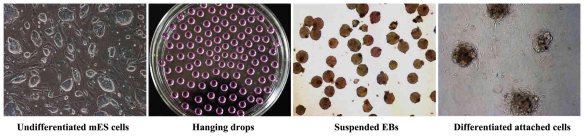
Differentiation of ES cells
As previously described, when undifferentiated mES
cells are incubated in vitro by the hanging
drop-suspension-adherence method in ES medium without LIF, the
cells can combine to form EBs (Fig.
1) (2,9,15).
In brief, ~1,000 mES cells in 20 µl droplets of cell suspension
were placed onto the inner side of the lid of a 10 cm Petri dish
(BD Biosciences, Franklin Lakes, NJ, USA) filled with 5 ml PBS and
then incubated at 37°C in 5% CO2 atmosphere. This
‘hanging drop’ culture was maintained in the absence of LIF to form
EBs. Following culturing for 3 days, these EBs were transferred
into sterile Petri dishes and cultured in suspension in 5 ml
differentiation medium for 2 days. Subsequently, these EBs were
seeded on 0.1% gelatin-coated 6-well plates at a density of 100 EBs
per well and incubated for an additional 5 days for differentiation
into beating cardiomyocytes. mES cells were exposed to the test
compound in appropriate concentrations from day 0 onwards over the
complete culture duration as described. In Phase I, five
concentrations were set for 5-fluorouracil (0.02, 0.04, 0.06, 0.08
and 0.1 µg/ml) and penicillin G (200, 400, 600, 800 and 1,000
µg/ml). In phase II, 5-fluorouracil served as positive control, and
the appropriate concentration was set as 0.037 µg/ml. In addition,
five concentrations were set for eugenol and carnosic acid (2, 4,
6, 8 and 10 µg/ml), as well as for procyanidin and dioctyl
phthalate (20, 40, 60, 80 and 100 µg/ml). The setting of the
concentrations of each test compound for differentiation assay was
based on the results of the cytotoxicity assay and preliminary
experiments. Untreated controls and the corresponding solvent
controls were included in each experiment.
RNA isolation and reverse
transcription-polymerase chain reaction (RT-PCR) analysis
On day 10, ~100 EBs per sample were harvested, and
total RNA was isolated with TRIzol reagent (15596–026; Invitrogen;
Thermo Fisher Scientific, Inc.). cDNA was synthesized from 1,000 ng
RNA per reaction with the Revert Aid First Strand cDNA Synthesis
kit (K1622; Thermo Fisher Scientific, Inc.). The α/β-MHC gene
expressed specially in cardiomyocyte differentiation was chosen as
a marker gene, and GAPDH was chosen as the housekeeping gene. The
polymerase chain reaction was performed in a T100 Thermal Cycler
(Bio-Rad Laboratories, Inc., Hercules, CA, USA) with specific
primers (Table I) by initial
denaturation at 94°C for 3 min, followed by 32 cycles of PCR
amplification: Denaturation at 94°C for 30 sec, annealing at 62°C
(α/β-MHC) or 56°C (GAPDH) for 30 sec, and completed by a final
extension of 72°C for 5 min (16).
PCR fragments were run on a 3% agarose gel containing 0.2 µg/ml
ethidium bromide (Sangon Biotech Co., Ltd., Shanghai, China),
visualized under UV light with a Molecular Imager ChemiDoc XRS
system (Bio-Rad Laboratories, Inc.) and analyzed with Quantity One
1-D analysis software version, 4.6.2 (Bio-Rad Laboratories, Inc.)
(39). The ID50 of the
test compound, expressed as the concentration that suppressed the
expression of MHC by 50% in comparison with control, was calculated
from a concentration-response curve. The differentiation assay of
each compound was repeated three times. The mean ID50
value of three times was set as the result.
 | Table I.Primer sequences used for reverse
transcription-quantitative polymerase chain reaction. |
Table I.
Primer sequences used for reverse
transcription-quantitative polymerase chain reaction.
| Gene name | Forward primer
sequence | Reverse primer
sequence |
|---|
| α/β-MHC |
CTTGTTGACCTGGGACTCGG |
ACCTGTCCAAGTTCCGCAAG |
| GAPDH |
GCCTTCTCCATGGTGGTGAA |
GCACAGTCAAGGCCGAGAAT |
Classification of the
embryotoxicity
The embryotoxic potential of each test compound was
classified into three grades (strong, weak and non-embryotoxic)
based on three values (IC503T3, IC50ES and
ID50), according to the PM proposed by ECVAM (2,19).
The values were as follows: I, 5.9157 lg (IC503T3)+3.500
lg (IC50ES)-5.307
[(IC503T3-ID50D3)/IC503T3]-15.72;
II, 3.651 lg (IC503T3)+2.394 lg
(IC50ES)-2.033
[(IC503T3-ID50D3)/IC503T3]-6.8;
and III, -0.125 lg (IC50 3T3)+1.917 lg
(IC50ES)+1.500
[(IC503T3-ID50D3)/IC503T3]-2.67.
The grades were classified as follows: Class 1, non-embryotoxicity,
If I>II and I>III; Class 2, weak embryotoxicity, If II>I
and II>III; and Class 3, strong embryotoxicity, If III>I and
III>II.
Statistical analysis
The statistical analysis was performed using SPSS
software (version, 19.0; IBM SPSS, Armonk, NY, USA) Data were
expressed as mean ± standard error of the mean. Each data point
represented the mean from three independent experiments. P<0.05
was considered to indicate a statistically significant
difference.
Results
Phase I: Evaluation of the feasibility
of the established EST model with control substances
The strongly embryotoxic 5-fluorouracil was used as
a positive control and the non-embryotoxic penicillin G was used as
a negative control; 5-fluorouracil and penicillin G are the classic
positive and negative substances routinely used to evaluate the
feasibility of the established EST model (2,5,6,20).
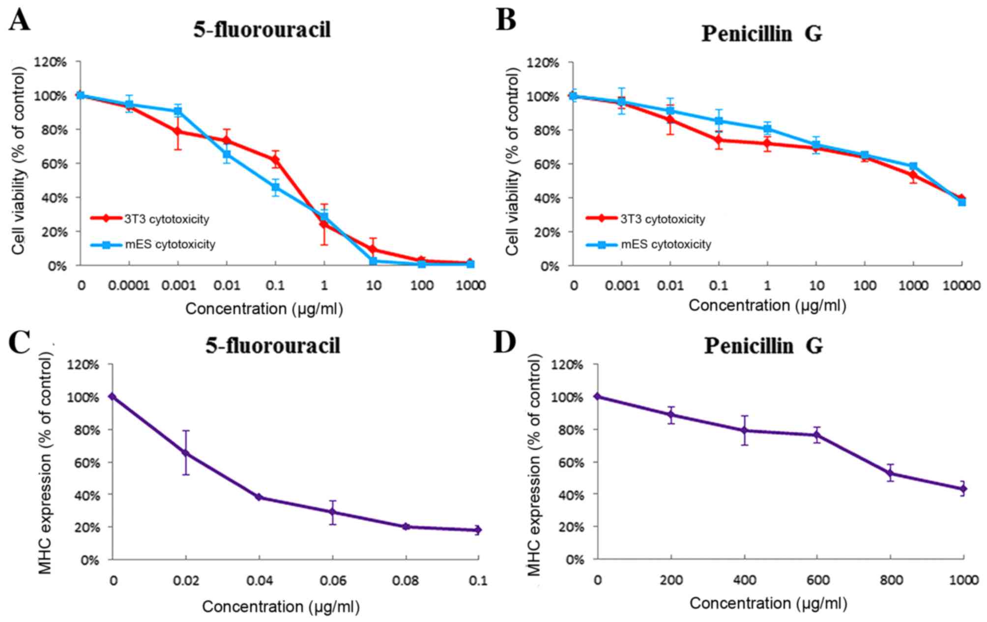
In the present study, 5-fluorouracil exhibited strong
embryotoxicity. When treated with 5-fluorouracil, the viability of
3T3 cells and mES cells decreased dose-dependently (Fig. 2A) and were influenced at very low
concentrations (IC503T3, 0.244±0.051 µg/ml;
IC50ES, 0.080±0.016 µg/ml). In the differentiation assay
(Figs. 2C, 3A and B), 5-fluorouracil exhibited strong
inhibition of the differentiation of ES cells into contracting
cardiomyoctyes (ID50, 0.037±0.006 µg/ml) as indicated by
the expression of marker gene (α/β-MHC) analyzed by RT-PCR. Test
results demonstrated that penicillin G exhibited little
cytotoxicity to 3T3 cells or mES cells (IC503T3,
1,160.667±69.07 µg/ml; IC50ES, 1,567.497±152.471 µg/ml;
Fig. 2B) and weak inhibition of ES
cells differentiation into contracting cardiomyoctyes
(ID50, 980.098±24.693 µg/ml; Figs. 2D and 3C). Even at the highest concentration
tested (1,000 µg/ml), only a minor inhibition was observed.
According to the PM, 5-fluorouracil and penicillin G were
classified as reagents with strong embryotoxicity and
non-embryotoxicity, respectively. The embryotoxicity
classifications were the same as those in the ECVAM validation
study.
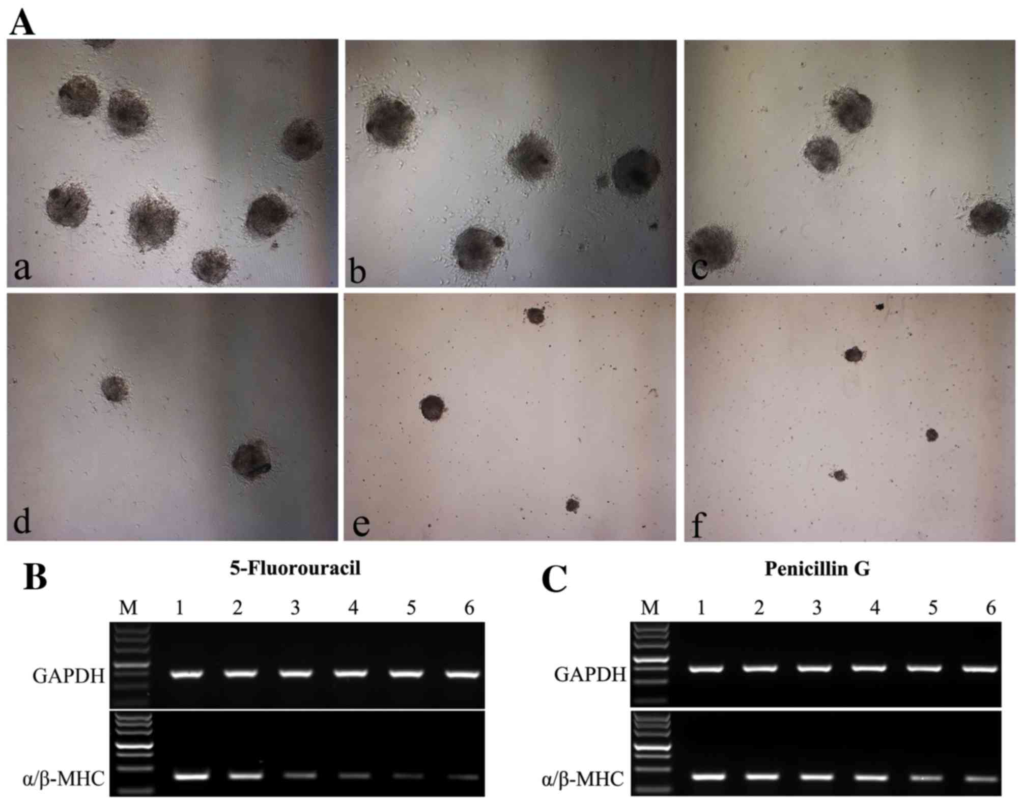 | Figure 3.(A) In the differentiation assay,
5-fluorouracil exhibited inhibition for mES cells differentiated
into cardiomyoctyes in a dose-dependent manner. a, Solvent control;
b, 0.02 µg/ml; c, 0.04 µg/ml; d, 0.06 µg/ml; e, 0.08 µg/ml; f, 0.1
µg/ml. Images were captured at ×40 magnification. (B and C)
Expression levels of the α/β-MHC in mES cells treated by different
concentrations of test compounds were analyzed by reverse
transcription quantitative-polymerase chain reaction, normalized to
GAPDH. (B) 5-fluorouracil electrophoresis. M, DL500 DNA Marker
(500, 400, 300, 200, 150, 100, 50 bp); 1, Solvent control; 2, 0.02
µg/ml; 3, 0.04 µg/ml; 4, 0.06 µg/ml; 5, 0.08 µg/ml; 6, 0.1 µg/ml.
(C) Penicillin G electrophoresis, M, DL500 DNA Marker; 1, Solvent
control; 2, 200 µg/ml; 3, 400 µg/ml; 4, 600 µg/ml; 5, 800 µg/ml; 6,
1,000 µg/ml. mES, murine embryonic stem cells; MHC, myosin heavy
chain. |
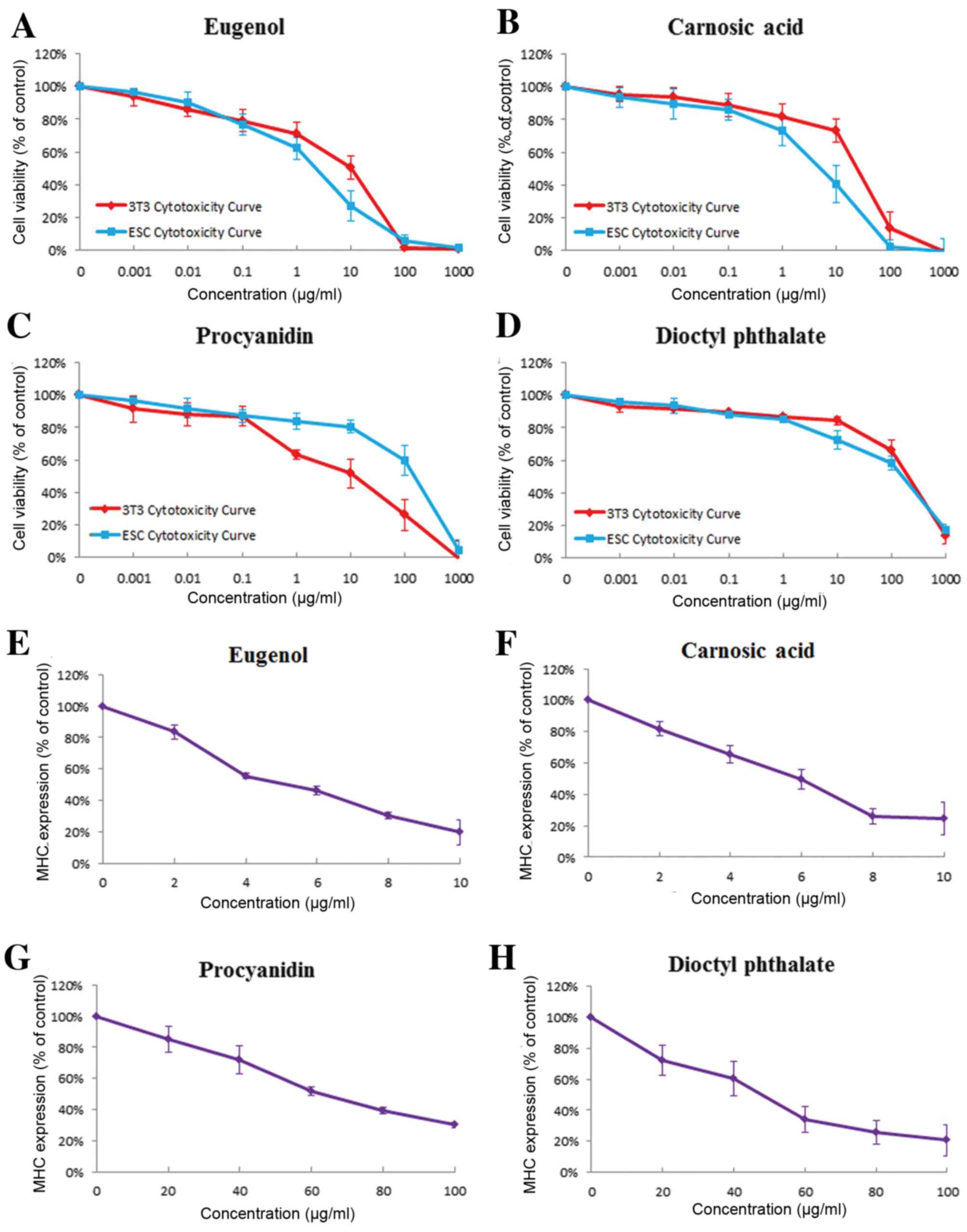
Phase II: Embryotoxicity assessment of
four compounds with the model of EST
In order to expand the application of EST, the
embryotoxic potentials of four selected compounds (eugenol,
carnosic acid, procyanidin and dioctyl phthalate) were assessed
with the model in phase II.
Eugenol
Test results demonstrated that eugenol presented
strong embryotoxicity in the EST. Both 3T3 cells and mES cells were
markedly sensitive to the cytotoxic effect of eugenol
(IC503T3, 9.441±2.849 µg/ml; IC50ES,
1.929±0.329 µg/ml; Fig. 4A). In
the differentiation assay (Figs.
4E and 5A), when the EBs were
exposed to eugenol, a concentration-dependent inhibition of
differentiation was observed. Treatment with eugenol resulted in
the direct inhibition of differentiation of mES-D3 cells at very
low concentrations (ID50, 5.434±0.715 µg/ml).
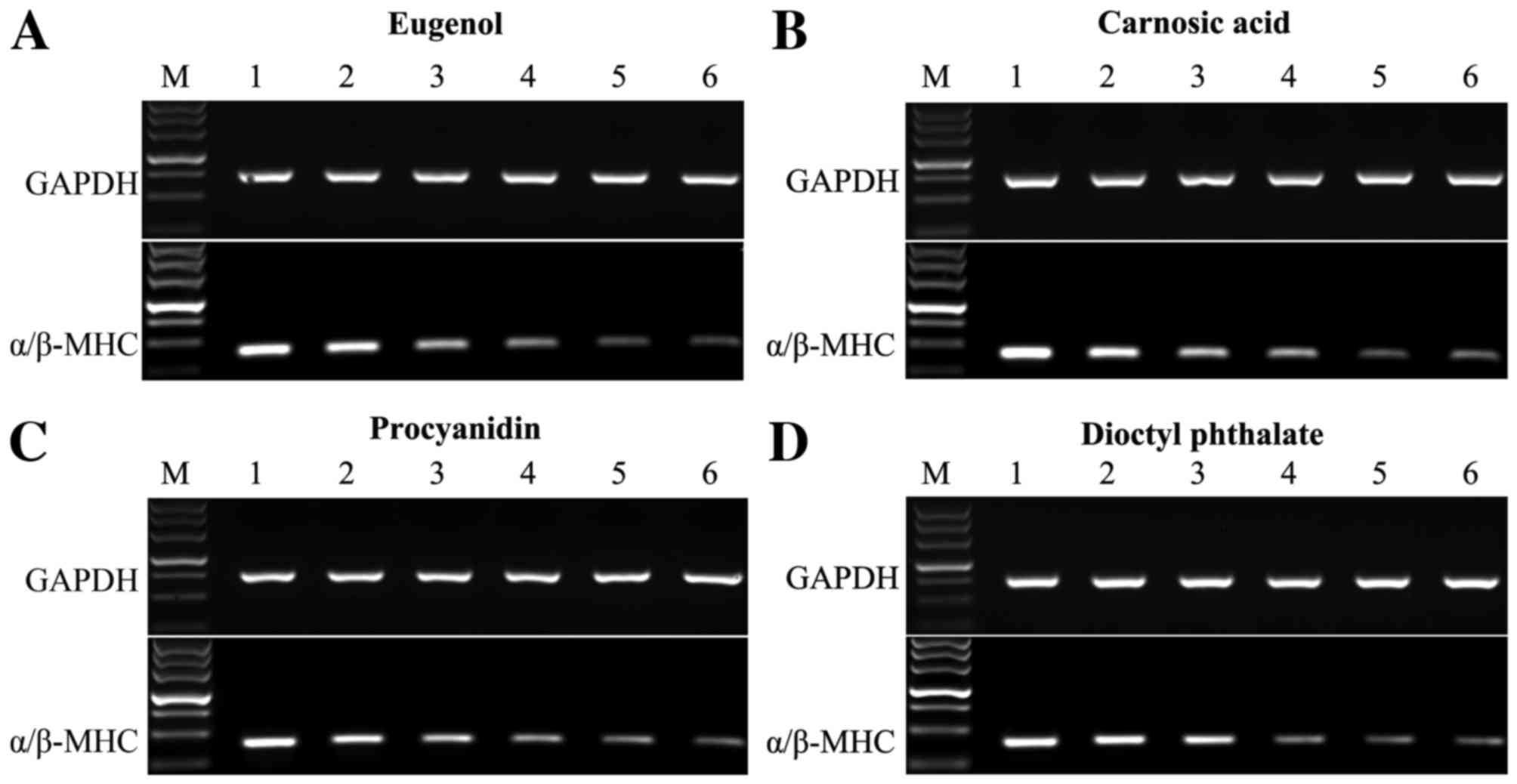 | Figure 5.Expression levels of the α/β-MHC at
different concentrations of test compounds were analyzed by reverse
transcription quantitative-polymerase chain reaction, normalized to
GAPDH expression. (A and B): Eugenol, carnosic acid. M, DL500 DNA
Marker; 1, Solvent control; 2, 2 µg/ml; 3, 4 µg/ml; 4, 6 µg/ml; 5,
8 µg/ml; 6, 10 µg/ml. (C and D): Procyanidin, dioctyl phthalate. M,
DL500 DNA Marker; 1, Solvent control; 2, 20 µg/ml; 3, 40 µg/ml; 4,
60 µg/ml; 5, 80 µg/ml; 6, 100 µg/ml. MHC, myosin heavy chain. |
Carnosic acid
In the cytotoxicity assay, the test results
indicated that carnosic acid exhibited greater cytotoxicity effects
on mES cells than on 3T3 cells (IC50 3T3, 26.28±3.861
µg/ml; IC50ES, 5.771±1.297 µg/ml; Fig. 4B). Exposure of the EBs to carnosic
acid, inhibition of differentiation into cardiomyocytes was
detected at similar concentrations as the IC50 test in
ES cells (Figs. 4F and 5B) and the ID50 was
6.143±0.575 µg/ml.
Procyanidin
As presented in Fig.
4C, 3T3 cells were notably sensitive to the cytotoxic effect of
procyanidin in comparison to mES cells (IC503T3,
12.1±1.828 µg/ml; IC50ES, 145.139±21.121 µg/ml) in the
cytotoxicity assay. The differentiation assay demonstrated that
procyanidin had weak inhibition of ES cell differentiation
(ID50, 72.493±2.706 µg/ml; Figs. 4G and 5C).
Dioctyl phthalate
In the EST, when the cells were treated with dioctyl
phthalate, greater cytotoxicity effects were identified on mES
cells than on 3T3 cells (IC50 3T3, 213.487±28.158 µg/ml;
IC50 ES, 123.587±24.944 µg/ml; Fig. 4D). Under dioctyl phthalate
treatment, the authors demonstrated that it could inhibit the
differentiation of mES cells at relatively low concentrations
(ID50, 60.116±5.39 µg/ml; Figs. 4H and 5D).
Subsequently, the mean values of IC503T3,
IC50ES and ID50 of each compound were
substituted into the PM, and the embryotoxic potentials of each
compound were successfully classified: Eugenol displayed strong
embryotoxicity, carnosic acid and dioctyl phthalate displayed weak
embryotoxicity, while procyanidin displayed non-embryotoxicity.
Summary results are presented in Table II.
| Table II.Summary of mean IC503T3,
IC50ES and ID50 values obtained in three
independent experiments and classification of test compounds
according to the prediction model. Data are presented as
IC50 and ID50 values (µg/ml) ± standard error
of the mean, and embryotoxicity was classified as strong, weak or
non-embryotoxic. |
Table II.
Summary of mean IC503T3,
IC50ES and ID50 values obtained in three
independent experiments and classification of test compounds
according to the prediction model. Data are presented as
IC50 and ID50 values (µg/ml) ± standard error
of the mean, and embryotoxicity was classified as strong, weak or
non-embryotoxic.
| Tested
compound | IC50 3T3
(µg/ml) | IC50 ES
(µg/ml) | ID50
(µg/ml) | Classification of
the embryotoxicity |
|---|
| 5-fluorouracil | 0.244±0.051 | 0.080±0.016 | 0.037±0.006 | Strong |
| Penicillin G |
1160.667±69.070 |
1567.497±152.471 | 980.098±24.693 | None |
| Eugenol | 9.441±2.849 | 1.929±0.329 | 5.434±0.715 | Strong |
| Carnosic acid | 26.280±3.861 | 5.771±1.297 | 6.143±0.575 | Weak |
| Procyanidin | 12.100±1.828 | 145.139±21.121 | 72.493±2.706 | None |
| Dioctyl
phthalate | 213.487±28.158 | 123.587±24.944 | 60.116±5.390 | Weak |
Discussion
In the early 1960s, >10,000 infants were born
with phocomelia (malformation of the limbs) due to exposure to
thalidomide throughout the world (40). The negative effects of thalidomide
have focused worldwide attention squarely on the embryotoxicity
caused by environmental insults (e.g., drugs, diet and
environmental toxic chemicals). At present, a wide range of
compounds needs to be tested, and, in particular, the development
of those compounds that may be used in pregnant women (those with
low toxic potency) must be prioritized. Currently, embryotoxicity
is mainly detected by in vivo tests. However, in vivo
detection requires a large number of experimental animals and
generous amounts of test chemical with long duration, which
altogether make the studies extremely costly (41,42).
The EST is currently the only test method that is completely free
from use of animals. The validation study of EST funded by ECVAM,
presented a good overall test accuracy of 78% for classification of
the 20 tested chemicals with known in vivo embryotoxic
potential (43). In particular,
the predictability of 100% for strongly embryotoxic chemicals was
obtained, and the precision was considered to be fairly good
(2,3,43).
In the present study, a model of EST was established
according to the standard EST system of ECVAM, and the validity of
the model was verified with 5-fluorouracil as a positive control
and penicillin G as a negative control. During pregnancy, the
developing embryo is very sensitive, and a variety of compounds
have been reported to be toxic or teratogenic for its development
(44,45). Eugenol, carnosic acid, procyanidin
and dioctyl phthalate, commonly-used compounds, have already been
used food, cosmetic and medical applications, within a specific
range of concentrations. However, little is known about their
influence on embryo development. To the best of the authors'
knowledge, there are no available studies describing the effect of
these compounds on the embryotoxicity in vitro. For this
reason, subsequently, the present study assessed the embryotoxicity
of these four compounds with the established EST model.
Domaracky et al (46) studied the influence of eugenol on
the development of mouse preimplantation embryos in vivo.
The study indicated that eugenol may induce a significantly
increased rate of cell death and affect the development of embryo.
For its isomer, isoeugenol, when received by pregnant rats (at the
highest dose of 1,000 mg/kg/day), it caused intrauterine growth
retardation and skeletal defects in fetuses (47). The current results indicated that
3T3 cells and mES cells were both sensitive to eugenol. A higher
incidence of cell death was observed following treatment with
eugenol. According to the test data, eugenol was classified as
strongly embryotoxic. Research on carnosic acid has demonstrated
that a short-term oral administration has a relatively low toxicity
profile, and the oral lethal dose for mice was 7,100 mg/kg of body
weight (48). The present study
suggested that carnosic acid was weakly embryotoxic. Therefore, for
the use of carnosic acid, pregnant women must be cautious. However,
toxicological studies have indicated that procyanidin is nontoxic
and does not cause any detrimental effects in vivo (49,50).
According to these results, procyanidin is safe and non-embryotoxic
under the conditions investigated in the present study. With
regards to dioctyl phthalate, many studies on animals clearly
demonstrated that dioctyl phthalate could cause a certain
tissue/organ toxicity (51–54),
developmental toxicity (55–58)
and reproductive toxicity (37,59–61)
in some species, such as rats, mice and marmosets (36,62,63).
To the best of the authors' knowledge, there are no data on the
embryotoxicity of dioctyl phthalate obtained by in vitro
animal-free tests. The present study employed the established EST
to predict the embryotoxicity of dioctyl phthalate in vitro,
and the results indicated that dioctyl phthalate exhibited weak
embryotoxicity.
Taken together, the authors successfully established
the model of EST, and further assessed embryotoxicity of four
selected compounds with this model. In the future, it will be
important to determine the embryotoxicity of the many commonly used
compounds. The EST system for embryotoxicity screening test is
rapid, simple and sensitive. It may be used for high-throughput
screening of embryotoxicity of test substance.
Acknowledgments
This work was supported by the National Natural
Science Foundation of China (grant nos. 30871246, 81070993,
81272972 and 81472355), the National Basic Research Program of
China (grant no. 2010CB833605), the Specialized Research Fund for
the Doctoral Program of Higher Education of China (grant no.
20120162110059), the Foundation of Hunan Provincial Science and
Technology Department (grant nos. 2010TD2026 and 2011FJ4180), the
Program for New Century Excellent Talents in University (grant no.
NCET-10-0790), the Key Program of Central South University (grant
no. 2010QYZD006) and the Open-End Fund for the Valuable and
Precision Instruments of Central South University (grant nos.
CSUZC201634 and CSUZC201638).
References
|
1
|
Scholz G, Genschow E, Pohl I, Bremer S,
Paparella M, Raabe H, Southee J and Spielmann H: Prevalidation of
the embryonic stem cell test (EST)-A new in vitro embryotoxicity
Test. Toxicol In Vitro. 13:675–681. 1999. View Article : Google Scholar : PubMed/NCBI
|
|
2
|
Genschow E, Spielmann H, Scholz G, Pohl I,
Seiler A, Clemann N, Bremer S and Becker K: Validation of the
embryonic stem cell test in the international ECVAM validation
study on three in vitro embryotoxicity tests. Altern Lab Anim.
32:209–244. 2004.PubMed/NCBI
|
|
3
|
Seiler AE and Spielmann H: The validated
embryonic stem cell test to predict embryotoxicity in vitro. Nat
Protoc. 6:961–978. 2011. View Article : Google Scholar : PubMed/NCBI
|
|
4
|
Chen R, Chen J, Cheng S, Qin J, Li W,
Zhang L, Jiao H, Yu X, Zhang X, Lahn BT and Xiang AP: Assessment of
embryotoxicity of compounds in cosmetics by the embryonic stem cell
test. Toxicol Mech Methods. 20:112–118. 2010. View Article : Google Scholar : PubMed/NCBI
|
|
5
|
Festag M, Viertel B, Steinberg P and
Sehner C: An in vitro embryotoxicity assay based on the disturbance
of the differentiation of murine embryonic stem cells into
endothelial cells. II. Testing of compounds. Toxicol In Vitro.
21:1631–1640. 2007. View Article : Google Scholar : PubMed/NCBI
|
|
6
|
Li L, Zhang X, Wang L, Chai Z, Shen X,
Zhang Z and Liu C: A toxicology study to evaluate the
embryotoxicity of metformin compared with the hypoglycemic drugs,
the anticancer drug, the anti-epileptic drug, the antibiotic, and
the cyclo-oxygenase (COX)-2 inhibitor. J Diabetes. 7:839–849. 2015.
View Article : Google Scholar : PubMed/NCBI
|
|
7
|
Paquette JA, Kumpf SW, Streck RD, Thomson
JJ, Chapin RE and Stedman DB: Assessment of the embryonic stem cell
test and application and use in the pharmaceutical industry. Birth
Defects Res B Dev Reprod Toxicol. 83:104–111. 2008. View Article : Google Scholar : PubMed/NCBI
|
|
8
|
Chen F, Cao F, Su Z, Li L, Huang A and Xu
H: Assessment of the developmental toxicity of epidermal growth
factor using embryonic stem cell test. Trop J Pharm Res.
13:6892014. View Article : Google Scholar
|
|
9
|
Fuegemann CJ, Samraj AK, Walsh S,
Fleischmann BK, Jovinge S and Breitbach M: Differentiation of mouse
embryonic stem cells into cardiomyocytes via the hanging-drop and
mass culture methods. Curr Protoc Stem Cell Biol Chapter. 1:Unit 1F
11. 2010. View Article : Google Scholar
|
|
10
|
Doetschman TC, Eistetter H, Katz M,
Schmidt W and Kemler R: The in vitro development of
blastocyst-derived embryonic stem cell lines: Formation of visceral
yolk sac, blood islands and myocardium. J Embryol Exp Morphol.
87:27–45. 1985.PubMed/NCBI
|
|
11
|
Sánchez A, Jones WK, Gulick J, Doetschman
T and Robbins J: Myosin heavy chain gene expression in mouse
embryoid bodies. An in vitro developmental study. J Biol Chem.
266:22419–22426. 1991.PubMed/NCBI
|
|
12
|
Robbins J, Gulick J, Sanchez A, Howles P
and Doetschman T: Mouse embryonic stem cells express the cardiac
myosin heavy chain genes during development in vitro. J Biol Chem.
265:11905–11909. 1990.PubMed/NCBI
|
|
13
|
de Jong E, Louisse J, Verwei M, Blaauboer
BJ, van de Sandt JJ, Woutersen RA, Rietjens IM and Piersma AH:
Relative developmental toxicity of glycol ether alkoxy acid
metabolites in the embryonic stem cell test as compared with the in
vivo potency of their parent compounds. Toxicol Sci. 110:117–124.
2009. View Article : Google Scholar : PubMed/NCBI
|
|
14
|
Deng SQ, Xu H, He Q, Jiang HX, Su BJ and
Zhang QH: Detecting the developmental toxicity of bFGF in the
embryonic stem cell test using differential gene expression of
differentiation-related genes. Toxicol Mech Methods. 24:323–331.
2014. View Article : Google Scholar : PubMed/NCBI
|
|
15
|
Schulpen SH, Pennings JL, Tonk EC and
Piersma AH: A statistical approach towards the derivation of
predictive gene sets for potency ranking of chemicals in the mouse
embryonic stem cell test. Toxicol Lett. 225:342–349. 2014.
View Article : Google Scholar : PubMed/NCBI
|
|
16
|
zur Nieden NI, Ruf LJ, Kempka G,
Hildebrand H and Ahr HJ: Molecular markers in embryonic stem cells.
Toxicol In Vitro. 15:455–461. 2001. View Article : Google Scholar : PubMed/NCBI
|
|
17
|
Nieden NI, Kempka G and Ahr HJ: Molecular
multiple endpoint embryonic stem cell test-a possible approach to
test for the teratogenic potential of compounds. Toxicol Appl
Pharmacol. 194:257–269. 2004. View Article : Google Scholar : PubMed/NCBI
|
|
18
|
Seiler A, Visan A, Buesen R, Genschow E
and Spielmann H: Improvement of an in vitro stem cell assay for
developmental toxicity: The use of molecular endpoints in the
embryonic stem cell test. Reprod Toxicol. 18:231–240. 2004.
View Article : Google Scholar : PubMed/NCBI
|
|
19
|
Genschow E, Scholz G, Brown N, Piersma A,
Brady M, Clemann N, Huuskonen H, Paillard F, Bremer S, Becker K and
Spielmann H: Development of prediction models for three in vitro
embryotoxicity tests in an ECVAM validation study. In Vitr Mol
Toxicol. 13:51–66. 2000.PubMed/NCBI
|
|
20
|
Buesen R, Genschow E, Slawik B, Visan A,
Spielmann H, Luch A and Seiler A: Embryonic stem cell test
remastered: Comparison between the validated EST and the new
molecular FACS-EST for assessing developmental toxicity in vitro.
Toxicol Sci. 108:389–400. 2009. View Article : Google Scholar : PubMed/NCBI
|
|
21
|
Jadhav BK, Khandelwal KR, Ketkar AR and
Pisal SS: Formulation and evaluation of mucoadhesive tablets
containing eugenol for the treatment of periodontal diseases. Drug
Dev Ind Pharm. 30:195–203. 2004. View Article : Google Scholar : PubMed/NCBI
|
|
22
|
Chami N, Chami F, Bennis S, Trouillas J
and Remmal A: Antifungal treatment with carvacrol and eugenol of
oral candidiasis in immunosuppressed rats. Braz J Infect Dis.
8:217–226. 2004. View Article : Google Scholar : PubMed/NCBI
|
|
23
|
Dip EC, Pereira NA and Fernandes PD:
Ability of eugenol to reduce tongue edema induced by Dieffenbachia
picta Schott in mice. Toxicon. 43:729–735. 2004. View Article : Google Scholar : PubMed/NCBI
|
|
24
|
Fischedick JT, Standiford M, Johnson DA
and Johnson JA: Structure activity relationship of phenolic
diterpenes from Salvia officinalis as activators of the
nuclear factor E2-related factor 2 pathway. Bioorg Med Chem.
21:2618–2622. 2013. View Article : Google Scholar : PubMed/NCBI
|
|
25
|
Bai N, He K, Roller M, Lai CS, Shao X, Pan
MH and Ho CT: Flavonoids and phenolic compounds from Rosmarinus
officinalis. J Agric Food Chem. 58:5363–5367. 2010. View Article : Google Scholar : PubMed/NCBI
|
|
26
|
Yu YM, Lin CH, Chan HC and Tsai HD:
Carnosic acid reduces cytokine-induced adhesion molecules
expression and monocyte adhesion to endothelial cells. Eur J Nutr.
48:101–106. 2009. View Article : Google Scholar : PubMed/NCBI
|
|
27
|
Mengoni ES, Vichera G, Rigano LA,
Rodriguez-Puebla ML, Galliano SR, Cafferata EE, Pivetta OH, Moreno
S and Vojnov AA: Suppression of COX-2, IL-1β and TNF-α expression
and leukocyte infiltration in inflamed skin by bioactive compounds
from Rosmarinus officinalis L. Fitoterapia. 82:414–421.
2011. View Article : Google Scholar : PubMed/NCBI
|
|
28
|
Xiang Q, Liu Z, Wang Y, Xiao H, Wu W, Xiao
C and Liu X: Carnosic acid attenuates lipopolysaccharide-induced
liver injury in rats via fortifying cellular antioxidant defense
system. Food Chem Toxicol. 53:1–9. 2013. View Article : Google Scholar : PubMed/NCBI
|
|
29
|
Ibarra A, Cases J, Roller M, Chiralt-Boix
A, Coussaert A and Ripoll C: Carnosic acid-rich rosemary
(Rosmarinus officinalis L.)leaf extract limits weight gain
and improves cholesterol levels and glycaemia in mice on a high-fat
diet. Br J Nutr. 106:1182–1189. 2011. View Article : Google Scholar : PubMed/NCBI
|
|
30
|
Birtic S, Dussort P, Pierre FX, Bily AC
and Roller M: Carnosic acid. Phytochemistry. 115:9–19. 2015.
View Article : Google Scholar : PubMed/NCBI
|
|
31
|
Santos-Buelga C and Scalbert A:
Proanthocyanidins and tannin-like compounds-nature, occurrence,
dietary intake and effects on nutrition and health. J Sci Food
Agriculture. 80:1094–1117. 2000. View Article : Google Scholar
|
|
32
|
Monagas M, Quintanilla-López JE,
Gómez-Cordovés C, Bartolomé B and Lebrón-Aguilar R: MALDI-TOF MS
analysis of plant proanthocyanidins. J Pharm Biomed Anal.
51:358–372. 2010. View Article : Google Scholar : PubMed/NCBI
|
|
33
|
Yamakoshi J, Saito M, Kataoka S and
Kikuchi M: Safety evaluation of proanthocyanidin-rich extract from
grape seeds. Food Chem Toxicol. 40:599–607. 2002. View Article : Google Scholar : PubMed/NCBI
|
|
34
|
Corder R, Mullen W, Khan NQ, Marks SC,
Wood EG, Carrier MJ and Crozier A: Oenology: Red wine procyanidins
and vascular health. Nature. 444:5662006. View Article : Google Scholar : PubMed/NCBI
|
|
35
|
Benson R: Hazard to the developing male
reproductive system from cumulative exposure to phthalate
esters-dibutyl phthalate, diisobutyl phthalate, butylbenzyl
phthalate, diethylhexyl phthalate, dipentyl phthalate, and
diisononyl phthalate. Regul Toxicol Pharmacol. 53:90–101. 2009.
View Article : Google Scholar : PubMed/NCBI
|
|
36
|
Kavlock R, Boekelheide K, Chapin R,
Cunningham M, Faustman E, Foster P, Golub M, Henderson R, Hinberg
I, Little R, et al: NTP center for the evaluation of risks to human
reproduction: Phthalates expert panel report on the reproductive
and developmental toxicity of di(2-ethylhexyl) phthalate. Reprod
Toxicol. 16:529–653. 2002. View Article : Google Scholar : PubMed/NCBI
|
|
37
|
Helal MA: Celery oil modulates
DEHP-induced reproductive toxicity in male rats. Reprod Biol.
14:182–189. 2014. View Article : Google Scholar : PubMed/NCBI
|
|
38
|
Latini G: Monitoring phthalate exposure in
humans. Clin Chim Acta. 361:20–29. 2005. View Article : Google Scholar : PubMed/NCBI
|
|
39
|
Li G, Ren C, Shi J, Huang W, Liu H, Feng
X, Liu W, Zhu B, Zhang C, Wang L, et al: Identification, expression
and subcellular localization of ESRG. Biochem Biophys Res Commun.
435:160–164. 2013. View Article : Google Scholar : PubMed/NCBI
|
|
40
|
van Dartel DA and Piersma AH: The
embryonic stem cell test combined with toxicogenomics as an
alternative testing model for the assessment of developmental
toxicity. Reprod Toxicol. 32:235–244. 2011. View Article : Google Scholar : PubMed/NCBI
|
|
41
|
Hartung T: Toxicology for the twenty-first
century. Nature. 460:208–212. 2009. View Article : Google Scholar : PubMed/NCBI
|
|
42
|
Rovida C and Hartung T: Re-evaluation of
animal numbers and costs for in vivo tests to accomplish REACH
legislation requirements for chemicals-a report by the
transatlantic think tank for toxicology (t(4)). ALTEX. 26:187–208.
2009. View Article : Google Scholar : PubMed/NCBI
|
|
43
|
Genschow E, Spielmann H, Scholz G, Seiler
A, Brown N, Piersma A, Brady M, Clemann N, Huuskonen H, Paillard F,
et al: The ECVAM international validation study on in vitro
embryotoxicity tests: Results of the definitive phase and
evaluation of prediction models. European centre for the validation
of alternative methods. Altern Lab Anim. 30:151–176.
2002.PubMed/NCBI
|
|
44
|
Peters AK, Steemans M, Hansen E, Mesens N,
Verheyen GR and Vanparys P: Evaluation of the embryotoxic potency
of compounds in a newly revised high throughput embryonic stem cell
test. Toxicol Sci. 105:342–350. 2008. View Article : Google Scholar : PubMed/NCBI
|
|
45
|
Jones KL, Lacro RV, Johnson KA and Adams
J: Pattern of malformations in the children of women treated with
carbamazepine during pregnancy. N Engl J Med. 320:1661–1666. 1989.
View Article : Google Scholar : PubMed/NCBI
|
|
46
|
Domaracky M, Rehák P, Juhás S and Koppel
J: Effects of selected plant essential oils on the growth and
development of mouse preimplantation embryos in vivo. Physiol Res.
56:97–104. 2007.PubMed/NCBI
|
|
47
|
George JD, Price CJ, Marr MC, Myers CB and
Jahnke GD: Evaluation of the developmental toxicity of isoeugenol
in Sprague-Dawley (CD) rats. Toxicol Sci. 60:112–120. 2001.
View Article : Google Scholar : PubMed/NCBI
|
|
48
|
Wang QL, Li H, Li XX, Cui CY, Wang R, Yu
NX and Chen LX: Acute and 30-day oral toxicity studies of
administered carnosic acid. Food Chem Toxicol. 50:4348–4355. 2012.
View Article : Google Scholar : PubMed/NCBI
|
|
49
|
Ray S, Bagchi D, Lim PM, Bagchi M, Gross
SM, Kothari SC, Preuss HG and Stohs SJ: Acute and long-term safety
evaluation of a novel IH636 grape seed proanthocyanidin extract.
Res Commun Mol Pathol Pharmacol. 109:165–197. 2001.PubMed/NCBI
|
|
50
|
Lluís L, Muñoz M, Nogués MR,
Sánchez-Martos V, Romeu M, Giralt M, Valls J and Solà R: Toxicology
evaluation of a procyanidin-rich extract from grape skins and
seeds. Food Chem Toxicol. 49:1450–1454. 2011. View Article : Google Scholar : PubMed/NCBI
|
|
51
|
Hazelton WI: A subchronic (4-week) dietary
oral toxicity study of di(2-ethylhexyl) phthalate in B6FC3F1 mice.
8D submission, microfische no. OTS0535433: Submited to US EPA.
Office of Toxic Substances, prepared for Eastman Kodak Company
Washington, DC: 1992
|
|
52
|
Poon R, Lecavalier P, Mueller R, Valli VE,
Procter BG and Chu I: Subchronic oral toxicity of di-n-octyl
phthalate and di(2-Ethylhexyl) phthalate in the rat. Food Chem
Toxicol. 35:225–239. 1997. View Article : Google Scholar : PubMed/NCBI
|
|
53
|
David RM, Moore MR, Cifone MA, Finney DC
and Guest D: Chronic peroxisome proliferation and hepatomegaly
associated with the hepatocellular tumorigenesis of
di(2-ethylhexyl)phthalate and the effects of recovery. Toxicol Sci.
50:195–205. 1999. View Article : Google Scholar : PubMed/NCBI
|
|
54
|
Li SG, Huang X, Zhang XW and Xu SH:
Effects of diethylhexyl phthalate on lipid peroxidation and the
life-span in Drosophila melanogaster. Zhonghua Yu Fang Yi
Xue Za Zhi. 39:111–114. 2005.(In Chinese). PubMed/NCBI
|
|
55
|
Tyl R, Jones-Price C, Marr MC and Kimmel
CA: Teratological evaluation of diethylhexylphthalate (CAS no.
117-81-7) in CD-1 mice. National Center for Toxicological Research.
1984.
|
|
56
|
Tyl RW, Price CJ, Marr MC and Kimmel CA:
Developmental toxicity evaluation of dietary
di(2-ethylhexyl)phthalate in Fischer 344 rats and CD-1 mice. Fundam
Appl Toxicol. 10:395–412. 1988. View Article : Google Scholar : PubMed/NCBI
|
|
57
|
Peters JM, Taubeneck MW, Keen CL and
Gonzalez FJ: Di(2-ethylhexyl) phthalate induces a functional zinc
deficiency during pregnancy and teratogenesis that is independent
of peroxisome proliferator-activated receptor-alpha. Teratology.
56:311–316. 1997. View Article : Google Scholar : PubMed/NCBI
|
|
58
|
Dobrzynska MM, Tyrkiel EJ, Derezińska E,
Pachocki KA and Ludwicki JK: Two generation reproductive and
developmental toxicity following subchronic exposure of pubescent
male mice to di(2-ethylhexyl)phthalate. Ann Agric Environ Med.
19:31–37. 2012.PubMed/NCBI
|
|
59
|
Erkekoglu P, Zeybek ND, Giray B, Asan E,
Arnaud J and Hincal F: Reproductive toxicity of di(2-ethylhexyl)
phthalate in selenium-supplemented and selenium-deficient rats.
Drug Chem Toxicol. 34:379–389. 2011. View Article : Google Scholar : PubMed/NCBI
|
|
60
|
Zhang XF, Zhang LJ, Li L, Feng YN, Chen B,
Ma JM, Huynh E, Shi QH, De Felici M and Shen W: Diethylhexyl
phthalate exposure impairs follicular development and affects
oocyte maturation in the mouse. Environ Mol Mutagen. 54:354–361.
2013. View Article : Google Scholar : PubMed/NCBI
|
|
61
|
Zhang XF, Zhang T, Han Z, Liu JC, Liu YP,
Ma JY, Li L and Shen W: Transgenerational inheritance of ovarian
development deficiency induced by maternal diethylhexyl phthalate
exposure. Reprod Fertil Dev. 27:1213–1221. 2015. View Article : Google Scholar : PubMed/NCBI
|
|
62
|
Hauser R and Calafat AM: Phthalates and
human health. Occup Environ Med. 62:806–818. 2005. View Article : Google Scholar : PubMed/NCBI
|
|
63
|
Swan SH: Environmental phthalate exposure
in relation to reproductive outcomes and other health endpoints in
humans. Environ Res. 108:177–184. 2008. View Article : Google Scholar : PubMed/NCBI
|