|
1
|
Seki E and Brenner DA: Recent advancement
of molecular mechanisms of liver fibrosis. J Hepatobiliary Pancreat
Sci. 22:512–518. 2015. View
Article : Google Scholar : PubMed/NCBI
|
|
2
|
Ramachandran P, Iredale JP and Fallowfield
JA: Resolution of liver fibrosis: Basic mechanisms and clinical
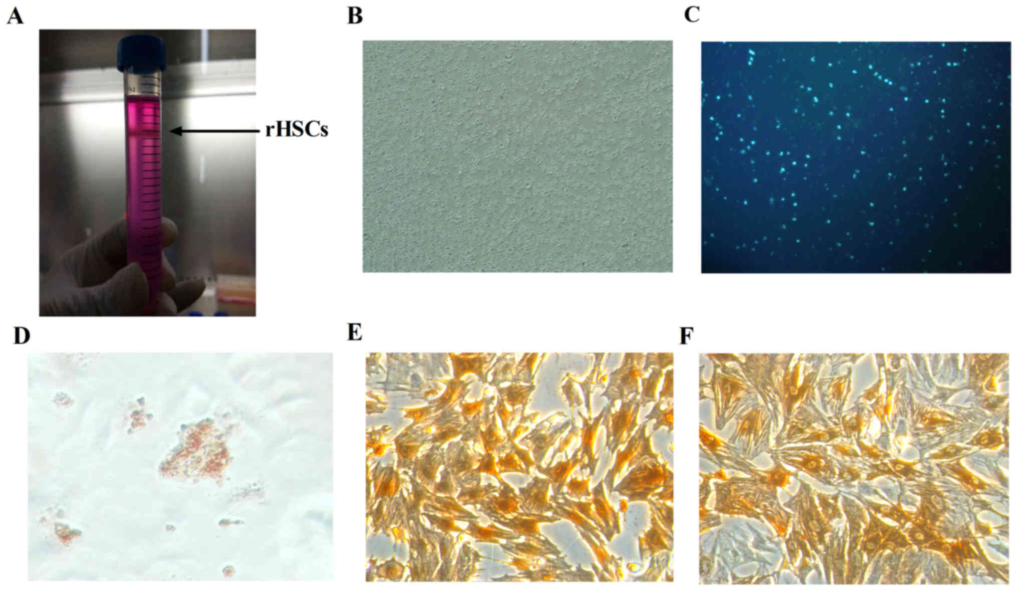
relevance. Semin Liver Dis. 35:119–131. 2015. View Article : Google Scholar : PubMed/NCBI
|
|
3
|
Krizhanovsky V, Yon M, Dickins RA, Hearn
S, Simon J, Miething C, Yee H, Zender L and Lowe SW: Senescence of
activated stellate cells limits liver fibrosis. Cell. 134:657–667.
2008. View Article : Google Scholar : PubMed/NCBI
|
|
4
|
Muñoz-Espín D and Serrano M: Cellular
senescence: From physiology to pathology. Nat Rev Mol Cell Bio.
15:482–496. 2014. View
Article : Google Scholar
|
|
5
|
Kojima H, Inoue T, Kunimoto H and Nakajima
K: IL-6-STAT3 signaling and premature senescence. JAKSTAT.
2:e257632014.
|
|
6
|
Kuilman T, Michaloglou C, Vredeveld LC,
Douma S, van Doorn R, Desmet CJ, Aarden LA, Mooi WJ and Peeper DS:
Oncogene-induced senescence relayed by an interleukin-dependent
inflammatory network. Cell. 133:1019–1031. 2008. View Article : Google Scholar : PubMed/NCBI
|
|
7
|
Li P, Gan Y, Xu Y, Song L, Wang L, Ouyang
B, Zhang C and Zhou Q: The inflammatory cytokine TNF-α promotes the
premature senescence of rat nucleus pulposus cells via the PI3K/Akt
signaling pathway. Sci Rep. 7:429382017. View Article : Google Scholar : PubMed/NCBI
|
|
8
|
Hung KS, Lee TH, Chou WY, Wu CL, Cho CL,
Lu CN, Jawan B and Wang CH: Interleukin-10 gene therapy reverses
thioacetamide-induced liver fibrosis in mice. Biochem Biophys Res
Commun. 336:324–331. 2005. View Article : Google Scholar : PubMed/NCBI
|
|
9
|
Huang YH, Shi MN, Zheng WD, Zhang LJ, Chen
ZX and Wang XZ: Therapeutic effect of interleukin-10 on
CCl4-induced hepatic fibrosis in rats. World J Gastroenterol.
12:1386–1391. 2006. View Article : Google Scholar : PubMed/NCBI
|
|
10
|
Huang YH, Chen YX, Zhang LJ, Chen ZX and
Wang XZ: Hydrodynamics-based transfection of rat interleukin-10
gene attenuates porcine serum-induced liver fibrosis in rats by
inhibiting the activation of hepatic stellate cells. Int J Mol Med.
34:677–686. 2014. View Article : Google Scholar : PubMed/NCBI
|
|
11
|
Shi MN, Huang YH, Zheng WD, Zhang LJ, Chen
ZX and Wang XZ: Relationship between transforming growth factor
beta1 and anti-fibrotic effect of interleukin-10. World J
Gastroenterol. 12:2357–2362. 2006. View Article : Google Scholar : PubMed/NCBI
|
|
12
|
Livak KJ and Schmittgen TD: Analysis of
relative gene expression data using real-time quantitative PCR and
the 2(-Delta Delta C(T)) method. Methods. 25:402–408. 2001.
View Article : Google Scholar : PubMed/NCBI
|
|
13
|
Weiskirchen R and Gressner AM: Isolation
and culture of hepatic stellate cells. Methods Mol Med. 117:99–113.
2005.PubMed/NCBI
|
|
14
|
Chang W, Yang M, Song L, Shen K, Wang H,
Gao X, Li M, Niu W and Qin X: Isolation and culture of hepatic
stellate cells from mouse liver. Acta Biochim Biophys Sin
(Shanghai). 46:291–298. 2014. View Article : Google Scholar : PubMed/NCBI
|
|
15
|
Mederacke I, Dapito DH, Affò S, Uchinami H
and Schwabe RF: High-yield and high-purity isolation of hepatic
stellate cells from normal and fibrotic mouse livers. Nat Protoc.
10:305–315. 2015. View Article : Google Scholar : PubMed/NCBI
|
|
16
|
Mederacke I, Hsu CC, Troeger JS, Huebener
P, Mu X, Dapito DH, Pradere JP and Schwabe RF: Fate tracing reveals
hepatic stellate cells as dominant contributors to liver fibrosis
independent of its aetiology. Nat Commun. 4:28232013. View Article : Google Scholar : PubMed/NCBI
|
|
17
|
Kim KH, Chen CC, Monzon RI and Lau LF:
Matricellular protein CCN1 promotes regression of liver fibrosis
through induction of cellular senescence in hepatic myofibroblasts.
Mol Cell Biol. 33:2078–2090. 2013. View Article : Google Scholar : PubMed/NCBI
|
|
18
|
Jin H, Lian N, Zhang F, Bian M, Chen X,
Zhang C, Jia Y, Lu C, Hao M, Yao S, et al: Inhibition of YAP
signaling contributes to senescence of hepatic stellate cells
induced by tetramethylpyrazine. Eur J Pharm Sci. 96:323–333. 2017.
View Article : Google Scholar : PubMed/NCBI
|
|
19
|
Kong X, Feng D, Wang H, Hong F, Bertola A,
Wang FS and Gao B: Interleukin-22 induces hepatic stellate cell
senescence and restricts liver fibrosis in mice. Hepatology.
56:1150–1159. 2012. View Article : Google Scholar : PubMed/NCBI
|
|
20
|
Chen J, Xu T, Zhu D, Wang J, Huang C, Lyu
L, Hu B, Sun W and Duan Y: Egg antigen p40 of Schistosoma japonicum
promotes senescence in activated hepatic stellate cells by
activation of the STAT3/p53/p21 pathway. Cell Death Dis.
7:e23152016. View Article : Google Scholar : PubMed/NCBI
|
|
21
|
He L, He X, Lowe SW and Hannon GJ:
microRNAs join the p53 network-another piece in the
tumour-suppression puzzle. Nat Rev Cancer. 7:819–822. 2007.
View Article : Google Scholar : PubMed/NCBI
|
|
22
|
Ahsan MK and Mehal WZ: Activation of
adenosine receptor A2A increases HSC proliferation and inhibits
death and senescence by down-regulation of p53 and Rb. Front
Pharmacol. 5:692014. View Article : Google Scholar : PubMed/NCBI
|
|
23
|
Kong D, Zhang F, Zhang Z, Lu Y and Zheng
S: Clearance of activated stellate cells for hepatic fibrosis
regression: Molecular basis and translational potential. Biomed
Pharmacother. 67:246–250. 2013. View Article : Google Scholar : PubMed/NCBI
|
|
24
|
Zhang Z, Yao Z, Zhao S, Shao J, Chen A,
Zhang F and Zheng S: Interaction between autophagy and senescence
is required for dihydroartemisinin to alleviate liver fibrosis.
Cell Death Dis. 8:e28862017. View Article : Google Scholar : PubMed/NCBI
|
|
25
|
Braumüller H, Wieder T, Brenner E, Aßmann
S, Hahn M, Alkhaled M, Schilbach K, Essmann F, Kneilling M,
Griessinger C, et al: T-helper-1-cell cytokines drive cancer into
senescence. Nature. 494:361–365. 2013. View Article : Google Scholar : PubMed/NCBI
|