Introduction
Ischemic brain damage has long been associated with
neuronal injury (1–4), and it is a result of combined action
of acute necrosis with delayed injury mechanisms. The damage
cascade is the recognized theory of the cerebral ischemia
physiological and pathological mechanism, predominantly including
excitotoxicity, peri-infarct depolarization, inflammation and
apoptosis (5). Metabolism of
arachidonic acid (AA) and its metabolic products have been
implicated to be associated with these pathophysiological
mechanisms (4). To a great extent,
cellular levels of AA are under the control of phospholipase A2
(PLA2) enzymes. PLA2 catalyze the membrane
phospholipid hydrolysis, and the resulting released AA is
metabolized into eicosanoids. PLA2 is a family of
esterases that catalyze the hydrolysis of phospholipids in the sn-2
position, and it is the crucial enzyme of AA, prostaglandin (PG)
and platelet activating factor (PAF) (6). Previous studies have indicated that
PLA2 is crucial in ischemic cell death (1,7,8).
Almost all human cells contain PLA2, and
numerous isotypes have been identified. There are three well known
major subfamilies: Secretory PLA2,
Ca2+-independent PLA2 and cytosolic
Ca2+-dependent PLA2 (cPLA2). The
cPLA2α (cPLA2α) form is a member of the cPLA2
class, and has unique characteristics, including its preference to
catalyze the hydrolysis of sn-2 position of phospholipid substrate
molecules to AA. Numerous studies demonstrated that cPLA2α is a key
component associated with the development of stroke injury
(2,3,9,10),
and it has been shown that cPLA2α has a marked role in neurological
injury caused by ischemic brain injury, and the inhibitor of cPLA2α
may reduce stroke injury (11).
RNA interference (RNAi) is an experimental technique
with a wide range of applications in gene function research and
gene therapy, and there is increasing focus on developing
therapeutic strategies based on RNAi (12). Since the discovery of mammalian
RNAi, >50,000 studies have described the use of small
interfering RNA (siRNA) (13).
RNAi is a gene silencing mechanism triggered by double-stranded RNA
with 9–23 base pairs, it activates an RNA-induced silencing complex
(RISC) and guides the degradation of homologous RNA by functional
RISC (13). Compared with other
antisense strategic tools, RNAi is a perfect technique for gene
silencing with sensitivity and specificity (14).
Silencing of target genes in dividing and
non-dividing cells is effectively via RNAi technology (15). In mammalian cells, RNAi efficiently
blocks specific gene expression to treat diseases caused by excess
protein expression. Due to the poor efficiency of retrovirus
vectors and liposomes in transfection, adenovirus-mediated gene
transfer was developed (16). The
transfection capacity of adenovirus vectors was identified to be
highly effective in vivo and in vitro. The expression
of foreign genes in brain tissues peaked 7 days after transfection
and terminated after 2 weeks (17). It has been demonstrated that a low
degree virus, limited within a certain range, prevents the body's
immune response. Low dosage adenovirus transfection to ischemic
brain tissue with is efficient and stable with low toxicity. To the
best of our knowledge, there are no studies reporting the effects
of cPLA2α RNAi on the development of ischemic brain damage in an
in vivo animal model. Therefore, the present study employed
the adenovirus-delivered RNAi technique to evaluate the effects of
cPLA2α knockdown in vivo in a mouse model, and hypothesized
that it may be used in further adenovirus intervention experimental
research.
Materials and methods
Cell culture
L929 and HEK293 cells were purchased from Shanghai
Bioleaf Biotech Co., Ltd. (Shanghai, China). Cells were maintained
in Dulbecco's modified Eagle's medium (DMEM) containing 10% fetal
bovine serum (both from Gibco; Thermo Fisher Scientific, Inc.,
Waltham, MA, USA) and 100 U/ml penicillin/streptomycin (AppliChem
GmbH, Darmstadt, Germany). At confluency, cells were split ~1:3.
Cultures were incubated at 37°C in a humidified atmosphere with 5%
CO2. The growth media was replaced every other day. The
engineered, stable L929-cPLA2α-siRNA cells were maintained in the
above-mentioned conditions. The L929 cells stably expressed
wild-type human cPLA2α.
Reagents and siRNA
If not otherwise stated, reagents were supplied by
Sigma-Aldrich (Merck KGaA, Darmstadt, Germany). Arachidonyl
trifluoromethyl ketone (ATK), an effective inhibitor of
cPLA2, was purchased from Cayman Chemical Co. (Ann
Arbor, MI, USA). The siRNAs targeting the mouse cPLA2α gene and the
sequences of RNAi were designed and synthesized by Guangzhou
RiboBio Co., Ltd. (Guangzhou, China). Three gene sequences were
selected for RNAi, which were verified by experiments to suppress
the expression of mouse cPLA2α in L929 cells (18). The siRNA sequences (sense strand)
were as follows: No. 1, 5′-GCGAACGAGACCTTCAAT-3′; no. 2,
5′-GGTGCATAACTTCATGCTG-3′; no. 3, 5′-GCACATCGTGAGTAATGAC-3′.
5′-TTCTCCGAACGTGTCACGT-3′ served as a negative control. A BLAST
search on the NCBI website was used to confirm that the sequences
did not have homology to any other mouse genes.
Generation and appraisal of
recombinant adenovirus encoding cPLA2α siRNA
(pAd-siRNA-cPLA2α)
To construct a plasmid expressing siRNA/cPLA2α, the
target sequences were subcloned into shuttle vector, pShuttle-H1,
then sequencing was performed by Thermo Fisher Scientific, Inc.
(19). The replication-deficient
adenovirus that contained the full-length cDNA of the RNAi sequence
was generated by homologous recombination through co-transfection
of plasmid, pShuttle-H1-RNAi with pAdEasy into HEK293 cells that
were grown in DMEM to 70% confluence in 60 mm wells using DOTAP
liposome reagent. The cells were incubated for 48 h at 37°C.
Following several rounds of plaque assay for purification (20), the adenovirus was purified from
cell lysates via banding twice in CsCl density gradients. The
harvested viral supernatant was desalted and added to
phosphate-buffered saline (PBS) containing 10% glycerol (v/v). The
infectious titer was determined via a standard plaque assay and the
titers averaged 2×10⁹ pfu/ml. The mRNA and protein
expression levels were detected by reverse
transcription-quantitative polymerase chain reaction and western
blot analyses.
RNAi target screening
At 24 h prior to transfection, L929 cells (90%
confluent) were digested for 5 min at 37°C with trypsin/EDTA
solution (Invitrogen; Thermo Fisher Scientific, Inc.). The cells
were inoculated in 12-well tissue culture plates with 1.5 ml cell
culture medium (DMEM containing 10% FBS and 100 U/ml
penicillin/streptomycin) and 5×10⁵ cells/well, and
incubated in culture medium overnight at 37°C. The cells were
transfected with Ad-siRNA-cPLA2α or negative control (NC) for
detecting the efficiency of silencing. The transfection efficiency
of the adenovirus had plateaued at 7 days post-transfection, and
cPLA2α protein and mRNA expression levels were analyzed by western
blotting and RT-qPCR 4 days after transfection. All transfections
were performed in triplicate.
Animals and treatment
Experimental mice (male C57BL/6 mice, aged 12–16
weeks) were purchased from Better Biotechnology Co., Ltd. (Jiangsu,
China). The mice were housed in a constant-temperature facility
with a 12-h dark/light cycle and given access to water and food
freely. All procedures performed with mice were according to the
National Institutes of Health Guide for the Care and Use of
Laboratory Animals (21), and the
experimental procedures were approved by the Ethics Committee of
Hebei, Cangzhou Central Hospital (Cangzhou, China).
The mice were randomly allocated to the sham, middle
cerebral artery occlusion and/or reperfusion (MCAO/R), NC and RNAi
groups. At 2 h before unilateral ischemia, freshly prepared
pAd-siRNA-cPLA2α and pAd-siRNA-control were injected into the RNAi
and NC animals, respectively. The sham and MCAO/R group animals
received only the carrier solution. Mice were sacrificed by cardiac
puncture following anesthetization by intraperitoneal injection of
pentobarbital sodium (45 mg/kg) 2 h, 1, 3, 7 or 14 days
post-surgery. Blood (500 µl) was collected by direct cardiac
puncture and indomethacin (10 µM) was added to inhibit PG synthase
activity immediately after the blood was drawn. Brains were removed
from the skull and prepared for the experience. Brain tissue
samples from each group were divided into two parts, some (n=3 of
each group) were fixed in 4% paraformaldehyde in PBS, and the
others (n=3 of each group) were stored at −80°C until use. At 14
days post-surgery, the mice (n=3 from each group) were anesthetized
and perfused through the left ventricle with 0.01 M PBS. Brains
were removed from the skull and the rhinencephalon, cerebellum and
brain stem were removed. The coronary brain regions of brain tissue
samples were divided into slices (~2 mm thick) and prepared for
triphenyltetrazolium chloride (TTC) staining.
Stereotactic surgery
Prior to the MCAO/R surgery, animals were
anaesthetized with 2% halothane in a mixture of 60% N2O
and 40% O2 using a face mask and fixed in stereotactic
apparatus, where a longitudinal incision was made in the middle of
the head. The skull was drilled into and the epidural thorn was
opened. A microsyringe (World Precision Instruments, LLC, Sarasota,
FL, USA) was used to slowly puncture to a corresponding depth
(0.4–0.6 mm), and pAd-siRNA-cPLA2α and pAd-siRNA-control, were
injected at a constant slow speed (200 nl/min; dose, 200 nl). The
wounds were stitched following surgery.
Focal cerebral ischemia model
The transient focal ischemic brain damage in mice
was performed using the intraluminal occlusion technique, as
described previously (22).
Occlusion of the right MCA was removed after 1 h, and the surgery
in the sham group mice was performed through a vertical cervical
incision. Thermostatically controlled heating pads and a heating
lamp were used to maintain a normal body temperature until the
mouse recovered from anesthesia. In addition, a laser Doppler
perfusion monitor was used to monitor the regional blood flow. The
cerebral blood flow (CBF) measurements in the mice were performed
with the probe affixed with glue to the surface of the cerebral
cortex perfused by the MCA.
Neurological deficit in the mice was evaluated using
a 5-point neurological deficit score (NDS) following surgery and
prior to euthanasia. Neurological impairment was assessed using the
scores between 0 and 4 as follows: 0, no neurological deficit; 1,
forelimb weakness; 2, circling to the affected side; 3, falling to
the affected side; and 4, unable to walk spontaneously (23). Animals that were scored 1–3
following reperfusion were used for subsequent experiments, and the
required number of mice were randomly selected to the experimental
group. The mice were scored on day 1, 3, 7 and 14 day after
reperfusion.
Evaluation of locomotor function
The rotarod test was used to analyze the motor
coordination and anti-fatigue characteristics of the mice. The
rotarod machine (Med Associates, Inc., Fairfax, VT, USA) adjusts
the initial rotating and accelerating speeds. The detecting
parameters in the present study were the latency time to the first
fall and the number of falls in 5 min. The experimental mice were
trained at 2–20 rpm in the days prior to commencing testing.
Assessment of their motor ability was performed 1 day before
surgery and on days 1, 3, 7 and 14 after surgery.
Enzyme-linked immunosorbent assay
(ELISA)
The levels of leukotriene B4 (LTB4) and
prostaglandin E2 (PGE2) were examined using commercially-available
ELISA kits (cat nos. 10009292-96S and 500141-96; Cayman Chemical
Co.). The experiment required 50 µl serum samples from the mice,
and all procedures were performed according to the manufacturer's
instructions.
High-performance thin-layer
chromatography (HPTLC)
Free fatty acids (FFAs) and lysophosphatidylcholine
(LPC) were determined and quantified using HPTLC. The required
lipids samples were extracted from the brain tissue samples using
the Folch method (24). FFA and
LPC levels were determined and quantified using HPTLC plates
according to previously described methods (25,26).
Histological examination
The tissue samples were fixed and embedded in
paraffin (4 µm sections) and processed in an alcohol series
(50–100%), stained with hematoxylin for 10 min and eosin for 5 min
at 25°C (H&E). The stained sections were observed under
inverted microscope (magnification, ×100) for histological
examination.
Terminal deoxynucleotidyl transferase
dUTP nick end-labeling (TUNEL) staining
To evaluate the degree of apoptosis, TUNEL staining
was performed on brain sections using a TUNEL Apoptosis Assay kit
(cat. no. 11684795910; Roche Diagnostics, Basel, Switzerland). The
tissue samples were fixed in 4% paraformaldehyde for 24 h at 4°C,
paraffin-embedded, dewaxed and the TUNEL assay was performed. All
procedures were conducted according to the manufacturer's
instructions. DAPI Fluoromount-G™ anti-fluorescence quenching
sealant (cat. no. 36308ES11; Shanghai Yeasen Biotechnology Co.,
Ltd., Shanghai, China) was used to seal slices. The observed
results were recorded using a fluorescence microscope equipped with
AxioVision software (version 4.1; Carl Zeiss AG, Oberkochen,
Germany). Four brain samples from each experimental group were used
for the analysis. At least four microscopic fields within the
ischemic penumbra of each brain sample were imaged, and a
double-blinded manner was used to count the TUNEL-positive cells.
The mean value of the TUNEL-positive cells counted from these areas
was calculated to represent each brain section.
Western blotting
Total proteins were extracted from the cells or
frozen tissue samples using radioimmunoprecipitation assay lysis
buffer (Sigma-Aldrich; Merck KGaA). The brain tissue lysates were
prepared as described previously (27). Briefly, brain tissue samples were
homogenized, centrifuged and the supernatants were collected. The
Bradford method was used to determine the protein concentrations
(28), and bovine serum albumin
standard protein served as the standard. Western blotting was
performed according to standard protocols. The proteins (40 µg)
were separated by 12% SDS-PAGE, then transferred to a
polyvinylidene fluoride membrane and blocked with 5% non-fat dried
milk at 37°C for 2 h. Membranes were incubated with the following
primary antibodies for 16 h at 4°C: Anti-cPLA2α antibody (cat. no.
sc-137069; 1:200; Santa Cruz Biotechnology, Inc., Dallas, TX, USA)
and anti-β-actin antibody (cat. no. sc-8432; 1:200; Santa Cruz
Biotechnology, Inc.). The membrane was subsequently incubated with
rabbit anti-mouse IgG secondary antibody (cat. no. A9044; 1:500;
Sigma-Aldrich; Merck KGaA) at 37°C for 1 h. Bands were visualized
with an enhanced chemiluminescence visualization reagent (cat. no.
GERPN2232; Sigma-Aldrich; Merck KGaA). The protein bands were
quantified using Quantity One software (version 4.4.0.36; Bio-Rad
Laboratories, Inc., Hercules, CA, USA).
RNA isolation and RT-qPCR
Total RNA extraction, and RT-qPCR were performed as
previously described (29). Total
RNA was isolated from the tissue samples or cells using TRIzol
reagent (Invitrogen; Thermo Fisher Scientific, Inc.) according to
the manufacturer's instructions. First-strand cDNA was synthesized
using PrimeScript reverse transcriptase (Takara Biotechnology Co.,
Ltd., Dalian, China) and oligo (dT). Following RT, qPCR was
performed according to the manufacturer's instructions of the SYBR
Premix Ex Taq™ II PCR kit (Takara Bio, Inc., Otsu, Japan). The
thermocycling conditions were as follows: 94°C for 3 min, followed
by 40 amplification cycles of 30 sec at 94°C, 30 sec at 58°C and 30
sec at 72°C. The relative expression levels of cPLA2α mRNA were
calculated using SYBR green on the ABI Prism 7300 Sequence
Detection System (Applied Biosystems; Thermo Fisher Scientific,
Inc.) using β-actin as the endogenous reference gene amplified from
the samples. The relative amounts of mRNA were calculated using the
2−ΔΔCq method (30).
The primers were as follows: cPLA2α forward,
5′-GGTGGGAGAGAAGAAAGAAGTC-3′ and reverse,
5′-AGGGATTTTGGATTGTGCGACC-3′; β-actin forward,
5′-GCTATGCTCTCCCTCACGCCAT-3′ and reverse,
5′-TCACGCACGATTTCCCTCTCAG-3′.
TTC staining
The infarct volume was evaluated by TTC staining as
previously described (31). The 2
mm brain sections were incubated with 2% TTC at 37°C for 30 min
with gentle shaking, then were fixed with 10% formalin in PBS. The
stained slices were photographed, and the size of the infarct
volume was determined by subtracting the area of the non-infarcted
ipsilateral hemisphere from that of the intact contralateral
hemisphere. The percentage of infarct volume was calculated by
dividing the sum of the area from all sections of infarction by the
total of that of contralateral hemisphere to avoid the influence of
tissue edema (32).
Statistical analysis
All values are presented as the mean ± standard
deviation. Each experiment was repeated at least three times.
Statistical analysis was performed by comparing two groups using
Student's t-test and P<0.05 was considered to indicate a
statistically significant difference.
Results
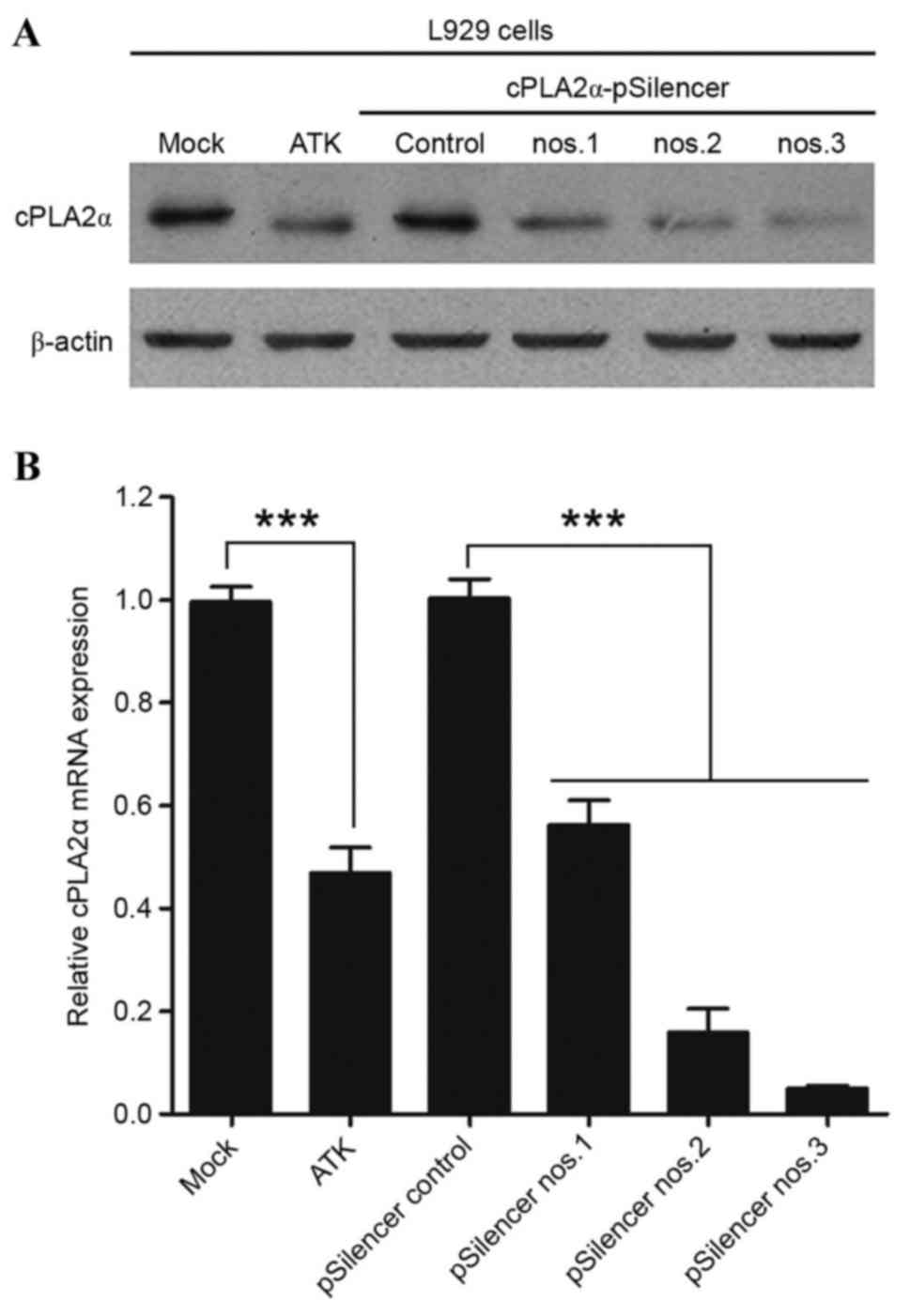
Selection of RNAi sequence for
knockdown of mouse cPLA2α in L929 cells
We examined the inhibitory effects of the three RNAi
sequences for mouse cPLA2α in L929, and attempted to establish the
most effective silencer. ATK effectively inhibited cPLA2α in
vitro and in vivo (33–37).
The treatment of L929 cells with 10 nM ATK every 24 h served as a
positive control. The level of cPLA2α in L929-cPLA2α-RNAi cells was
examined by qPCR and western blotting. The effects of three
predesigned RNAi sequences for mouse cPLA2α (silencer nos. 1–3) on
cPLA2α protein and activity (Fig.
1) were confirmed. Treatment with silencer-2 or −3 decreased
the expression level and activity of cPLA2α markedly, while
Silencer-1 only exerted a partial inhibitory effect. Results showed
that cells under silencer-3 treatment almost entirely inhibited the
expression level of cPLA2α protein without altering β-actin
expression. These results indicate that the Silencer-3 sequence is
an effective RNAi sequence for inhibiting mouse cPLA2α expression.
Subsequently, the most effective RNAi sequence was used to
knockdown the expression of cPLA2α in mice.
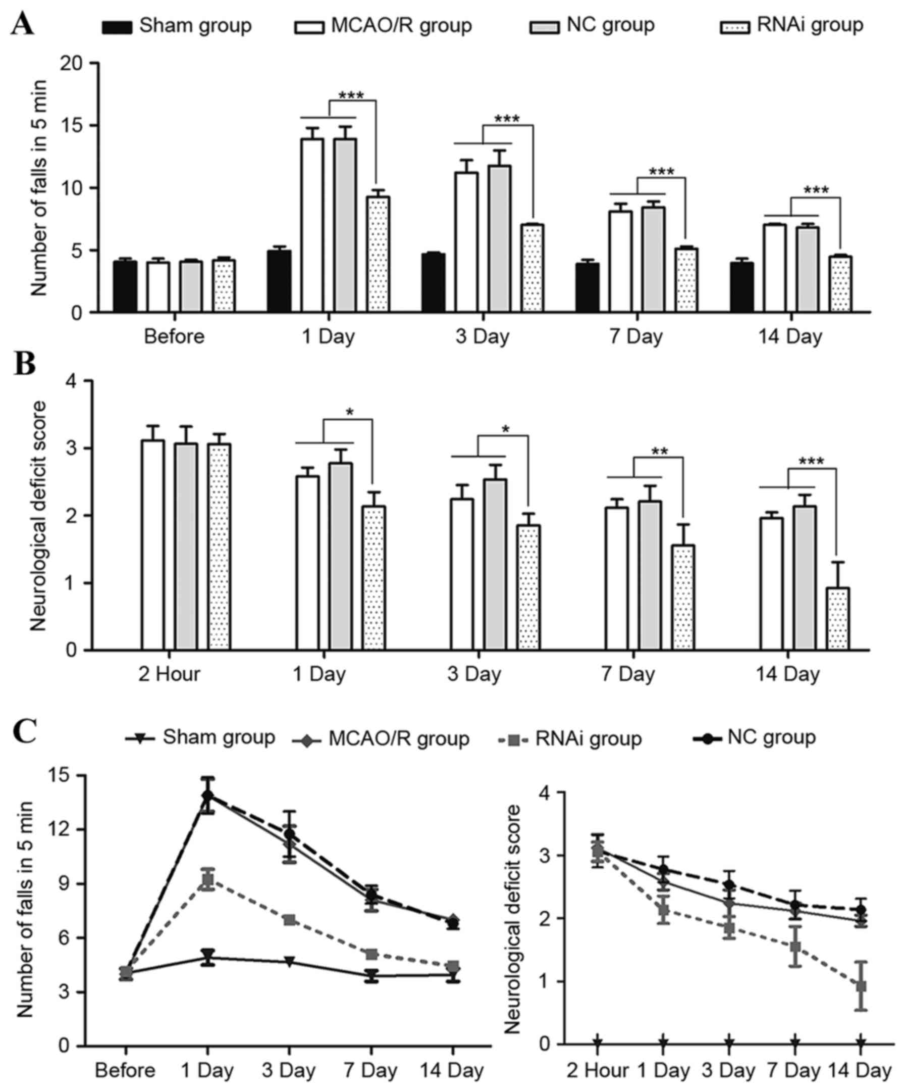
Neurological function and motor
function evaluation in animals following MCAO/R surgery
To investigate the role of cPLA2α in focal ischemic
brain damage, MCAO/R surgery was performed on male C57BL/6 mice
(age, 12–16 weeks). Neurological deficits of these mice were
assessed and 2 h after reperfusion, all mice exhibited neurological
injury. Neurological deficit was evaluated again on days 1, 3, 7
and 14 after reperfusion. The RNAi group demonstrated no
significant difference compared with the NC group and MCAO/R group
at 2 h post-surgery (P>0.05); however, the NDSs of the RNAi
group animals were significantly lower than those of the control
group mice at 7 and 14 days after stroke (Fig. 2B; P<0.01).
Ischemic brain damage patients are prone to motor
function impairment, losing partial or complete motor ability
(clinically termed paralysis). To monitor the motor function change
of mice following ischemic stroke, rotarod tests were performed at
1, 3, 7 and 14 days after ischemic stroke surgery. Prior to
surgery, the mice demonstrated no differences during the rotarod
test (Fig. 2). However, the number
of falls in 5 min was significantly higher in the MCAO/R surgery
mice compared with the sham mice when evaluated following surgery
(Fig. 2A). Seven days after
surgery, the RNAi group exhibited a good recovery as assessed by
the number of falls in 5 min, whereas the MCAO/R group and NC group
mice continued to demonstrate evident deficits at that time-point.
These data indicate that pAd-siRNA-cPLA2α treatment in mice
alleviates exacerbated motor dysfunction induced by ischemic brain
damage.
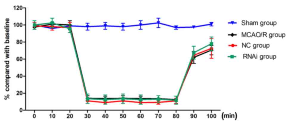
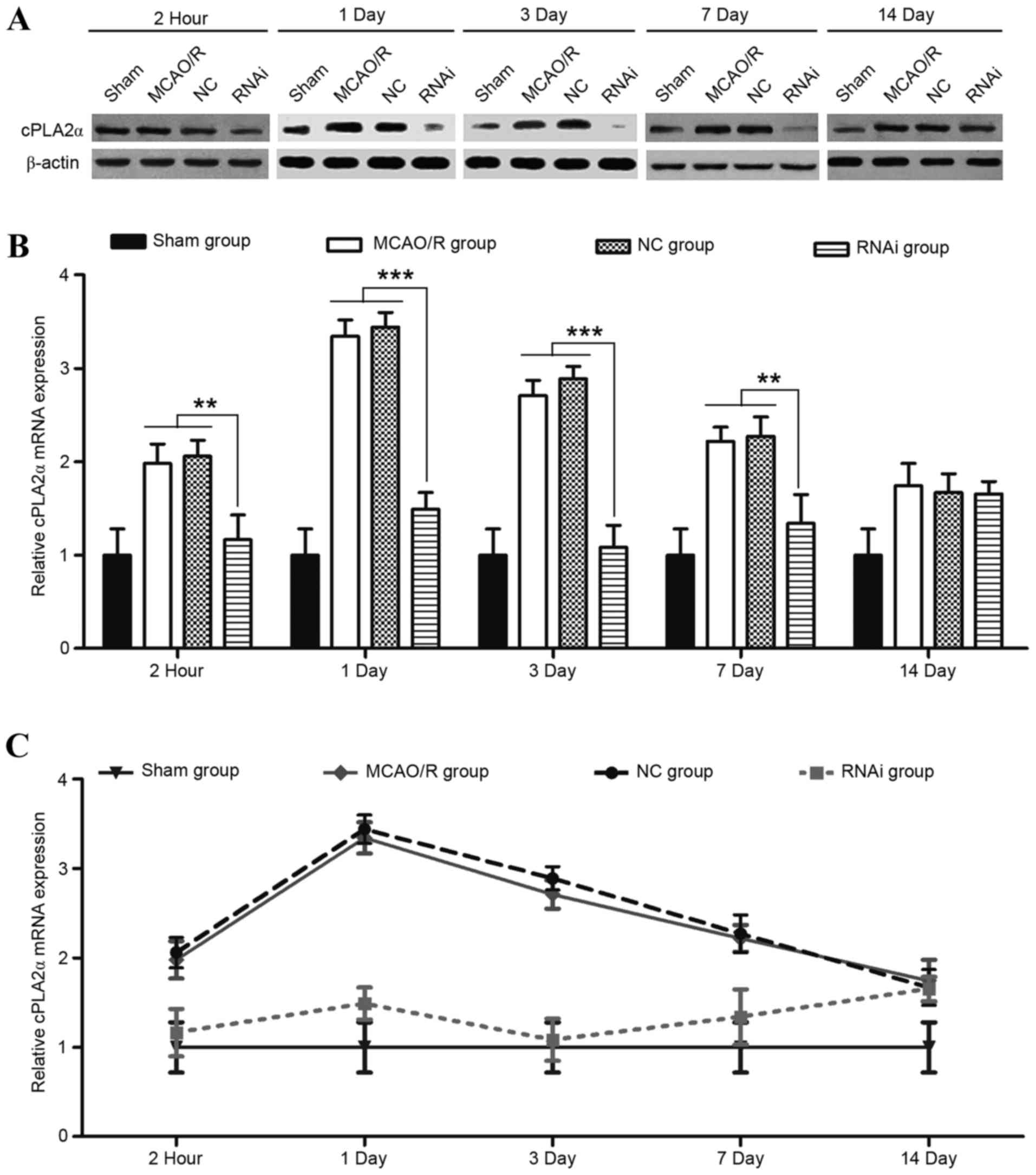
Effects of pAd-siRNA-cPLA2α on the
expression levels of cPLA2α, FFA, LPC, PGE2 and LTB4
The role of cPLA2α in exacerbating transient focal
ischemic brain damage has been previously demonstrated in
cPLA2α-knockout mice (11). In the
present study, whether the use of pAd-siRNA-cPLA2α attenuates
ischemic brain damage was evaluated in a mouse model of MCAO/R. A
laser Doppler perfusion monitor was used to monitor the regional
blood flow and this was continued until 20 min after reperfusion.
No difference in blood flow was detected in the mice from the
different groups at any time (Fig.
6). Compared with the NC group, the RNAi group effectively
decreased the cPLA2α expression levels in the ischemic damage mice
at 2 h, 1, 3 and 7 days after surgery (Fig. 3; P<0.01).
To evaluate whether pAd-siRNA-cPLA2α treatment
decreased the levels of phospholipid degradation products,
cPLA2α-derived injurious lipid mediators in the brain tissue
samples were assessed. The results indicated that pAd-siRNA-cPLA2α
treatment significantly reduced the levels of FFA and LPC, while
the levels of FFA and LPC exhibited a marked increase in the NC
group (Fig. 4A and B). At the
measurement time-points, the levels of FFA and LPC in the RNAi
group did not recover to the normal levels of the sham group (data
not shown). cPLA2 tend to catalyze the hydrolysis of
phospholipids in the sn-2 position (35), and it is the crucial enzyme of AA,
PG and PAF. PGE2 and LTB4 are proinflammatory eicosanoids
metabolized from free AA. Serum from the RNAi group mice exhibited
a reduced level of PGE2 compared with the sham, MCAO/R and NC group
animals at 24 h following surgery (Fig. 4C; P<0.001). Similarly, reduced
levels of LTB4 were observed in the serum of the RNAi group animals
(Fig. 4D; P<0.001). PGE2 and
LTB4 in the serum were significantly decreased following
pAd-siRNA-cPLA2α treatment (Fig.
4).
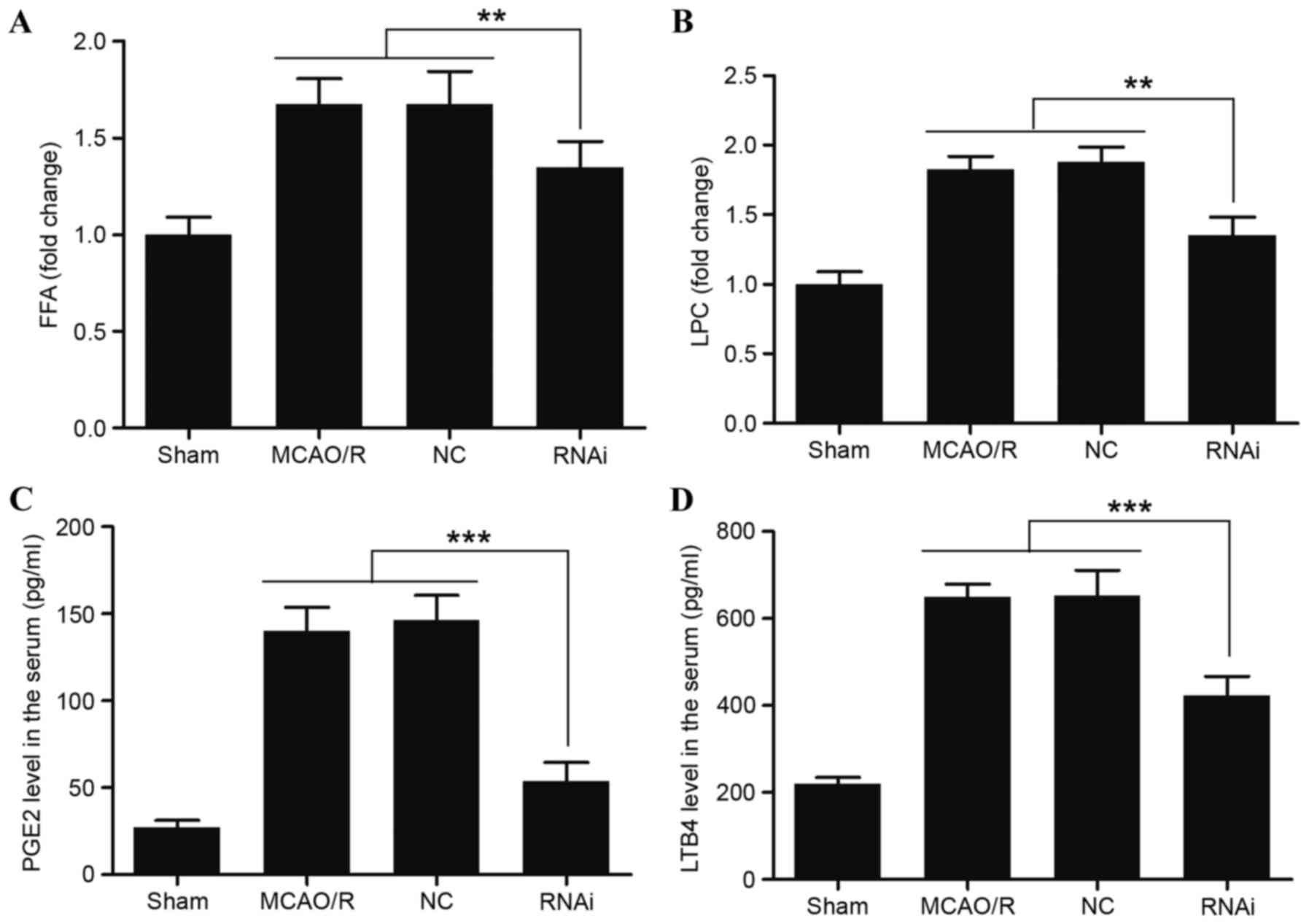 | Figure 4.Effects of siRNA-cPLA2α on the
expression of FFA, LPC, PGE2 and LTB4 in mice after MCAO/R.
Expression levels of (A) FFA and (B) LPC in the brain tissue from
mice 24 h after MCAO/R surgery were measured by high performance
thin layer chromatography. Expression levels of (C) PGE2 and (D)
LTB4 in the serum of mice 24 h after MCAO/R surgery were measured
using ELISA. pAd-siRNA-cPLA2α treatment decreased the expression
levels of FFA, LPC, PGE2 and LTB4. Data are presented as means ±
standard deviation of three independent experiments. **P<0.01
and ***P<0.001 (Student's t-test). siRNA, small interfering RNA;
cPLA2α, cytosolic phospholipase A2α; FFA, free fatty acids; LPC,
lysophosphatidylcholine; PGE2, prostaglandin E2; LTB4, leukotriene
B4; MCAO/R, middle cerebral artery occlusion and/or reperfusion;
NC, negative control. |
Effects of cPLA2α on pathological
changes
H&E staining effectively demonstrated the
pathological changes. The examination of these pathological
sections demonstrated that the morphological features of the brain
tissue samples were normal 14 days after surgery in the sham group
(Fig. 5A), while the morphological
features of the brain tissue samples from the other three groups
exhibited significant changes. MCAO/R tissue samples exhibited
nerve cell necrosis, loose structures and visible intercellular
edema to different degrees, which validated that the MCAO/R surgery
had been successfully performed. However, compared with the MCAO/R
group or NC group, the histological structure changes of the brain
tissue samples were less marked in the RNAi group. The results
demonstrate that the treatment of cPLA2α RNAi ameliorated
pathological changes following ischemic brain damage in mice.
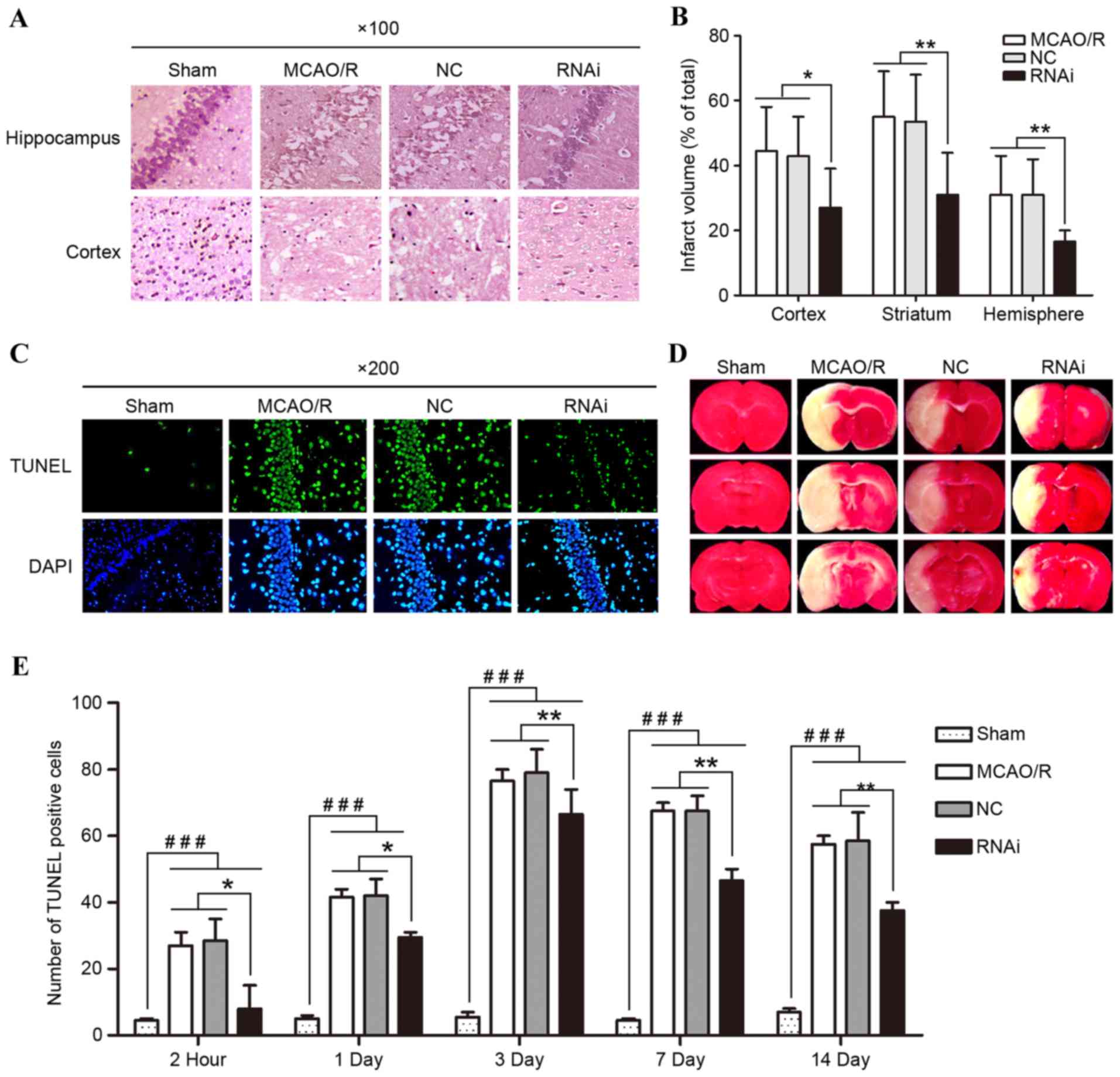 | Figure 5.Effects of siRNA-cPLA2α on
pathological changes, cell apoptosis, and infarct volume of mice
following MCAO/R. (A) Pathological changes of hippocampal and
cortical tissues following pAd-siRNA-cPLA2α treatment
(magnification, ×100). (B) PAd-siRNA-cPLA2α treatment protects mice
from brain infarction 14 days after MCAO/R. (C) Representative
images of TUNEL staining from mice brain tissue samples 14 days
after MCAO/R (magnification, ×200). (D) Triphenyltetrazolium
chloride staining images from mice brain tissue samples 14 days
after MCAO/R. (E) Number of TUNEL-positive cells was counted. Data
are presented as means ± standard deviation of three independent
experiments. *P<0.05 and **P<0.01, RNAi group vs. the MCAO/R
and NC group; ###P<0.001, sham group vs. the other
three groups (Student's t-test). siRNA, small interfering RNA;
cPLA2α, cytosolic phospholipase A2α; MCAO/R, middle cerebral artery
occlusion and/or reperfusion; RNAi, RNA interference; TUNEL,
terminal deoxynucleotidyl transferase dUTP nick end-labeling; NC,
negative control. |
Effects of cPLA2α on reducing
ischemia-induced brain infarct volume
Brain infarct was assessed following the experiment
using laboratory standard volumetric analysis of the anterior and
posterior views of coronal slabs stained with TTC, and corrected
for swelling. No infarction was observed in the sham group, whereas
extensive lesions were observed in the model and NC groups. RNAi
treatment effectively decreased the cerebral injury with a
significant reduction in infarct volume of the ischemic hemisphere
when compared with the NC or model group mice (Fig. 5B; P<0.01).
Effects of cPLA2α on apoptosis in
brain cells
TUNEL staining was performed to evaluate whether
pAd-siRNA-cPLA2α treatment alleviates cell apoptosis by decreasing
cPLA2α expression levels (Fig. 5C and
E). TUNEL+ cell number in the brain tissue of mice following
MCAO/R surgery exhibited a significant increase when compared with
the sham group mice at each time-point. The RNAi group had a
smaller number of TUNEL-positive cells in comparison to the NC
group on days 7 and 14. Such results indicate that pAd-siRNA-cPLA2α
treatment may improve pathological changes by inhibiting apoptosis
in mice with focal ischemic brain damage.
Discussion
The treatment of cerebral ischemia should be
selected according to the principle of damage cascades. The aim of
clinical treatment of cerebral ischemia is to recover the blood and
oxygen supply, suppress inflammation of the ischemic area, and
maintain the integrity of the neuron structure and function. In the
present study, it was validated and confirmed that cPLA2α
significantly influences the speed of recovery from cerebral
ischemia. In addition, cPLA2α-knockout effectively suppresses
inflammation and helps to maintain the integrity of the neuron
structure and function. The present results provide a reference for
the potential use of adenoviruses-mediated RNAi targeting cPLA2α in
the clinical setting.
Numerous studies demonstrated that the level of
cPLA2a was closely associated with the functional recovery in
stroke (11), Alzheimer's disease
(38) and multiple sclerosis
(34), which indicated the
causative role of cPLA2a in neurodegeneration. cPLA2α has a major
role in rat cerebral ischemia, and upregulated expression of cPLA2α
following trauma may be significant in secondary injury, such as
inflammation, nociception and functional deficits (11). Certain studies indicated that
certain inhibitors of cPLA2α inhibit the activity of cPLA2α, but
not the protein expression and that the effects are short term
(22,39). In the present study, three
adenoviruses-mediated RNAi sequences were established and their
effects in L929 cells were identified. An RNAi sequence that
inhibits the expression and activity of mouse cPLA2α was
successfully identified (Fig. 1).
The MCAO/R mice were treated with pAd-siRNA-cPLA2α prior to injury,
and it was observed that it effectively reduced the expression
levels of cPLA2α (Fig. 3).
pAd-siRNA-cPLA2α affects the expression of cPLA2α in the long-term
(14 days) following only one injection (Fig. 3C).
The fact that inhibition of cPLA2α attenuates focal
ischemic brain damage in mice indicates that the activity of cPLA2α
contributes markedly to the injury cascade following MCAO/R surgery
(9,10). The effects of cPLA2α on focal
ischemic brain damage go beyond the phase of acute injury. Indeed,
the present and previous studies have demonstrated that the
secondary injury mechanism is closely associated with cPLA2α
(11), as the cPLA2α RNAi
treatment reduced the injury following 3 days of reperfusion. It
has been reported previously that upregulation of cPLA2α is
relevant to blood-brain barrier disruption in rats at 24 h of
reperfusion (40).
A total of 75% of patients with cerebral ischemia
exhibit varying degrees of neurological deficit (41), and motor function deficits are an
important manifestation of neurological deficits (42). Neurological deficits are a
characteristic of cerebral ischemia. Thus, motor function recovery
is an important index for evaluating the therapeutic efficacy of
medications. An apparent neurological deficit was observed from 2 h
after MCAO/R surgery in the present study, which validated the
cerebral ischemia model. However, the finding that the NDS was
reduced in the RNAi group compared with the NC group supports the
conclusion that pAd-siRNA-cPLA2α treatment protects against
functional injury (Fig. 2A). The
motor function recovery was restored faster in the RNAi group,
which was assessed by the number of falls by mice following MCAO/R
surgery (Fig. 2B).
Inhibition of cPLA2α to treat cerebral ischemia has
certain anti-inflammatory and neuroprotective effects, expediting
the processes involved in physiological function recovery. Previous
studies regarding the link between cPLA2α and cerebral ischemia
have revealed that levels of cyclooxygenase (COX)-2 in the basal
and stimulated central nervous system are reduced in cPLA2α
knock-out animals (43,44). Proinflammatory lipid mediators,
such as PGE2, LTB4, LPC and FFA, which are regulated by the
activity of cPLA2α are closely associated with ischemic brain
damage. COX-2 and 5-lipoxygenase (5-LOX), as well as their
products, were involved in neuron damage and neurodegeneration. FFA
represents predominantly free AA, and was catalyzed to
prostaglandins and leukotrienes by COX-2 and 5-LOX, respectively.
In the present study, PGE2 was a representative product of COX-2,
and LTB4 was a representative product of 5-LOX. LPC is an important
signaling molecule correlated with chronic inflammation and tissue
damage (45). In the present
study, MCAO/R surgery mice treated with pAd-siRNA-cPLA2α
demonstrated effectively reduced expression levels of those
proinflammatory lipid mediators (Fig.
4), indicating that they originated from cPLA2α. H&E
staining demonstrated that the RNAi group changes were less marked
when compared with the NC group. Similarly, mice treatment with
pAd-siRNA-cPLA2α exhibited a decreased number of TUNEL+ cells and
the brain infarct volume was reduced. These findings indicate that
treatment with pAd-siRNA-cPLA2α may alleviate the pathological
damage by reducing inflammation and inhibiting apoptosis following
MCAO/R surgery. Thus, these data suggest that treatment with
pAd-siRNA-cPLA2α inhibits cPLA2α effectively and efficiently
protects cells from ischemic-induced adverse effects.
In conclusion, the present study demonstrates the
therapeutic potential of the adenoviruses-mediated RNAi targeting
cPLA2α in a cerebral ischemia animal model. The efficacy of the
treatment was predominantly attributed to the inhibition of cPLA2α
and the reduction of cPLA2α-derived proinflammatory lipid
mediators. The neuroprotective effects of pAd-siRNA-cPLA2α
treatment in the MCAO/R mice indicated that a long-term effect
cPLA2α inhibitor may be used in the future as a therapeutic
strategy for cerebral ischemia. However, further investigations are
required to evaluate the adenoviruses-mediated RNAi targeting
cPLA2α in human cerebral ischemia patients.
Acknowledgements
The authors would like to thank the Cangzhou Central
Hospital (Hebei, China) for their support.
Funding
No funding was received.
Availability of data and materials
The analyzed datasets generated during the study are
available from the corresponding author on reasonable request.
Authors' contributions
HW and LZ conceived and designed the study. HW, HL
and FZ performed the experiments. HW wrote the study. LZ reviewed
and edited the study. All authors read and approved the study.
Ethics approval and consent to
participate
All procedures performed with mice were according to
the National Institutes of Health Guide for the Care and Use of
Laboratory Animals, and the experimental procedures were approved
by the Ethics Committee of Cangzhou Central Hospital (Hebei,
China).
Consent for publication
Not applicable.
Competing interests
The authors declare that they have no competing
interests.
References
|
1
|
Arai K, Ikegaya Y, Nakatani Y, Kudo I,
Nishiyama N and Matsuki N: Phospholipase A2 mediates ischemic
injury in the hippocampus: A regional difference of neuronal
vulnerability. In Eur J Neurosci. 13:2319–2323. 2001. View Article : Google Scholar
|
|
2
|
Bonventre JV, Huang Z, Taheri MR, O'Leary
E, Li E, Moskowitz MA and Sapirstein A: Reduced fertility and
postischaemic brain injury in mice deficient in cytosolic
phospholipase A2. Nature. 390:622–625. 1997. View Article : Google Scholar : PubMed/NCBI
|
|
3
|
Brady KM, Texel SJ, Kishimoto K, Koehler
RC and Sapirstein A: Cytosolic phospholipase A alpha modulates NMDA
neurotoxicity in mouse hippocampal cultures. Eur J Neurosci.
24:3381–3386. 2006. View Article : Google Scholar : PubMed/NCBI
|
|
4
|
Ward NC, Croft KD, Blacker D, Hankey GJ,
Barden A, Mori TA, Puddey IB and Beer CD: Cytochrome P450
metabolites of arachidonic acid are elevated in stroke patients
compared with healthy controls. Clin Sci (Lond). 121:501–507. 2011.
View Article : Google Scholar : PubMed/NCBI
|
|
5
|
Moskowitz MA, Lo EH and Iadecola C: The
science of stroke: Mechanisms in search of treatments. Neuron.
67:181–198. 2010. View Article : Google Scholar : PubMed/NCBI
|
|
6
|
Menschikowski M, Hagelgans A and Siegert
G: Secretory phospholipase A2 of group IIA: Is it an offensive or a
defensive player during atherosclerosis and other inflammatory
diseases? Prostaglandins Other Lipid Mediat. 79:1–33. 2006.
View Article : Google Scholar : PubMed/NCBI
|
|
7
|
Gabryel B, Chalimoniuk M, Stolecka A and
Langfort J: Activation of cPLA2 and sPLA2 in astrocytes exposed to
simulated ischemia in vitro. Cell Biol Int. 31:958–965. 2007.
View Article : Google Scholar : PubMed/NCBI
|
|
8
|
Williams SD and Gottlieb RA: Inhibition of
mitochondrial calcium-independent phospholipase A2 (iPLA2)
attenuates mitochondrial phospholipid loss and is cardioprotective.
Biochem J. 362:23–32. 2002. View Article : Google Scholar : PubMed/NCBI
|
|
9
|
Kishimoto K, Li RC, Zhang J, Klaus JA,
Kibler KK, Doré S, Koehler RC and Sapirstein A: Cytosolic
phospholipase A2 alpha amplifies early cyclooxygenase-2 expression,
oxidative stress and MAP kinase phosphorylation after cerebral
ischemia in mice. J Neuroinflammation. 7:422010.PubMed/NCBI
|
|
10
|
Shen Y, Kishimoto K, Linden DJ and
Sapirstein A: Cytosolic phospholipase A(2) alpha mediates
electrophysiologic responses of hippocampal pyramidal neurons to
neurotoxic NMDA treatment. Proc Natl Acad Sci USA. 104:pp.
6078–6083. 2007; View Article : Google Scholar : PubMed/NCBI
|
|
11
|
Zhang J, Barasch N, Li RC and Sapirstein
A: Inhibition of cytosolic phospholipase A(2) alpha protects
against focal ischemic brain damage in mice. Brain Res.
1471:129–137. 2012. View Article : Google Scholar : PubMed/NCBI
|
|
12
|
Leung RK and Whittaker PA: RNA
interference: From gene silencing to gene-specific therapeutics.
Pharmacol Ther. 107:222–239. 2005. View Article : Google Scholar : PubMed/NCBI
|
|
13
|
Elbashir SM, Harborth J, Lendeckel W,
Yalcin A, Weber K and Tuschl T: Duplexes of 21-nucleotide RNAs
mediate RNA interference in cultured mammalian cells. Nature.
411:494–498. 2001. View
Article : Google Scholar : PubMed/NCBI
|
|
14
|
Dykxhoorn DM and Lieberman J: The silent
revolution: RNA interference as basic biology, research tool, and
therapeutic. Annu Rev Med. 56:401–423. 2005. View Article : Google Scholar : PubMed/NCBI
|
|
15
|
Morris KV and Rossi JJ:
Lentiviral-mediated delivery of siRNAs for antiviral therapy. Gene
Ther. 13:553–558. 2006. View Article : Google Scholar : PubMed/NCBI
|
|
16
|
Oualikene W, Lamoureux L, Weber JM and
Massie B: Protease-deleted adenovirus vectors and complementing
cell lines: Potential applications of single-round replication
mutants for vaccination and gene therapy. Hum Gene Ther.
11:1341–1353. 2000. View Article : Google Scholar : PubMed/NCBI
|
|
17
|
Abea K, Setoguchib Y, Hayashia T and
Itoyama Y: In vivo adenovirus-mediated gene transfer and the
expression in ischemic and reperfused rat brain. Brain Res.
763:191–201. 1997. View Article : Google Scholar : PubMed/NCBI
|
|
18
|
Shimizu M, Matsumoto Y, Kurosawa T, Azuma
C, Enomoto M, Nakamura H, Hirabayashi T, Kaneko M, Okuma Y and
Murayama T: Release of arachidonic acid induced by tumor necrosis
factor-alpha in the presence of caspase inhibition: Evidence for a
cytosolic phospholipase A2alpha-independent pathway. Biochem
Pharmacol. 75:1358–1369. 2008. View Article : Google Scholar : PubMed/NCBI
|
|
19
|
Shen C, Buck AK, Liu X, Winkler M and
Reske SN: Gene silencing by adenovirus-delivered siRNA. FEBS Lett.
539:111–114. 2003. View Article : Google Scholar : PubMed/NCBI
|
|
20
|
Sambrook J, Fritsch EF and Maniatis T:
Molecular Cloning: A Laboratory Manual. 2nd. Cold Spring Harbor
Laboratory Press; New York: 1989
|
|
21
|
National Research Council, . Guide for the
Care and Use of Laboratory Animals. The National Academies Press;
Washington DC: 1996, https://doi.org/10.17226/5140September
6–2014
|
|
22
|
Zhang J, Barasch N, Li RC and Sapirstein
A: Inhibition of cytosolic phospholipase A(2) alpha protects
against focal ischemic brain damage in mice. Brain Res.
1471:129–137. 2012. View Article : Google Scholar : PubMed/NCBI
|
|
23
|
Longa EZ, Weinstein PR, Carlson S and
Cummins R: Reversible middle cerebral artery occlusion without
craniectomy in rats. Stroke. 20:84–91. 1989. View Article : Google Scholar : PubMed/NCBI
|
|
24
|
Khan M, Singh J and Singh I: Plasmalogen
deficiency in cerebral adrenoleukodystrophy and its modulation by
lovastatin. J Neurochem. 106:1766–1779. 2008.PubMed/NCBI
|
|
25
|
Khan M, Contreras M and Singh I:
Endotoxin-induced alterations of lipid and fatty acid compositions
in rat liver peroxisomes. J Endotoxin Res. 6:41–50. 2000.
View Article : Google Scholar : PubMed/NCBI
|
|
26
|
Weerheim AM, Kolb AM, Sturk A and
Nieuwland R: Phospholipid composition of cell-derived
microparticles determined by one-dimensional high-performance
thin-layer chromatography. Anal Biochem. 302:191–198. 2002.
View Article : Google Scholar : PubMed/NCBI
|
|
27
|
Weissman L, Jo DG, Sorensen MM, de
Souza-Pinto NC, Markesbery WR, Mattson MP and Bohr VA: Defective
DNA base excision repair in brain from individuals with Alzheimer's
disease and amnestic mild cognitive impairment. Nucleic Acids Res.
35:5545–5555. 2007. View Article : Google Scholar : PubMed/NCBI
|
|
28
|
Bradford MM: A rapid and sensitive method
for the quantitation of microgram quantities of protein utilizing
the principle of protein-dye binding. Anal Biochem. 72:248–254.
1976. View Article : Google Scholar : PubMed/NCBI
|
|
29
|
Hu N, Zhang J, Cui W, Kong G, Zhang S, Yue
L, Bai X, Zhang Z, Zhang W, Zhang X and Ye L: miR-520b regulates
migration of breast cancer cells by targeting hepatitis B
X-interacting protein and interleukin-8. J Biol Chem.
286:13714–13722. 2011. View Article : Google Scholar : PubMed/NCBI
|
|
30
|
Livak KJ and Schmittgen TD: Analysis of
relative gene expression data using real-time quantitative PCR and
the 2(Delta Delta C(T)) method. Methods. 25:402–408. 2001.
View Article : Google Scholar : PubMed/NCBI
|
|
31
|
Arumugam TV, Phillips TM, Cheng A, Morrell
CH, Mattson MP and Wan R: Age and energy intake interact to modify
cell stress pathways and stroke outcome. Ann Neurol. 67:41–52.
2010. View Article : Google Scholar : PubMed/NCBI
|
|
32
|
Swanson RA and Sharp FR: Infarct
measurement methodology. J Cereb Blood Flow Metab. 14:697–698.
1994. View Article : Google Scholar : PubMed/NCBI
|
|
33
|
Ackermann EJ, Conde-Frieboes K and Dennis
EA: Inhibition of macrophage Ca(2+)-independent phospholipase A2 by
bromoenol lactone and trifluoromethyl ketones. J Biol Chem.
270:445–450. 1995. View Article : Google Scholar : PubMed/NCBI
|
|
34
|
Kalyvas A and David S: Cytosolic
phospholipase A2 plays a key role in the pathogenesis of multiple
sclerosis-like disease. Neuron. 41:323–335. 2004. View Article : Google Scholar : PubMed/NCBI
|
|
35
|
Myou S, Sano H, Fujimura M, Zhu X,
Kurashima K, Kita T, Nakao S, Nonomura A, Shioya T, Kim KP, et al:
Blockade of eosinophil migration and airway hyperresponsiveness by
cPLA2-inhibition. Nat Immunol. 2:145–149. 2001. View Article : Google Scholar : PubMed/NCBI
|
|
36
|
Nagase T, Uozumi N, Aoki-Nagase T,
Terawaki K, Ishii S, Tomita T, Yamamoto H, Hashizume K, Ouchi Y and
Shimizu T: A potent inhibitor of cytosolic phospholipase A2,
arachidonyl trifluoromethyl ketone, attenuates LPS-induced lung
injury in mice. Am J Physiol Lung Cell Mol Physiol. 284:L720–L726.
2003. View Article : Google Scholar : PubMed/NCBI
|
|
37
|
Riendeau D, Guay J, Weech PK, Laliberte F,
Yergey J, Li C, Desmarais S, Perrier H, Liu S, Nicoll-Griffith D,
et al: Arachidonyl trifluoromethyl ketone, a potent inhibitor of
85-kDa phospholipase A2, blocks production of arachidonate and
12-hydroxyeicosatetraenoic acid by calcium ionophore-challenged
platelets. J Biol Chem. 269:15619–15624. 1994.PubMed/NCBI
|
|
38
|
Sanchez-Mejia RO, Newman JW, Toh S, Yu GQ,
Zhou Y, Halabisky B, Cissé M, Scearce-Levie K, Cheng IH, Gan L, et
al: Phospholipase A2 reduction ameliorates cognitive deficits in a
mouse model of Alzheimer's disease. Nat Neurosci. 11:1311–1318.
2008. View Article : Google Scholar : PubMed/NCBI
|
|
39
|
Khan M, Shunmugavel A, Dhammu TS, Matsuda
F, Singh AK and Singh I: Oral administration of cytosolic PLA2
inhibitor arachidonyl trifluoromethyl ketone ameliorates cauda
equina compression injury in rats. J Neuroinflammation. 12:942015.
View Article : Google Scholar : PubMed/NCBI
|
|
40
|
Nito C, Kamada H, Endo H, Niizuma K, Mye
DJ and Chan PH: Role of the p38 mitogen-activated protein
kinase/cytosolic phospholipase A2 signaling pathway in bolld-brain
barrier disruption after focal cerebral ischemia and reperfusion. J
Cereb Blood Flow Metab. 28:1686–1696. 2008. View Article : Google Scholar : PubMed/NCBI
|
|
41
|
Cramer SC: Repairing the human brain after
stroke. II. Restorative therapies. Ann Neurol. 63:549–560. 2008.
View Article : Google Scholar : PubMed/NCBI
|
|
42
|
Cramer SC and Crafton KR: Somatotopy and
movement representation sites following cortical stroke. Exp Brain
Res. 168:25–32. 2006. View Article : Google Scholar : PubMed/NCBI
|
|
43
|
Bosetti F and Weerasinghe GR: The
expression of brain cyclooxygenase-2 is down-regulated in the
cytosolic phospholipase A2 knockout mouse. J Neurochem.
87:1471–1477. 2003. View Article : Google Scholar : PubMed/NCBI
|
|
44
|
Sapirstein A, Saito H, Texel SJ, Samad TA,
O'Leary E and Bonventre JV: Cytosolic phospholipase A2alpha
regulates induction of brain cyclooxygenase-2 in a mouse model of
inflammation. Am J Physiol Regul Integr Comp Physiol.
288:R1774–R1782. 2005. View Article : Google Scholar : PubMed/NCBI
|
|
45
|
Sevastou I, Kaffe E, Mouratis MA and
Aidinis V: Lysoglycerophospholipids in chronic inflammatory
disorders: The PLA(2)/LPC and ATX/LPA axes. Biochim Biophys Acta.
1831:42–60. 2013. View Article : Google Scholar : PubMed/NCBI
|