Introduction
Glioma is the most common primary brain tumor in
adults and the second most common malignant tumor in children, with
an overall annual incidence rate of 3–8/100,000. According to the
histological tumor classification standard established by the World
Health Organization (WHO), glioma can be classified into four
grades (I–IV) and three histological types, namely astrocytoma,
oligodendroglioma and oligoastrocytoma (1). Despite the advances in surgical
resection, chemotherapy and radiotherapy, the prognosis of patients
with glioma remains poor, resulting in an overall 5-year survival
rate of <10%. Currently, glioma ranks third worldwide as the
cause of cancer-associated mortality, after pancreatic cancer and
lung cancer (2). The standard
therapy for newly diagnosed glioma is surgical resection followed
by adjuvant radiotherapy and/or chemotherapy. However, surgical
resection is often inappropriate due to the unclear boundaries
between the primary tumor and adjacent anatomical structures.
Furthermore, tumor cells can often rapidly develop resistance to
radiotherapy and chemotherapy, leading to low therapeutic efficacy.
In addition, therapeutic effects may be affected by the blood-brain
barrier and the adverse effects of anticancer drugs. Therefore, the
development of novel, effective therapies with fewer adverse
effects for the treatment of glioma is imperative.
In recent years, gene therapy using small-molecule
inhibitors has garnered attention. It has been reported that
members of the signal transducer and activator of transcription
(STAT) family are involved in various regulatory processes, such as
cellular proliferation, differentiation and apoptosis (3). STAT-1, which was the first member of
the STAT family to be discovered, is able to inhibit cell
proliferation and promote apoptosis (3). STAT-1 has been identified as a
tumor-inhibiting factor, and reduced STAT-1 expression is
associated with the occurrence and progression of malignant tumors
(4). In addition, it has been
demonstrated that the activation of STAT-1 expression in tumor
cells by exogenous or endogenous stimuli can improve the
sensitivity of these cells to chemotherapy (5). The p53 gene has been classified as a
tumor suppressor gene that can effectively inhibit cellular growth
and response to stress in various cell types by inducing temporary
or permanent suppression of proliferation, or by activating cell
death processes. It has previously been suggested that the
quinoline derivative chloroquine induces apoptosis in human glioma
cells by activating the p53 pathway (6), suggesting the potential of p53-based
cancer therapies. Furthermore, a p53-dependent activation pathway
of STAT1 has been identified (7),
indicating a strong association between STAT1 and p53. However, the
association between STAT1 and p53 in glioma has not been
clarified
Wild-type p53 has been well recognized as a tumor
suppressor gene, and p53 is the most frequently mutated gene in
human cancer. Evidence has demonstrated that mutant p53 proteins
not only lose the ability to suppress tumorigenesis but also gain
tumor-promoting functions (8).
Therefore, the present study examined the expression of STAT-1 and
mutant p53 in glioma of different grades, and analyzed their
correlation in order to investigate their effects on the
pathogenesis and development of glioma.
Materials and methods
Subjects
The present study included 50 patients who underwent
glioma resection at the First Affiliated Hospital of Inner Mongolia
Medical University (Hohhot, China) between December 2007 and
December 2011. The patients comprised 29 men and 21 women, with a
mean age of 55 years (range, 43–70 years). All glioma specimens
were evaluated by an experienced neuropathologist based on the
histological classification standard for tumors of the central
nervous system established by the WHO in 2007 (1). When the pathological type of a
specimen was between grade I–II, grade II–III or grade III–IV, it
was classified as belonging to the higher grade. As a result, there
were 10 cases of grade I, 14 cases of grade II, 14 cases of grade
III and 12 cases of grade IV glioma. A total of 10 patients with
acute cerebral contusion, without other nervous system diseases,
who underwent intracerebral hematoma removal at the same hospital
between January 2013 and January 2014 were selected as controls.
The control group comprised 6 men and 4 women with a mean age of 52
years (range, 48–65 years). Brain tissue was collected during the
surgery and stored at −70°C until further use.
The study was approved by the Ethics Committee of
the First Affiliated Hospital of Inner Mongolia Medical University.
Written informed consent was obtained from all patients prior to
enrollment in the present study.
Reagents
Rat anti-human STAT-1 polyclonal antibody (cat. no.
sc-592) and anti-mutant p53 antibody (cat. no. ab32049) were
purchased from Abcam (Cambridge, UK). Antibody dilution buffer was
purchased from Wuhan Boster Biological Technology, Ltd. (Wuhan,
China). UltraSensitive™ S-P kits (C reagent, biotinyated sheep
anti-mouse immunoglobulin G; D reagent, streptavidin-peroxidase
complex), antigen retrieval solution, hematoxylin and eosin
staining solution, and peroxidase/diaminobenzidine (DAB) chromogen
(rabbit/mouse) were purchased from Fuzhou Maixin Biotech Co., Ltd.
(Fuzhou, China).
Immunohistochemical detection of
STAT-1 and mutant p53 expression
All tissue samples were fixed with 10% formalin for
24 h at room temperature, embedded in paraffin, and sliced into
4-µm sections. The sections were mounted, cleared 3 times with
xylene, and rehydrated through serial ethanol dilutions (100, 90,
80, and 70%) and Tris-buffered saline. Subsequently, the sections
were heated in antigen retrieval solution (Fuzhou Maxim Biotech,
Inc.) in a pressure cooker for 5 min. The sections were incubated
in 3% hydrogen peroxide for 30 min to block endogenous peroxidases,
and goat serum (Fuzhou Maxim Biotech, Inc.) was used to minimize
non-specific binding. The sections were then incubated with rabbit
anti-human STAT-1 antibody (1:350 dilution) and anti-mutant p53
antibody (1:300 dilution) at 37°C for 1 h. Chromogenic detection of
the substrate was performed using the UltraSensitive™ S-P kit
according to the manufacturer's protocol, followed by color
development with DAB. The sections were counterstained with
hematoxylin, and examined for the extent and intensity of nuclear
and non-nuclear staining using a light microscope by two
independent observers in a blinded manner.
Determination of positive results
Nuclear and/or cytoplasmic yellow or brown staining
was identified as positive staining. Under ×200 magnification, 4
observation fields were randomly chosen for each section. A
proportion score, which described the estimated percentage of
positively-stained tumor cells (0, none; 1, <10%; 2, 10–50%; 3,
50–80%; 4, >80%), and an intensity score, representing the
estimated staining intensity (0, no staining; 1, weak; 2, moderate;
3, strong), was determined for each sample. A final
immunohistochemical score was obtained by multiplying the
proportion score and the intensity score, and STAT-1 and mutant p53
expression was classified into 4 subgroups: 0, negative (−); 1–4,
weakly positive (+); 5–8, moderately positive (++); and 9–12,
strongly positive (+++).
Statistical analysis
All statistical analyses were performed using the
SPSS statistical package, version 13.0 (SPSS, Inc., Chicago, IL,
USA). Correlations between STAT-1 expression and pathological
glioma grades, and between STAT-1 and mutant p53 expression were
assessed using Spearman's correlation coefficient. Differences
among groups were compared by one-way ANOVA followed by a Tukey's
multiple comparison test. The expression ratio of STAT-1 and mutant
p53 was compared by χ2 test. P<0.05 was considered to
indicate a statistically significant difference.
Results
Expression characteristics of STAT-1
and mutant p53 in glioma and normal brain tissue
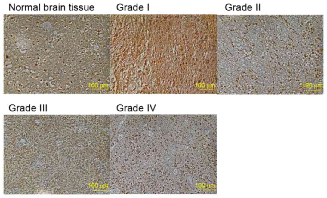
STAT-1 was mainly expressed in the cytoplasm of
glioma cells, with some expression also present in the nucleus
(Fig. 1). Intense STAT-1 staining
was observed in tumor-associated macrophages; however, no staining
was present in microglia and capillary endothelial cells. As shown
in Table I, although no
significant difference was detected between the positive expression
rate in normal brain tissue and that in glioma (χ2=1.38,
P>0.05), the expression score in glioma (5.32±3.25) was
significantly lower compared with that in normal brain tissue
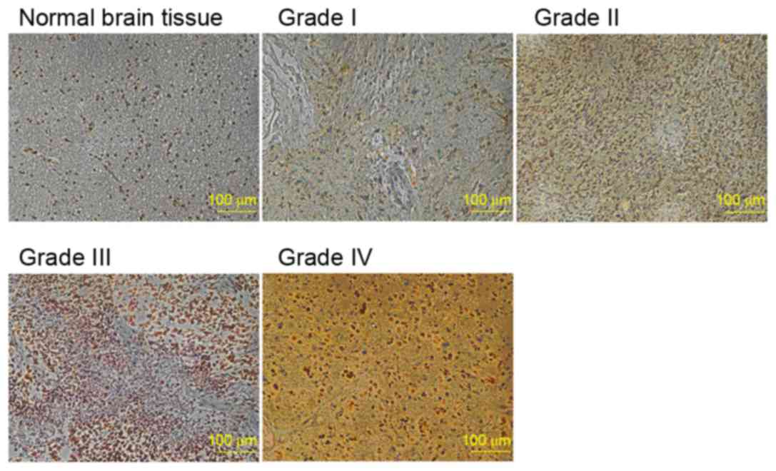
(10±1.76, P<0.05). Mutant p53 expression was stronger in the
nucleus and was associated with yellow, tan or brown staining
(Fig. 2). The positive expression
rate and expression score of mutant p53 were significantly higher
in glioma compared with normal brain tissue (χ2=31.27,
P<0.05), as shown in Table
II.
 | Table I.Expression of STAT-1 in glioma and
normal brain tissue. |
Table I.
Expression of STAT-1 in glioma and
normal brain tissue.
|
| STAT-1 |
|---|
|
|
|
|---|
| Group | Positive (n) | Positive expression
rate (%) | Expression score
(mean ± SD) |
|---|
| Glioma | 46 | 92 |
5.32±3.25a |
| Normal brain
tissue | 10 | 100 | 10.00±1.76 |
| P-value |
| >0.05 | <0.05 |
| χ2 |
| 1.38 |
|
| Table II.Expression of mutant p53 in glioma and
normal brain tissue. |
Table II.
Expression of mutant p53 in glioma and
normal brain tissue.
|
| Mutant p53 |
|---|
|
|
|
|---|
| Group | Positive (n) | Positive expression
rate (%) | Expression score
(mean ± SD) |
|---|
| Glioma | 46 | 92 |
3.10±1.28a |
| Normal brain
tissue | 6 | 60 | 0.80±0.79 |
| P-value |
| <0.05 | <0.05 |
| χ2 |
| 31.27 |
|
Correlation between STAT-1 expression
and glioma grade
As shown in Table
III, the expression score of STAT-1 in glioma was reduced as
the tumor grade increased. Spearman's correlation analysis
(Table IV) revealed a negative
correlation between STAT-1 expression and tumor grade (r=−0.767,
P<0.01).
| Table III.Expression scores of STAT-1 and mutant
p53 in glioma of different grades. |
Table III.
Expression scores of STAT-1 and mutant
p53 in glioma of different grades.
| Group | Case number | STAT-1 expression
scores (mean ± SD) | Mutant p53 expression
scores (mean ± SD) |
|---|
| Control | 10 | 10.00±1.76 | 0.80±0.79 |
| Glioma |
|
|
|
| Grade
I | 10 |
8.50±2.07a |
1.20±0.79a |
| Grade
II | 14 |
6.93±3.08a,b |
3.14±2.44c,d |
| Grade
III | 14 |
3.21±2.08c–e |
3.93±2.56c,d |
| Grade
IV | 12 |
3.17±1.99c–e |
3.67±1.87c,d |
| Table IV.Correlation analysis of STAT-1
expression and different grades of glioma. |
Table IV.
Correlation analysis of STAT-1
expression and different grades of glioma.
| Comparison | n | Correlation
coefficient | Bilateral
P-value |
|---|
| Grade vs.
STAT-1 | 60 | −0.767a | <0.01 |
Correlation between STAT-1 and mutant
p53 expression in glioma
The expression score of mutant p53 in glioma is
presented in Table III. The
mutant p53 expression score in glioma (grade I–IV) was
significantly higher compared with the control group (P<0.05).
Furthermore, the mutant p53 expression score in grade II, III and
IV glioma was significantly higher than in grade I glioma
(P<0.01). Spearman's correlation analysis (Table V) revealed a negative correlation
between STAT-1 and mutant p53 expression in glioma (r=−0.876,
P<0.01).
| Table V.Correlation analysis of STAT-1 and
mutant p53 expression. |
Table V.
Correlation analysis of STAT-1 and
mutant p53 expression.
| Comparison | n | Correlation
coefficient | Bilateral
P-value |
|---|
| STAT-1 vs. mutant
p53 | 60 | −0.876a | <0.01 |
Discussion
The gene coding for STAT-1 has been classified as a
tumor suppressor gene. A previous study demonstrated that STAT-1
overexpression in squamous cell carcinoma inhibits cell growth
(9), whereas transfection with
STAT-1 was revealed to inhibit the proliferation and promote the
apoptosis of U87MG cells in vitro (10). These results suggested that STAT-1
may participate in inducing the apoptosis of glioma cells.
Furthermore, STAT-1 inhibits the activity of proliferating cell
nuclear antigen by stimulating p21 expression, thus suppressing DNA
replication and cell cycle progression in U87MG cells, and
decreasing in vitro cell survival. In addition, a mouse
study demonstrated that STAT-1 defects inhibit interferon
(IFN)-mediated immune responses to bacteria and viruses.
Conversely, transfection with STAT-1 may result in a significant
decline in the growth and metastatic rate of highly tumorigenic
cancer cells in nude mice (11).
Most of the antitumor effects of radiotherapy and
chemotherapy are mediated by the cell cycle checkpoint, a mechanism
normally activated to repair DNA damage. Apoptosis of damaged cells
is activated when DNA damage cannot be repaired. In a previous
study regarding ataxia telangiectasia mutated (ATM) checkpoint
pathways, STAT-1 was reported to upregulate the transcription of
two important intermediary factors, namely p53 connexin 1 (53BP1)
and DNA damage checkpoint 1 (MDC1) (12). In the present study, STAT-1
expression was reduced in patients with a higher glioma grade,
whereas it has previously been demonstrated that the effects of ATM
on tumor radiosensitivity are decreased with increasing glioma
grade. Therefore, it may be hypothesized that decreased STAT-1
expression results in reduced tumor radiosensitivity via the ATM
checkpoint. Similar studies have revealed that STAT-1-positive
cancer may improve chemosensitivity of the tumor (10,13,14),
whereas STAT-1 activation by IFN-α/β and antineoplastic drugs may
exert a synergistic effect on the induction of apoptosis (5). In addition, the functions of the
topoisomerase I inhibitor irinotecan on squamous cell carcinoma,
and those of the antimetabolite raltitrexed, appear to depend on
the expression of STAT-1 (15),
thus suggesting that similar mechanisms may exist in glioma.
STAT-1 and anticancer drugs may interact through
cell cycle checkpoints, a possibility that needs to be addressed in
further studies. Numerous anticancer drugs trigger the DNA damage
checkpoint and exert their antineoplastic effects through the
induction of DNA damage and genotoxic stress, as well as through
the activation of ATM serine/threonine kinase and ataxia
telangiectasia and Rad3-related protein. A previous study suggested
that STAT-1 participates in ATM checkpoint activation through
upregulating two important mediators, namely 53BP1 and MDC1, a
finding that proposes a role for STAT-1 in enhancing the effect of
DNA-damaging agents through activation of ATM checkpoint pathways
(16).
STAT-1 can also be used as an indicator for the
efficacy of immunotherapy, since STAT-1 defects are associated with
immune escape mechanisms exploited by invasive tumors (17). Since some antineoplastic drugs are
able to enhance the antitumor immune response (18), the higher chemosensitivity of
STAT-1-expressing gliomas may be associated with the activation of
immune surveillance mechanisms by STAT-1 signaling pathways
(19). In addition, a high
percentage of patients with low intratumoral STAT-1 expression
exhibited reduced sensitivity to adjuvant chemotherapy. In the
present study, the negative correlation between STAT-1 expression
and the pathological grade of glioma suggested that STAT-1, as well
as being a valuable biomarker for evaluating the degree of glioma
malignancy, also exhibits potential as a molecular target for
enhancing the radio- and chemosensitivity of tumors.
The p53 gene is a tumor suppressor gene located on
chromosome 17 in humans. Although the pathogenesis of glioma
remains to be elucidated, studies have suggested that it is
associated with p53 gene mutation or loss (20,21).
It has also been demonstrated that although p53 mutations may be
present regardless of the glioma grade, the mutation rate in
high-grade glioma is significantly higher compared with that in
low-grade glioma (22). The p53
gene can temporarily or permanently suppress cell growth, or
activate stress-associated cell death mechanisms, thus providing a
theoretical basis for p53-based cancer therapy (23). In addition, a role for p53 in the
radio- and chemosensitivity of tumors has been suggested (24,25).
A recent study regarding gene therapy targeting p53 revealed that
survival rates of the human glioma cell line U87MG were markedly
reduced following transfection with the wild-type p53 gene using an
adenoviral vector (26). In
addition, another study reported that gliomas of a higher grade
exhibited increased mutant p53 gene expression (27), thus suggesting an association
between mutant p53 gene expression and the development and
progression of glioma. p53 holds potential as a target for gene
therapies that aim to increase the sensitivity of glioma cells to
chemotherapy or radiotherapy.
Anticancer drugs, such as fludarabine, doxorubicin
and cisplatin, are known to activate the tumor suppressor gene p53
(28). Furthermore, it has been
confirmed that STAT-1 and p53 synergistically promote tumor cell
death (29). Notably, anticancer
drugs can activate STAT-1 in p53-positive cells, but fail to work
in p53-null cells (7), thus
suggesting that p53 serves a necessary role in STAT-1 activation.
This finding is further supported by studies demonstrating that
anti-p53 small interfering RNAs, as well as overexpression of the
mouse double minute 2 homolog (MDM2) gene, coding the E3
ubiquitin-protein ligase Mdm2, which is a negative regulator of
p53, can deactivate STAT-1 expression (29). Overall, these findings suggested
that STAT-1 activation by genotoxic drugs depends on p53 (7). The present study revealed a negative
correlation between STAT-1 and mutant p53 expression in glioma
cells, indicating a crucial role of both proteins in the occurrence
and development of glioma.
In conclusion, the present study revealed a negative
correlation between the expression of the STAT-1 gene and the
glioma grade, as well as between STAT-1 and mutant p53 expression.
The negative correlation between STAT-1 and the pathological level
of glioma suggested that STAT-1 may be associated with the
occurrence and development of glioma, and may be a diagnostic
biomarker and therapeutic target for the malignancy of glioma.
Acknowledgements
The authors of the present study would like to thank
all the members of the Inner Mongolia Medical Molecular Pathology
Laboratory for their contribution to this experiment. This study
was funded by the Inner Mongolia Natural Science Foundation Project
(grant nos. 2014MS0836 and 2014MS08121).
Competing interests
The authors declare that they have no competing
interests.
References
|
1
|
Louis DN, Ohgaki H, Wiestler OD, Cavenee
WK, Burger PC, Jouvet A, Scheithauer BW and Kleihues P: The 2007
WHO classification of tumors of the central nervous system. Acta
Neuropathol. 114:97–109. 2007. View Article : Google Scholar : PubMed/NCBI
|
|
2
|
Xu L, Li Z, Tao Y, Li RH, Fang F, Zhao H,
Li G, Li YH, Wang J, Feng X and Pan J: Histone acetyltransferase
inhibitor II induces apoptosis in glioma cell lines via the p53
signaling pathway. J Exp Clin Cancer Res. 33:1082014. View Article : Google Scholar : PubMed/NCBI
|
|
3
|
Kim HS and Lee MS: STAT1 as a key
modulator of cell death. Cell Signal. 19:454–465. 2007. View Article : Google Scholar : PubMed/NCBI
|
|
4
|
Bruyère C, Madonna S, Van Goietsenoven G,
Mathieu V, Dessolin J, Kraus JL, Lefranc F and Kiss R: JLK1486, a
Bis 8-hydroxyquinoline-substituted benzylamine, displays cytostatic
effects in experimental gliomas through MyT1 and STAT1 activation
and, to a lesser extent, PPARγ activation. Transl Oncol. 4:126–137.
2011. View Article : Google Scholar : PubMed/NCBI
|
|
5
|
Strobl B and Moriggl R: Recovery from
chemotherapy depends on STAT1 for replenishment of B lymphopoiesis.
J Leukoc Biol. 95:849–851. 2014. View Article : Google Scholar : PubMed/NCBI
|
|
6
|
Kim EL, Wüstenberg R, Rübsam A,
Schmitz-Salue C, Warnecke G, Bücker EM, Pettkus N, Speidel D, Rohde
V, Schulz-Schaeffer W, et al: Chloroquine activates the p53 pathway
and induces apoptosis in human glioma cells. Neuro Oncol.
12:389–400. 2010. View Article : Google Scholar : PubMed/NCBI
|
|
7
|
Youlyouz-Marfak I, Gachard N, Le Clorennec
C, Najjar I, Baran-Marszak F, Reminieras L, May E, Bornkamm GW,
Fagard R and Feuillard J: Identification of a novel p53-dependent
activation pathway of STAT1 by antitumour genotoxic agents. Cell
Death Differ. 15:376–385. 2008. View Article : Google Scholar : PubMed/NCBI
|
|
8
|
Yue X, Zhao Y, Xu Y, Zheng M, Feng Z and
Hu W: Mutant p53 in cancer: Accumulation, gain-of-function, and
therapy. J Mol Biol. 429:1595–1606. 2017. View Article : Google Scholar : PubMed/NCBI
|
|
9
|
Xi S, Dyer KF, Kimak M, Zhang Q, Gooding
WE, Chaillet JR, Chai RL, Ferrell RE, Zamboni B, Hunt J and Grandis
JR: Decreased STAT1 expression by promoter methylation in squamous
cell carcinogenesis. J Natl Cancer Inst. 98:181–189. 2006.
View Article : Google Scholar : PubMed/NCBI
|
|
10
|
Ju H, Li X, Li H, Wang X, Wang H, Li Y,
Dou C and Zhao G: Mediation of multiple pathways regulating cell
proliferation, migration, laboratory investigation. J Neurosurg.
118:1239–1247. 2013. View Article : Google Scholar : PubMed/NCBI
|
|
11
|
Huang S, Bucana CD, Van Arsdall M and
Fidler IJ: Stat1 negatively regulates angiogenesis, tumorigenicity
and metastasis of tumor cells. Oncogene. 21:2504–2512. 2002.
View Article : Google Scholar : PubMed/NCBI
|
|
12
|
Barry SP, Townsend PA, Knight RA,
Scarabelli TM, Latchman DS and Stephanou A: STAT3 modulates the DNA
damage response pathway. Int J Exp Pathol. 91:506–514. 2010.
View Article : Google Scholar : PubMed/NCBI
|
|
13
|
Thomas M, Finnegan CE, Rogers KM, Purcell
JW, Trimble A, Johnston PG and Boland MP: STAT1: A modulator of
chemotherapy-induced apoptosis. Cancer Res. 64:8357–8364. 2004.
View Article : Google Scholar : PubMed/NCBI
|
|
14
|
Zhu H, Wang Z, Xu Q, Zhang Y, Zhai Y, Bai
J, Liu M, Hui Z and Xu N: Inhibition of STAT1 sensitizes renal cell
carcinomacells to radiotherapy and chemotherapy. Cancer Biol Ther.
13:401–407. 2012. View Article : Google Scholar : PubMed/NCBI
|
|
15
|
McDermott U, Longley DB, Galligan L, Allen
W, Wilson T and Johnston PG: Effect of p53 status and STAT1 on
chemotherapy-induced, Fas-mediated apoptosis in colorectal cancer.
Cancer Res. 65:8951–8960. 2005. View Article : Google Scholar : PubMed/NCBI
|
|
16
|
Duan X, Ponomareva L, Veeranki S,
Panchanathan R, Dickerson E and Choubey D: Differential roles for
the interferon-inducible IFI16 and AIM2 innate immune sensors for
cytosolic DNA in cellular senescence of human fibroblasts. Mol
Cancer Res. 9:589–602. 2011. View Article : Google Scholar : PubMed/NCBI
|
|
17
|
Liu K, Caldwell SA and Abrams SI: Immune
selection and emergence of aggressive tumor variants as negative
consequences of Fas-mediatedcytotoxicity and altered
IFN-gamma-regulated gene expression. Cancer Res. 65:4376–4388.
2005. View Article : Google Scholar : PubMed/NCBI
|
|
18
|
Lake RA and Robinson BW: Immunotherapy and
chemotherapy-a practical partnership. Nat Rev Cancer. 5:397–405.
2005. View
Article : Google Scholar : PubMed/NCBI
|
|
19
|
Dunn GP, Koebel CM and Schreiber RD:
Interferons, immunity and cancer immunoediting. Nat Rev Immunol.
6:836–848. 2006. View
Article : Google Scholar : PubMed/NCBI
|
|
20
|
Watanabe T, Katayama Y, Yoshino A, Komine
C and Yokoyama T: Deregulation of the TP53/p14ARF tumor suppressor
pathway in low-grade diffuse astrocytomas and its influenceon
clinical course. Clin Cancer Res. 9:4884–4890. 2003.PubMed/NCBI
|
|
21
|
Wang YY, Zhang T, Li SW, Qian TY, Fan X,
Peng XX, Ma J, Wang L and Jiang T: Mapping p53 mutations in
low-grade glioma: A voxel-based neuroimaging analysis. AJNR Am J
Neuroradiol. 36:70–76. 2015. View Article : Google Scholar : PubMed/NCBI
|
|
22
|
Cohen AL and Colman H: Glioma biology and
molecular markers. Cancer Treat Res. 163:15–30. 2015. View Article : Google Scholar : PubMed/NCBI
|
|
23
|
Bell HS and Ryan KM: Targeting the p53
family for cancer therapy: ‘Big brother’ joins the fight. Cell
Cycle. 6:1995–2000. 2007. View Article : Google Scholar : PubMed/NCBI
|
|
24
|
Fedrigo CA, Grivicich I, Schunemann DP,
Chemale IM, dos Santos D, Jacovas T, Boschetti PS, Jotz GP, Filho
Braga A and da Rocha AB: Radioresistance of human glioma spheroids
and expression of HSP70, p53 and EGFr. Radiat Oncol. 6:1562011.
View Article : Google Scholar : PubMed/NCBI
|
|
25
|
Biddlestone-Thorpe L, Sajjad M, Rosenberg
E, Beckta JM, Valerie NC, Tokarz M, Adams BR, Wagner AF, Khalil A,
Gilfor D, et al: ATM kinase inhibition preferentially sensitizes
p53 mutant glioma to ionizing radiation. Clin Cancer Res.
19:3189–3200. 2013. View Article : Google Scholar : PubMed/NCBI
|
|
26
|
Lang FF, Yung WK, Raju U, Libunao F, Terry
NH and Tofilon PJ: Enhancement of radiosensitivity of wild-type p53
human glioma cells by adenovirus-mediated delivery of the p53 gene.
J Neurosurg. 89:125–132. 1988. View Article : Google Scholar
|
|
27
|
Broaddus WC, Liu Y, Steele LL, Gillies GT,
Lin PS, Loudon WG, Valerie K, Schmidt-Ullrich RK and Fillmore HL:
Enhanced radiosensitivity of malignant glioma cell after adenoviral
p53 transduction. J Neurosurg. 91:997–1004. 1999. View Article : Google Scholar : PubMed/NCBI
|
|
28
|
Xiang W, Zhu XL and Zhao HY: Therapeutic
effect of p53 gene combined with cisplatin on experimental glioma.
Chin J Clin Neurosur. 11:154–156. 2006.(In Chinese).
|
|
29
|
Townsend PA, Scarabelli TM, Davidson SM,
Knight RA, Latchman DS and Stephanou A: STAT-1 interacts with p53
to enhance DNA damage-induced apoptosis. J Biol Chem.
279:5811–5820. 2004. View Article : Google Scholar : PubMed/NCBI
|