Introduction
Inflammatory bowel diseases (IBD), including Crohn's
disease (CD) and ulcerative colitis (UC), are chronic relapsing and
non-resolving inflammatory disorders that are characterized
pathologically by gastrointestinal inflammation and epithelial
injury (1,2). The pathogenesis and etiology of IBD
are still unclear and wildly thought to involve genetic factors,
the intestinal microbiota, immune dysfunction, and environmental
factors (3). IBD has become a
global disease with an increasing incidence worldwide (4), and it has a significant effect on
morbidity and quality of life (5).
Since currently available treatments for IBD are unsatisfactory,
new therapeutic strategies are desirable (6,7).
The gastrointestinal tract is the primary site of
interaction between the host immune system and microorganisms, both
symbiotic and pathogenic. The balance in the community structure of
gut bacteria may be intimately associated with the proper function
of the immune system (8–11). Numerous studies have revealed the
close relationship between the composition of the gut microbiota
and IBD (12,13). We previously used 16S-rRNA genome
sequencing to detect differences in the intestinal microbiota
between CD patients and healthy controls (HCs), and found that the
species R. intestinalis (R.I.) was significantly decreased
in CD patients. In agreement with our findings, a number of other
studies have also shown that the abundance of R.
intestinalis was decreased to varying degrees in IBD patients
(14,15), indicating that this species is
closely related to the development of IBD.
Cytokines also have a crucial role in the
pathogenesis of IBD, as they regulate multiple aspects of the
inflammatory response. In particular, the imbalance between
pro-inflammatory and anti-inflammatory cytokines that occurs in IBD
impedes the resolution of inflammation and instead leads to disease
perpetuation and tissue destruction (16). On the other hand, regulatory T
cells (Treg), a suppressive T cell population, can restrain the
progression of inflammation (17,18).
Based on our previous findings and other reports, we hypothesize
that R. intestinalis protects the intestinal mucosa from
inflammation by regulating the secretion of cytokines and the
differentiation of Treg. To investigate this, we evaluated the
potential therapeutic effects of R. intestinalis on
intestinal inflammation both in vivo and in vitro.
R. intestinalis increased the level of interleukin (IL)-17
secretion and Treg differentiation and protected colon epithelial
cells from pathological damage in an animal model of chemically
induced inflammation. These findings suggest that R.
intestinalis could be a potential treatment for IBD.
Materials and methods
Ethics approval
All animal experiments were approved by the Ethical
Committee of Medical Research, Third Xiangya Hospital, Affiliated
Hospital of Central South University.
R. intestinalis culture and
preparation
R. intestinalis (DSMZ-14610) was purchased
from Deutsche Sammlung von Mikroorganismen und Zellkulturen GmbH
(Braunschweig, Germany) and grown anaerobically at 37°C in Lytic/10
Anaerobic/F Medium (BD Biosciences, Franklin Lakes, NJ, USA). The
number of live bacteria (colony-forming units; CFU) was determined
according to the absorbance at 600 nm (A600). For in vitro
studies, bacterial cells were washed and resuspended at
1×10⁹ cells/ml in medium. For animal experiments, the
bacterial suspension (1×10⁹ CFU in 100 µl) was
administered to mice by intragastric gavage.
Animals and
2,4,6-trinitrobenzenesulfonic acid solution (TNBS)-induced
colitis
Mice (BALB/c, 6 weeks old, male) were obtained from
the Animal Center, Xiangya School of Medicine (Hunan, China), and
animal experiments were performed at the same facility. The mice
were maintained under specific pathogen-free conditions according
to the Animal Regulations of Hunan Province, China. Mice were
acclimatized to the facility before experiments were initiated. The
mice were then randomly assigned to four groups (n=6): A control
group without colitis, a group in which mice were preconditioned
with R.I. prior to the induction of colitis with TNBS (R.I. Pre), a
group in which colitis was induced but which did not receive R.I.
(TNBS), and a group in which R.I. was administered after the
induction of colitis with TNBS (R.I. Treat). Starting on day 1, the
preconditioned group received R. intestinalis
intragastrically for 2 days. On day 3, the mice in the groups in
which colitis was induced were given 100 µl of TNBS (a 1:1 mixture
by volume of 5% TNBS and absolute ethanol) intrarectally, while the
control group received normal saline. Bacteria were administered to
the R.I. Pre and R.I. Treat groups by intragastric gavage on days 5
and 7. Mice were observed and weighed, and fecal occult blood was
measured daily and used calculate the disease activity index (DAI)
using a previously published grading system (19) (Table
I). On day 9, the mice were weighed and sacrificed. Serum was
collected and colon tissues were removed, washed and opened, fixed
in 10% neutral buffered formalin solution, embedded in paraffin,
cut into tissue sections, and stained with hematoxylin and eosin
(H&E). Inflammation grading was carried out by two independent
blinded observers, and lesions were analyzed using histological
scoring criteria, as previously described (20) (Table
II).
 | Table I.Criteria for diseases activity index
scores in mice. |
Table I.
Criteria for diseases activity index
scores in mice.
| Weight loss
(%) | Stool
characters | Hematochezia | Score |
|---|
| 0 | Normal | OB negative | 0 |
| 1–5 |
|
| 1 |
| 5–10 | Loose | OB positive | 2 |
| 10–15 |
|
| 3 |
| >15 | Sloppy stools | Bloody stools | 4 |
 | Table II.Criteria for assessment of
microscopic colonic damage. |
Table II.
Criteria for assessment of
microscopic colonic damage.
| Score | Criteria |
|---|
| 0 | No
inflammation |
| 1 | Low level of
lymphocyte infiltration with infiltration seen in a <10% hpf, no
structural changes observed |
| 2 | Moderate lymphocyte
infiltration with infiltration seen in 10–25% hpf, crypt
elongation, bowel wall thickening which does not extend beyond
mucosal layer, no evidence of ulceration |
| 3 | High level of
lymphocyte infiltration with infiltration seen in 25–50% hpf, high
vascular density, thickening of bowel wall which extends beyond
mucosal layer |
| 4 | Marked degree of
lymphocyte infiltration with infiltration seen in >50% hpf, high
vascular density, crypt elongation with distortion, transmural
bowel wall-thickening with ulceration |
Immunohistochemistry
The paraffin-embedded samples were cut into
4-µm-thick sections, which were boiled in sodium citrate solution
(pH 6.0; Goodbio Technology, Wuhan, China) for 18 min and then
cooled at room temperature. The sections were incubated with an
IL-17 antibody (Boosen, Beijing, China) at 4°C overnight and then
with the corresponding secondary antibody (Goodbio Technology) for
30 min, followed by staining with 3,3′-diaminobenzidine (DAB; Mai
New Biotechnology Development Company, Fuzhou, China). The samples
were observed under a microscope by two independent blinded
observers.
Flow cytometric analysis of Treg in
murine peripheral blood
Mononuclear cells were isolated from murine
peripheral blood by Ficoll-Isopaue density gradient centrifugation
(Ficoll-Paque; GE Healthcare Bio-Sciences AB, Uppsala, Sweden). The
cells (2×10⁶ cells/sample) were labeled with FITC
anti-mouse CD4 (BD Bioscience), APC anti-mouse CD25 (BD
Bioscience), and PE anti-mouse Foxp3 (BD Bioscience). The stained
cells were analyzed by flow cytometry (BD Bioscience) using Cell
Quest software (BD Bioscience).
Experiments on NCM460 cells
The human colon epithelial cell line NCM460 was
obtained from the Cancer Research Institute of Central South
University (Changsha, China). NCM460 cells were cultured in
RPMI-1640 medium supplemented with 10% fetal bovine serum,
penicillin, and streptomycin at 37°C with 5% CO2 and
grown to 70–80% confluence. Cells were stimulated with
lipopolysaccharide (LPS) (1 µg/ml) and then co-cultured with R.
intestinalis (1×10⁹ CFU/ml in 30 µl) for 24 h.
Quantitative polymerase chain reaction
(qPCR)
Total RNA was extracted and reverse transcribed into
cDNA, which was then amplified by qPCR for detecting the mRNA
levels of targeted genes. The primers used are shown in Table III. The amplified PCR products
were identified by agarose gel electrophoresis. The results were
quantitated using the 2−ΔΔCq method, with expression of
GAPDH mRNA as an internal reference.
 | Table III.List of quantitative polymerase chain
reaction primers. |
Table III.
List of quantitative polymerase chain
reaction primers.
| Primer | Forward
(3′-5′) | Reverse
(5′-3′) |
|---|
| IL-17 |
TACAACCGATCCACCTCACCTT |
AGCCCACGGACACCAGTATCT |
| GAPDH |
GGAAGCTTGTCATCAATGGAAATC |
TGATGACCCTTTTGGCTCCC |
Protein extraction and western
blotting
Total protein was extracted with
radioimmunoprecipitation assay (RIPA) buffer containing phosphatase
and protease inhibitors. The protein concentration was determined
using the BCA Protein Assay kit (Beyotime, Shanghai, China). After
quantification, the proteins were separated by 10% SDS-PAGE and
transferred to polyvinylidene difluoride (PVDF) membranes (Merck
KGaA, Darmstadt, Germany). Membranes were blocked with 5% nonfat
dried milk, immunoblotted with a GAPDH polyclonal Ab (1:1,000) and
an IL-17 rabbit polyclonal Ab (1:1,000) at 4°C overnight, incubated
with secondary antibodies for 1 h at 37°C, and then developed with
an enhanced chemiluminescence (ECL) detection system (Bio-Rad
Laboratories, Inc., Hercules, CA, USA).
Cytokine detection in serum and cell
supernatants
Cytokine concentrations in mouse serum and cell
culture supernatants were quantified using IL-17 ELISA kits
according to the manufacturers recommendations.
Statistical analyses
Data were expressed as the standard deviation of the
mean and analyzed by one-way ANOVA (SPSS 18.0; SPSS, Inc., Chicago,
IL, USA) with an SNK post hoc test. P<0.05 was considered to
indicate a statistically significant difference. All reported
results are the average of three independent experiments.
Results
R. intestinalis exerts anti-inflammatory
effects in mice. Male, 6-week-old BALB/c mice were randomly divided
into four groups (n=6): A control group without colitis, a group in
which colitis was induced (TNBS), a group with colitis that was
treated with R. intestinalis (R.I. Treat), and a group that
was preconditioned with R.I. prior to the induction of colitis with
TNBS (R.I. Pre). In the R.I. Pre group, R. intestinalis was
administered intragastrically daily for 2 days before colitis was
induced with TNBS, while the R.I. Treat group was given R.
intestinalis after the induction of colitis with TNBS, on days
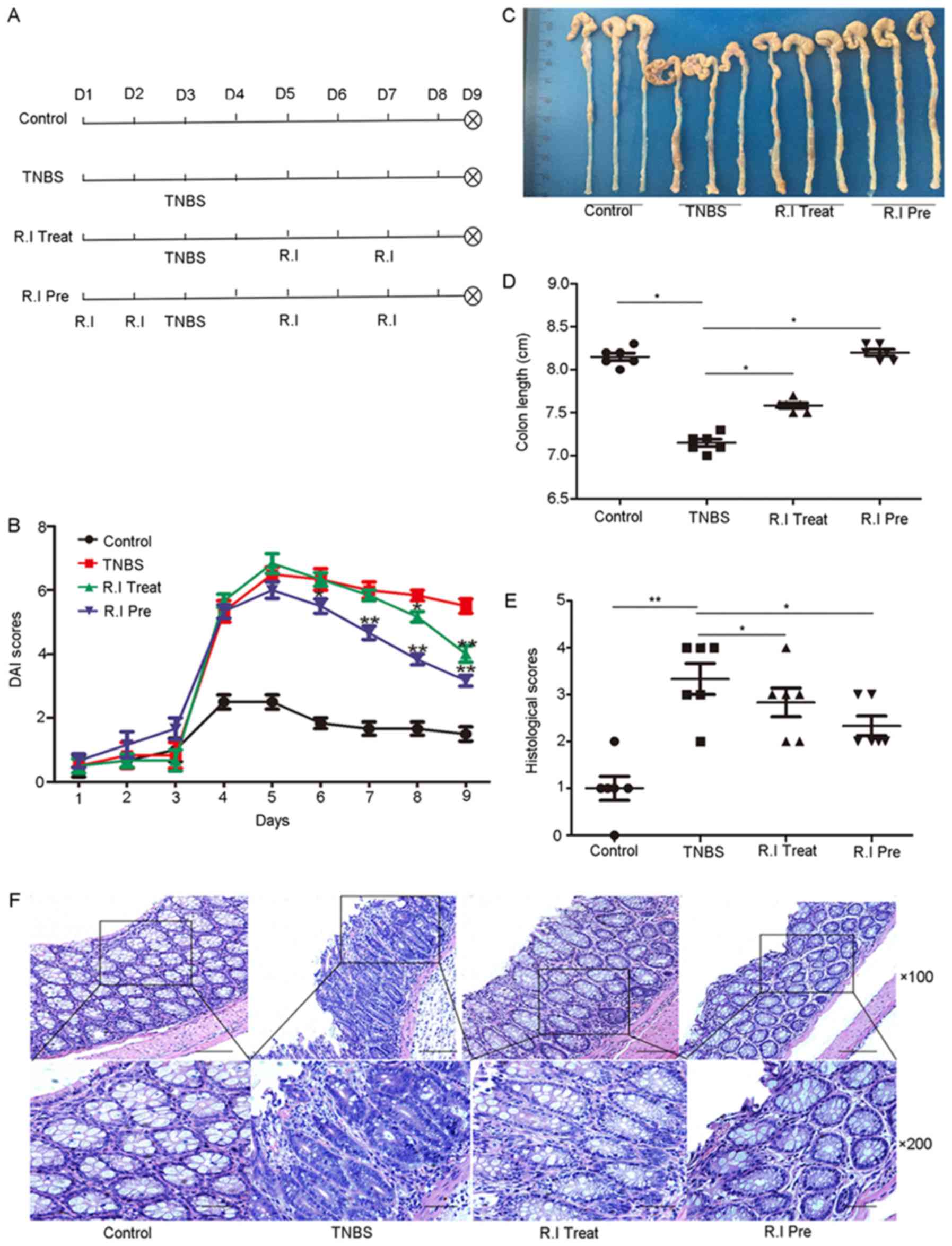
5 and 7 (Fig. 1A). At the end of
the experiment, the TNBS group mice had significantly higher DAI
scores (Fig. 1B), shorter colon
lengths (Fig. 1C and D), and
higher histological scores (Fig.
1E) than the control group. These symptoms were significantly
ameliorated by the administration of R. intestinalis. In
addition, the R.I. Pre group showed improvement of inflammatory
symptoms earlier (from day 6) and a greater anti-inflammatory
effect overall than the R.I. Treat group (Fig. 1B-F), suggesting that early
administration of R. intestinalis preparations could lead to
better anti-inflammatory effects. Moreover, histological
examination showed that the TNBS mice developed extensive
ulceration in the colon, with large numbers of infiltrating
neutrophils and some infiltrating mononuclear cells, while R.
intestinalis-treated mice displayed only mild mucosal
inflammation with a relatively a low level of neutrophil
infiltration (Fig. 1F).
IL-17 is upregulated in human IBD specimens, and
R. intestinalis inhibits the expression of IL-17 in mice
with TNBS-induced colitis. IL-17 is mainly produced by Th17 cells,
macrophages, and neutrophils (21). A number of recent studies have
suggested that IL-17 plays an important role in the pathogenesis of
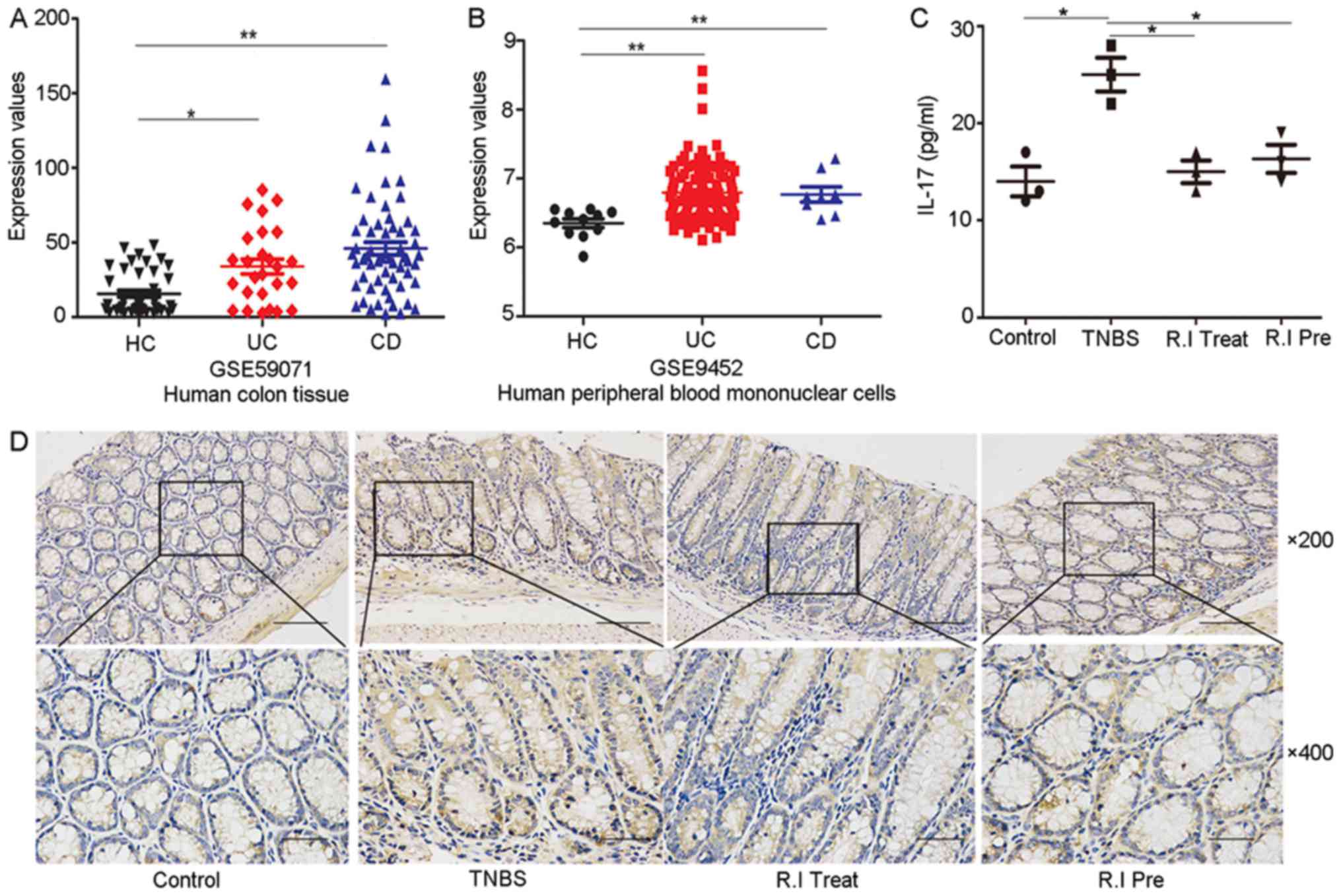
IBD (22,23). Therefore, we evaluated IL-17 gene
expression in a large cohort of HC, UC, and CD tissues (colon
tissue and human peripheral blood mononuclear cells) using data
deposited into the National Center for Biotechnology Information
(NCBI) Gene Expression Omnibus (GEO) database [no. GSE59071
(24) and no. GSE9452 (25)]. This analysis revealed that IL-17
mRNA levels were significantly upregulated in UC and CD compared to
the HC (Fig. 2A and B). This
finding was confirmed in our animal experiment, which revealed
higher IL-17 levels in both the serum and colon tissue of mice with
TNBS-induced colitis. Furthermore, the elevated IL-17 was decreased
by treatment with R. intestinalis (Fig. 2C and D).
 | Figure 2.IL-17 expression in human specimens,
and in mouse serum and colon tissue. Relative expression of IL-17
mRNA in HC, UC and CD tissues, based on data obtained from the
NCBI's GEO database (A) GSE59071 and (B) GSE9452). (C) IL-17
concentrations in mouse serum. (D) Representative
immunohistochemical staining of IL-17 in mouse colon mucosa. The
upper and lower panels are magnification, ×200 and ×400,
respectively. *P<0.05, **P<0.01. HC, healthy control; UC,
ulcerative colitis; CD, Crohn's disease; IL, interleukin. |
R. intestinalis inhibits the expression of
IL-17 in the human colon epithelial cell line NCM460. To verify the
function of R. intestinalis in vitro, the human colon
epithelial cell line NCM460 was stimulated with LPS to create a
model of cellular inflammation. NCM460 cells were divided into four
groups: A control group, an LPS group, a group treated with LPS and
R. intestinalis (R.I. Treat), and a R.I. preconditioned
group (R.I. Pre), in which NCM460 cells were co-cultured with R.
intestinalis 12 h before inflammation was induced with LPS. The
expression of IL-17 was detected by RT-qPCR, ELISA, and western
blotting. In agreement with our in vivo results, the IL-17
mRNA levels were upregulated in the LPS-stimulated cells, and the
induction of IL-17 could be decreased by either preconditioning or
co-culturing with R. intestinalis (Fig. 3A). The real-time PCR results were
confirmed by ELISA (Fig. 3B) and
western blotting (Fig. 3C).
R. intestinalis promotes regulatory T cell
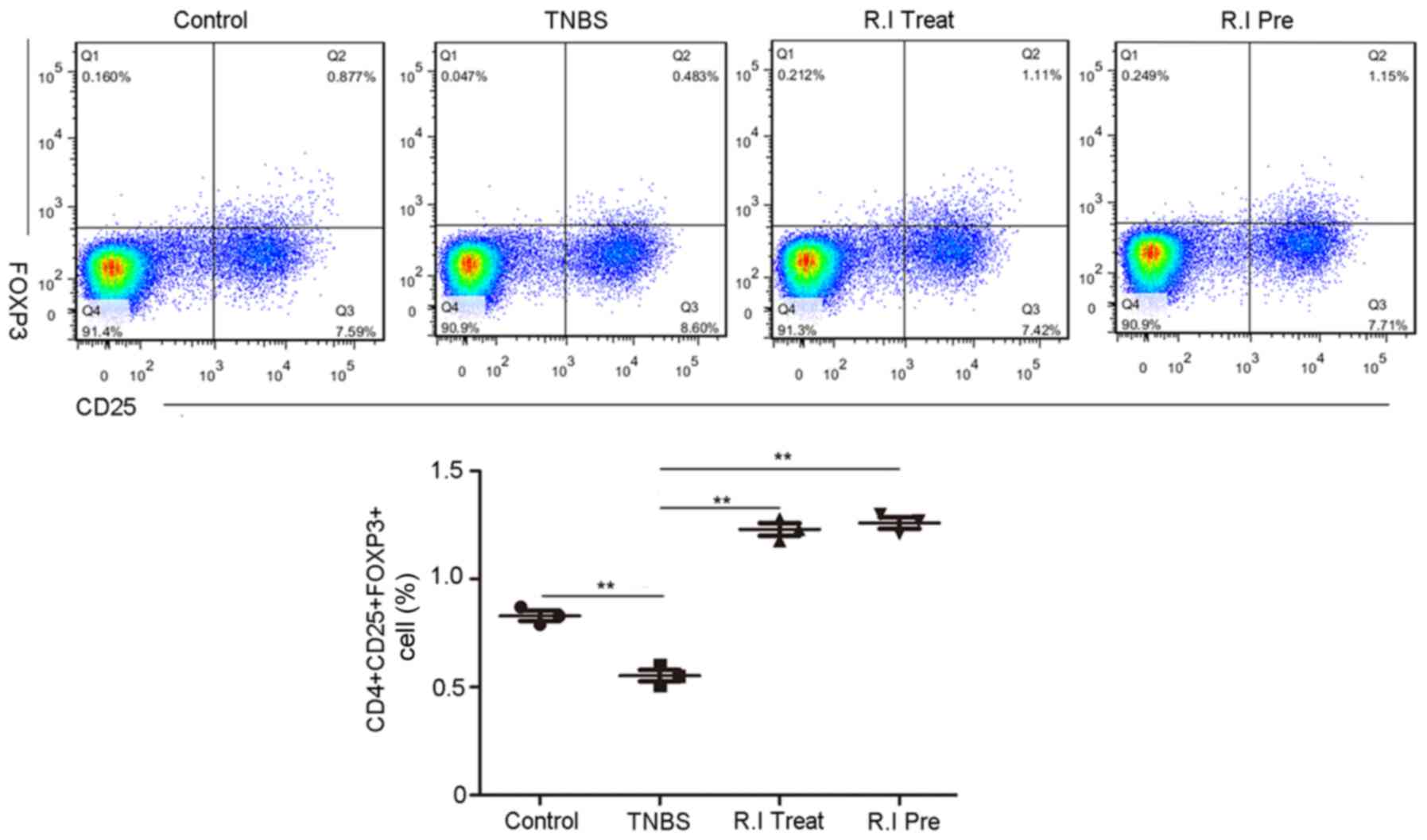
differentiation in the mouse peripheral blood. In the in
vivo studies, the numbers of CD25+Foxp3+ regulatory T cells
(Treg) in the peripheral blood of the TNBS group mice were
statistically lower than in the control mice without colitis. After
treatment with R. intestinalis, the numbers of
CD4+CD25+Foxp3+ Treg cells in the peripheral blood cells increased
compared with the TNBS group. Furthermore, the R.I. Pre group
showed a greater increase in the frequency of CD4+CD25+Foxp3+ Treg
than the R.I. Treat group (Fig.
4).
Discussion
The causes of IBD are multifactorial, but it is well
recognized that disturbed intestinal bacterial homeostasis may
contribute to the onset and recurrence of IBD (26). Although more and more bacterial
species have been shown to be associated with IBD and tested in
animal models and clinical trials, the molecular mechanisms of the
protective effects of probiotics are largely unknown. Recently, a
growing number of studies have shown that probiotics play a
protective role against colitis by effectively regulating the
secretion of cytokines (upregulating the secretion of
anti-inflammatory cytokines and inhibiting the secretion of
pro-inflammatory cytokines) (27,28)
and promoting the differentiation of Treg (29).
R. intestinalis is composed of Gram-positive
to Gram-variable rods (30) and
belongs to the family Clostridium cluster XIVa, which has a
strong regulatory effect on the polarization of Treg cells
(29). A number of studies have
demonstrated that the abundance of R. intestinalis decreased
to varying degrees in IBD patients. In agreement with these
findings, our previous research using 16S-rRNA genome sequencing
revealed that R. intestinalis decreased significantly in CD
patients, leading to the hypothesis that the presence of R.
intestinalis protects the intestine from inflammatory
damage.
IL-17 is a pro-inflammatory cytokine that is
reported to be closely related to IBD development (31). Consistent with these reports, we
evaluated IL-17 gene expression in data sets deposited into the GEO
database and found that it was significantly upregulated in UC and
CD patients compared to HCs, confirming the association between
IL-17 secretion and colon inflammation. To evaluate whether R.I.
could inhibit colon inflammation, we measured the IL-17 levels in
an animal model of chemically induced colitis and in an in
vitro model of cellular inflammation in which LPS-treated
NCM460 colon cells were co-cultured with R. intestinalis.
Treg, a suppressive subset of CD4+ T cells, also play a critical
role in the maintenance of intestinal homeostasis and
self-tolerance (32). Our in
vivo and in vitro results demonstrate that R.
intestinalis can inhibit the secretion of IL-17 and promote the
differentiation of Treg in colorectal colitis. IL-10, which is the
major effector cytokine secreted by Treg cells, plays crucial role
during the resolution phase of infection (33). Some probiotics, including
Bacteroides fragilis and Parabacteroides distasonis,
reduce intestinal inflammation through the production of IL-10
(34,35), which suggests a potential mechanism
through which R. intestinalis could act as a probiotic in
the treatment of IBD.
Interestingly, in our study, when R.
intestinalis was administered to the animals 2 days before the
induction of colitis with TNBS, the protective effect was stronger
and was apparent earlier than in the mice in which R.I. was
administered after the induction of colitis, demonstrating that
early feeding of R. intestinalis preparations could lead to
better anti-inflammatory effects. This finding may be explained by
data obtained with the Kaede transgenic mice, which revealed a
constant trafficking of immune cells between the intestine and
other parts of the body (36).
Therefore, early intake of the probiotic may have anti-inflammatory
effects on the immune cells trafficking through the intestine even
before the inflammatory stimulus is administered. Jun Li and
colleagues also found that a novel probiotic mixture effectively
reduced hepatocellular carcinoma (HCC) growth in mice, especially
when the probiotics were administrated before the implantation of
the tumor. This probiotic mixture, when given 1 week in advance of
tumor implantation, resulted in a strong antitumor effect that was
associated with reduced secretion of IL-17 and other
anti-inflammatory factors (37).
In this study, R. intestinalis exerted
significant anti-inflammatory effects in colorectal colitis in
vivo and in vitro by inhibiting the secretion of IL-17.
Furthermore, R.I. promoted the differentiation of Treg in the
peripheral blood in a mouse model of TNBS-induced colitis. The
detailed mechanisms through which R. intestinalis regulates
cytokine secretion and T cell differentiation are being
investigated in our ongoing studies. In conclusion, R.
intestinalis could be a candidate probiotic for the treatment
or prevention of IBD, and further research will be necessary to
elucidate the safety, efficacy, optimum dose, and mechanism of this
bacterium in the clinical practice.
Acknowledgements
Not applicable.
Funding
This study was funded by grants from the National
Natural Science Foundation of China (grant nos. 81670504 and
81472287) and the New Xiangya Talent Project of the Third xiangya
hospital of Central South University (grant no. 20150308).
Availability of data and materials
The datasets analyzed during the current study are
available in the National Center for Biotechnology Information Gene
Expression Omnibus database (nos. GSE59071 and GSE9452; ncbi.nlm.nih.gov/geo/). The rest of the data used and
analyzed during the current study are available from the
corresponding author on reasonable request.
Authors' contributions
CZ performed experiments and wrote the article. KS
contributed to the design of the study and revised the manuscript.
ZS and YQ performed the data analysis and revised the manuscript.
BT, WL, SW, KT and ZY performed the western blot analysis and
immunohistochemistry experiments and revised the manuscript. XW
contributed to the conception of the study and gave final approval
for publication. All the authors read and approved the final
version.
Ethics approval and consent to
participate
All animal experiments were approved by the Ethical
Committee of Medical Research, Third Xiangya Hospital, Affiliated
Hospital of Central South University.
Consent for publication
Not applicable.
Competing interests
The authors declare that they have no competing
interests.
References
|
1
|
Baumgart DC and Sandborn WJ: Crohn's
disease. Lancet. 380:1590–1605. 2012. View Article : Google Scholar : PubMed/NCBI
|
|
2
|
Danese S and Fiocchi C: Ulcerative
colitis. N Engl J Med. 365:1713–1725. 2011. View Article : Google Scholar : PubMed/NCBI
|
|
3
|
Ananthakrishnan AN: Epidemiology and risk
factors for IBD. Nat Rev Gastroenterol Hepatol. 12:205–217. 2015.
View Article : Google Scholar : PubMed/NCBI
|
|
4
|
Ng SC, Shi HY, Hamidi N, Underwood FE,
Tang W, Benchimol EI, Panaccione R, Ghosh S, Wu JCY, Chan FKL, et
al: Worldwide incidence and prevalence of inflammatory bowel
disease in the 21st century: A systematic review of
population-based studies. Lancet. 390:2769–2778. 2018. View Article : Google Scholar : PubMed/NCBI
|
|
5
|
Høivik ML, Moum B, Solberg IC, Henriksen
M, Cvancarova M and Bernklev T: IBSEN Group: Work disability in
inflammatory bowel disease patients 10 years after disease onset:
Results from the IBSEN Study. Gut. 62:368–375. 2013. View Article : Google Scholar : PubMed/NCBI
|
|
6
|
Gionchetti P, Dignass A, Danese S, Dias
Magro FJ, Rogler G, Lakatos PL, Adamina M, Ardizzone S, Buskens CJ,
Sebastian S, et al: 3rd European Evidence-based Consensus on the
Diagnosis and Management of Crohn's Disease 2016: Part 2: Surgical
management and special situations. J Crohns Colitis. 11:135–149.
2017. View Article : Google Scholar : PubMed/NCBI
|
|
7
|
Kaplan GG and Ng SC: Understanding and
preventing the global increase of inflammatory bowel disease.
Gastroenterology. 152:313–321.e2. 2017. View Article : Google Scholar : PubMed/NCBI
|
|
8
|
Belkaid Y and Hand TW: Role of the
microbiota in immunity and inflammation. Cell. 157:121–141. 2014.
View Article : Google Scholar : PubMed/NCBI
|
|
9
|
Blander JM, Longman RS, Iliev ID,
Sonnenberg GF and Artis D: Regulation of inflammation by microbiota
interactions with the host. Nat Immunol. 18:851–860. 2017.
View Article : Google Scholar : PubMed/NCBI
|
|
10
|
Caballero S and Pamer EG:
Microbiota-mediated inflammation and antimicrobial defense in the
intestine. Annu Rev Immunol. 33:227–256. 2015. View Article : Google Scholar : PubMed/NCBI
|
|
11
|
Cullen TW, Schofield WB, Barry NA, Putnam
EE, Rundell EA, Trent MS, Degnan PH, Booth CJ, Yu H and Goodman A:
Gut microbiota. Antimicrobial peptide resistance mediates
resilience of prominent gut commensals during inflammation.
Science. 347:170–175. 2015. View Article : Google Scholar : PubMed/NCBI
|
|
12
|
Miyoshi J and Chang EB: The gut microbiota
and inflammatory bowel diseases. Transl Res. 179:38–48. 2017.
View Article : Google Scholar : PubMed/NCBI
|
|
13
|
Eppinga H, Fuhler GM, Peppelenbosch MP and
Hecht GA: Gut microbiota developments with emphasis on inflammatory
bowel disease: Report from the Gut Microbiota for Health World
Summit 2016. Gastroenterology. 151:e1–e4. 2016. View Article : Google Scholar : PubMed/NCBI
|
|
14
|
Machiels K, Joossens M, Sabino J, De
Preter V, Arijs I, Eeckhaut V, Ballet V, Claes K, Van Immerseel F,
Verbeke K, et al: A decrease of the butyrate-producing species
Roseburia hominis and Faecalibacterium prausnitzii defines
dysbiosis in patients with ulcerative colitis. Gut. 63:1275–1283.
2014. View Article : Google Scholar : PubMed/NCBI
|
|
15
|
Tilg H and Danese S: Roseburia hominis: A
novel guilty player in ulcerative colitis pathogenesis? Gut.
63:1204–1205. 2014. View Article : Google Scholar : PubMed/NCBI
|
|
16
|
Neurath MF: Cytokines in inflammatory
bowel disease. Nat Rev Immunol. 14:329–342. 2014. View Article : Google Scholar : PubMed/NCBI
|
|
17
|
van der Veeken J, Gonzalez AJ, Cho H,
Arvey A, Hemmers S, Leslie CS and Rudensky AY: Memory of
inflammation in regulatory T cells. Cell. 166:977–990. 2016.
View Article : Google Scholar : PubMed/NCBI
|
|
18
|
Himmel ME, Yao Y, Orban PC, Steiner TS and
Levings MK: Regulatory T-cell therapy for inflammatory bowel
disease: More questions than answers. Immunology. 136:115–122.
2012. View Article : Google Scholar : PubMed/NCBI
|
|
19
|
Krieglstein CF, Cerwinka WH, Laroux FS,
Grisham MB, Schürmann G, Brüwer M and Granger DN: Role of appendix
and spleen in experimental colitis. J Surg Res. 101:166–175. 2001.
View Article : Google Scholar : PubMed/NCBI
|
|
20
|
Neurath MF, Fuss I, Kelsall BL, Stüber E
and Strober W: Antibodies to interleukin 12 abrogate established
experimental colitis in mice. J Exp Med. 182:1281–1290. 1995.
View Article : Google Scholar : PubMed/NCBI
|
|
21
|
Cua DJ and Tato CM: Innate IL-17-producing
cells: The sentinels of the immune system. Nat Rev Immunol.
10:479–489. 2010. View
Article : Google Scholar : PubMed/NCBI
|
|
22
|
Rosen MJ, Karns R, Vallance JE, Bezold R,
Waddell A, Collins MH, Haberman Y, Minar P, Baldassano RN, Hyams
JS, et al: Mucosal expression of type 2 and type 17 immune response
genes distinguishes ulcerative colitis from colon-only Crohn's
disease in treatment-naive pediatric patients. Gastroenterology.
152:1345–1357.e7. 2017. View Article : Google Scholar : PubMed/NCBI
|
|
23
|
Geremia A, Arancibia-Cárcamo CV, Fleming
MP, Rust N, Singh B, Mortensen NJ, Travis SP and Powrie F:
IL-23-responsive innate lymphoid cells are increased in
inflammatory bowel disease. J Exp Med. 208:1127–1133. 2011.
View Article : Google Scholar : PubMed/NCBI
|
|
24
|
Vanhove W, Peeters PM, Staelens D,
Schraenen A, Van der Goten J, Cleynen I, De Schepper S, Van Lommel
L, Reynaert NL, Schuit F, et al: Strong upregulation of AIM2 and
IFI16 inflammasomes in the mucosa of patients with active
inflammatory bowel disease. Inflamm Bowel Dis. 21:2673–2682. 2015.
View Article : Google Scholar : PubMed/NCBI
|
|
25
|
Olsen J, Gerds TA, Seidelin JB, Csillag C,
Bjerrum JT, Troelsen JT and Nielsen OH: Diagnosis of ulcerative
colitis before onset of inflammation by multivariate modeling of
genome-wide gene expression data. Inflamm Bowel Dis. 15:1032–1038.
2009. View Article : Google Scholar : PubMed/NCBI
|
|
26
|
Sartor RB and Wu GD: Roles for intestinal
bacteria, viruses, and fungi in pathogenesis of inflammatory bowel
diseases and therapeutic approaches. Gastroenterology.
152:327–339.e4. 2017. View Article : Google Scholar : PubMed/NCBI
|
|
27
|
Tsilingiri K, Barbosa T, Penna G, Caprioli
F, Sonzogni A, Viale G and Rescigno M: Probiotic and postbiotic
activity in health and disease: Comparison on a novel polarised
ex-vivo organ culture model. Gut. 61:1007–1015. 2012. View Article : Google Scholar : PubMed/NCBI
|
|
28
|
Zhang M, Qiu X, Zhang H, Yang X, Hong N,
Yang Y, Chen H and Yu C: Faecalibacterium prausnitzii inhibits
interleukin-17 to ameliorate colorectal colitis in rats. PLoS One.
9:e1091462014. View Article : Google Scholar : PubMed/NCBI
|
|
29
|
Atarashi K, Tanoue T, Oshima K, Suda W,
Nagano Y, Nishikawa H, Fukuda S, Saito T, Narushima S, Hase K, et
al: Treg induction by a rationally selected mixture of Clostridia
strains from the human microbiota. Nature. 500:232–236. 2013.
View Article : Google Scholar : PubMed/NCBI
|
|
30
|
Duncan SH, Hold GL, Barcenilla A, Stewart
CS and Flint HJ: Roseburia intestinalis sp. nov., a novel
saccharolytic, butyrate-producing bacterium from human faeces. Int
J Syst Evol Microbiol. 52:1615–1620. 2002. View Article : Google Scholar : PubMed/NCBI
|
|
31
|
Calderón-Gómez E, Bassolas-Molina H,
Mora-Buch R, Dotti I, Planell N, Esteller M, Gallego M, Martí M,
Garcia-Martín C, Martínez-Torró C, et al: Commensal-specific CD4(+)
cells from patients with Crohn's disease have a T-Helper 17
inflammatory profile. Gastroenterology. 151:489–500.e3. 2016.
View Article : Google Scholar : PubMed/NCBI
|
|
32
|
Kryczek I, Wang L, Wu K, Li W, Zhao E, Cui
T, Wei S, Liu Y, Wang Y, Vatan L, et al: Inflammatory regulatory T
cells in the microenvironments of ulcerative colitis and colon
carcinoma. Oncoimmunology. 5:e11054302016. View Article : Google Scholar : PubMed/NCBI
|
|
33
|
Laidlaw BJ, Cui W, Amezquita RA, Gray SM,
Guan T, Lu Y, Kobayashi Y, Flavell RA, Kleinstein SH, Craft J and
Kaech SM: Production of IL-10 by CD4(+) regulatory T cells during
the resolution of infection promotes the maturation of memory
CD8(+) T cells. Nat Immunol. 16:871–879. 2015. View Article : Google Scholar : PubMed/NCBI
|
|
34
|
Kverka M, Zakostelska Z, Klimesova K,
Sokol D, Hudcovic T, Hrncir T, Rossmann P, Mrazek J, Kopecny J,
Verdu EF and Tlaskalova-Hogenova H: Oral administration of
Parabacteroides distasonis antigens attenuates experimental murine
colitis through modulation of immunity and microbiota composition.
Clin Exp Immunol. 163:250–259. 2011. View Article : Google Scholar : PubMed/NCBI
|
|
35
|
Arpaia N, Campbell C, Fan X, Dikiy S, van
der Veeken J, deRoos P, Liu H, Cross JR, Pfeffer K, Coffer PJ and
Rudensky AY: Metabolites produced by commensal bacteria promote
peripheral regulatory T-cell generation. Nature. 504:451–455. 2013.
View Article : Google Scholar : PubMed/NCBI
|
|
36
|
Ding Y, Xu J and Bromberg JS: Regulatory T
cell migration during an immune response. Trends Immunol.
33:174–180. 2012. View Article : Google Scholar : PubMed/NCBI
|
|
37
|
Li J, Sung CY, Lee N, Ni Y, Pihlajamäki J,
Panagiotou G and El-Nezami H: Probiotics modulated gut microbiota
suppresses hepatocellular carcinoma growth in mice. Proc NatlAcad
Sci USA. 113:E1306–E1315. 2016. View Article : Google Scholar
|


