Introduction
Lung cancer is a very common and highly lethal
malignant tumor worldwide (1,2).
Non-small cell lung cancer (NSCLC) is a major type of lung cancer.
As a result of its complex tumorigenesis and other mechanisms, it
is exceedingly difficult to treat and predict the prognosis of
NSCLC. In order to identify therapeutic targets and prognostic
biomarkers, numerous studies regarding NSCLC have focused on
specific key molecules, particularly growth factor receptors,
including epidermal growth factor receptor and insulin-like growth
factor 1 receptor (3,4). Previous studies have indicated that
interleukin (IL)-12 may be involved in specific regulatory pathways
and may serve vital biological roles via the cross-interaction
between interstitial inflammatory factors and IL-12 receptors
(IL-12Rs); therefore, it may be considered a target for lung cancer
treatment (5,6).
It is well known that the biological functions of
human IL-12 are mediated by IL-12Rs, which are composed of two
subunits, the β1 and β2 chains, which confer high-affinity binding
and responsiveness to IL-12. The β2 chains may be considered the
main molecules encoding the IL-12R chain, and IL-12Rβ2 is essential
for IL-12 signaling transduction and functions as a tumor
suppressor (7,8). In previous studies (9,10),
the roles of IL-12Rβ2 were investigated and IL-12Rβ2-deficient mice
were demonstrated to develop lung adenocarcinoma. However, the
mechanism by which IL-12Rβ2 acts is unclear; it has been reported
to be potentially associated with certain immunological factors or
cells, including interferon-γ or natural killer cells (9–11).
In addition, other studies have identified that by binding to
IL-12Rs, including IL-12Rβ2, IL-12 activates downstream molecules
or interstitial inflammatory factors and regulates NSCLC
progression (11–14).
p38 mitogen-activated protein kinase (p38MAPK) can
be activated by various environmental insults and inflammatory
cytokines, and controls specific cell functions, including the cell
cycle, apoptosis and proliferation (15–17).
It has been demonstrated that activation of p38MAPK in multiple
myeloma promotes destructive osteoclast differentiation and
recruitment (18). Furthermore,
p38MAPK has been identified as a regulator of dickkopf-related
protein 1 expression (18,19). In various tumor types, including
prostate and breast cancer, p38MAPK activity is deregulated and it
has been reported to present both tumorigenic and tumor-suppressor
roles (20). With respect to lung
cancer, certain studies (8,13,14)
have demonstrated that IL-12 can activate downstream signaling
pathways via specific molecular interactions, including with
p38MAPK, and it is possible that further interactions exist between
p38MAPK and IL-12.
In the present study, the expression and
distribution of IL-12Rβ2 and p38MAPK were analyzed, and their
association was observed via western blotting, immunohistochemistry
(IHC) and immunofluorescence (IF). Furthermore, through
Kaplan-Meier and Cox proportional hazard models, the association
between these proteins and overall survival (OS) was observed. The
data from the present study provided novel insights into
interpreting the mechanisms underlying NSCLC.
Materials and methods
Human tissue samples and
antibodies
The present study was approved by the medical ethics
committee of First Affiliated Hospital, Sun Yat-sen University
(Guangzhou, China). A total of 230 patients with NSCLC that
underwent thoracic surgical procedures at the General Thoracic
Surgery Department of First Affiliated Hospital, Sun Yat-sen
University were recruited to the present study between April 2007
and October 2016. A total of 72 benign pulmonary (BPL) tissue
samples (Of the 72 specimens from the paraplastic lung tissue
distant from the tumor tissue >5 cm, 42 were male, 30 were
female) and 102 NSCLC frozen tissue samples (Of the 102 subsequent
frozen specimens from April 2016 and October 2016, there were 60
males and 42 females (74 cases of lung adenocarcinoma, 28 squamous
cell carcinoma) were also collected between April 2016 and October
2016. Written informed consent was obtained from all patients. None
of the patients included in the present study received radiotherapy
or chemotherapy. The relevant clinicopathological data were
collected including age, gender, tumor node metastasis (TNM)
staging, smoking index (Smoking index=number of cigarettes per day
and number of years of smoking), histological type and OS, as
illustrated in Table I. Anti-human
IL-12Rb2 (cat. no. ABIN1828221; Shanghai univ-bio Co., Ltd.,
Shanghai, China) and anti-p38MAPK (cat. no. AN1020; Abgent, Inc.,
San Diego, CA, USA) antibodies were demonstrated to be highly
specific in IHC, WB and IF assays.
 | Table I.Association between IL-12Rβ2 and
p38MAPK, and clinicopathological parameters. |
Table I.
Association between IL-12Rβ2 and
p38MAPK, and clinicopathological parameters.
|
| IL-12Rβ2 |
| p38MAPK |
|
|---|
|
|
|
|
|
|
|---|
| Variable | − | + | P-value | − | + | P-value |
|---|
| Sex |
| Male | 59 | 53 | 0.0168 | 65 | 47 | 0.0082 |
|
Female | 43 | 75 |
| 47 | 71 |
|
| Age (years) |
|
<55 | 56 | 52 | 0.0341 | 56 | 52 | 0.4280 |
| ≥55 | 46 | 76 |
| 56 | 66 |
|
| Smoking index |
|
<400 | 65 | 66 | 0.0813 | 70 | 61 | 0.1108 |
| ≥400 | 37 | 62 |
| 42 | 57 |
|
| Histological
type |
| SCC | 70 | 53 | <0.0001 | 62 | 61 | 0.5989 |
| AC | 32 | 75 |
| 50 | 57 |
|
| TNM staging |
|
IA-IIB | 22 | 73 | <0.0001 | 28 | 67 | <0.0001 |
|
IIIA-IV | 80 | 55 |
| 84 | 51 |
|
IHC
Following dewaxing and hydration and antigen
retrieval: All tissues placed 4% polyformaldehyde (pH 7.4), fixed
for 12 h in the refrigerator at 4 C, embedded in paraffin and made
of continuous slice (4 um), and then randomly sampled from the
front and back with equidistance. Each specimen was extracted with
10 slices. The slices were dewaxing for 10 min × 2 times in xylene
and hydrated in the gradient concentration alcohol (100% alcohol
I→100% alcohol II→95% alcohol I→95% alcohol II, each 5 min, 90%
alcohol →80% alcohol, each 3 min, 70% alcohol, 2 min). The antigen
was repaired, and the pre-prepared antigen retrieval solution was
heated to 95 C in the water bath, and the slice was immersed into
the 0.01M citrate buffer solution (pH 6.0) repair solution, and 5
min was carried out. After the repair, the slice was removed, the
temperature was cooled at room temperature for 40 min, and then
0.01M PBS wash, 5 min 3 times. The rest is described as IHC steps
in the method. Subsequently, sections were treated with 3% hydrogen
peroxide for 15 min to inactivate endogenous enzymes and blocked in
5% bovine serum albumin (BSA, 36101ES25, YE SEN Bio Technology Co.,
Ltd., ShangHai, China) for 20 min at room temperature. Sections
were then incubated with the following primary antibodies:
Anti-IL-12Rβ2 (1:100) and anti-p38MAPK (1:250) overnight at 4°C.
Negative control staining consisted of sections incubated with PBS
instead of primary antibodies. Sections were then washed in PBS and
incubated with secondary antibodies (A0277, Biotinlabeled Goat
AntiRabbit IgG (H+L), 1:100; Beyotime, Shanghai, China) at room
temperature for 20 min. After further washing in PBS, sections were
treated with a streptavidin-biotin complex (xy-PRO-283; X-Y
Biotechnology, Shanghai, China; shxysw.biomart.cn) at room
temperature for 20 min, washed in PBS and treated with
3,3′-diaminobenzidine (DAB). Following dehydration, clearing and
mounting, observation was performed under a light microscope.
IHC evaluation
The results of the IHC were evaluated in a
double-blind manner. Cells that were positive for IL-12Rβ2 and
p38MAPK exhibited red-brown or brown cellular granules. To count
the positive cells, 10 fields of view were randomly selected at a
magnification of ×200, and 100 cancer cells were counted in each
field (a total of 1,000 cells). Under a light microscope, the
product of the staining intensity score and the proportion of
positive cells was used as the end standard score. The percentage
of positive cells was recorded as follows: ≤20%, 1; 20–50%, 2;
50–75%, 3 and >75%, 4. The staining intensity scores were
recorded as follows: Negative staining, 1; weak staining, 2;
moderate staining, 3; and strong staining, 4. These two results
were then multiplied to obtain the Allred score (1 to 16), and
negative (−), 1–4; (+), 5 to 8; (++), 9 to 12; (+++), 13 to 16.
Western blot analyses
Proteins were extracted from frozen lung cancer
tissues in 100 mmol/l Tris (pH 7.5), 300 mmol/l NaCl, 4 mmol/l
EDTA, 2% NP40, 0.5% Na deoxycholate and 1 mmol/l sodium
orthovanadate. The protein samples were quantified by BCA, Laemmli
buffer (3 ul) was added to 10 µg protein/lane and the samples were
boiled for 5 min. Proteins were resolved by SDS-PAGE (4–15%
gradient polyacrylamide gels) and transferred onto a Hybond-ECL
membrane. Membranes were blocked at room temperature for 2 h and
antibodies were diluted in PBS containing 5% milk and 0.1% Tween 20
and were washed in PBS containing 0.1% Tween 20. The membranes were
incubated at 4°C for 20 h with anti-IL-12Rβ2 (1:300), anti-p38MAPK
(1:1,000) and mouse anti-β-actin (clone AC-15; 1:10,000;
Sigma-Aldrich; Merck KGaA, Darmstadt, Germany). Following further
washing, blots were incubated with horseradish
peroxidase-conjugated goat anti mouse immunoglobulin G (cat. no.
20060105; Beijing Bayer Corporation, BeiJing, China; 1:1,000).
Gelpro 32 analysis was performed for gel image analysis and
download from internet (www.bioon.com/Soft/Class1/Class16/200408/155.html).
Cell culture
Human NSCLC cell lines, H358 and a549 were obtained
from shanghai institute of cell biology. They were cultured in
RPMI-1640 medium (Invitrogen; Thermo Fisher Scientific, Inc.,
Waltham, MA, USA) supplemented with 10% fetal bovine serum (FBS,
10099141, Thermo Fisher scientific, Inc.) at 37°C in humidified
atmosphere of 5% CO2.
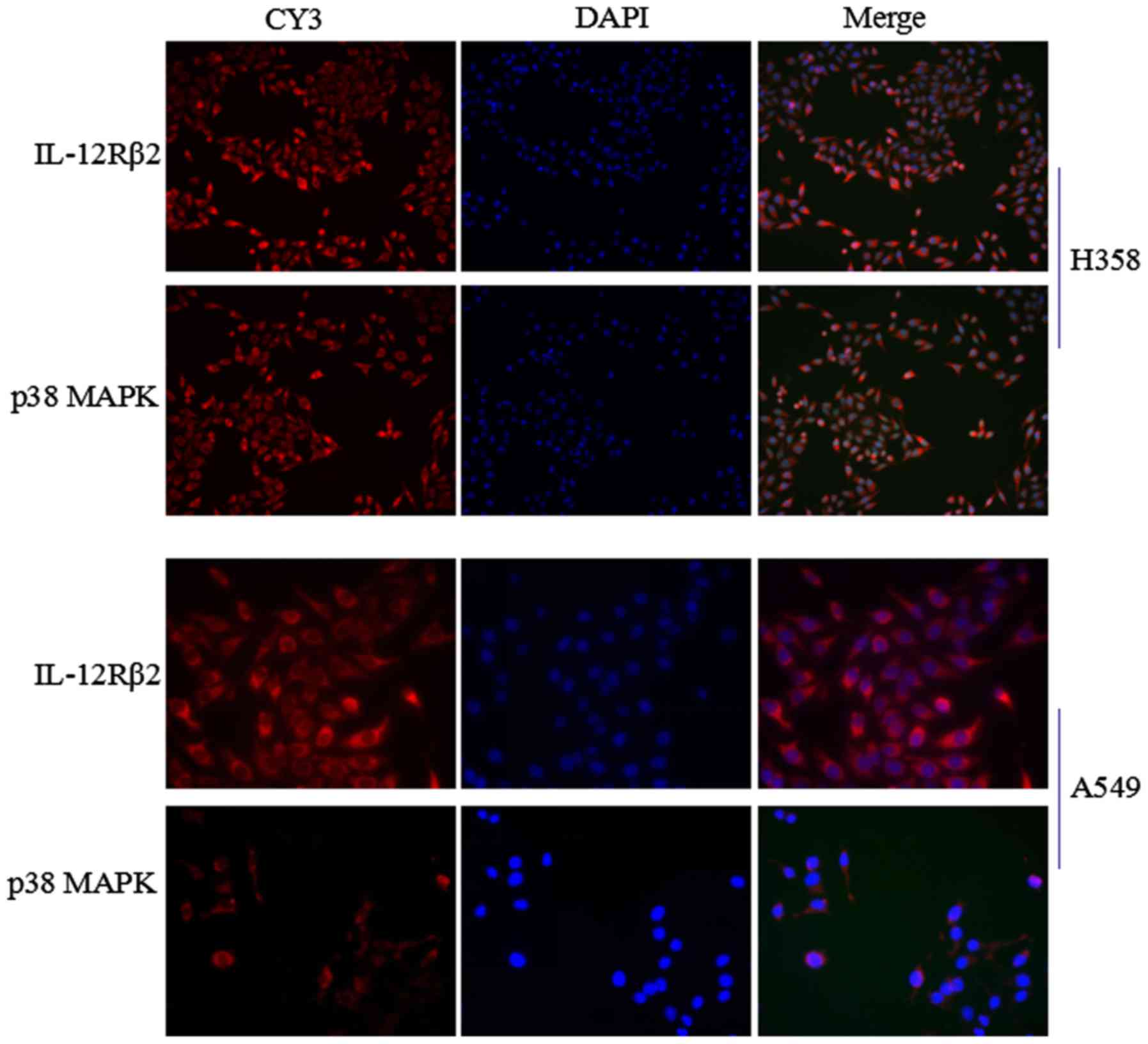
IF
A total of 5 ×10⁴/ml cells (H358 and A549 cells)
were fixed in 4% paraformaldehyde for 20 min and permeabilized in
0.5% Triton X-100 at room temperature for 20 min. Cells were then
incubated with anti-IL-12Rβ2 (1:50) and anti-p38MAPK (1:250) in 10%
sheep serum No: 005-000-121, Amyjet scientific, WuHan, China;
amyjet.bioon.com.cn) overnight at 4°C,
washed with PBS and incubated with GY3-conjugated anti-mouse
immunoglobulin G (1:100; Abcam, Cambridge, UK) for 1 h at 20°C.
Subsequently, cells were washed with PBS and counterstained with
DAPI for 4 min at 4°C. A fluorescence microscope (Leica DM1000;
Leica Microsystems GmbH, Wetzlar, Germany) was used for
examination.
Statistical analysis
Graphpad Prism6.0 was used for analysis. Continuous
variables (WB results) were compared among the groups using the
Kruskal-Wallis test, with a post hoc Mann-Whitney U test and
Bonferroni correction, and the Pearson χ2 test was used to analyze
categorical variables. The correlation of protein expression was
determined using Spearman correlation tests. OS curves were
calculated using Kaplan-Meier analyses. Univariate survival
analysis was conducted with the log-rank test and Cox proportional
hazards regression which were used to assess the prognostic power
of these parameters. P<0.05 was considered to indicate a
statistically significant difference.
Results
Expression of IL-12Rb2 and p38MAPK in
NSCLC
In the IHC analysis, overexpression of IL-12Rβ2 and
p38MAPK proteins was detected in BPL tissues and early pTNM stage
NSCLC. In H358 and A549 cells the expression of two proteins was
also observed (Figs. 1A and
2).
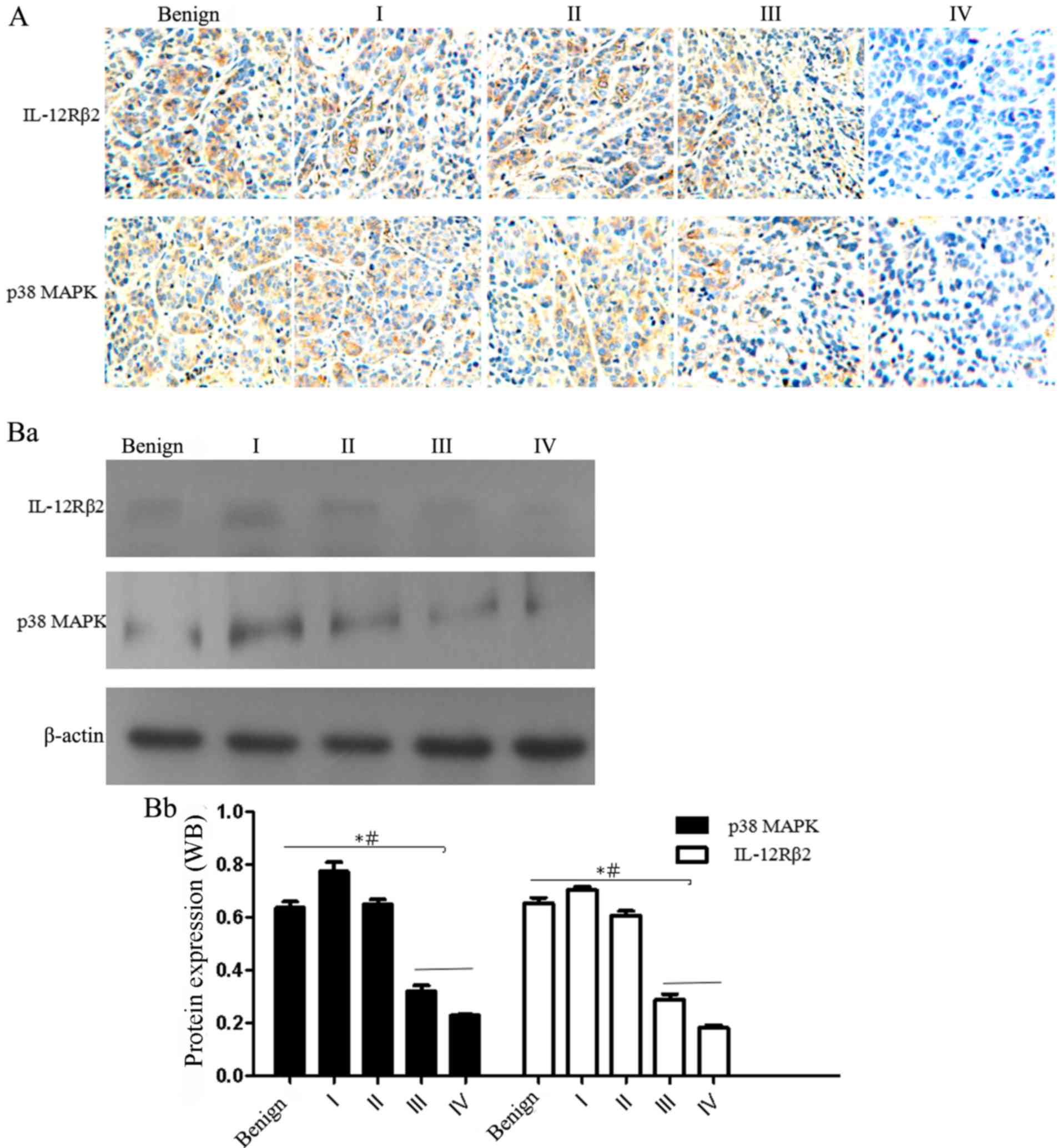 | Figure 1.Expression of IL-12Rb2 and p38MAPK in
NSCLC and BPL tissues, as determined by IHC and western blotting.
The immunopositive signal of each protein was graded according to
the Allred scoring system. Only an Allred score greater than the
cut-off point was considered positive. (A) BPL and NSCLC tissues
(TNM stage I–IV) were analyzed by IHC (streptavidin-biotin complex;
magnification, ×200). (Ba) IL-12Rb2 and p38MAPK expression was
detected in BPL and NSCLC frozen tissues using western blotting. As
TNM stage advanced, protein expression levels were gradually
reduced. (Bb) Semi-quantification of western blotting. Compared
with in III+IV stage tissues, protein expression levels were
significantly increased in BPL and I+II stage tissues. *P<0.05
vs. benign, #P indicates P<0.05 vs. I+II BPL, benign pulmonary;
IHC, immunohistochemistry; IL-12R, interleukin-12 receptor; NSCLC,
non-small cell lung cancer; p38MAPK, p38 mitogen-activated protein
kinases; TMN, tumor-node-metastasis. |
The expression levels of IL-12Rβ2 and p38MAPK were
markedly decreased in pTNM stage III+IV NSCLC tissues compared with
in the BPL and pTNM I+II stage NSCLC tissues, as determined by IIHC
and western blotting (P<0.05) (Fig.
1A and B). In addition, with increasing pTNM stage the Allred
scores of IL-12Rβ2 and p38MAPK expression were significantly
decreased (Table II). The number
of cases (Patients with IL-12Rβ2 or p38MAPK positive expression)
with positive expression (Allred score-vs. +, ++ and +++ as cut-off
for negative/positive expression). Of IL-12Rβ2 and p38MAPK was
decreased (both P<0.0001; Table
II). Further analysis via Spearman's correlation analyses
demonstrated a significant correlation between IL-12Rβ2 and p38MAPK
(r=0.415, P=0.0143; Table
III).
| Table II.Association between IL-12Rβ2 and
p38MAPK expression, and TNM stage. |
Table II.
Association between IL-12Rβ2 and
p38MAPK expression, and TNM stage.
|
| IL-12Rβ2 | p38MAPK |
|---|
| Variable Allred
score | I | II | III | IV | I | II | III | IV |
|---|
| – | 10 | 12 | 30 | 50 | 13 | 15 | 30 | 54 |
| + | 6 | 12 | 24 | 9 | 6 | 11 | 22 | 7 |
| ++ | 12 | 16 | 9 | 3 | 11 | 18 | 14 | 3 |
| +++ | 14 | 13 | 5 | 3 | 12 | 9 | 4 | 1 |
| Positive (%) | 76.19 | 77.35 | 55.88 | 23.07 | 69.04 | 71.69 | 57.14 | 16.92 |
| χ2 | 67.49 | 65.22 |
| P-value | <0.0001 | <0.0001 |
| Table III.Spearman correlation analysis between
IL-12Rb2 and p38MAPK expression. |
Table III.
Spearman correlation analysis between
IL-12Rb2 and p38MAPK expression.
| Comparison | Patients (n) | r | P-value |
|---|
| IL-12Rβ2 vs.
p38MAPK | 230 | 0.415 | 0.0143 |
Association between IL-12Rb2 and
p38MAPK expression, and patient clinical characteristics
The association between IL-12Rβ2 and p38MAPK
expression, and a range of standard clinicopathological parameters
were measured. Using the χ2 test, significant associations between
gender (P=0.0168), age (P=0.0341), histological type (P<0.0001),
TNM staging (P<0.0001) and IL-12Rβ2 expression were demonstrated
(Table I). Similar results were
observed when the associations between p38MAPK expression and
clinicopathological factors were analyzed. For example, gender
(P=0.0082) and TNM staging (P<0.0001) were significantly
associated with p38MAPK expression (Table I). In addition, with advanced TNM
staging, the expression of both proteins was significantly
decreased, particularly in the III+IV stage tissues compared with
in the I+II and BPL tissues (P<0.0001; Fig. 1B; Tables I and II).
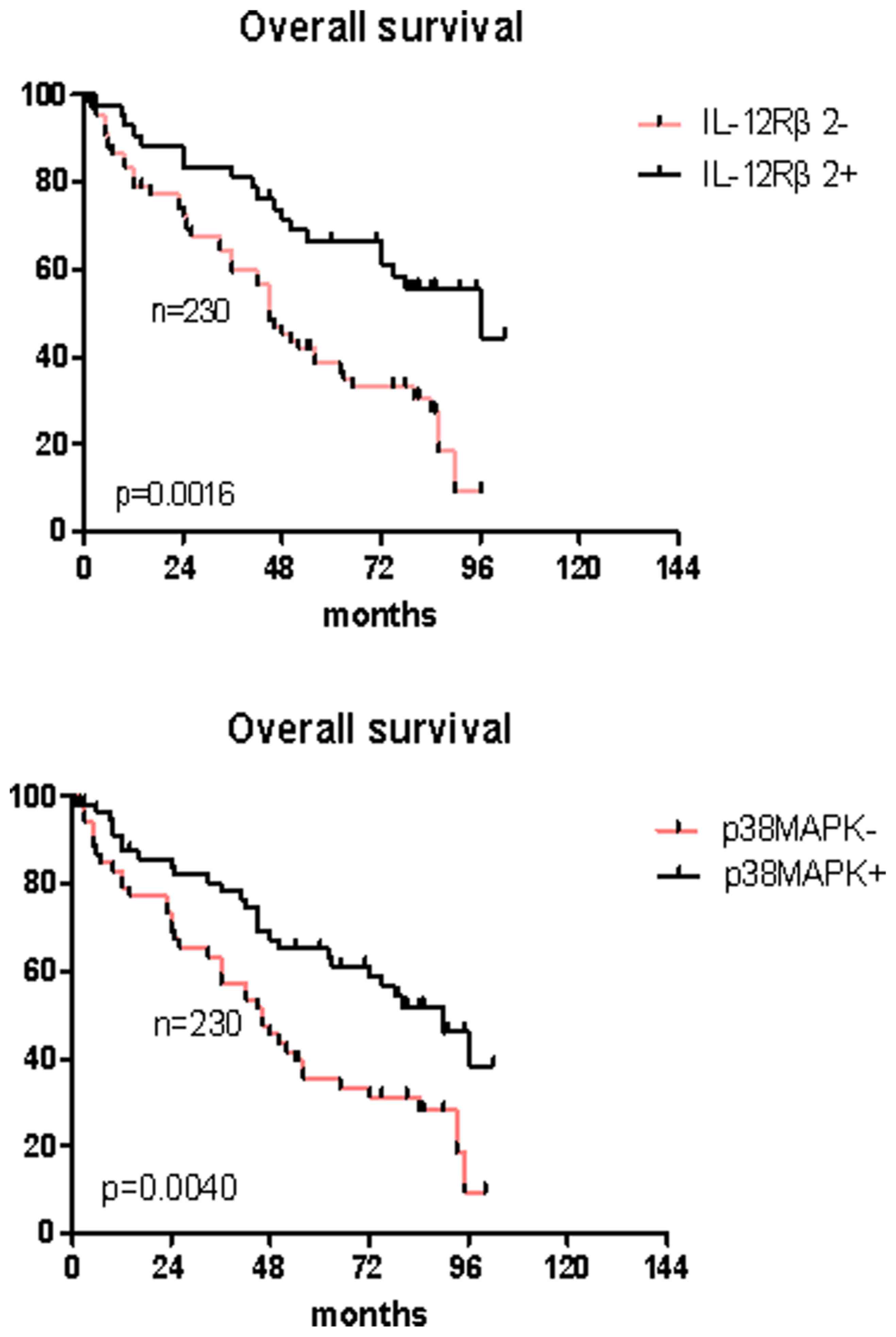
Correlation analysis between IL-12Rb2,
p38MAPK and OS
The Kaplan-Meier method was used to investigate the
association between patient survival and IL-12Rβ2- and
p38MAPK-positive expression compared with negative expression in a
univariate model. The results indicated that IL-12Rβ2- and
p38MAPK-positive expression were significantly associated with
longer OS (IL-12Rβ2: Log-rank, 5.203 and P=0.0016; p38MAPK:
Log-rank, 4.503 and P=0.0040; Fig.
3 and Table IV). In addition,
a similar result was observed from the TNM stage analysis
(log-rank, 5.565 and P=0.0021; Table
IV).
| Table IV.Kaplan-Meier and Cox multivariate
proportional hazard analysis. |
Table IV.
Kaplan-Meier and Cox multivariate
proportional hazard analysis.
|
| Univarivate
analysis | Multivariate
analysis |
|---|
|
|
|
|
|---|
| Factor | Log-rank | P-value | Hazard ratio
(95%) | P-value |
|---|
| Age (years) |
|
<55 | 2.014 | 0.1302 |
|
|
|
≥55 |
| Sex |
|
Male | 1.245 | 0.4521 |
|
|
|
Female |
| IL-12Rβ2 |
|
Negative | 5.203 | 0.0016 | 4.32
(3.02–6.33) | 0.0221 |
|
Positive |
| p38MAPK |
|
Negative | 4.503 | 0.0040 | 3.59
(1.03–5.28) | 0.0457 |
|
Positive |
| pTNM stage |
|
I–II | 5.565 | 0.0021 | 4.03
(2.26–6.74) | 0.0203 |
|
III–IV |
A Cox regression model was performed to determine
whether IL-12Rβ2 and p38MAPK are significant, independent
prognostic factors. The analysis demonstrated that high expression
of IL-12Rβ2 (P=0.0221) and p38MAPK (P=0.0457), as well as TNM stage
(P=0.0203), were significant, independent prognostic factors
(Table IV).
Discussion
Lung carcinoma is a frequent, lethal malignancy in
humans worldwide (21,22). NSCLC is an important type of lung
cancer, which comprises two major categories: Lung squamous cell
carcinoma and adenocarcinoma. Previous studies have focused on
IL-12 in NSCLC; however, the current understanding of the mechanism
underlying IL-12R binding to IL-12, and the activated downstream
signaling pathways, is incomplete and a number of uncertainties
remain (23,24). The present study aimed to elucidate
the expression and roles of IL-12Rβ2, and its possible association
with p38MAPK, in NSCLC via IHC, WB and IF analyses.
In previous studies (8,9),
investigations have been conducted regarding the role of IL-12Rβ2,
and notable results have been obtained regarding the development of
lung adenocarcinoma in IL-12Rβ2-deficient mice. However, the
mechanism by which IL-12Rβ2 performs its' roles remains unclear. In
the present study, similar analyses demonstrated that IL-12Rβ2 was
overexpressed in BPL and early TNM stage tissues, according to IHC
and western blot analyses; the results confirmed that IL-12Rβ2 may
be negatively correlated with NSCLC progression, which is
consistent with the results of previous studies (7–10)
and indicated that IL-12Rβ2 may be involved in complex cellular
biological functions, including proliferation, apoptosis and
metastasis. Through further analyses, a close correlation was
demonstrated between IL-12Rβ2 and p38MAPK expression, thus
suggesting that the mechanism by which IL-12Rβ2 acts in NSCLC is
closely associated with the p38MAPK signaling pathway.
The results of the IHC analysis revealed that a
close correlation may exist between IL-12Rβ2 and p38MAPK
expression, according to the Spearman's correlation test, which is
consistent with the previous literature (25). In addition, IL-12Rβ2 and p38MAPK
were identified as not only being overexpressed in early NSCLC but
were also associated with TNM stage and gender; furthermore, it was
demonstrated that compared with in stage III+IV NSCLC tissues, the
number of cases in which both proteins were positively expressed
was increased in stage I+II and BPL tissues. Similar results were
also observed via western blotting; protein expression was
decreased with increasing TNM stage, particularly at the III+IV
stage compared with the I+II stage, thus indicating that the
expression of these proteins is negatively associated with the TNM
stage, which is in agreement with previous studies on IL-12Rβ2
(7–9,26–28).
Notably, differential expression of IL-12Rβ2 and p38MAPK was
detected between the genders. The number of cases of positive
expression was increased in female patients for both IL-12Rβ2
(female; 61.86%, 75 out of 118) and p38MAPK (female; 60.16%, 71 out
of 118) compared with in male patients. In addition, differences
between IL-12Rβ2, and tumor types and age were observed;
inconsistencies in IL-12Rβ2 expression between patients of
different ages, tumor types and gender may imply certain hypotheses
e.g., whether IL-12Rβ2 affects tumor progression through the
endocrine system, immune environment or intracellular environmental
factors. In addition, the present study revealed that IL-12Rβ2- and
p38MAPK-positive expression may be strong predictors of OS in
NSCLC. The results of a Kaplan-Meier analysis demonstrated that the
positive expression of IL-12Rβ2 and p38MAPK was associated with a
good OS. In addition, a Cox regression model was performed; both
proteins were confirmed as being independent prognostic factors of
survival. Based on these results, the correlation between the
expression of both proteins, their associations with tumor
characteristics (i.e., TNM staging and OS) and the results of
previous studies, a close association may exist between IL-12Rβ2
and p38MAPK, which may indicate the presence of cross-talk between
IL-12Rβ2 and p38MAPK signaling mechanisms and raise the question as
to whether IL-12Rβ2 influences NSCLC via the p38MAPK signaling
pathway.
The results of the present study confirmed that
IL-12Rβ2 and p38MAPK may be key signaling molecules with important
biological functions. However, the biological mechanisms of
IL-12Rβ2 are complex and there is a lack of understanding about how
these two proteins interact in NSCLC. Although the mechanisms
underlying the interaction between these proteins remain unclear,
the effects of IL-12Rβ2 on p38MAPK expression is interesting in
NSCLC and may imply possible novel methods of diagnosis and
treatment in NSCLC.
In conclusion, the correlation between IL-12Rβ2 and
p38MAPK expression may help to further explain the mechanisms
underlying the effects of IL-12Rβ2 on NSCLC, which may possibly be
regulated by p38MAPK. The results of the present study demonstrated
that IL-12Rβ2 and p38MAPK may be prognostic factors for survival in
NSCLC. Although the detection of IL-12Rβ2 and p38MAPK expression
has improved the understanding of the roles of IL-12Rβ2 to a
certain extent, several uncertainties remain; in particular, how
IL-12Rβ2 exerts its' biological effects via p38MAPK. Therefore,
in vitro and in vivo experiments are required, which
will be the focus of future experiments.
Acknowledgements
Not applicable.
Funding
The present study was supported by the National
Nature Science Foundation of China (Grant no. 81501964).
Availability of data and materials
The datasets used and/or analyzed during the current
study are available from the corresponding author on reasonable
request.
Authors' contributions
ZL designed the study, selected surgical specimens
and wrote the manuscript. WY performed the statistical analysis and
the IF assay. SY performed the western blotting and IHC assay. KC
wrote the manuscript, participated in the control of clinical cases
and experimental process quality, and coordinated all experimental
progress.
Ethics approval and consent to
participate
The present study was approved by the medical ethics
committee of First Affiliated Hospital, Sun Yat-sen University
(Guangzhou, China). Written informed consent was obtained from all
patients.
Patient consent for publication
Written informed consent was obtained from all
patients.
Competing interests
The authors declare that they have no competing
interests.
References
|
1
|
Kamangar F, Dores GM and Anderson WF:
Patterns of cancer incidence, mortality, and prevalence across five
continents: Defining priorities to reduce cancer disparities in
different geographic regions of the world. J Clin Oncol.
24:2137–2150. 2006. View Article : Google Scholar : PubMed/NCBI
|
|
2
|
Suzuki M, Iizasa T, Nakajima T, Kubo R,
Iyoda A, Hiroshima K, Nakatani Y and Fujisawa T: Aberrant
methylation of IL-12Rβ2 gene in lung cancer. J Thorac oncol. 2 8
Suppl 4:S4932007. View Article : Google Scholar
|
|
3
|
Orlova A, Hofström C, Strand J, Varasteh
Z, Sandstrom M, Andersson K, Tolmachev V and Gräslund T:
[99mTc(CO)3]+-(HE)3-ZIGF1R:4551, a new Affibody conjugate for
visualization of insulin-like growth factor-1 receptor expression
in malignant tumours. Eur J Nucl Med Mol Imaging. 40:439–449. 2013.
View Article : Google Scholar : PubMed/NCBI
|
|
4
|
Nose N, Uramoto H, Iwata T, Hanagiri T and
Yasumoto K: Expression of estrogen receptor beta predicts a
clinical response and longer progression-free survival after
treatment with EGFR-TKI for adenocarcinoma of the lung. Lung
Cancer. 71:350–355. 2011. View Article : Google Scholar : PubMed/NCBI
|
|
5
|
Torres-Arzayus MI, Zhao J, Bronson R and
Brown M: Estrogen-dependent and estrogen-independent mechanisms
contribute to AIB1-mediated tumor formation. Cancer Res.
70:4102–4111. 2010. View Article : Google Scholar : PubMed/NCBI
|
|
6
|
Airoldi I, Cocco C, Di Carlo E, Disarò S,
Ognio E, Basso G and Pistoia V: Methylation of the IL-12Rbeta2 gene
as novel tumor escape mechanism for pediatric B-acute lymphoblastic
leukemia cells. Cancer Res. 66:3978–3980. 2006. View Article : Google Scholar : PubMed/NCBI
|
|
7
|
Airoldi I, Di Carlo E, Banelli B, Moserle
L, Cocco C, Pezzolo A, Sorrentino C, Rossi E, Romani M, Amadori A
and Pistoia V: The IL-12Rbeta2 gene functions as a tumor suppressor
in human B cell malignancies. J Clin Invest. 113:1651–1659. 2004.
View Article : Google Scholar : PubMed/NCBI
|
|
8
|
Airoldi I, Di Carlo E, Cocco C, Caci E,
Cilli M, Sorrentino C, Sozzi G, Ferrini S, Rosini S, Bertolini G,
et al: IL-12 can target human lung adenocarcinoma cells and normal
bronchial epithelial cells surrounding tumor lesions. PLoS One.
4:e61192009. View Article : Google Scholar : PubMed/NCBI
|
|
9
|
Suzuki M, Iizasa T, Nakajima T, Kubo R,
Iyoda A, Hiroshima K, Nakatani Y and Fujisawa T: Aberrant
methylation of IL-12Rbeta2 gene in lung adenocarcinoma cells is
associated with unfavorable prognosis. Ann Surg Oncol.
14:2636–2642. 2007. View Article : Google Scholar : PubMed/NCBI
|
|
10
|
Li H, Cao MY, Lee Y, Lee V, Feng N,
Benatar T, Jin H, Wang M, Der S, Wright JA and Young AH: Virulizin,
a novel immunotherapy agent, activates NK cells through induction
of IL-12 expression in macrophages. Cancer Immunol Immunother.
54:1115–1126. 2005. View Article : Google Scholar : PubMed/NCBI
|
|
11
|
Lecocq M, Detry B, Guisset A and Pilette
C: FcαRI-mediated inhibition of IL-12 production and priming by
IFN-γ of human monocytes and dendritic cells. J Immunol.
190:2362–2371. 2013. View Article : Google Scholar : PubMed/NCBI
|
|
12
|
Kontoyiannis D, Kotlyarov A, Carballo E,
Alexopoulou L, Blackshear PJ, Gaestel M, Davis R, Flavell R and
Kollias G: Interleukin-10 targets p38 MAPK to modulate
ARE-dependent TNF mRNA translation and limit intestinal pathology.
EMBO J. 20:3760–3770. 2001. View Article : Google Scholar : PubMed/NCBI
|
|
13
|
Fahmi A, Smart N, Punn A, Jabr R, Marber M
and Heads R: p42/p44-MAPK and PI3K are sufficient for IL-6 family
cytokines/gp130 to signal to hypertrophy and survival in
cardiomyocytes in the absence of JAK/STAT activation. Cell Signal.
25:898–909. 2013. View Article : Google Scholar : PubMed/NCBI
|
|
14
|
Korhonen R, Huotari N, Hömmö T, Leppänen T
and Moilanen E: The expression of interleukin-12 is increased by
MAP kinase phosphatase-1 through a mechanism related to interferon
regulatory factor 1. Mol Immunol. 51:219–226. 2012. View Article : Google Scholar : PubMed/NCBI
|
|
15
|
Cuenda A and Rousseau S: p38 MAP-kinases
pathway regulation, function and role in human diseases. Biochim
Biophys Acta. 1773:1358–1375. 2007. View Article : Google Scholar : PubMed/NCBI
|
|
16
|
He J, Liu Z, Zheng Y, Qian J, Li H, Lu Y,
Xu J, Hong B, Zhang M, Lin P, et al: p38 MAPK in myeloma cells
regulates osteoclast and osteoblast activity and induces bone
destruction. Cancer Res. 72:6393–6402. 2012. View Article : Google Scholar : PubMed/NCBI
|
|
17
|
Tanaka Y, Gavrielides MV, Mitsuuchi Y,
Fujii T and Kazanietz MG: Protein kinase C promotes apoptosis in
LNCaP prostate cancer cells through activation of p38 MAPK and
inhibition of the Akt survival pathway. J Biol Chem.
278:33753–33762. 2003. View Article : Google Scholar : PubMed/NCBI
|
|
18
|
Slawinska-Brych A, Zdzisinska B,
Mizerska-Dudka M and Kandefer-Szerszen M: Induction of apoptosis in
multiple myeloma cells by a statin-thalidomide combination can be
enhanced by p38 MAPK inhibition. Leuk Res. 37:586–594. 2013.
View Article : Google Scholar : PubMed/NCBI
|
|
19
|
Shen KH, Hung SH, Yin LT, Huang CS, Chao
CH, Liu CL and Shih YW: Acacetin, a flavonoid, inhibits the
invasion and migration of human prostate cancer DU145 cells via
inactivation of the p38 MAPK signaling pathway. Mol Cell Biochem.
333:279–291. 2010. View Article : Google Scholar : PubMed/NCBI
|
|
20
|
Koul HK, Pal M and Koul S: Role of p38 MAP
kinase signal transduction in solid tumors. Genes Cancer.
4:342–359. 2013. View Article : Google Scholar : PubMed/NCBI
|
|
21
|
Pietras RJ, Marquez DC, Chen HW, Tsai E,
Weinberg O and Fishbein M: Estrogen and growth factor receptor
interactions in human breast and non-small cell lung cancer cells.
Steroids. 70:372–381. 2005. View Article : Google Scholar : PubMed/NCBI
|
|
22
|
Kawasaki H, Altieri DC, Lu CD, Toyoda M,
Tenjo T and Tanigawa N: Inhibition of apoptosis by survivin
predicts shorter survival rates in colorectal cancer. Cancer Res.
58:5071–5074. 1998.PubMed/NCBI
|
|
23
|
Shaaban AM, Green AR, Karthik S, Alizadeh
Y, Hughes TA, Harkins L, Ellis IO, Robertson JF, Paish EC, Saunders
PT, et al: Nuclear and cytoplasmic expression of ERbeta1, ERbeta2,
and ERbeta5 identifies distinct prognostic outcome for breast
cancer patients. Clin Cancer Res. 14:5228–5235. 2008. View Article : Google Scholar : PubMed/NCBI
|
|
24
|
Chi A, Chen X, Chirala M and Younes M:
Differential expression of estrogen receptor beta isoforms in human
breast cancer tissue. Anticancer Res. 23:211–216. 2003.PubMed/NCBI
|
|
25
|
Liu ZG, Jiao XY, Chen ZG, Feng K and Luo
HH: Estrogen receptorβ2 regulates interlukin-12 receptorβ2
expression via p38 mitogen-activated protein kinase signaling and
inhibits non-small-cell lung cancer proliferation and invasion. Mol
Med Rep. 12:248–254. 2015. View Article : Google Scholar : PubMed/NCBI
|
|
26
|
Kondadasula SV, Roda JM, Parihar R, Yu J,
Lehman A, Caligiuri MA, Tridandapani S, Burry RW and Carson WE III:
Colocalization of the IL-12 receptor and FcgammaRIIIa to natural
killer cell lipid rafts leads to activation of ERK and enhanced
production of interferon-gamma. Blood. 111:4173–4183. 2008.
View Article : Google Scholar : PubMed/NCBI
|
|
27
|
Jana M, Dasgupta S, Pal U and Pahan K:
IL-12 p40 homodimer, the so-called biologically inactive molecule,
induces nitric oxide synthase in microglia via IL-12R beta 1. Glia.
57:1553–1565. 2009. View Article : Google Scholar : PubMed/NCBI
|
|
28
|
Pan SH, Chao YC, Hung PF, Chen HY, Yang
SC, Chang YL, Wu CT, Chang CC, Wang WL, Chan WK, et al: The ability
of LCRMP-1 to promote cancer invasion by enhancing filopodia
formation is antagonized by CRMP-1. J Clin Invest. 121:3189–3205.
2011. View
Article : Google Scholar : PubMed/NCBI
|