Introduction
Breast cancer is the most common malignant tumor and
the leading cause of cancer mortality in women, resulting in 14% of
cancer-related deaths (1,2). Besides, Breast cancer is a major
malignant tumor threatening women's health in China (3). According to statistics, the new cases
and dead cases of breast cancer account for 12.2 and 9.6% annually,
respectively (4). Although the
incidence of breast cancer in China is low, it is noteworthy that
the incidence of breast cancer in China grows rapidly at 3% per
year in recent years, which has become one of the leading causes of
death in Chinese cities (5).
However, the cause of breast cancer is not clear yet, and even
about 50% of the causes with breast cancer can not be explained at
all, which seriously threatens the life and health of women
(6).
Along with the increasing study of molecular
mechanisms of breast cancer, the level of comprehensive treatment
for breast cancer has been greatly improved. In recent years,
multiple treatments have been developed for breast cancer, such as
local surgical treatment, radiotherapy and chemotherapy, endocrine
therapy, molecular targeted treatment, auxiliary treatment of
traditional Chinese medicine and so on, which significantly
improves the quality of life and prolongs the survival period of
patients (7–11). A large number of clinical data and
experimental studies have shown that the adjuvant treatment of
breast cancer patients with traditional Chinese medicine can
improve the body's metabolism, reduce the toxic and side effects of
radiotherapy and chemotherapy, improve the surgical resection rate
and endocrine therapy efficacy, and so on (12,13).
The development of traditional Chinese medicine plays an important
role in reducing the recurrence and metastasis of tumor, prolonging
the survival time, and improving the quality of patients' life
(14). Moreover, traditional
Chinese medicine still has many positive advantages of rich
resources, relatively less cytotoxicity, easy access and low cost.
Therefore, the study of the targets and mechanisms of traditional
Chinese medicine in the comprehensive treatment for breast cancer
has been an important subject in the study of breast cancer
prevention and treatment.
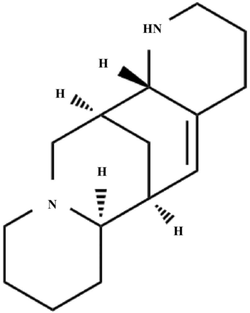
Aloperine (Alo) (Fig.
1) is an alkaloid extracted from the traditional Chinese
medicine Sophora alopecuroides (S. alopecuroides)
(15,16). It has been reported that Alo
induced apoptosis and inhibited invasion in MG-63 and U2OS human
osteosarcoma cells, and induced G2/M phase cell cycle arrest and
apoptosis in HCT116 human colon cancer cells (17,18).
However, the impact of Alo on breast cancer has not been reported
yet. Therefore, the article was designed to investigate the
antitumor potential and the underlying molecular mechanisms of Alo
on breast cancer.
Materials and methods
Cell culture
Human breast cancer cells MCF-7 and MDA-MB-231 were
purchased from the American Type Culture Collection (Manassas, VA,
USA). MCF-7 cells were cultured in Dulbecco's modified Eagle's
medium (DMEM) (Gibco; Thermo Fisher Scientific, Inc., Waltham, MA,
USA) supplemented with 10% fetal bovine serum (FBS; Tianhang,
Hangzhou, China), 100 mg/ml streptomycin and 100 U/ml penicillin in
5% CO2 atmosphere at 37°C. MDA-MB-231 cells were
cultured in L-15 medium (both Gibco; Thermo Fisher Scientific,
Inc.) medium supplied with 10% FBS, 100 mg/ml streptomycin and 100
U/ml penicillin in normal air atmosphere at 37°C.
Cell counting kit-8 (CCK-8) assay
The viabilities of MCF-7 and MDA-MB-231 cells were
measured by CCK-8 assay. Briefly, MCF-7 and MDA-MB-231 cells at a
density of 1×10⁴ cells/well in 100-µl of complete
culture medium were seeded in 96-well plates. After culturing for
24 h, the medium was replaced with serum-free media or serum-free
media containing various concentrations of Alo (0, 0.1, 0.2 and 0.4
mM) according to previous studies (17,18),
and incubated in a humidified incubator at 37°C for 12, 24 and 48
h, respectively. After incubation, 10 µl of CCK-8 (Sigma-Aldrich;
Merck KGaA, Darmstadt, Germany) was added to each well for another
2 h at 37°C. The optical density (OD) was recorded at 450 nm using
a microplate reader (Dojindo Molecular Technology, Rockville, MD,
USA).
Colony formation test
Colony formation assay was conducted to evaluate the
role of Alo in the proliferative potential of MCF-7 and MDA-MB-231
cells. MCF-7 and MDA-MB-231 cells (1×10³ cells/well)
were plated in 6-well plates and cultured at 37°C with 5%
CO2. The medium was replaced with fresh culture media or
fresh culture media containing Alo (0, 0.1, 0.2 and 0.4 mM) every
2–3 days. After two weeks, the plates were fixed with 4%
paraformaldehyde for 20 min and stained using 10% crystal violet
for 30 min. Then the number of stained colonies was manually
counted.
Hoechst 33342 staining
MCF-7 and MDA-MB-231 cells at a density of
2×10⁴ cells/well were seeded in 24-well plates, and
after incubation for 24 h, fresh culture media or fresh culture
media containing Alo (0, 0.1, 0.2 and 0.4 mM) were added, and
incubated in a humidified incubator at 37°C for 24 h. Then the
cells were stained with Hoechst 33342 (10 mg/ml) in culture medium
at room temperature in the dark for 20 min. Subsequently, the cells
were washed twice with PBS and immediately evaluated by a
microscope.
Apoptosis analysis
Cell apoptosis was performed using Annexin V
Apoptosis Detection kit I (BD Biosciences, Franklin Lakes, NJ,
USA). Briefly, MCF-7 and MDA-MB-231 at a density of
2×10⁴ cells were treated with indicated doses of Alo (0,
0.1, 0.2 and 0.4 mM) for 24 h. Then the treated cells were digested
with trypsin and washed in cold 1X PBS (4°C) twice, followed by
resuspending the cell pellet with 300 µl of 1X Binding Buffer.
Next, 5 µl of Annexin V-PE were added to the cell suspension for 15
min in the dark at room temperature, according to the
manufacturer's instructions. 7-AAD solution (5 µl) was added in the
cell suspension 5 min before flow cytometry analysis and then 200
µl of 1X Binding Buffer was added for flow cytometry analysis. The
percentage of apoptotic cells was evaluated by FACS Calibur (BD
Biosciences).
Wound healing assay
MCF-7 and MDA-MB-231 cells were cultured in fresh
culture media to full confluence. After that, we created a wound
using a plastic scraper. After being washed with PBS, the medium
was replaced with fresh culture media or fresh culture media
containing Alo (0, 0.1, 0.2 and 0.4 mM) and incubated at 37°C for
48 h. Then, the cells were washed twice with PBS and the wound was
observed under a microscope (Nikon, Tokyo, Japan).
Transwell migration and invasion
assay
Cell migration and invasion assays were performed
using Trans-well chambers (8 µm pore-size, Corning Co., Corning,
NY, USA). In migration assay, 5×10⁴ cells in fresh
culture media were added into the upper chamber. In invasion assay,
Matrigel was purchased from BD Biosciences and stored at −20°C.
After thawing at 4°C overnight, the Matrigel was diluted in
serum-free medium, and 30 µl of the diluted Matrigel were evenly
inoculated into the upper chamber to form a gel at 37°C. Cells
(1×10⁵) suspended in 300 µl of fresh culture media were
seeded into the upper compartments. For trans-well migration and
invasion assay, the lower compartments were filled with 600 µl of
medium with 20% FBS. After incubation for 24 h, the non-migrate or
non-invasive cells were removed from the upper surface of the
membrane by scrubbing. The cells that migrated or invaded to the
lower surface of the membrane were fixed with 4% paraformaldehyde,
stained in 10% crystal violet, and cells were counted under a
microscope (Olympus, Tokyo, Japan).
Western blot analysis
The total protein of cells was extracted according
to the manufacturer's recommended protocol (Vazyme, Piscataway, NJ,
USA). The protein concentrations were determined using the BCA
Protein Assay kit (Vazyme, Piscataway, NJ, USA). Samples with equal
amounts of protein (50 µg) were fractionated on 10% SDS
polyacrylamide gels, transferred to polyvinylidene difluoride
membranes (PVDF), and blocked in 5% skim milk in TBST for 1.5 h at
25±1°C. The membranes were then incubated at 4°C overnight with
1:1,000 dilutions (v/v) of the primary antibodies. After washing
the membranes with TBST, incubations with 1:1,000 dilutions (v/v)
of the secondary antibodies were conducted for 2 h at 25±1°C.
Protein expression was detected using an Enhanced Chemiluminescence
Detection System. GAPDH was used as a loading control. Antibodies
in western blot were purchased Cell Signaling Technology (Beverly,
MA, USA), including matrix metalloproteinase (MMP)-2 (cat. no.
4022), MMP-9 (cat. no. 3852), caspase-3 (cat. no. 9662), caspase-9
(cat. no. 9508), Bax (cat. no. 2774), Bcl-2 (cat. no. 2872), Ras
(cat. no. 3965), p-Raf1 (cat. no. 9421), Raf1 (cat. no. 4432),
p-Erk1/2 (cat. no. 4370), Erk1/2 (cat. no. 9102), GAPDH (cat. no.
8884).
Statistical analyses
GraphPad Prism 5.0 statistical software (GraphPad
Software, Inc., La Jolla, CA, USA) was utilized to analyze the
above experimental data. Measurement data were represented as the
mean ± standard deviation. Statistical differences between means
among multiple groups were analyzed by one-way analysis of variance
followed by a Bonferroni post hoc analysis. P<0.05 was
considered to indicate a statistically significant difference.
Results
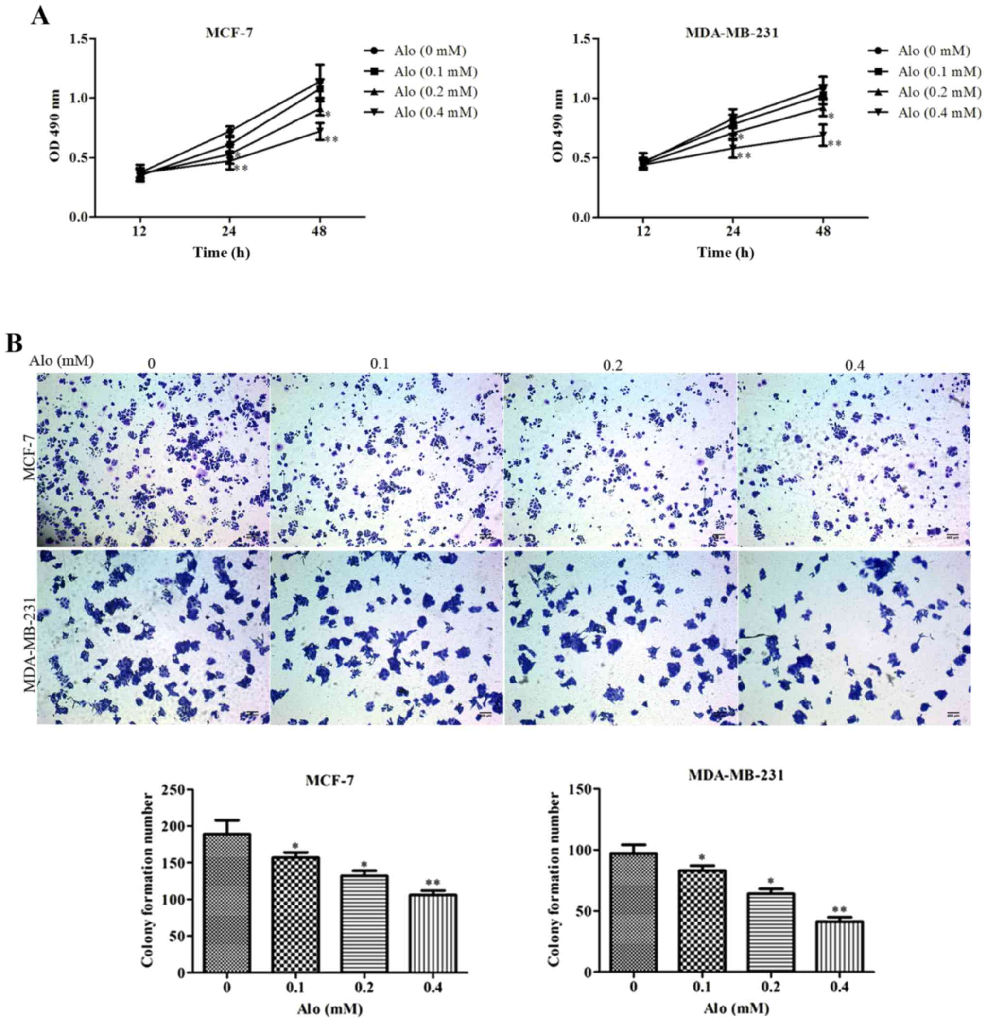
Alo inhibited human breast cancer
cells proliferation and colony formation
To investigate the impact of Alo on human breast
cancer MCF-7 and MDA-MB-231 cells proliferation, MCF-7 and
MDA-MB-231 cells were treated with Alo (0.1, 0.2 and 0.4 mM) for
12, 24 and 48 h, respectively. The results of CCK-8 assay showed
that Alo suppressed cell proliferation in a dose and time dependent
manner (Fig. 2A). Subsequently, to
evaluate whether Alo regulated the colony growth of human breast
cancer cells, MCF-7 and MDA-MB-231 cells were treated with Alo
(0.1, 0.2 and 0.4 mM), and incubated for two weeks. The results
revealed that Alo reduced colony formation of MCF-7 and MDA-MB-231
cells compared with the untreated group (Fig. 2B).
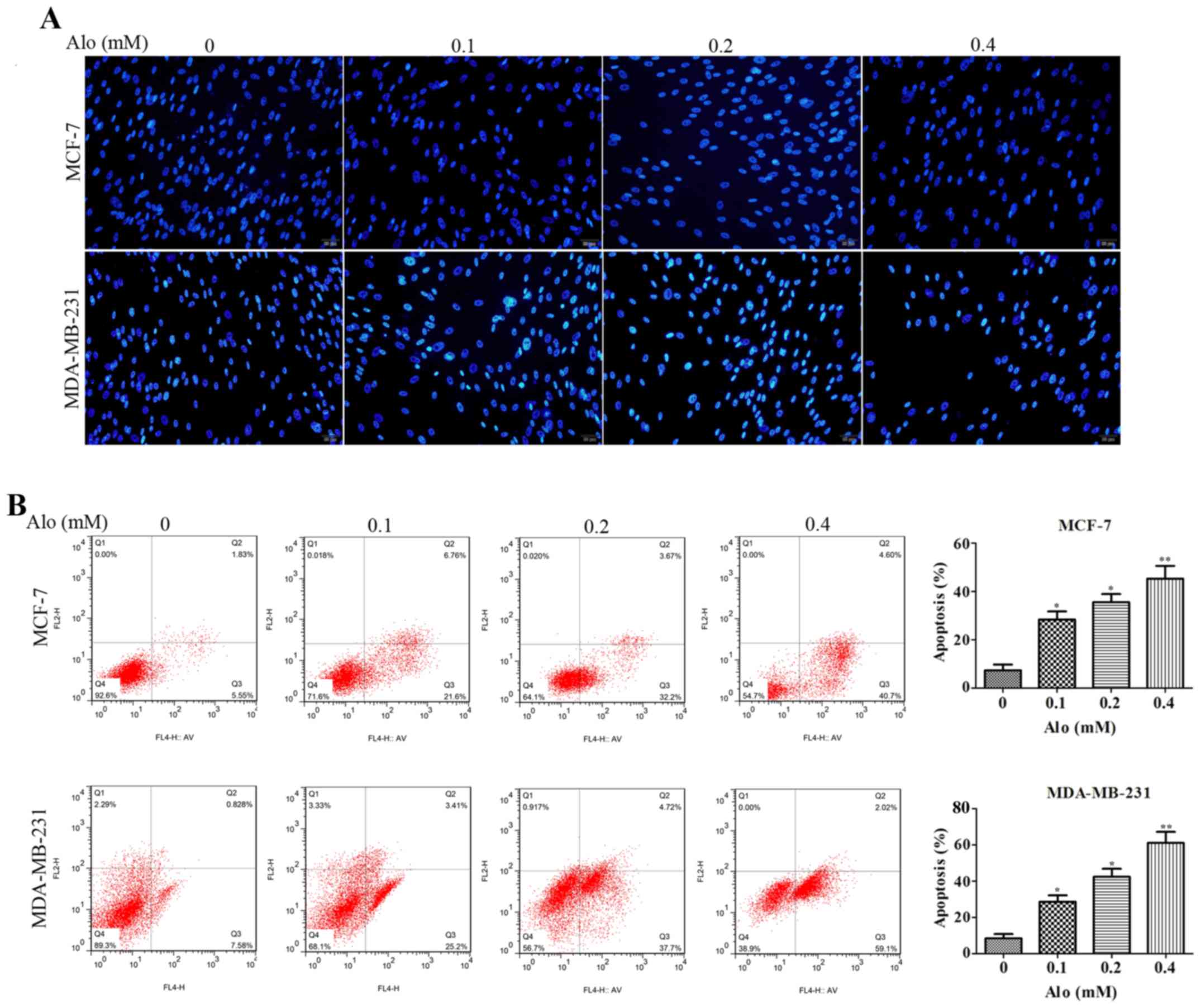
Alo induced human breast cancer cells
apoptosis
In order to determine whether Alo induced human
breast cancer MCF-7 and MDA-MB-231 cells apoptosis, first Hoechst
33342 staining was adapted to evaluate cell apoptosis by
morphological examination. The results demonstrated the nuclei of
MCF-7 and MDA-MB-231 cells were uniformly stained in the untreated
group, indicating these cells had intact cell membrane morphology.
However, both MCF-7 and MDA-MB-231 cells treated with Alo (0.1, 0.2
and 0.4 mM) for 24 h clearly exhibited significant morphological
changes compared with the untreated group, showing that MCF-7 and
MDA-MB-231 cells apoptosis remarkably increased (Fig. 3A). Additionally, the effect of Alo
on MCF-7 and MDA-MB-231 cells apoptosis was evaluated by flow
cytometry with Annexin V-FITC/PI staining. After treatment of MCF-7
and MDA-MB-231 cells with Alo (0.1, 0.2 and 0.4 mM), we found
degree of apoptosis in the Alo group was higher than that in the
untreated group (Fig. 3B). These
findings suggested that Alo dramatically abated MCF-7 and
MDA-MB-231 cells proliferation rate in a dose dependent manner,
which was possibly associated with increasing apoptosis.
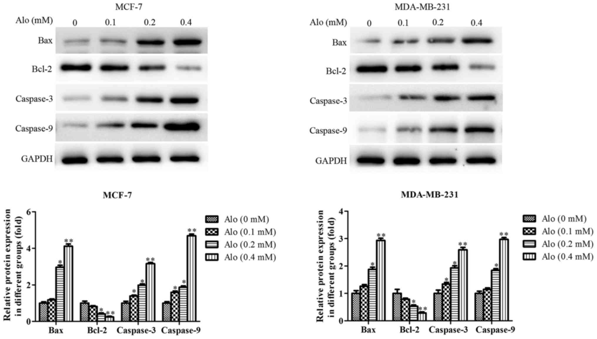
Furthermore, the expressions of apoptosis-related
proteins, including caspase-3, caspase-9, Bax and Bcl-2, were
detected by western blotting analysis. The results showed that Alo
could downregulate the expression of Bcl-2 and upregulate the
expressions of caspase-3, caspase-9 and Bax compared with the
untreated group in MCF-7 and MDA-MB-231 cells (Fig. 4).
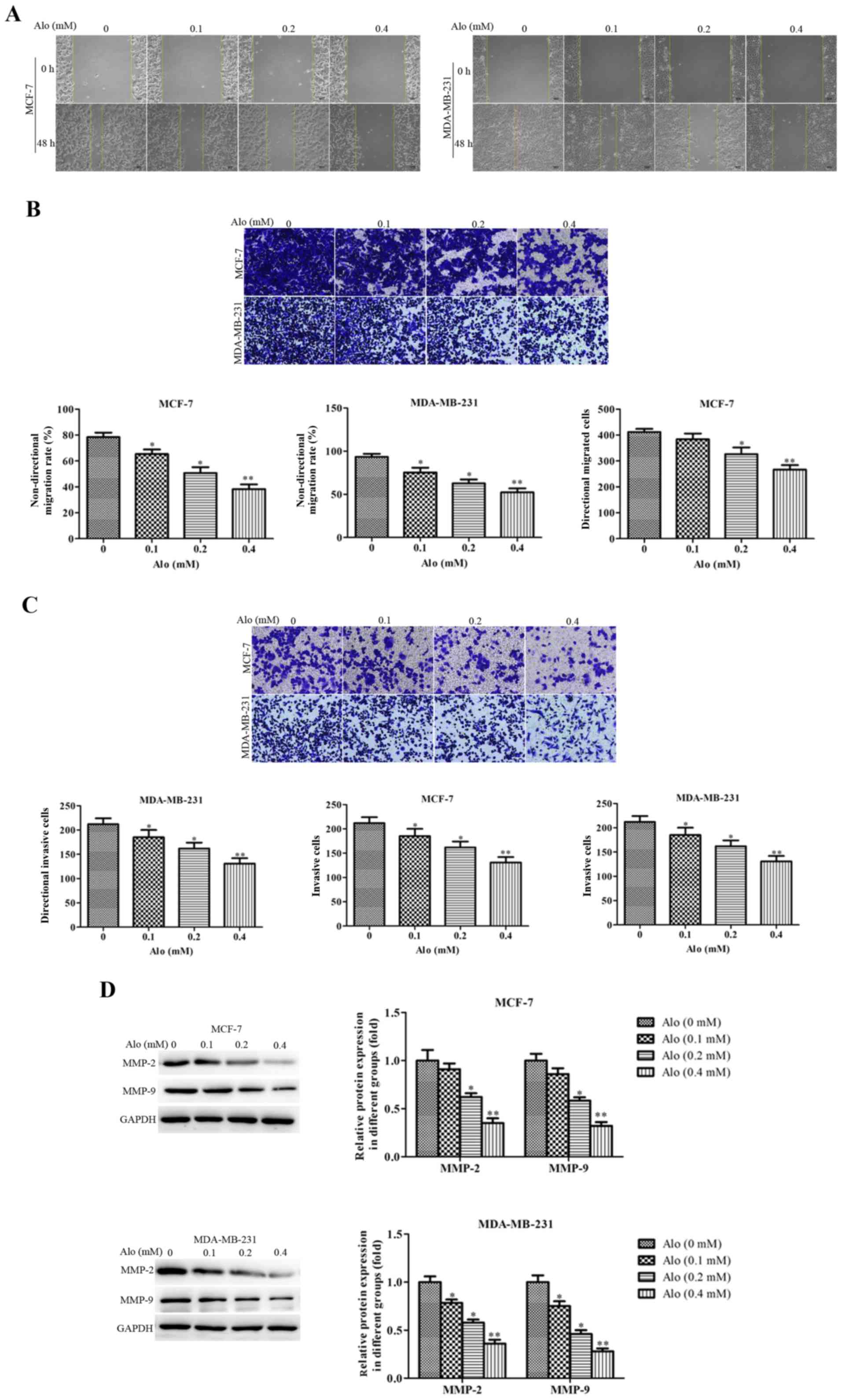
Alo suppressed human breast cancer
cells migration and invasion
To further confirm the effects of Alo on cell
motility, wound healing, trans-well migration and invasion assays
were conducted. The results of wound healing and Trans-well
migration assays revealed that Alo could significantly suppress
MCF-7 cells migration and also remarkably inhibit MDA-MB-231 cells
migration in a dose dependent manner compared with the untreated
group (Fig. 5A and B). Besides, a
similar results in cell invasion was observed. Alo treatment in
MCF-7 and MDA-MB-231 cells led to a decreased percent of invasion
cells compared with the untreated group (Fig. 5C). These data indicated Alo
dramatically depressed cell migration and invasion in MCF-7 and
MDA-MB-231 cells.
MMPs are zinc-dependent proteolytic enzymes of the
extracellular matrix (ECM), widely involved in cell migration and
invasion (19,20). And as the members of MMPs, MMP-2
and MMP-9 are closely related to the migration and invasion of
various types of cancer cells (21,22).
Thus, the protein expressions of MMP-2 and MMP-9 were detected to
evaluate the effect of Alo on the motility of MCF-7 and MDA-MB-231
cells. It showed that Alo significantly suppressed the protein
expressions of MMP-2 and MMP-9 (Fig.
5D).
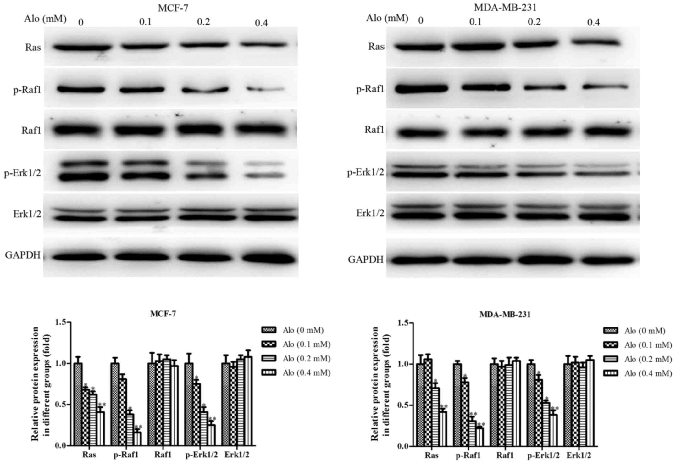
Alo blocked Ras in human breast cancer
cells
Ras family is usually recognized as an oncogene,
including H-Ras, Ha-Ras, K-Ras, Ki-Ras and N-Ras, and
Ras/Raf1/ERK1/2 pathway is considered the be the downstream of
epidermal growth factor receptor (EGFR), which is closely
associated to the incidence and prognosis of various cancers
(23). Activated ERK1/2 regulates
gene expression and ultimately mediates cell growth,
differentiation, migration, invasion and other processes through
phosphorylation on transcription factors in the cell cytoplasm and
nucleus (24,25). Thus, in this study, we observed
that, after treatment of human breast cancer cells MCF-7 and
MDA-MB-231 with Alo (0.1, 0.2 and 0.4 mM), the level of Ras was
downregulated, and the levels of phosphorylation of Raf1 and Erk1/2
were decreased as well (Fig. 6),
which indicated that Alo could block the Ras/Raf1/Erk1/2 signaling
pathway.
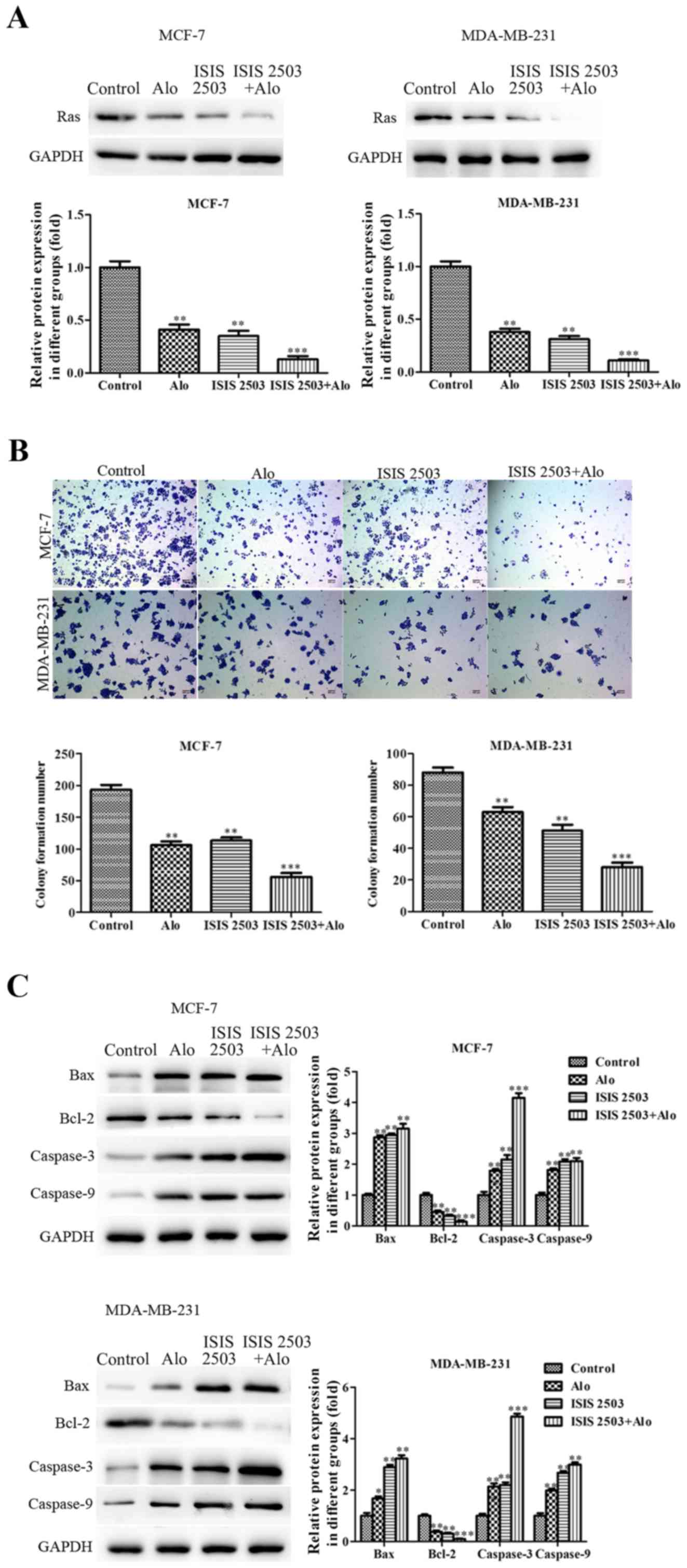
Alo inhibited proliferation, migration
and invasion and induced apoptosis via Ras pathway in human breast
cancer cells
After demonstrating decreased level of Ras following
incubation of MCF-7 and MDA-MB-231 cells with Alo (0.1, 0.2 and 0.4
mM), further, we evaluated whether activation of these kinases
contributed to the progress of MCF-7 and MDA-MB-231 cells. To
further verify the involvement of Ras signaling in inhibiting
proliferation, migration and invasion and inducing apoptosis of
human breast cancer cells, ISIS 2503, a novel Ras inhibitor with
selective activity against H-Ras, was employed to inhibit the Ras
expression (26,27). First, we found both Alo (0.4 mM)
and ISIS 2503 inhibited the expression of Ras, and co-treatment of
Alo and ISIS 2503 inhibited the level of Ras more than either one
of them in MCF-7 and MDA-MB-231 cells (Fig. 7A). Then the results demonstrated
that Alo (0.4 mM) and ISIS 2503 reduced colony formation (Fig. 7B), and could apparently
downregulate the expression of Bcl-2 and upregulate the expressions
of caspase-3, caspase-9 and Bax (Fig.
7C). However, co-treatment of Alo (0.4 mM) and ISIS 2503 had
better inhibitory effect on the cell colony formation and apoptosis
compared with that of either agent alone in MCF-7 and MDA-MB-231
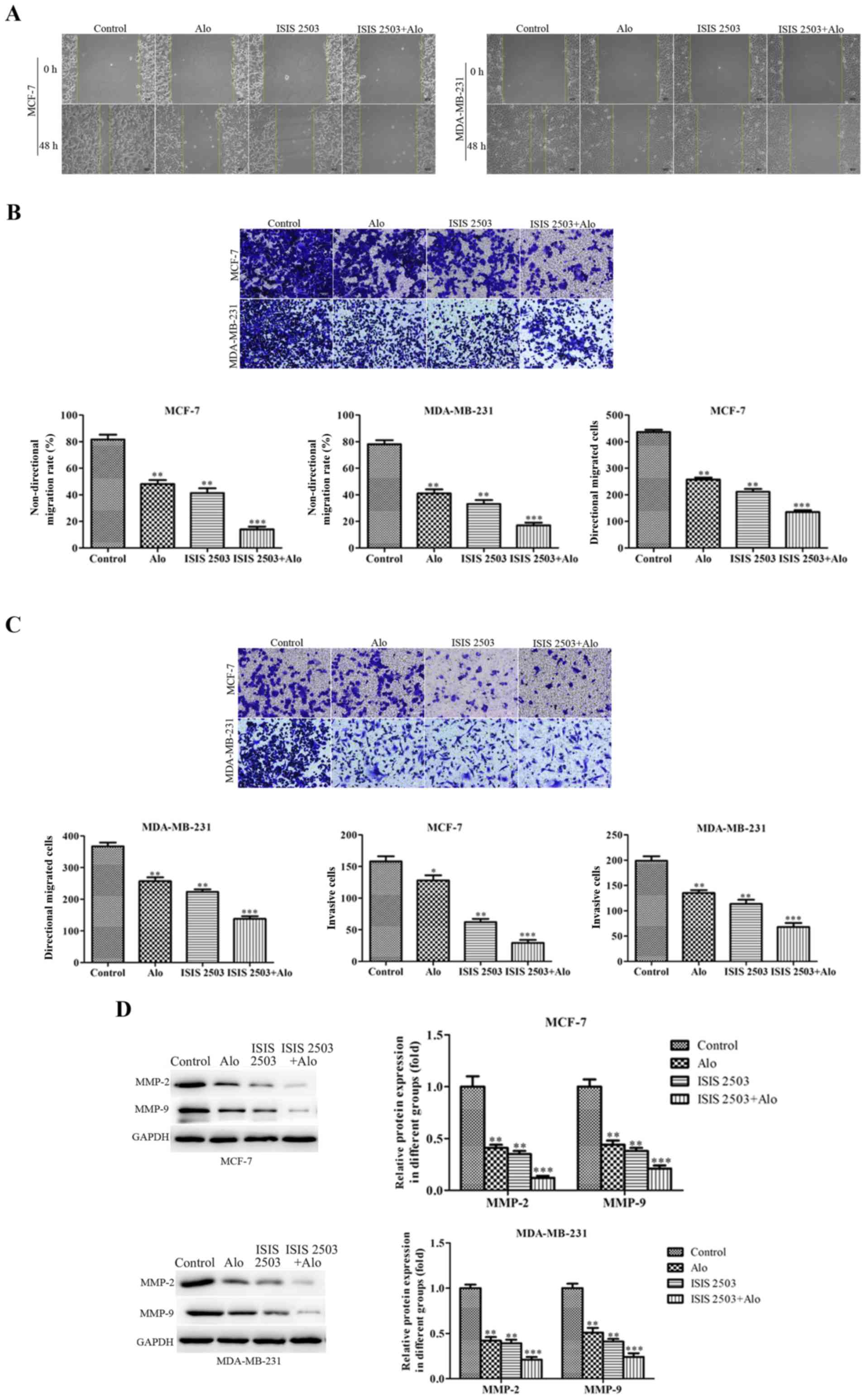
cells. Moreover, we adapted the wound healing (Fig. 8A), trans-well migration (Fig. 8B) and invasion (Fig. 8C) assays to evaluate the impact of
ISIS 2503 on cell motility, and the results demonstrated
co-treatment of Alo (0.4 mM) and ISIS 2503 remarkably inhibited
migration and invasion more than either one of them in MCF-7 and
MDA-MB-231 cells. In addition, co-treatment of Alo (0.4 mM) and
ISIS 2503 showed more positive activity of inhibition of MMP-2 and
MMP-9 levels (Fig. 8D).
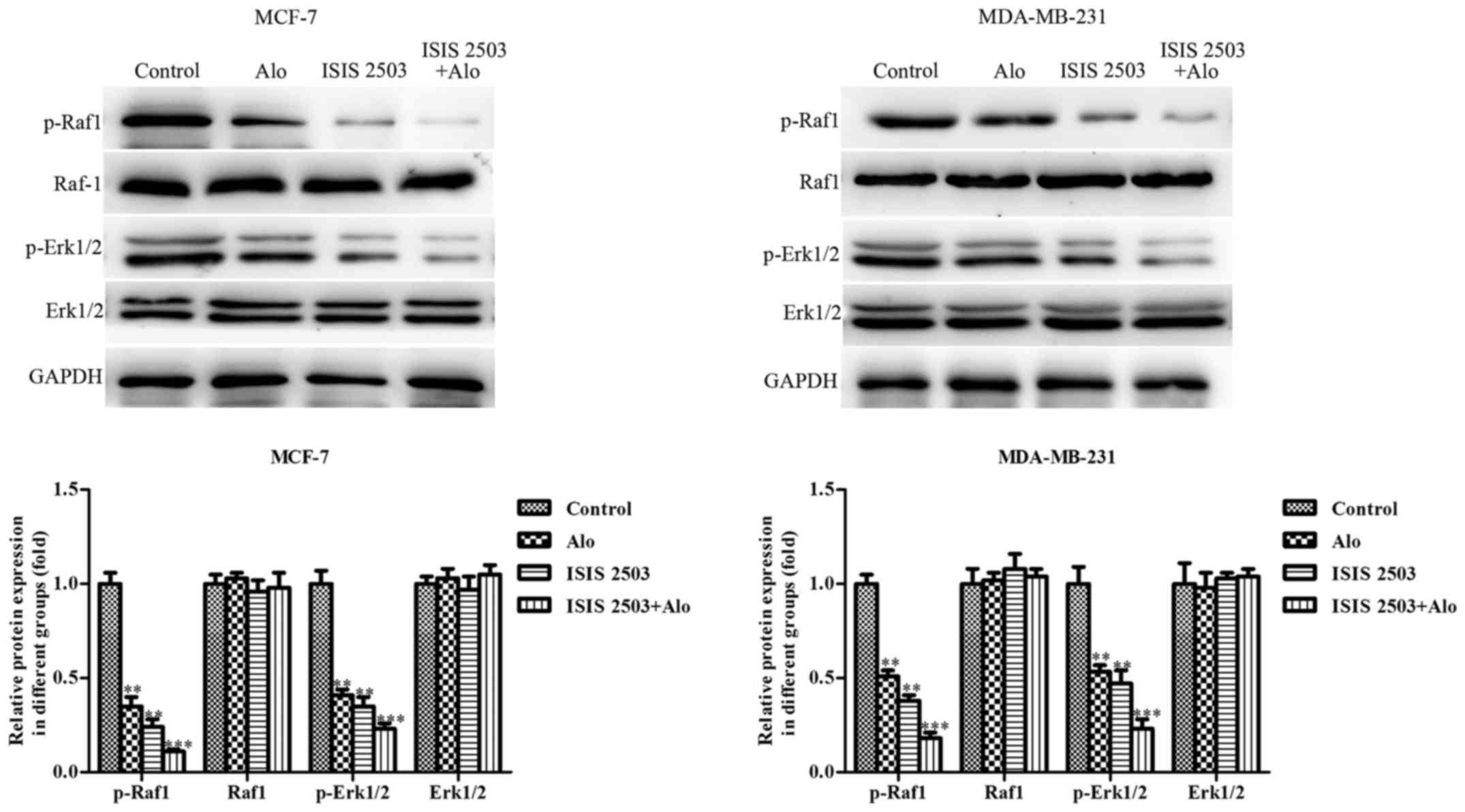
Furthermore, we found co-treatment of Alo (0.4 mM) and ISIS 2503
had better effects on the decrease of the phosphorylation of Raf1
and Erk1/2 (Fig. 9). These
findings suggested that Alo inhibited proliferation, migration and
invasion and induced apoptosis maybe through Ras pathway in human
breast cancer cells.
Discussion
Breast cancer is one of the most common malignant
tumors that leads to the death of women, and hematogenous spread
occurs easily in the early stage, which threatens the heath of
women seriously. At present, the treatments of breast cancer
include surgery, chemotherapy, radiotherapy, targeted biotherapy
and so on, which is certain to have to cancer cell kill and wound
action. However, there are still 25–30% of early breast cancer
patients with distant metastasis after 10–15 years of follow-up
(28). At the same time, the drug
resistance of tumor cells and the side effects of chemotherapeutic
drugs are also another difficult problem in the process of cancer
treatment. Therefore, the natural compounds from animal and plant
have become the main research direction of anticancer drugs
(29). Currently, traditional
Chinese medicine is still the mainstream of the prevention and
treatment of tumor, and the characteristics of multi-component,
multi-link and multi-target play a significant role in polygene
regulation, complicated pathogenesis, and prevention and treatment
of various tumors (30). Moreover,
traditional Chinese medicine has been paid more attention because
of its unique antitumor effect, sensitivity of radiotherapy and
chemotherapy, little toxic and side effects and prolonged the
survival time of patients (31).
Therefore, this article was designed to explore the effects and
mechanisms of Chinese medicine monomer Alo on proliferation,
apoptosis, migration and invasion of breast cancer cells. In this
study, we found Alo inhibited MCF-7 and MDA-MB-231 cells
proliferation and colony formation in a dose dependent manner,
changed cell membrane morphology and promoted the apoptosis in
MCF-7 and MDA-MB-231 cells. Bcl-2 family includes pro-apoptotic
proteins and anti-apoptotic proteins. A pro-apoptotic bcl-2 family
protein, Bax, promotes cell apoptosis by activation of caspase and
the release of cytochrome c from mitochondria, while Bcl-2, an
anti-apoptotic bcl-2 family protein, restrains cell apoptosis via
blocking the release of cytochrome c (32,33).
Our date showed that Alo could dramatically downregulate the
expression of Bcl-2 and upregulate the expression of caspase-3,
caspase-9 and Bax in MCF-7 and MDA-MB-231 cells. These results were
similar to previous studies (17,18),
which further confirmed that Alo had positive inhibitory activity
on the progression and development of many types of cancer.
The main cause of death in patients with breast
cancer is the invasion and metastasis of the tumor (34). Wound healing, trans-well migration
and invasion assays were conducted to further confirm the effects
of Alo on cell motility, and the results revealed that Alo could
significantly suppress MCF-7 and MDA-MB-231 cells migration and
invasion. In the process of tumor invasion and metastasis, the
degradation of ECM plays an important role. Moreover, the
degradation of ECM mainly depends on proteolytic enzymes, and the
most important is MMPs, which are highly conserved zinc dependent
endonuclease protease family with the positive activities on
degrading most proteins of basement membrane and ECM (35). Furthermore, MMP-2 and MMP-9 have
been strongly correlated with the invasiveness of many types of
cancer cell (36). Thus, the
protein expressions of MMP-2 and MMP-9 were detected to evaluate
the effect of Alo on the motility of MCF-7 and MDA-MB-231 cells and
we found that Alo significantly suppressed the protein expressions
of MMP-2 and MMP-9.
At present, it is believed that the occurrence of
human tumor is the result of multiple gene mutations that control
the proliferation, differentiation and apoptosis of normal cells,
and these mutations include the activation of the oncogenes and the
inactivation of the anti-oncogenes. It has been found that mutation
or activation of Ras gene and abnormal over-expression of Ras
protein exist in about 30% human tumors (37,38).
Therefore, research on the regulation of Ras signal transduction
pathway plays an important role in the design of antitumor drugs
targeting the cellular signal transduction pathway (39). Ras protein is considered as an
important element to regulate the signal pathway of cell growth and
proliferation. If Ras protein is activated continuously, it can
bind downstream effective proteins and transmit signals to
downstream signaling elements, which may cause abnormal
proliferation of cells and lead to the occurrence of tumors
(40). Therefore, we evaluated the
role of Alo in Ras and its downstream signaling (41). From the results, we found Alo could
significantly downregulate the level of Ras and suppress the levels
of phosphorylation of Raf1 and Erk1/2, indicating that Alo could
block the Ras/Raf1/Erk1/2 signaling pathway. To further verify role
of Ras signaling in occurrence and development of human breast
cancer cells (42), we chose a Ras
inhibitor, ISIS 2503, to inhibit the Ras signaling pathway, and we
found that ISIS 2503 could remarkably inhibit the expression of
Ras. Moreover, we also found that co-treatment of Alo and ISIS 2503
had better inhibitory effects on proliferation, migration, invasion
and apoptosis of MCF-7 and MDA-MB-231 cells than either one of
them. These resulted indicated that Alo had better inhibitory
effects on the occurrence and development of breast cancer via
blockage of Ras pathway.
The current research is a preliminary study on the
antitumor effect of Alo, the other effects of Alo and the
mechanisms of Alo in breast cancer are not very clear. Based on the
current findings, we will further explore the role of Alo in
development of cancer both in vivo and in vitro, and
explore the anticancer mechanism of Alo by genomics, proteomics and
metabolomics in future. Taken together, the present data suggest
that Alo possesses anticancer effects in human breast cancer cells
via inhibiting the potential of cell proliferation, migration and
invasion, and inducing apoptosis. Furthermore, Alo might act its
role in suppressing human breast cancer cells via Ras inactivation.
Based on our findings, Alo could be considered as a potential
candidate to treat breast cancer.
Acknowledgements
Not applicable.
Funding
No funding was received.
Availability of data and materials
All data generated or analyzed during this study are
included in this published article.
Authors' contributions
DT and YL designed the experiments. DT, YL, XL and
ZT performed the experiments. DT and YL wrote the manuscript. All
authors reviewed the manuscript.
Ethics approval and consent to
participate
Not applicable.
Patient consent for publication
Not applicable.
Competing interests
The authors declare that they have no competing
interests.
References
|
1
|
Torre LA, Bray F, Siegel RL, Ferlay J,
Lortet-Tieulent J and Jemal A: Global cancer statistics, 2012. CA
Cancer J Clin. 65:87–108. 2015. View Article : Google Scholar : PubMed/NCBI
|
|
2
|
Siegel RL, Miller KD and Jemal A: Cancer
statistics, 2015. CA Cancer J Clin. 65:5–29. 2015. View Article : Google Scholar : PubMed/NCBI
|
|
3
|
DeSantis C, Ma J, Bryan L and Jemal A:
Breast cancer statics, 2013. CA Cancer J Clin. 64:52–62. 2014.
View Article : Google Scholar : PubMed/NCBI
|
|
4
|
DeSantis CE, Bray F, Ferlay J,
Lortet-Tieulent J, Anderson BO and Jemal A: International variation
in female breast cancer incidence and mortality rates. Cancer
Epidemiol Biomarkers Prev. 24:1495–1506. 2015. View Article : Google Scholar : PubMed/NCBI
|
|
5
|
Fan L, Zheng Y, Yu KD, Liu GY, Wu J, Lu
JS, Shen KW, Shen ZZ and Shao ZM: Breast cancer in a transitional
society over 18 years: Trends and present status in Shanghai,
China. Breast Cancer Res Treat. 117:409–416. 2009. View Article : Google Scholar : PubMed/NCBI
|
|
6
|
Kamal A, Lakshma Nayak V, Nagesh N,
Vishnuvardan MV and Subba Reddy NV: Benzo[b]furan derivatives
induces apoptosis by targeting the PI3K/Akt/mTOR signaling pathway
in human breast cancer cells. Bioorg Chem. 66:124–131. 2016.
View Article : Google Scholar : PubMed/NCBI
|
|
7
|
Khpal M, Miller JRC, Petrovic Z and
Hassanally D: Local anesthetic delivery via surgical drain provides
improved pain control versus direct skin infiltration following
axillary node dissection for breast cancer. Breast Cancer.
25:185–190. 2018. View Article : Google Scholar : PubMed/NCBI
|
|
8
|
Lauberg T, Tramm T, Nielsen T, Alsner J,
Nord S, Myhre S, Sørlie T, Leung S, Fan C, Perou C, et al:
Intrinsic subtypes and benefit from postmastectomy radiotherapy in
node-positive premenopausal breast cancer patients who received
adjuvant chemotherapy-results from two independent randomized
trials. Acta Oncol. 25:38–43. 2018. View Article : Google Scholar
|
|
9
|
Ito K, Park SH, Katsyv I, Zhang W, De
Angelis C, Schiff R and Lrie HY: PTK6 regulates growth and survival
of endocrine therapy-resistant ER+ breast cancer cells. NPJ Breast
Cancer. 3:452017. View Article : Google Scholar : PubMed/NCBI
|
|
10
|
Sui Y, Zhang X, Yang H, Wei W and Wang M:
MicroRNA-133a acts as a tumour suppressor in breast cancer through
targeting LASP1. Oncol Rep. 39:473–482. 2018.PubMed/NCBI
|
|
11
|
Zhang J, Yu K, Han X, Zhen L, Liu M, Zhang
X, Ren Y and Shi J: Paeoniflorin influences breast cancer cell
proliferation and invasion via inhibition of the Notch-1 signaling
pathway. Mol Med Rep. 17:1321–1325. 2018.PubMed/NCBI
|
|
12
|
Wu C, Sun Z, Guo B, Ye Y, Han X, Qin Y and
Liu S: Osthole inhibits bone metastasis of breast cancer.
Oncotarget. 8:58480–58493. 2017.PubMed/NCBI
|
|
13
|
Huang C, Lu CK, Tu MC, Chang JH, Chen YJ,
Tu YH and Huang HC: Polyphenol-rich Avicennia marina leaf extracts
induce apoptosis in human breast and liver cancer cells and in a
nude mouse xenograft model. Oncotarget. 7:35874–35893.
2016.PubMed/NCBI
|
|
14
|
Zhou WJ, Wang S, Hu Z, Zhou ZY and Song
CJ: Angelica sinensis polysaccharides promotes apoptosis in human
breast cancer cells via CREB-regulated caspase-3 activation.
Biochem Biophys Res Commun. 467:562–569. 2015. View Article : Google Scholar : PubMed/NCBI
|
|
15
|
Wang H, Guo S, Qian D, Qian Y and Duan JA:
Comparative analysis of quinolizidine alkaloids from different
parts of Sophora alopecuroides seeds by UPLC-MS/MS. J Pharm Biomed
Anal 67–68. 1–21. 2012. View Article : Google Scholar
|
|
16
|
Yuan XY, Liu W, Zhang P, Wang RY and Guo
JY: Effects and mechanisms of aloperine on 2,
4-dinitrofluorobenzene-induced allergic contact dermatitis in
BALB/c mice. Eur J Pharmacol. 629:147–152. 2010. View Article : Google Scholar : PubMed/NCBI
|
|
17
|
Chen S, Jin Z, Dai L, Wu H, Wang J, Wang
L, Zhou Z, Yang L and Gao W: Aloperine induces apoptosis and
inhibits invasion in MG-63 and U2OS human osteosarcoma cells.
Biomed Pharmacother. 97:45–52. 2018. View Article : Google Scholar : PubMed/NCBI
|
|
18
|
Zhang L, Zheng Y, Deng H, Liang L and Peng
J: Aloperine induces G2/M phase cell cycle arrest and apoptosis in
HCT116 human colon cancer cells. Int J Mol Med. 33:1613–1620. 2014.
View Article : Google Scholar : PubMed/NCBI
|
|
19
|
Brown GT and Murray GI: Current
mechanistic insights into the roles of matrix metalloproteinases in
tumour invasion and metastasis. J Pathol. 237:273–281. 2015.
View Article : Google Scholar : PubMed/NCBI
|
|
20
|
Folgueras AR, Pendás AM, Sánchez LM and
López-Otín C: Matrix metalloproteinases in cancer: From new
functions to improved inhibition strategies. Int J Dev Biol.
48:411–424. 2004. View Article : Google Scholar : PubMed/NCBI
|
|
21
|
Yang Q, Ji M, Guan H, Shi B and Hou P:
Shikonin inhibits thyroid cancer cell growth and invasiveness
through targeting major signaling pathways. J Clin Endocrinol
Metab. 98:E1909–E1917. 2013. View Article : Google Scholar : PubMed/NCBI
|
|
22
|
Zhang XG, Lu XF, Jiao XM, Chen B and Wu
JX: PLK1 gene suppresses cell invasion of undifferentiated thyroid
carcinoma through the inhibition of CD44v6, MMP-2 and MMP-9. Exp
Ther Med. 4:1005–1009. 2012. View Article : Google Scholar : PubMed/NCBI
|
|
23
|
Downward J: Targeting RAS signalling
pathways in cancer therapy. Nat Rev Cancer. 3:11–22. 2003.
View Article : Google Scholar : PubMed/NCBI
|
|
24
|
Hilger RA, Scheulen ME and Strumberg D:
The Ras-Raf-MEK-ERK pathway in the treatment of cancer. Onkologie.
25:511–518. 2002.PubMed/NCBI
|
|
25
|
Friday BB and Adjei AA: Advances in
targeting the Ras/Raf/MEK/Erk mitogen-activated protein kinase
cascade with MEK inhibitors for cancer therapy. Clin Cancer Res.
14:342–346. 2008. View Article : Google Scholar : PubMed/NCBI
|
|
26
|
Dancey JE: Agents targeting ras signaling
pathway. Curr Pharm Des. 8:2259–2267. 2002. View Article : Google Scholar : PubMed/NCBI
|
|
27
|
Adjei AA, Dy GK, Erlichman C, Reid JM,
Sloan JA, Pitot HC, Alberts SR, Goldberg RM, Hanson LJ, Atherton
PJ, et al: A phase I trial of ISIS 2503, an antisense inhibitor of
H-ras, in combination with gemcitabine in patients with advanced
cancer. Clin Cancer Res. 9:115–123. 2003.PubMed/NCBI
|
|
28
|
Brackstone M, Townson JL and Chambers AF:
Tumor dormancy in breast cancer: An update. Breast Cancer Res.
9:2082007. View
Article : Google Scholar : PubMed/NCBI
|
|
29
|
Huang KW, Wu HL, Lin HL, Liang PC, Chen
PJ, Chen SH, Lee HI, Su PY, Wu WH, Lee PH, et al: Combining
antiangiogenic therapy with immunotherapy exerts better
therapeutical effects on large tumors in a woodchuck hepatoma
model. Proc Natl Acad Sci USA. 107:pp. 14769–14774. 2010;
View Article : Google Scholar : PubMed/NCBI
|
|
30
|
Chen Y, Hao H, He S, Cai L, Li Y, Hu S, Ye
D, Hoidal J, Wu P and Chen X: Lipoxin A4 and its analogue suppress
the tumor growth of transplanted H22 in mice: The role of
antiangiogenesis. Mol Cancer Ther. 9:2164–2174. 2010. View Article : Google Scholar : PubMed/NCBI
|
|
31
|
Banerjee S, A'Hern R, Detre S,
Littlewood-Evans AJ, Evans DB, Dowsett M and Martin LA: Biological
evidence for dual antiangiogenic-antiaromatase activity of the
VEGFR inhibitor PTK787/ZK222584 in vivo. Clin Cancer Res.
16:4178–4187. 2010. View Article : Google Scholar : PubMed/NCBI
|
|
32
|
Vaux DL and Korsmeyer SJ: Cell death in
development. Cell. 96:245–254. 1999. View Article : Google Scholar : PubMed/NCBI
|
|
33
|
Ackler S, Mitten MJ, Foster K, Oleksijew
A, Refici M, Tahir SK, Xiao Y, Tse C, Frost DJ, Fesik SW, et al:
The Bcl-2 inhibitor ABT-263 enhances the response of multiple
chemotherapeutic regimens in hematologic tumors in vivo. Cancer
Chemother Pharmacol. 66:869–880. 2010. View Article : Google Scholar : PubMed/NCBI
|
|
34
|
Edelstein ML, Abedi MR and Wixon J: Gene
therapy clinical trials worldwide to 2007-an update. J Gene Med.
9:833–842. 2007. View
Article : Google Scholar : PubMed/NCBI
|
|
35
|
Westermarck J and Kähäri VM: Regulation of
matrix metalloproteinase expression in tumor invasion. FASEB J.
13:781–792. 1999. View Article : Google Scholar : PubMed/NCBI
|
|
36
|
Liu X, Zhao W, Wang W, Lin S and Yang L:
Puerarin suppresses LPS-induced breast cancer cell migration,
invasion and adhesion by blockage NF-κB and Erk pathway. Biomed
Pharmacother. 92:429–436. 2017. View Article : Google Scholar : PubMed/NCBI
|
|
37
|
Kim D, Kim SY, Lee JS, Hong YS, Kim JE,
Kim KP, Kim J, Jang SJ, Yoon YK and Kim TW: Primary tumor location
predicts poor clinical outcome with cetuximab in RAS wild-type
metastatic colorectal cancer. BMC Gastroenterol. 17:1212017.
View Article : Google Scholar : PubMed/NCBI
|
|
38
|
Zhou J, Zhang S, Chen X, Zheng X, Yao Y,
Lu G and Zhou J: Palbociclib, a selective CDK4/6 inhibitor,
enhances the effect of selumetinib in RAS-driven non-small cell
lung cancer. Cancer Lett. 408:130–137. 2017. View Article : Google Scholar : PubMed/NCBI
|
|
39
|
Wang Z, Ma L, Su M, Zhou Y, Mao K, Li C,
Peng G, Zhou C, Shen B and Dou J: Baicalin induces cellular
senescence in human colon cancer cells via upregulation of DEPP and
the activation of Ras/Raf/MEK/ERK signaling. Cell Death Dis.
9:2172018. View Article : Google Scholar : PubMed/NCBI
|
|
40
|
Pan Q, Liu R, Banu H, Ma L and Li H:
Inhibition of isoprenylcysteine carboxylmethyltransferase
sensitizes common chemotherapies in cervical cancer via
Ras-dependent pathway. Biomed Pharmacother. 99:169–175. 2018.
View Article : Google Scholar : PubMed/NCBI
|
|
41
|
Yu SH, Wang TH and Au LC: Specific
repression of mutant K-Ras by 10–23 DNAzyme: Sensitizing cancer
cell to anti-cancer therapies. Biochem Biophys Res Commun.
378:230–234. 2009. View Article : Google Scholar : PubMed/NCBI
|
|
42
|
Bolchi C, Pallavicini M, Rusconi C,
Diomede L, Ferri N, Corsini A, Fumagalli L, Pedretti A, Vistoli G
and Valoti E: Peptidomimetic inhibitors of farnesyltransferase with
high in vitro activity and significant cellular potency. Bioorg Med
Chem Lett. 17:6192–6196. 2007. View Article : Google Scholar : PubMed/NCBI
|