Introduction
Postoperative cognitive dysfunction (POCD) refers to
alterations in cognitive abilities experienced by patients
following anesthesia, affecting functions such as orientation,
memory, attention and insight (1).
Furthermore, changes in personality, social skills and other
cognitive functions may have a negative impact upon patient
recovery, such as extension of hospital stays and regression of
quality of life (2). POCD is a
common central nervous system complication affecting elderly people
following surgery and presents as a mild cognitive dysfunction
(2).
Learning and memory are closely associated with the
central nervous system, particularly with the function of the
hippocampus. The hippocampus is a major component of the limbic
system and is an important structure for learning, memory and the
regulation of behavior (3).
Neuronal nitric oxide synthase (nNOS) in the hippocampus is a key
enzyme involved in the synthesis of the neurotransmitter nitric
oxide (NO), which participates in mechanisms of learning and memory
(4). Furthermore, the long-term
potentiation of synaptic transmission efficiency in the hippocampus
is recognized as the synaptic plasticity model of memory, which
underpins the neuronal mechanisms responsible for behavior,
learning and memory (5).
Oxidative stress refers to the process in which the
body has been exposed to harmful stimuli and an imbalance between
antioxidative and oxidative systems in the body develops, thus
predisposing the body towards oxidation (3). Oxidative stress results in the
infiltration of neutrophils, increased secretion of proteases and
the production of a large number of oxide intermediates, including
reactive nitrogen species (RNS) and reactive oxygen species (ROS)
(6). Furthermore, excessive levels
of oxidation intermediates have been reported to cause tissue
damage, which is considered to be an important factor leading to
aging and the development of disease (7). RNS include NO, nitrogen dioxide and
peroxide nitride, while ROS include the ultra-oxygen anion, oxygen
free radicals and hydrogen peroxide (8).
Highly enriched in green tea, polyphenols account
for ~22–30% of the dry weight of tea leaves and are primarily
composed of four components: Catechin, flavonoids, phenolic acids
and flower pigment composition. (−)Epigallocatechin-3-gallate
(EGCG) is an antioxidant that protects the adenosine triphosphate
enzyme in erythrocytes from oxidative damage (9). Previous studies have indicated that
when used as a food additive, EGCG may attenuate the effects of
POCD. Green tea leaves contain numerous natural active compounds
(9,10). For example, as the predominant
catechin in green tea in terms of content, EGCG exhibits important
physiological and pharmacological effects (11). As a result, further research is
required in order to investigate the therapeutic potential of EGCG
(11). Furthermore, numerous plant
polyphenols with diverse bioactivities require further
investigation in order to determine their therapeutic potential
(11). Considering this, the
present method of investigation into the effects of EGCG
administration may represent a valuable reference for future
research aiming to investigate the therapeutic potentials of other
polyphenols. The present study investigated whether EGCG
administration may attenuate anesthesia-induced memory deficit in
young mice, as well the potential underlying mechanisms.
Materials and methods
Experimental animals
All experiments were performed with the approval of
the Ethics Committee of Yinzhou People's Hospital (Ningbo, China).
Following the purchasing at postnatal day 14, C57BL/6J mice (male;
weight, 4–7 g, n=110) were purchased from Animal laboratory of
Zhejiang University, housed in polypropylene cages at 22–24°C and
55–60% humidity with free access to food and water on a 12 h
light/dark cycle. A total of 110 mice were used as follows: 30 in
the first set of experiments followed by 40 (n=10 each for control,
model, EGCG treatment and EGCG + L-arginine treatment) and 40 (n=10
each for control, model, EGCG treatment and EGCG + 7-nitroindazole
treatment) in the second set of experiments.
A total of 30 mice were randomly divided into three
groups (n=10 per group): Control group, anesthesia model group and
EGCG treatment group. In the anesthesia model and EGCG treatment
groups, mice were administered 2% sevoflurane delivered via
humidified 30% O2 carrier gas for 5 h. Following this,
mice belonging to the EGCG treatment group were intragastrically
administered EGCG (2 mg/kg/day, Sigma-Aldrich; Merck KGaA,
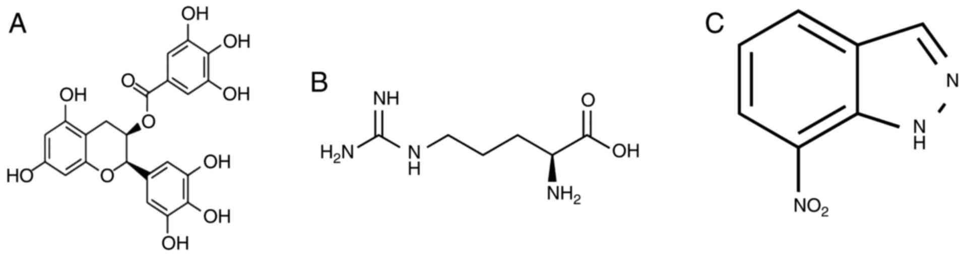
Darmstadt, Germany) for a total of 4 weeks. The chemical structure
of EGCG is presented in Fig.
1A.
Following this, a total of 40 mice were randomly
divided into four groups (n=10 per group): Control group,
anesthesia model group, EGCG treatment group (2 mg/kg/day of EGCG)
and EGCG + L-arginine (Sigma-Aldrich; Merck KGaA) group (50 mg/kg
once a week administered intragastrically; L-arginine for a total
of 4 weeks).
Following this, a total of 40 mice were randomly
divided into four groups (n=10 per group): Control group,
anesthesia model group, EGCG treatment group (2 mg/kg/day of EGCG)
and EGCG + L-arginine (Sigma-Aldrich; Merck KGaA) or
7-nitroindazole (Sigma-Aldrich; Merck KGaA) group (50 mg/kg once a
week administered intragastrically; 7-nitroindazole administered
for a total of 4 weeks). The chemical structures of 7-nitroindazole
and L-arginine are presented in Fig.
1B and C, respectively.
Morris water maze
A round pool (90×50 cm) was filled with warm water
(25–27°C) and painted black in order to appear opaque. An escape
platform (10 cm diameter) was fixed 0.5 cm below the water line in
one of four quadrants of the round pool. Mice (n=3/group) were
placed in a fixed position of one quadrant facing the wall of the
pool. Mice were allowed 120 sec to swim and discover the hidden
platform in order to escape. Mice that discovered the hidden
platform were permitted to remain on the platform for a further 30
sec. The duration of the escape latencies (finding the hidden
platform) was recorded for each mouse and the experiment was
repeated three times for each mouse. The mean time was considered
to represent the result for any given mouse for that day. Following
this, mice were wiped dry before being returned to their cages. On
the fifth day of experimentation, the platform was removed and mice
were permitted to swim in the pool for a 1 min time period. The
number of times mice crossed the original platform site and the
time spent in the target quadrant were recorded.
ELISA
Brain tissue was homogenized using a
radioimmunoprecipitation assay (Beyotime Institute of
Biotechnology, Haimen, China) and protein concentrations of the
homogenates were determined using a bicinchoninic assay (Beyotime
Institute of Biotechnology). A total of 5 µg protein samples were
analyzed for the detection of oxidative stress via determination of
malondialdehyde (MDA; cat. no. A003-1), superoxide dismutase (SOD;
cat. no. A001-1-1) and glutathione (GSH; cat. no. A006-2)
expression levels, while caspase-3/9 activities (cat. nos.
G015/G018) were analyzed to determine apoptosis and
acetylcholinesterase (AChE; cat. no. A024) activity was also
measured, using ELISA kits (Nanjing Jiancheng Bioengineering
Institute, Nanjing, China).
Western blot analysis
Brain tissue was homogenized using a
radioimmunoprecipitation assay. Total protein was determined using
a bicinchoninic assay. A total of 50 µg protein was subjected to
8–12% SDS-PAGE analysis and transferred to nitrocellulose
membranes. Membranes were blocked using 5% skimmed milk powder in
TBS with 0.1% Tween-20 for 1 h at room temperature and then
incubated at 4°C overnight with the following primary antibodies:
nNOS (cat. no. c-17825, 1:1,000, Santa Cruz Biotechnology, Inc.,
Dallas, TX, USA), β-amyloid (Aβ; cat. no. 8243, 1:2,000, Cell
Signaling Technology, Inc.), amyloid precursor protein (APP; cat.
no. sc-374527, 1:5,000, Santa Cruz Biotechnology, Inc.) and GAPDH
(cat. no. sc-51631, 1:5,000, Santa Cruz Biotechnology, Inc.).
Following this, membranes were incubated at 37°C for 1 h with
corresponding horseradish peroxidase-conjugated secondary
antibodies (cat. no. sc-2004, 1:2,000; Santa Cruz Biotechnology,
Inc.). Membranes were visualized using the ECL Plus western
blotting detection system (Beyotime Institute of Biotechnology),
developed on a C-DiGit Blot Scanner (LI-COR Biosciences, Lincoln,
NE, USA) and quantified using Image Lab 3.0 (Bio-Rad Laboratories,
Inc., Hercules, CA, USA).
Statistical analysis
Data are presented as the mean ± standard error
(n=3) and were analyzed by one-way analysis of variance followed by
Tukey's post-hoc tests using SPSS 17.0 (SPSS, Inc., Chicago, IL,
USA). P<0.05 was considered to indicate a statistically
significant difference.
Results
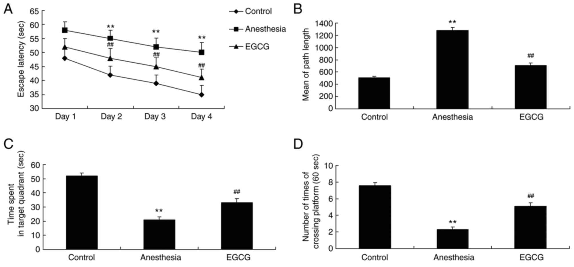
EGCG administration attenuates memory
deficit in anesthesia-induced mice
After EGCG treatment, mice from each group were
subjected to Morris water maze analysis in order to investigate the
effects of EGCG administration. As revealed in Fig. 2A and B, the duration of escape
latency and the mean path length (on day 5) exhibited by the
anesthesia model group were significantly increased compared with
the control group. In addition, the duration of time spent in the
target quadrant and the number of times crossing the platform
exhibited by the anesthesia model group were significantly
decreased compared with the control group (Fig. 2C and D). However, treatment with
EGCG was demonstrated to significantly decrease the escape latency
duration and the mean path length, and significantly increase the
time spent in the target quadrant and the number of times crossing
the platform, compared with the anesthesia model group (Fig. 2).
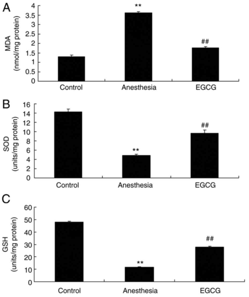
EGCG administration attenuates
oxidative stress in anesthesia-induced mice
In order to investigate the effect of EGCG
administration on oxidative stress levels in mice, samples were
subjected to ELISA analysis in order to determine the expression
levels of MDA, SOD and GSH. The results demonstrated that there was
a significant increase in the expression of MDA, and a significant
suppression of SOD and GSH expression, in the anesthesia model
group compared with the control group (Fig. 3). However, administration of EGCG
was revealed to significantly suppress the expression of MDA, and
increase the expression of SOD and GSH, compared with the
anesthesia model group (Fig.
3).
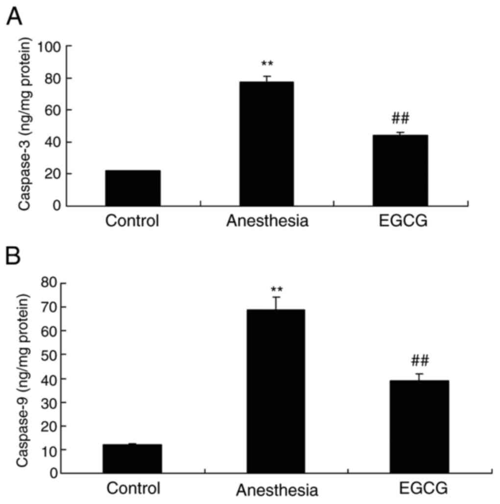
EGCG administration attenuates
apoptosis in anesthesia-induced mice
The effects of EGCG on apoptosis in
anesthesia-induced mice were investigated by ELISA. It was
demonstrated that caspase-3 and caspase-9 activities were
significantly enhanced in the anesthesia model group compared with
the control group (Fig. 4).
However, treatment with EGCG significantly suppressed both
caspase-3 and caspase-9 activity compared with the anesthesia model
group (Fig. 4).
EGCG administration suppresses nNOS,
Aβ and APP expression and AChE activity in anesthesia-induced
mice
In order to investigate the biological alterations
underlying cognitive impairment induced by anesthesia, nNOS, Aβ and
APP expression, as well as AChE activity, were analyzed by western
blotting and ELISA, respectively. Notably, in the anesthesia model
group, nNOS, Aβ and APP protein expression levels, as well as AChE
activity, were enhanced compared with the control group (Fig. 5). However, EGCG administration
significantly suppressed nNOS, Aβ and APP protein expression
levels, as well as AChE activity, compared with the anesthesia
model group (Fig. 5).
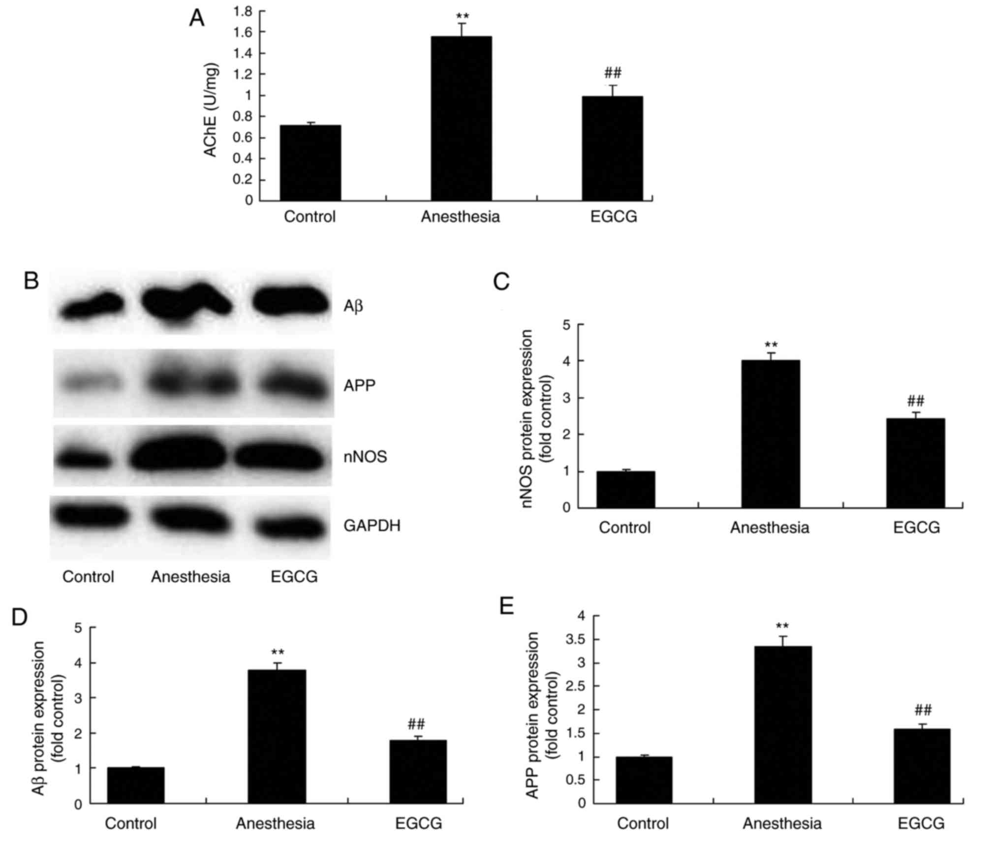 | Figure 5.Effects of EGCG administration on the
activity of AChE, and nNOS, Aβ and APP protein expression, in
anesthesia-induced mice. (A) ELISA was performed to determine the
effect of EGCG administration on AChE activity in
anesthesia-induced mice. (B) Representative protein bands for nNOS,
Aβ and APP protein expression by western blotting analysis.
Densitometric analysis of western blotting results was performed to
quantify and statistically analyze the protein expression of (C)
nNOS, (D) Aβ and (E) APP. **P<0.01 vs. control group;
##P<0.01 vs. anesthesia model group. EGCG,
(−)epigallocatechin-3-gallate; AChE, acetylcholinesterase; nNOS,
nitric oxide synthase; Aβ, β-amyloid; APP, amyloid precursor
protein. |
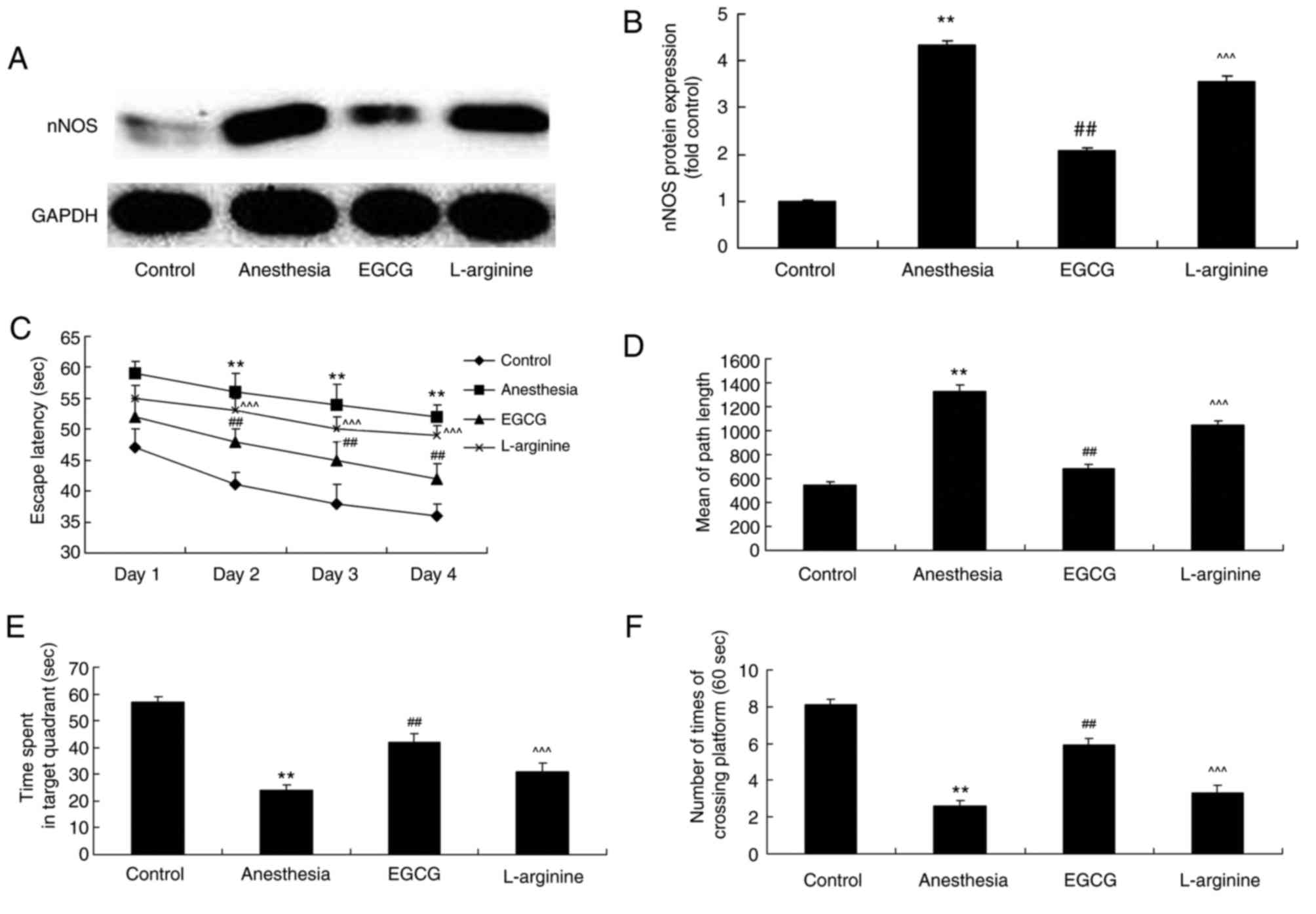
L-arginine administration reverses the
protective effects of EGCG on memory deficit in anesthesia-induced
mice
The role of nNOS expression in ECGC-induced
attenuation of memory deficit was investigated via administration
of L-arginine (an nNOS substrate) alongside EGCG. L-arginine was
revealed to significantly enhance nNOS protein expression in
anesthesia-induced mice following EGCG treatment, compared with the
EGCG-only treatment group (Fig. 6A and
B). Furthermore, following treatment with L-arginine, the
effects of EGCG treatment on the results of the Morris water maze
experiment for the investigation of memory deficit in
anesthesia-induced mice were significantly attenuated (Fig. 6C-F).
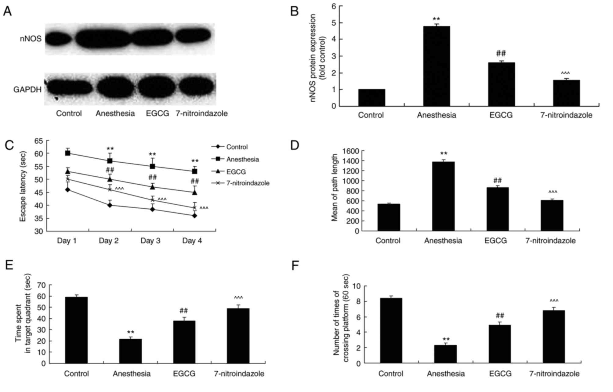
Inhibition of nNOS with
7-nitroindazole enhances the effects of EGCG administration with
regards to memory deficit
The effect of the inhibition of nNOS on memory
deficit exhibited by anesthesia-induced mice treated with EGCG was
also investigated. 7-nitroindazole was employed as an nNOS
inhibitor, and the results of western blotting demonstrated that
7-nitroindazole significantly inhibited nNOS protein expression in
anesthesia-induced mice treated with EGCG (Fig. 7A and B). However, the inhibition of
nNOS via administration of 7-nitroindiazole significantly enhanced
the effects of EGCG treatment with regards to memory deficit in
anesthesia-induced mice, as measured by the Morris water maze tests
(Fig. 7C-F).
Discussion
The elderly population represents a high-risk group
for the development of POCD following clinical anesthesia (12). As a result, ketamine increases the
incidence of POCD in elderly people, thus limiting the clinical
application of ketamine for anesthetic purposes with regards to
elderly patients (12).
Sevoflurane is a frequently used intravenous anesthetic in clinical
practice. Previous studies have also demonstrated that a
combinatory administration of propofol and ketamine reduces the
required dosage of ketamine as well as the associated adverse side
effects, such as mental and cardiovascular excitation (13,14).
In the present study, it was demonstrated that EGCG administration
attenuated memory deficit in anesthesia-induced mice. He et
al (15) revealed that EGCG
administration attenuated acrylamide-induced apoptosis and
astrogliosis in the cerebral cortex of rats (12). These results indicate that EGCG may
represent a novel therapeutic agent for the treatment of
anesthesia-induced memory deficit; however, determining the exact
molecular mechanism underlying this process requires further
investigation.
Sevoflurane has previously been demonstrated to
attenuate cell apoptosis meditated by tumor necrosis factor through
the suppression of caspase-3 activity and inhibition of apoptosis
via regulation of apoptosis-associated protein expression, such as
affecting the Bcl-2/Bcl-2-associated X protein ratio (16). The caspase family refers to a group
of regulator genes closely associated with apoptosis, and caspase-3
represents an important apoptotic protein responsible for the
induction and execution of cell apoptosis in mammals (17). Hippocampal neurons are the most
responsive to total cerebral ischemia and thus are the most
susceptible to apoptotic induction (18). The present study revealed that
treatment with EGCG significantly suppressed caspase-3 and
caspase-9 activity in anesthesia-induced mice.
Previous studies have demonstrated that isoflurane
administration affects the formation and degradation process of
APP, which may lead to alterations in the secondary structure of Aβ
(19,20). Following this, Aβ aggregates form
oligomers and eventually plaque formation surrounding brain neurons
(19). Furthermore, Aβ sediments
were reported to trigger immune inflammatory responses and activate
neurotoxic pathways, resulting in the degeneration and induction of
apoptosis in neurons (20). In
addition, Aβ polypeptides inhibit the release of endogenous
acetylcholine (19). Based on the
aforementioned factors, it may be hypothesized that the use of
isoflurane in anesthesia may damage hippocampal cholinergic
neurons, inhibit the release of acetylcholine in the hippocampus
and induce the development of POCD (21). The present study demonstrated that
EGCG treatment significantly inhibited the activity of AChE and the
protein expression of Aβ and APP in mice exposed to anesthesia. In
the present study, the regulation of AChE activity by EGCG, as well
as the modulation of Aβ and APP expression, was a potentially
important mechanism for the attenuation of anesthesia-induced
memory deficit in mice. Furthermore, we will investigate the
underlying regulatory mechanisms of EGCG administration in
anesthesia-induced mice with regards to AChE activity, as well as
Aβ and APP expression, in a future study.
Oxidative stress is a predominant factor involved in
the aging process. In 1956, a study demonstrated that free radicals
were implicated in the aging process of the body (22). Numerous studies have since
demonstrated that free radical damage represents part of the aging
process, and oxidative stress causes damage to cells and may induce
numerous diseases, including brain damage, arteriosclerosis,
cognitive dysfunction and dementia (3). As oxidative stress has been reported
to accelerate neurodegeneration in Alzheimer's disease, it may be
hypothesized that oxidative stress represents part of the neural
pathological process associated with POCD (23). Therefore, oxidative stress may
represent a predominant source of the damage experienced by neurons
and ROS may be considered an important causal factor for the
development of POCD and numerous other neuropathies (23). In addition, the present study
revealed that treatment with EGCG significantly suppressed MDA
levels and significantly enhanced SOD and GSH levels in
anesthesia-induced mice. Furthermore, He et al (24) demonstrated that EGCG administration
inhibited acrylamide-induced oxidative stress and apoptosis in PC12
cells via the reduction of oxidative stress.
As a relatively recently identified
neurotransmitter, NO administration is effective in POCD to a
certain extent as it aids learning and memory processing via the
regulation of synaptic transmission as well as the induction and
maintenance of long-term potentiation (25). Despite this, a dual role for NO in
POCD has been reported. A low concentration of NO was reported to
boost nerve growth and protect against apoptosis, while a high
concentration of NO exerted neurotoxic effects via the induction of
mitochondrial dysfunction and the activation of apoptotic pathways
(26). However, other studies have
indicated that different types of NOS have different effects on
POCD (27). The present study
demonstrated that EGCG treatment significantly suppressed nNOS
expression in anesthesia-induced mice. In addition, the present
study also demonstrated that treatment with L-arginine, an nNOS
substrate, and 7-nitroindazole, an nNOS inhibitor, inhibited and
potentiated the effects of EGCG, respectively, on
anesthesia-induced memory deficit in mice. Furthermore, Wei et
al (28) revealed that EGCG
attenuated NADPH-diaphorase/nNOS expression in the motor neurons of
rats. In addition, the present study demonstrated that EGCG
administration modulated nNOS levels in brain tissues and
attenuated memory deficit in anesthesia-induced mice. However, the
underlying therapeutic mechanisms of EGCG administration on nNOS
levels have not yet been determined and require further
investigation.
In conclusion, the results of the present study
demonstrated that EGCG administration attenuated memory deficit,
and suppressed apoptosis and oxidative stress, in
anesthesia-induced mice. Furthermore, it was revealed that the
mechanism underlying this process may involve the regulation of
nNOS expression. Thus, the protective effects of EGCG, functioning
as a memory deficit suppressor during anesthesia, may represent a
novel therapeutic agent for the treatment of POCD.
Acknowledgements
Not applicable.
Funding
No funding was received.
Availability of data and materials
The analyzed data sets generated during the present
study are available from the corresponding author on reasonable
request.
Authors' contributions
SY designed the study; LD, XG and JH performed the
experiments; SY analyzed the data; LD, XG and JH wrote the
manuscript.
Ethics approval and consent to
participate
All experiments were performed with the approval of
the Ethics Committee of Yinzhou People's Hospital (Ningbo,
China).
Patient consent for publication
Not applicable.
Competing interests
The authors declare that they have no competing
interests.
References
|
1
|
Qiao Y, Feng H, Zhao T, Yan H, Zhang H and
Zhao X: Postoperative cognitive dysfunction after inhalational
anesthesia in elderly patients undergoing major surgery: The
influence of anesthetic technique, cerebral injury and systemic
inflammation. BMC Anesthesiol. 15:1542015. View Article : Google Scholar : PubMed/NCBI
|
|
2
|
Zhu YZ, Yao R, Zhang Z, Xu H and Wang LW:
Parecoxib prevents early postoperative cognitive dysfunction in
elderly patients undergoing total knee arthroplasty: A
double-blind, randomized clinical consort study. Medicine
(Baltimore). 95:e40822016. View Article : Google Scholar : PubMed/NCBI
|
|
3
|
Ali MR, Abo-Youssef AM, Messiha BA and
Khattab MM: Tempol and perindopril protect against
lipopolysaccharide-induced cognition impairment and amyloidogenesis
by modulating brain-derived neurotropic factor, neuroinflammation
and oxido-nitrosative stress. Naunyn Schmiedebergs Arch Pharmacol.
389:637–656. 2016. View Article : Google Scholar : PubMed/NCBI
|
|
4
|
Gadek-Michalska A, Tadeusz J, Rachwalska P
and Bugajski J: Cytokines, prostaglandins and nitric oxide in the
regulation of stress-response systems. Pharmacol Rep. 65:1655–1662.
2013. View Article : Google Scholar : PubMed/NCBI
|
|
5
|
Shen F, Li YJ, Shou XJ and Cui CL: Role of
the NO/sGC/PKG signaling pathway of hippocampal CA1 in
morphine-induced reward memory. Neurobiol Learn Mem. 98:130–138.
2012. View Article : Google Scholar : PubMed/NCBI
|
|
6
|
Sohanaki H, Baluchnejadmojarad T, Nikbakht
F and Roghani M: Pelargonidin improves memory deficit in amyloid
β25–35 rat model of Alzheimer's disease by inhibition of glial
activation, cholinesterase, and oxidative stress. Biomed
Pharmacother. 83:85–91. 2016. View Article : Google Scholar : PubMed/NCBI
|
|
7
|
Nieradko-Iwanicka B and Borzęcki A:
Subacute poisoning of mice with deltamethrin produces memory
impairment, reduced locomotor activity, liver damage and changes in
blood morphology in the mechanism of oxidative stress. Pharmacol
Rep. 67:535–541. 2015. View Article : Google Scholar : PubMed/NCBI
|
|
8
|
De Moura AC, Brito VB, Porawski M, Saffi J
and Giovenardi M: Low maternal care is associated with increased
oxidative stress in the brain of lactating rats. Brain Res.
1655:17–22. 2017. View Article : Google Scholar : PubMed/NCBI
|
|
9
|
Koeberle D, Betticher DC, von Moos R,
Dietrich D, Brauchli P, Baertschi D, Matter K, Winterhalder R,
Borner M, Anchisi S, et al: Bevacizumab continuation versus no
continuation after first-line chemotherapy plus bevacizumab in
patients with metastatic colorectal cancer: A randomized phase III
non-inferiority trial (SAKK 41/06). Ann Oncol. 26:709–714. 2015.
View Article : Google Scholar : PubMed/NCBI
|
|
10
|
Malcomson FC, Willis ND, McCallum I, Xie
L, Lagerwaard B, Kelly S, Bradburn DM, Belshaw NJ, Johnson IT and
Mathers JC: Non-digestible carbohydrates supplementation increases
miR-32 expression in the healthy human colorectal epithelium: A
randomized controlled trial. Mol Carcinog. 56:2104–2111. 2017.
View Article : Google Scholar : PubMed/NCBI
|
|
11
|
Sha D, Lee AM, Shi Q, Alberts SR, Sargent
DJ, Sinicrope FA and Diasio RB: Association study of the let-7
miRNA-complementary site variant in the 3′ untranslated region of
the KRAS gene in stage III colon cancer (NCCTG N0147 Clinical
Trial). Clin Cancer Res. 20:3319–3327. 2014. View Article : Google Scholar : PubMed/NCBI
|
|
12
|
Silbert BS, Evered LA and Scott DA:
Incidence of postoperative cognitive dysfunction after general or
spinal anaesthesia for extracorporeal shock wave lithotripsy. Br J
Anaesth. 113:784–791. 2014. View Article : Google Scholar : PubMed/NCBI
|
|
13
|
Saleh AJ, Tang GX, Hadi SM, Yan L, Chen
MH, Duan KM, Tong J and Ouyang W: Preoperative cognitive
intervention reduces cognitive dysfunction in elderly patients
after gastrointestinal surgery: A randomized controlled trial. Med
Sci Monit. 21:798–805. 2015. View Article : Google Scholar : PubMed/NCBI
|
|
14
|
Tang N, Ou C, Liu Y, Zuo Y and Bai Y:
Effect of inhalational anaesthetic on postoperative cognitive
dysfunction following radical rectal resection in elderly patients
with mild cognitive impairment. J Int Med Res. 42:1252–1261. 2014.
View Article : Google Scholar : PubMed/NCBI
|
|
15
|
He Y, Tan D, Bai B, Wu Z and Ji S:
Epigallocatechin-3-gallate attenuates acrylamide-induced apoptosis
and astrogliosis in rat cerebral cortex. Toxicol Mech Methods.
27:298–306. 2017. View Article : Google Scholar : PubMed/NCBI
|
|
16
|
Tagawa T, Sakuraba S, Kimura K and
Mizoguchi A: Sevoflurane in combination with propofol, not
thiopental, induces a more robust neuroapoptosis than sevoflurane
alone in the neonatal mouse brain. J Anesth. 28:815–820. 2014.
View Article : Google Scholar : PubMed/NCBI
|
|
17
|
Jia Z, Geng L, Xie G, Chu Q and Zhang W:
Sevoflurane impairs acquisition learning and memory function in
transgenic mice model of Alzheimer's disease by induction of
hippocampal neuron apoptosis. Int J Clin Exp Med. 8:15490–15497.
2015.PubMed/NCBI
|
|
18
|
Zhang DX, Zhang LM, Zhao XC and Sun W:
Neuroprotective effects of erythropoietin against
sevoflurane-induced neuronal apoptosis in primary rat cortical
neurons involving the EPOR-Erk1/2-Nrf2/Bach1 signal pathway. Biomed
Pharmacother. 87:332–341. 2017. View Article : Google Scholar : PubMed/NCBI
|
|
19
|
Harach T, Marungruang N, Duthilleul N,
Cheatham V, McCoy KD, Frisoni G, Neher JJ, Fåk F, Jucker M, Lasser
T and Bolmont T: Reduction of Abeta amyloid pathology in APPPS1
transgenic mice in the absence of gut microbiota. Sci Rep.
7:418022017. View Article : Google Scholar : PubMed/NCBI
|
|
20
|
Somers C, Goossens J, Engelborghs S and
Bjerke M: Selecting Aβ isoforms for an Alzheimer's disease
cerebrospinal fluid biomarker panel. Biomark Med. 11:169–178. 2017.
View Article : Google Scholar : PubMed/NCBI
|
|
21
|
Wei L, Lv S, Huang Q, Wei J, Zhang S,
Huang R, Lu Z and Lin X: Pratensein attenuates Aβ-induced cognitive
deficits in rats: Enhancement of synaptic plasticity and
cholinergic function. Fitoterapia. 101:208–217. 2015. View Article : Google Scholar : PubMed/NCBI
|
|
22
|
Mehta V, Parashar A and Udayabanu M:
Quercetin prevents chronic unpredictable stress induced behavioral
dysfunction in mice by alleviating hippocampal oxidative and
inflammatory stress. Physiol Behav. 171:69–78. 2017. View Article : Google Scholar : PubMed/NCBI
|
|
23
|
Soodi M, Saeidnia S, Sharifzadeh M,
Hajimehdipoor H, Dashti A, Sepand MR and Moradi S: Satureja
bachtiarica ameliorate beta-amyloid induced memory impairment,
oxidative stress and cholinergic deficit in animal model of
Alzheimer's disease. Metab Brain Dis. 31:395–404. 2016. View Article : Google Scholar : PubMed/NCBI
|
|
24
|
He Y, Tan D, Mi Y, Bai B, Jiang D, Zhou X
and Ji S: Effect of epigallocatechin-3-gallate on
acrylamide-induced oxidative stress and apoptosis in PC12 cells.
Hum Exp Toxicol. 36:1087–1099. 2017. View Article : Google Scholar : PubMed/NCBI
|
|
25
|
Zhou XY, Zhang F, Ying CJ, Chen J, Chen L,
Dong J, Shi Y, Tang M, Hu XT, Pan ZH, et al: Inhibition of iNOS
alleviates cognitive deficits and depression in diabetic mice
through downregulating the NO/sGC/cGMP/PKG signal pathway. Behav
Brain Res. 322:70–82. 2017. View Article : Google Scholar : PubMed/NCBI
|
|
26
|
Orzelska J, Talarek S, Listos J and
Fidecka S: Effects of NOS inhibitors on the benzodiazepines-induced
memory impairment of mice in the modified elevated plus-maze task.
Behav Brain Res. 244:100–106. 2013. View Article : Google Scholar : PubMed/NCBI
|
|
27
|
Yu SY, Zhang M, Luo J, Zhang L, Shao Y and
Li G: Curcumin ameliorates memory deficits via neuronal nitric
oxide synthase in aged mice. Prog Neuropsychopharmacol Biol
Psychiatry. 45:47–53. 2013. View Article : Google Scholar : PubMed/NCBI
|
|
28
|
Wei IH, Tu HC, Huang CC, Tsai MH, Tseng CY
and Shieh JY: (−)-Epigallocatechin gallate attenuates NADPH-d/nNOS
expression in motor neurons of rats following peripheral nerve
injury. BMC Neurosci. 12:522011. View Article : Google Scholar : PubMed/NCBI
|