Introduction
Colorectal cancer (CRC) is one of the leading causes
of cancer-associated mortality worldwide. Despite advances in
treatment methods, patients with CRC have a poor 5-year survival
rate (1). Currently,
5-fluorouracil (5-FU) and oxaliplatin serve pivotal roles in
treatment regimens for CRC (2).
5-FU increases DNA damage via inhibition of thymidylate synthase
(TYMS). Oxaliplatin is a third-generation platinum-containing
compound that may induce DNA cross-links, leading to DNA
double-strand breaks (DSBs) (3).
DSBs are one of the most important factors that threaten the
integrity of the genome.
Mitogen-activated protein kinase kinase 1 (MEK1),
also known as MAP2K1, is a protein kinase that is a known
downstream target of Raf-1 proto-oncogene serine/threonine kinase
and is upstream of extracellular signal-regulated kinase (ERK). A
variety of small molecule inhibitors of MEK are currently
investigated in preclinical or clinical trials for the treatment of
malignancies (4). The
small-molecule compound U0126 has been identified as a MEK1/2
inhibitor that directly suppresses MEK1/2 activation with well
characterized off-target effects (5). Although the pharmacological
characteristics of U0126 indicate render it unsuitable for clinical
use, it has been demonstrated to be an efficient inhibitor in
vitro and in vivo to study the functions of MEK1/2
(6).
Combination therapy is a common approach in cancer
chemotherapy. However, the effect of an MEK inhibitor combined with
oxaliplatin or 5-FU in MEK-mutant colorectal cells and the
underlying mechanism remain unclear. Therefore, the present study
aimed to evaluate the combined effect of the MEK inhibitor U0126
with oxaliplatin and 5-FU in CRC cells, and to further explore the
underlying mechanisms involved.
Materials and methods
Reagents and cell culture
The human SW48 (cat. no. CCL-231) and NCI-H508 (cat.
no. CCL-253) cell lines were obtained from the ATCC (Manassas, VA,
USA). SW48 cells were cultured in Leibovitz's L-15 medium (Gibco;
Thermo Fisher Scientific, Inc., Waltham, MA, USA) supplemented with
10% fetal bovine serum (FBS), 100 U/ml penicillin and 50 µmol/l
β-mercaptoethanol, and were maintained in a tissue-culture
incubator (Thermo Fisher Scientific, Inc.) without CO2.
NCI-H508 cells were cultured in an environment of 5% CO2
at 37°C in Gibco RPMI-1640 medium (Thermo Fisher Scientific, Inc.)
supplemented with 10% FBS.
U0126 was purchased from Selleck Chemicals (cat. no.
S1102; Houston, TX, USA). 5-FU and oxaliplatin were obtained from
Tongtai Medicine Co., Ltd. (Shandong, China) and Chenxin Medicine
Co., Ltd. (Shandong, China), respectively. Antibodies against Akt
(cat. no. 4691S; dilution, 1:2,000), phospho-Akt (cat. no. 4060S;
dilution, 1:2,000), p44/42 MAPK (cat. no. 4695; dilution, 1:2,000),
phospho-p44/42 MAPK (cat. no. 4370; dilution, 1:2,000), H2AX (cat.
no. 9718; dilution, 1:1,000) and phospho-H2AX (cat. no. 7631;
dilution, 1:1,000) were obtained from Cell Signaling Technology,
Inc. (Danvers, MA, USA). β-actin polyclonal antibody (cat. no.
AP0060; dilution, 1:2,000) was purchased from Bioworld Technology,
Inc. (St. Louis Park, MN, USA), and goat anti-rabbit secondary
antibody was obtained from Signalway Antibody LLC (cat. no. L3012;
College Park, MD, USA). Excision repair cross-complementation group
1 (ERCC1) rabbit polyclonal antibody (cat. no. 14586-1-AP) and TYMS
rabbit polyclonal antibody (cat. no. 15047-1-AP) were purchased
from ProteinTech Group, Inc. (Wuhan, China).
Patient samples
A total of 120 CRC patients, including 72 males and
48 females with a mean age of 62.3, were enrolled into the present
study. Tumor tissues were obtained in Jiangsu Institute of Cancer
Research (Nanjing, Jiangsu, China) between 2016 and 2017. The
histological diagnosis of all samples was confirmed by
pathologists. TNM classification of malignant tumors was used to
determine tumor stage (7).
Inclusion criteria were: i) Histologically confirmed diagnosis of
CRC, ii) age ≥18 years, iii) availability of tumor tissue for
next-generation analyses and iv) no prior therapy for CRC except
surgery or radiotherapy, or any adjuvant chemotherapy had ceased
for >12 months. Patients with incomplete records, no available
tumor tissue or any other malignancy during the last 5 years were
also excluded. All detailed information was recorded and summarized
in Table I. All patients who
participated in the study provided signed informed consent. The
research using human tissue received approval from the Ethics
Committee of Jiangsu Institute of Cancer Research.
 | Table I.Patient characteristics (n=120). |
Table I.
Patient characteristics (n=120).
| Variable | N |
|---|
| Sex |
|
|
Male | 72 |
|
Female | 48 |
| Age, years |
|
|
<60 | 45 |
|
≥60 | 75 |
| Histopathological
grading |
|
|
High/moderate | 39 |
|
Low | 81 |
| TNM staging |
|
|
I–II | 46 |
|
III–IV | 74 |
| Distant
metastasis |
|
|
Yes | 33 |
| No | 87 |
Mutation detection of cells and
tissues by next-generation sequencing
Gene mutations in formalin-fixed and
paraffin-embedded tissues of 120 CRC patients were detected using
Ion AmpliSeq™ Colon and Lung Cancer Panel (Ion AmpliSeq™ Community
4571815; Thermo Fisher Scientific, Inc.), including mutations in
KRAS, EGFR, BRAF, PIK3CA, ALK, NRAS, ERBB2, MET, MEK1, PTEN,
SMAD4, STK11, FBXW7, ERBB4, DDR2, CTNNB1, AKT1, NOTCH1, FGFR1,
FGFR2 and FGFR3. Next, DNA was extracted with
E.Z.N.A.® Tissue DNA kit (Omega Bio-Tek Inc., Norcross,
GA, USA), and DNA concentration was determined by Qubit®
2.0 fluorometer dsDNA HS assay kit (Thermo Fisher Scientific,
Inc.). A total of 15 ng DNA was then amplified, fragmented, ligated
to adapters, barcoded and clonally amplified onto beads to create
DNA libraries using Ion PGM™ ampliSeq kit 2.0 and
IonXpress barcode adapters kit (Thermo Fisher Scientific, Inc.),
according to the manufacturer's protocol. Subsequently, library
mixtures were enriched on an Ion OneTouch system with Ion
PGM™ Hi-Q OT2 kit (Thermo Fisher Scientific, Inc.).
Finally, the library pool was sequenced with Ion PGM™
Hi-Q sequencing kit (Thermo Fisher Scientific, Inc.) using Ion
Torrent PGM system. The Ion Torrent variant caller plugin (version
4.0) was used to align reads to the reference genome hg19. The
sequencing coverage of the tested genomic regions was >1,000 and
the uniformity was >90%.
Cell viability assay
The cells were plated onto 96-well plates at a
density of ~5,000 cells per well. After 24 h, different
concentrations of oxaliplatin (0.5, 1,5, 10, 20 and 50 µg/ml), 5-FU
(0.5, 1,5, 10, 20 and 50 µg/ml) or U0126 (0.1, 0.5, 1,5, 10 and 20
µM) were added to the cells for 72 h. Cell Counting Kit-8 (CCK-8)
reagent (Dojindo Molecular Technologies, Inc., Kumamoto, Japan) was
then added to each well, and the optical density (OD) value was
measured at a wavelength of 450 nm with an absorbance reader
(BioTek ELx800; BioTek Instruments, Inc., Winooski, VT, USA). The
viability of untreated cells was set to 100%, and the data of
treated cells are expressed as a percentage of the control.
The calculation of combination index
(CI)
The interactions between drugs were presented in
terms of the combination index (CI), which was calculated by
dividing the expected growth inhibition rate by the observed growth
inhibition rate. A value of CI<1.0 was considered to indicate a
synergistic interaction, while CI>1.0 indicated antagonistic
drug effects. The CI analysis was performed using CalcuSyn software
(version 1.0, Biosoft, Cambridge, UK).
Flow cytometry analysis
SW48 cells were seeded in 6-well plates at a
concentration of 1×10⁶ cells per well. Next, the cells
were incubated with oxaliplatin, 5-FU or U0126 alone, or with a
combination of the drugs. Finally, the cells were stained with a
FITC-Annexin V apoptosis detection kit (BD Biosciences, San Jose,
CA, USA), and the apoptosis rate was detected with a BD Accuri C6
flow cytometer (BD Biosciences).
Western blot analysis
Subsequent to harvesting, cells were lysed in
radioimmunoprecipitation assay buffer (Beyotime Institute of
Biotechnology, Jiangsu, China), and then lysates were centrifuged
at 12,000 × g for 20 min at 4°C. Protein content was
determined by DC Protein Assay kit (Bio-Rad Laboratories, Inc.,
Hercules, CA, USA) and protein extracts (50 µg) were subjected to
electrophoresis on a NuPAGE 10% Bis-Tris gel (Thermo Fisher
Scientific, Inc.). Following protein transfer onto polyvinylidene
difluoride membranes (EMD Millipore, Billerica, MA, USA), membranes
were incubated in 5% bovine serum albumin for 1 h and then
incubated overnight at 4°C with the primary antibodies mentioned in
the reagents section. Subsequently, the membranes were incubated
for 1 h at room temperature with a horseradish
peroxidase-conjugated secondary antibody and visualized with an
enhanced chemiluminescence solution (EMD Millipore) and a BioRad
ChemiDoc™ XRS+ system (Bio-Rad Laboratories, Inc.).
Reverse transcription-quantitative
polymerase chain reaction (RT-qPCR) assay
Total RNA was extracted from cells with
TRIzol® reagent (Thermo Fisher Scientific, Inc.) and RNA
concentration was measured by OD-1000+ (Wuyi Technology Co., Ltd.,
Nanjing, China). Following RT with Takara PrimeScript™
RT Master Mix kit (cat. no. RR036Q; Takara Bio Inc., Otsu, Japan),
the PowerUp™ SYBR® Green Master Mix (cat. no.
A25742; Thermo Fisher Scientific, Inc.) and an Applied Biosystems
7300 Real-Time PCR system were applied for qPCR analysis. The
cycling conditions comprised 2 min at 50°C, 10 min at 95°C and 40
cycles at 95°C for 15 sec and 60°C for 60 sec. Experiments were
conducted in triplicate, and β-actin was used as an internal
control. The primer sequences used in this assay were as follows:
ERCC1, 5′-GGCGACGTAATTCCCGACTAT-3′ (forward) and
5′-GGATGTAGTCTGGGTGCAGGTT-3′ (reverse); TYMS,
5′-TTTGGAGGAGTTGCTGTGGTT-3′ (forward) and
5′-GATCCATTGGCATCCCAGAT-3′ (reverse); and β-actin,
5′-TTCTACAATGAGCTGCGTGTG-3′ (forward) and
5′-CAGCCTGGATAGCAACGTACA-3′ (reverse). The relative expression of
RNA was calculated using the comparative Cq method (8). The experiments were replicated three
times.
Immunofluorescence staining
Cells were seeded on coverslips, which were kept in
a 24-well plate at a concentration of 1×10⁴ cells for 24
h before treatment, and treated with different drugs for 72 h.
Next, the cells were washed and fixed in 4% paraformaldehyde in PBS
for 1 h at 37°C. The coverslips were then washed three times with
PBS and blocked in immunofluorescence staining blocking buffer
(cat. no. P0102; Beyotime Institute of Biotechnology) for 1 h.
Subsequently, cells were incubated with a primary antibody against
rabbit anti-phospho-H2AX (1:200) at 4°C overnight and washed three
times with 0.3% Triton X-100 in PBS. FITC-labeled Goat Anti-Rabbit
IgG (cat. no. A0562; JingAn Biological, Jiangsu, China) was used
for visualization of phospho-H2AX staining, while the nuclei were
stained with 4′,6-diamidino-2-phenylindole. The samples were
immediately examined using a fluorescence microscope (Carl Zeiss
AG, Oberkochen, Germany).
Statistical analysis
Statistical analysis was performed with IBM SPSS
software (version 20.0; IBM Corp., Armonk, NY, USA). Comparisons
between pairs were performed using a Student's t-test, while
multiple comparisons between the groups were analyzed using one-way
analysis of variance followed by a Student-Newman-Keuls test. All
the results are presented as the mean ± standard deviation of at
least three independent experiments. P<0.05 was considered to
indicate a statistically significant difference.
Results
MEK1 gene mutations in CRC cell lines
and patients
The colon and lung panel was used to screen gene
mutations in SW48 and NCI-H508 cell lines. All gene alterations are
listed in Table II. The
MEK1 Q56P mutation was identified in SW48 cells. In
addition, the presence of this mutation of MEK in patients
with CRC was examined in the current study by retrospectively
summarizing the genetic test results of 120 patients with CRC.
Genomic profiling of these 120 samples revealed two MEK1
mutations in the included CRC patients, including p.D67N and
p.Q56P. The total mutation rate of MEK1 was 1.67%.
| Table II.Gene mutations detected by
next-generation sequencing. |
Table II.
Gene mutations detected by
next-generation sequencing.
| Cell line | Gene | Mutation site | Protein
position |
|---|
| SW48 | EGFR | c.2155G>A | p.G719S |
|
| CTNNB1 | c.98C>A | p.S33Y |
|
| MEK1 | c.167A>C | p.Q56P |
| NCI-H508 | BRAF | c.1786G>C | p.G596R |
|
| PIK3CA | c.1633G>A | p.E545K |
U0126 effectively inhibits the growth
of SW48 cells
To verify the role of MEK1 mutation, SW48 and
NCI-H508 cells were stimulated with a concentration gradient of
U0126 (1, 5, 10 and 20 µM) for 72 h, and the cell viability was
measured by a CCK-8 assay. Cell growth profiles demonstrated that
inhibition of MEK by U0126 treatment significantly decreased the
growth of SW48 cells, whereas U0126 exerted little effect on the
growth of NCI-H508 cells (Fig.
1A). Approximately 82.8% of NCI-H508 cells survived with
stimulation of 20 µM U0126. Therefore, the SW48 cell line was
selected for use in subsequent investigations. Western blot
analysis revealed that U0126 exposure decreased the phosphorylation
of ERK in a dose-dependent manner, whereas Akt phosphorylation was
not evidently affected (Fig. 1B).
Furthermore, treatment with various concentrations of oxaliplatin
or 5-FU, the most frequently used chemotherapeutic agents in CRC,
was found to induce dose-dependent growth inhibition in SW48 cells
(Fig. 1C and D).
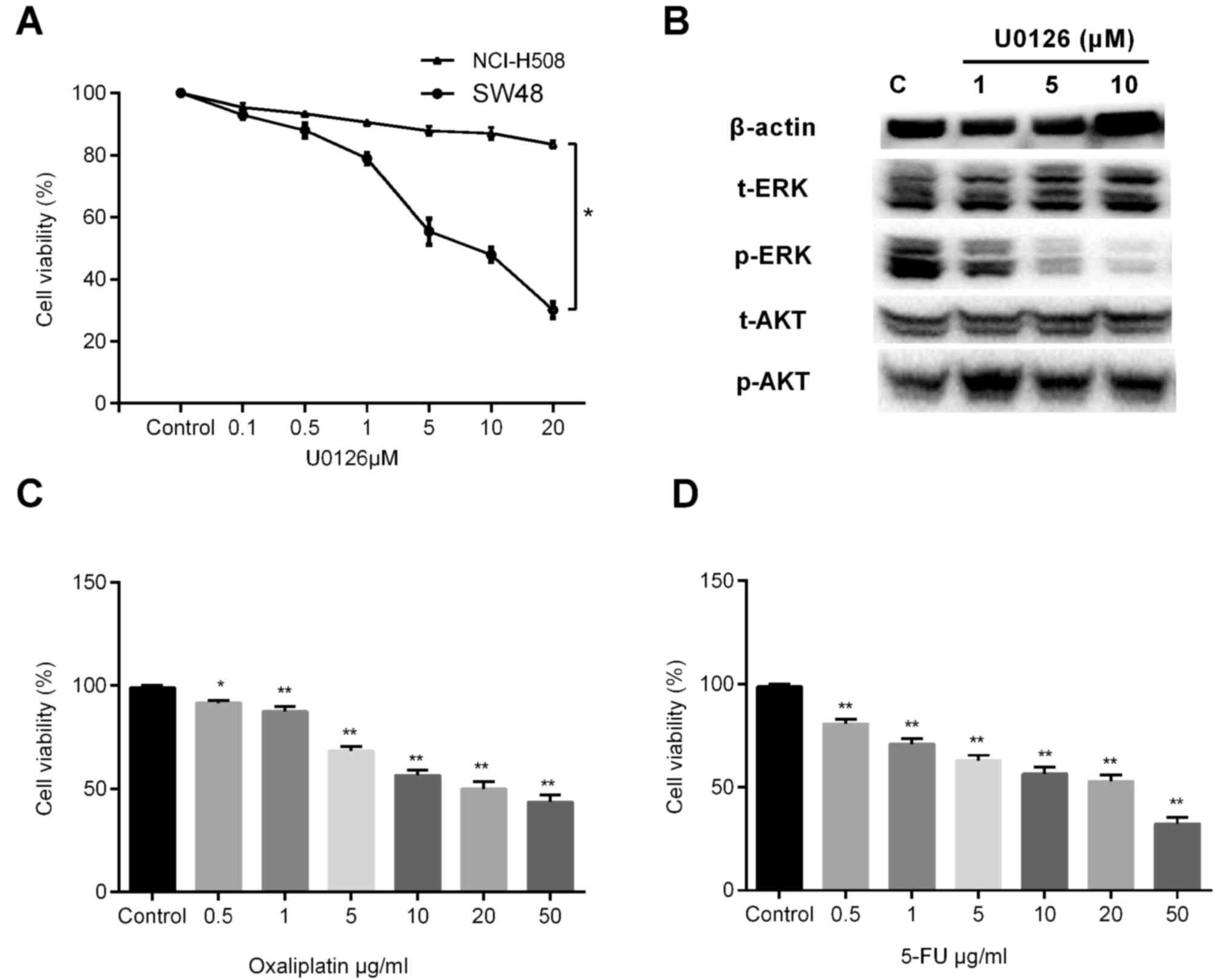 | Figure 1.Effects of U0126, oxaliplatin and
5-FU on SW48 cells. Cell viability was measured using CCK-8 assay
and is represented as the percentages of the untreated group value.
(A) CCK-8 was performed following the treatment of SW48 and
NCI-H508 cells with increasing concentrations of U0126 for 72 h.
There was a statistical difference between the two groups
(*P<0.05). (B) After 72 h of U0126 exposure, the cells were
lysed and subjected to western blot analysis with relevant
antibodies. Cell viability of cells treated with a concentration
gradient of (C) oxaliplatin (0.5, 1, 5, 10, 20 and 50 µg/ml) and
(D) 5-FU (0.5, 1, 5, 10, 20 and 50 µg/ml) for 3 days was assessed
by CCK-8 assay. Values are expressed as the mean ± standard
deviation of three individual measurements. *P<0.05 and
**P<0.01, vs. the untreated control group. 5-FU, 5-fluorouracil;
CCK-8, Cell Counting Kit-8; ERK, extracellular signal-regulated
kinase; t-, total; p-, phosphorylated. |
Combined effect of MEK1 inhibitor with
oxaliplatin and 5-FU
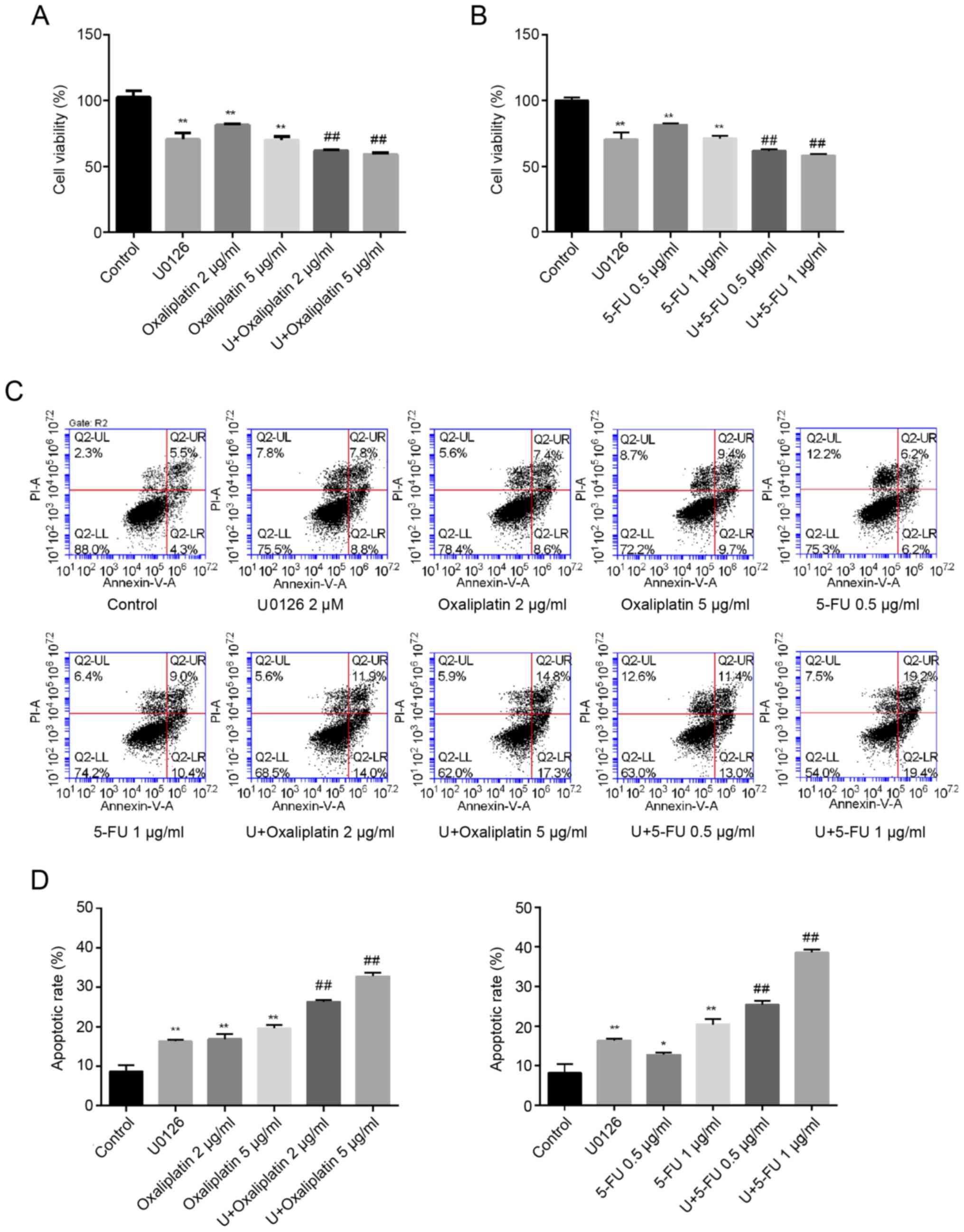
Compared with the control group (100%), the cell
viability after stimulation with 2 and 5 µg/ml oxaliplatin
decreased to 81.43±0.95 and 70.03±2.61%, respectively. However, the
combination of U0126 (2 µM) with 2 or 5 µg/ml oxaliplatin
significantly reduced cellular proliferation to 62.07±0.65 and
59.17±1.16%, respectively (Fig.
2A). Similarly, the cytotoxic effect in cells co-treated with
U0126 and 5-FU (0.5 and 1 µg/ml) was increased compared with that
in cells treated with either U0126 or 5-FU alone (Fig. 2B). Furthermore, the CI values,
shown in Table III, were both
<1.0 for combined treatment with U0126 and oxaliplatin, and
combined treatment with U0126 and 5-FU, indicating synergism
between the MEK inhibitor and two drugs.
| Table III.Combination index (CI) values for
combination of U0126 with oxaliplatin or 5-FU.a |
Table III.
Combination index (CI) values for
combination of U0126 with oxaliplatin or 5-FU.a
| Drug 1 | Drug 2 | CI |
|---|
| U0126 | Oxaliplatin 2
µg/ml | 0.72±0.03 |
|
| Oxaliplatin 2
µg/ml | 0.89±0.07 |
| U0126 | 5-FU 0.5 µg/ml | 0.77±0.06 |
|
| 5-FU 1 µg/ml | 0.86±0.02 |
The results of the apoptosis assay (Fig. 2C and D) were similar to those of
the CCK-8 experiment. Compared with the control group (8.70±1.56%),
single treatment with U0126, 2 or 5 µg/ml oxaliplatin, and 0.5 or 1
µg/ml 5-FU induced significant cell apoptosis (16.30±0.42,
16.90±1.27, 19.65±0.78, 12.95±0.78 and 20.40±1.41%, respectively).
However, the combination of U0126 (2 µM) and oxaliplatin (2 or 5
µg/ml) significantly enhanced cell apoptosis (26.30±0.57 and
32.75±0.92%, respectively). Similarly, treatment with 0.5 or 1
µg/ml 5-FU combined with U0126 markedly increased the cell
apoptosis rate (25.43±0.90 and 38.63±0.75%, respectively; Fig. 2C and D).
Combination of U0126 and oxaliplatin
or 5-FU triggers the formation of γH2AX foci
To further reveal potential mechanisms underlying
the effect of U0126 on oxaliplatin/5-FU therapeutic efficacy in
SW48 cells, the present study detected the effect of combination
therapy on DNA repair. According to the results of CCK-8 and flow
cytometry analysis, the doses of 2 µM U0126 combined with 5 µg/ml
oxaliplatin or 1 µg/ml 5-FU were selected for subsequent
investigations. Histone H2AX is a variant of the H2A histone family
that is involved in chromosomal stability (9). H2AX is phosphorylated at serine 139
when cells are induced by irradiation or cytotoxic drugs, and is a
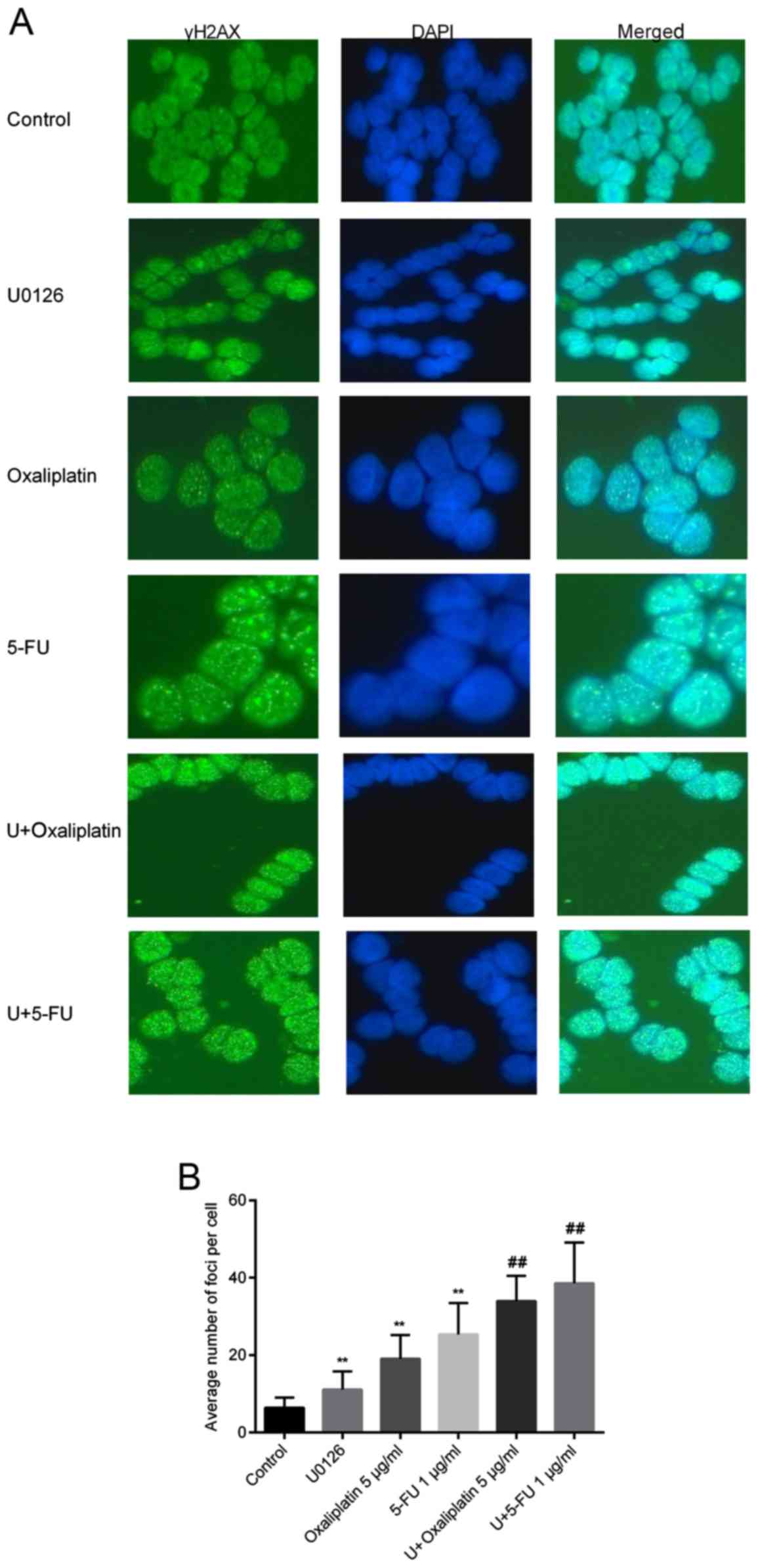
key protein for DNA repair and genomic stability (10). In the current study, γH2AX foci
were analyzed by immunofluorescence staining following single or
combined treatment with U0126, oxaliplatin or 5-FU. There were
significant differences between the cells treated with
U0126/oxaliplatin/5-FU alone and control cells, suggesting that all
three drugs were able to induce DSBs. Additionally, cells exposed
to oxaliplatin or 5-FU combined with U0126 exhibited significantly
more γH2AX foci compared with cells treated with monotherapy
(Fig. 3A). The number of foci in
100 cells of each sample was calculated, and the mean number of
foci per cell is shown in Fig. 3B.
Treatment with U0126, oxaliplatin and 5-FU induced comparable
amounts of γH2AX foci per cell (11.13±4.65 for U0126, 19.07±6.09
for oxaliplatin and 25.36±8.12 for 5-FU). Furthermore, the
induction of γH2AX was accelerated in SW48 cells treated with U0126
and oxaliplatin/5-FU together. The average number of foci following
stimulation with U0126 and oxaliplatin was 33.96±6.53, whereas the
number of foci was 38.58±10.53 following U0126 and 5-FU treatment
(Fig. 3B).
U0126 decreases ERCC1 expression
induced by oxaliplatin and enhances the inhibition of TYMS
expression when combined with 5-FU
Removal of adducts from genomic DNA is mediated by
the enzyme ERCC1, which serves an important role in the
rate-limiting step or regulation of nucleotide excision repair
(11). Increased expression of
ERCC1 caused by platinum in several cancer types has been
associated with improvement of DNA repair capacity and resistance
to platinum-based drugs (12,13).
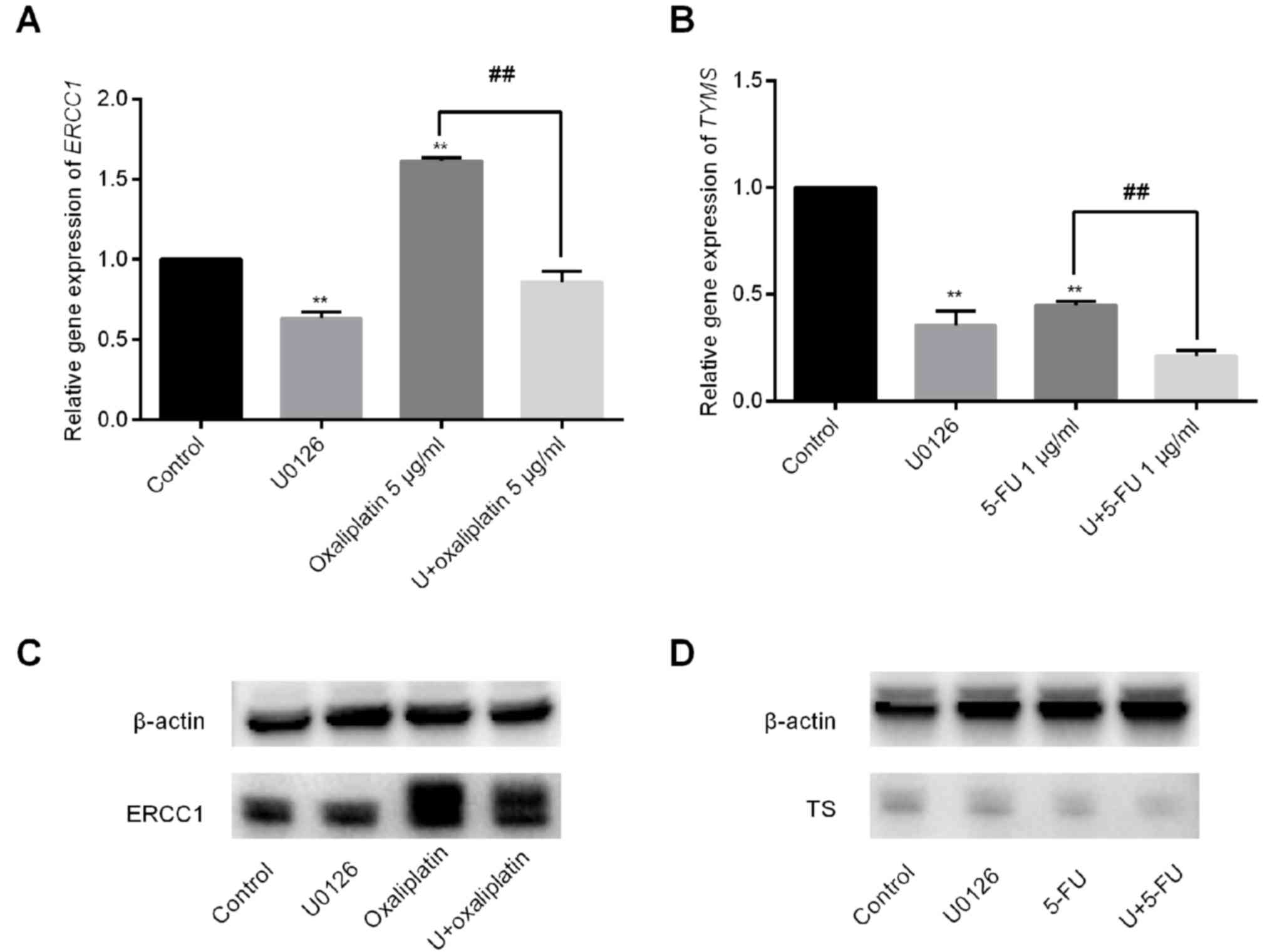
In the current study, it was observed that oxaliplatin exposure
increased ERCC1 expression by 1.62±0.02-fold. By contrast,
the combination of U0126 and oxaliplatin significantly reduced
ERCC1 mRNA levels to 0.86±0.06-fold (Fig. 4A). U0126 and 5-FU treatment alone
inhibited TYMS expression to 0.36±0.07-fold and
0.45±0.02-fold, respectively. Furthermore, the combination of U0126
and 5-FU decreased TYMS expression by 0.21±0.03-fold
(Fig. 4B). The results of protein
expression were consistent with the mRNA expression (Fig. 4C and D).
Discussion
Individualized therapy facilitates the selection of
the most suitable drug therapy for each patient according to
differences in the gene composition or alterations in expression
levels. Molecular targeted drugs selectively destroy tumor cells
with specific mutated genes, leading to their death, without
damaging the cells of the surrounding normal tissue (14). Selection of the appropriate
molecular targeted drug subsequent to relevant gene testing can
result in accurate and timely individualized treatment for
patients.
In 2014, the United States Food and Drug
Administration (https://www.fda.gov/) approved the
combination of the MEK inhibitor trametinib with the BRAF inhibitor
dabrafenib to treat patients with inoperable or metastatic melanoma
with BRAF V600E or V600K mutation. MEK1 mutations are
present in 1.5% of CRCs and the majority of mutations cause
constitutive activation of this kinase (15). A comprehensive study of MEK1
somatic mutations in lung adenocarcinoma revealed that
overexpression of MEK1 proteins with mutations in F53L, Q56P and
K57N causes phosphorylation of ERK and increased colony formation,
which may be inhibited by a MEK1/2 inhibitor (16). Transfection with MEK1 expression
vectors illustrated that mutations in this gene, including Q56P and
S72G, induced the phosphorylation of ERK1/2 and had a transforming
potential, enhancing the tumorigenicity. It was also observed that
use of a MEK inhibitor decreased the phosphorylation of ERK1/2 and
induced apoptosis of cell lines with MEK1 mutations
(17). In the present study, the
mutation rate of MEK1 was observed to be 1.67%. MEK
inhibition by U0126 significantly decreased the growth of SW48
cells that harbored a MEK1 Q56P mutation, although the
effect on the growth of NCI-H508 cells with MEK1 wild-type
was not marked. The results, to a certain extent, suggested that
CRC patients with such oncogenic MEK1 mutations may be
suitable for targeted therapy with MEK inhibitors.
Studies have reported that activation of MEK/ERK
signaling is associated with increased resistance to
fluoropyrimidines in breast and hepatocellular carcinoma (18,19).
In recent years, chemotherapy regimens consisting of 5-FU in
combination with oxaliplatin or irinotecan have served as the
first-line options for treatment of metastatic CRC. However, 5-FU
and oxaliplatin therapy exhibits problems with chemosensitivity
(20). A MEK1/2 inhibitor,
GSK1120212, exhibited an additive effect in combination with 5-FU
or oxaliplatin, and a synergistic effect in combination with SN-38
in HT-29 cells (21). GSK1120212
and other retinoblastoma gene-reactivating agents have been
reported to reduce thymidylate synthase expression and enhance the
efficacy of 5-FU in BRAF-mutated CRC (22). Furthermore, MEK/ERK pathway
inhibition enhanced the sensitivity to 5-FU in KRAS-mutated
and murine colorectal tumor xenograft models (23–25).
Consistent with the findings of previous studies, the current study
demonstrated that the MEK1 inhibitor U0126 increased sensitivity to
5-FU and oxaliplatin treatment in MEK1-mutated CRC. The
purpose of the present study was to provide experimental evidence
for the combination of a MEK1 inhibitor and chemotherapeutic agents
in patients with CRC harboring mutations in MEK1.
γH2AX foci were also detected by immunofluorescence
staining to further investigate the mechanism of action of the
investigated drugs. γH2AX is a topic of primary research interest
in the study of DNA damage stress responses. In 1998, Rogakou et
al (26) reported that
irradiation and other treatments may cause rapid phosphorylation of
Ser residues in H2XA, termed γH2AX. γH2AX foci may be used to
estimate the kinetics of DSBs rejoining and has become the gold
standard for the detection of DSBs. It has further been revealed
that there is no cell specificity for the formation of γH2AX foci,
and that induced DSBs are accompanied by H2AX phosphorylation and
clustering (27). In the present
study, SW48 cells treated with U0126, oxaliplatin or 5-FU alone
exhibited an increased number of γH2AX foci compared with the
control cells. Combination treatment of U0126 with produced more
γH2AX foci as compared with the single drug treatments, which may
be associated with the induction of DSBs. Phosphorylation of H2AX
indicated the presence of a large number of DSBs and slow repair in
SW48 cells exposed to combination therapy.
The present study demonstrated that combination
treatment with the MEK inhibitor and oxaliplatin/5-FU increased DNA
damage. Therefore, the expression levels of ERCC1 and TYMS, two
critical enzymes involved in the nucleotide excision repair
pathway, were detected. High expression of ERCC1 is associated with
resistance to platinum-based chemotherapy (28). In addition, TYMS is considered an
important predictor of 5-FU sensitivity (29) and it is important for maintaining
the dTMP pool for DNA synthesis and repair. TYMS inhibitors,
including fluorinated pyrimidine derivatives, are capable of
inhibiting the activity of TYMS; thus, TYMS expression is
associated with in vivo chemosensitivity to such inhibitors.
Improved efficacy of 5-FU may be achieved by increasing and
prolonging thymidylate synthase inhibition (30). The present study results confirmed
that oxaliplatin increased ERCC1 expression, while 5-FU
inhibited TYMS expression. Furthermore, U0126 decreased
ERCC1 expression induced by oxaliplatin and enhanced the
inhibition of TYMS expression when combined with 5-FU,
indicating that the inhibition of ERCC1 and TYMS expression levels
by U0126 may contribute to increased sensitivity to
oxaliplatin/5-FU therapy and severe DNA damage.
However, the present study has certain limitations.
Only one cell line was used to study the combination effect of MEK1
inhibitor and chemotherapeutic agents in vitro. Further
investigations are required to examine the detailed underlying
mechanisms.
In conclusion, a MEK1 inhibitor may be an effective
candidate for use with oxaliplatin and 5-FU, particularly for
MEK1-mutated cases of CRC. However, the future clinical
utility of MEK1 inhibitors in combination with chemotherapeutic
agents is limited by a lack of in vivo experimental results.
In the future, further investigations should be conducted to
explore the synergistic effect of MEK inhibitors with
oxaliplatin/5-FU in vivo.
Acknowledgements
Not applicable.
Funding
This study was supported by the Program of the
Department of Health in Jiangsu Province (grant no. Z201602) and
the Science Foundation of Jiangsu Province (grant no.
BE2016795).
Availability of data and material
The materials described in the manuscript, including
all relevant raw data, will be freely available to any researcher
wishing to use them for non-commercial purposes, without breaching
participant confidentiality. The datasets used and analyzed during
the current study are available from the corresponding author on
reasonable request.
Authors' contributions
CJ and HL wrote the manuscript and designed the
study. YD and HC performed the PCR and western blotting
experiments. SL, RM and ZW performed immunofluorescence staining
and flow cytometry FCM. JW and JF contributed to the design of the
study. All authors read and approved the final version of the
manuscript.
Ethics approval and consent
All patients who participated in the study provided
signed informed consent. The research using human tissue received
approval from Jiangsu Institute of Cancer Research Ethics
Committee.
Patient consent for publication
All patients participated in the study signed
informed consent.
Competing interests
The authors declare that they have no competing
interests.
References
|
1
|
Siegel RL, Miller KD and Jemal A: Cancer
statistics, 2016. CA Cancer J Clini. 66:7–30. 2016. View Article : Google Scholar
|
|
2
|
Meyerhardt JA and Mayer RJ: Systemic
therapy for colorectal cancer. N Engl J Med. 352:476–487. 2005.
View Article : Google Scholar : PubMed/NCBI
|
|
3
|
Chiu SJ, Lee YJ, Hsu TS and Chen WS:
Oxaliplatin-induced gamma-H2AX activation via both p53-dependent
and -independent pathways but is not associated with cell cycle
arrest in human colorectal cancer cells. Chem Biol Interact.
182:173–182. 2009. View Article : Google Scholar : PubMed/NCBI
|
|
4
|
Fremin C and Meloche S: From basic
research to clinical development of MEK1/2 inhibitors for cancer
therapy. J Hematol Oncol. 3:82010. View Article : Google Scholar : PubMed/NCBI
|
|
5
|
Favata MF, Horiuchi KY, Manos EJ, Daulerio
AJ, Stradley DA, Feeser WS, Van Dyk DE, Pitts WJ, Earl RA, Hobbs F,
et al: Identification of a novel inhibitor of mitogen-activated
protein kinase kinase. J Biol Chem. 273:18623–18632. 1998.
View Article : Google Scholar : PubMed/NCBI
|
|
6
|
Stepanenko A, Andreieva S, Korets K,
Mykytenko D, Huleyuk N, Vassetzky Y and Kavsan V: Step-wise and
punctuated genome evolution drive phenotype changes of tumor cells.
Mutat Res. 771:56–69. 2015. View Article : Google Scholar : PubMed/NCBI
|
|
7
|
Rusch VW, Rice TW, Crowley J, Blackstone
EH, Rami-Porta R and Goldstraw P: The seventh edition of the
American joint committee on cancer/international union against
cancer staging manuals: The new era of data-driven revisions. J
Thorac Cardiovasc Surg. 139:819–821. 2010. View Article : Google Scholar : PubMed/NCBI
|
|
8
|
Livak KJ and Schmittgen TD: Analysis of
relative gene expression data using real-time quantitative PCR and
the 2(-Delta Delta C(T)) method. Methods. 25:402–408. 2001.
View Article : Google Scholar : PubMed/NCBI
|
|
9
|
Motoyama N and Naka K: DNA damage tumor
suppressor genes and genomic instability. Curr Opin Genet Dev.
14:11–16. 2004. View Article : Google Scholar : PubMed/NCBI
|
|
10
|
Rogakou EP, Nieves-Neira W, Boon C,
Pommier Y and Bonner WM: Initiation of DNA fragmentation during
apoptosis induces phosphorylation of H2AX histone at serine 139. J
BiolChem. 275:9390–9395. 2000.
|
|
11
|
Niedernhofer LJ, Odijk H, Budzowska M, van
Drunen E, Maas A, Theil AF, de Wit J, Jaspers NG, Beverloo HB,
Hoeijmakers JH and Kanaar R: The structure-specific endonuclease
Ercc1-Xpf is required to resolve DNA interstrand cross-link-induced
double-strand breaks. Mol Cell Biol. 24:5776–5787. 2004. View Article : Google Scholar : PubMed/NCBI
|
|
12
|
Ronchi CL, Sbiera S, Kraus L, Wortmann S,
Johanssen S, Adam P, Willenberg HS, Hahner S, Allolio B and
Fassnacht M: Expression of excision repair cross complementing
group 1 and prognosis in adrenocortical carcinoma patients treated
with platinum-based chemotherapy. Endocr-Relat Cancer. 16:907–918.
2009. View Article : Google Scholar : PubMed/NCBI
|
|
13
|
Lu M, Gao J, Wang XC and Shen L:
Expressions of thymidylate synthase, thymidine phosphorylase, class
III beta-tubulin, and excision repair cross-complementing group
1predict response in advanced gastric cancer patients receiving
capecitabine plus paclitaxel or cisplatin. Chin J Cancer Res.
23:288–294. 2011. View Article : Google Scholar : PubMed/NCBI
|
|
14
|
Grady WM and Pritchard CC: Molecular
alterations and biomarkers in colorectal cancer. Toxicol Pathol.
42:124–139. 2014. View Article : Google Scholar : PubMed/NCBI
|
|
15
|
Cancer Genome Atlas Network: Comprehensive
molecular characterization of human colon and rectal cancer.
Nature. 487:330–337. 2012. View Article : Google Scholar : PubMed/NCBI
|
|
16
|
Arcila ME, Drilon A, Sylvester BE, Lovly
CM, Borsu L, Reva B, Kris MG, Solit DB and Ladanyi M: MAP2K1 (MEK1)
mutations define a distinct subset of lung adenocarcinoma
associated with smoking. Clin Cancer Res. 21:1935–1943. 2015.
View Article : Google Scholar : PubMed/NCBI
|
|
17
|
Sogabe S, Togashi Y, Kato H, Kogita A,
Mizukami T, Sakamoto Y, Banno E, Terashima M, Hayashi H, de Velasco
MA, et al: MEK inhibitor for gastric cancer with MEK1 gene
mutations. Mol Cancer Ther. 13:3098–3106. 2014. View Article : Google Scholar : PubMed/NCBI
|
|
18
|
Jin W, Wu L, Liang K, Liu B, Lu Y and Fan
Z: Roles of the PI-3K and MEK pathways in Ras-mediated
chemoresistance in breast cancer cells. Br J Cancer. 89:185–191.
2003. View Article : Google Scholar : PubMed/NCBI
|
|
19
|
Yan Y, Li J, Han J, Hou N, Song Y and Dong
L: Chlorogenic acid enhances the effects of 5-fluorouracil in human
hepatocellular carcinoma cells through the inhibition of
extracellular signal-regulated kinases. Anticancer Drugs.
26:540–546. 2015. View Article : Google Scholar : PubMed/NCBI
|
|
20
|
Yu X, Li Z, Yu J, Chan MT and Wu WK:
MicroRNAs predict and modulate responses to chemotherapy in
colorectal cancer. Cell Prolif. 48:503–510. 2015. View Article : Google Scholar : PubMed/NCBI
|
|
21
|
Yamaguchi T, Kakefuda R, Tajima N, Sowa Y
and Sakai T: Antitumor activities of JTP-74057 (GSK1120212), a
novel MEK1/2 inhibitor, on colorectal cancer cell lines in vitro
and in vivo. Int J Oncol. 39:23–31. 2011.PubMed/NCBI
|
|
22
|
Watanabe M, Sowa Y, Yogosawa M and Sakai
T: Novel MEK inhibitor trametinib and other retinoblastoma gene
(RB)-reactivating agents enhance efficacy of 5-fluorouracil on
human colon cancer cells. Cancer Sci. 104:687–693. 2013. View Article : Google Scholar : PubMed/NCBI
|
|
23
|
Pereira DM, Simões AE, Gomes SE, Castro
RE, Carvalho T, Rodrigues CM and Borralho PM: MEK5/ERK5 signaling
inhibition increases colon cancer cell sensitivity to
5-fluorouracil through a p53-dependent mechanism. Oncotarget.
7:34322–34340. 2016. View Article : Google Scholar : PubMed/NCBI
|
|
24
|
Urick ME, Chung EJ, Shield WP III, Gerber
N, White A, Sowers A, Thetford A, Camphausen K, Mitchell J and
Citrin DE: Enhancement of 5-fluorouracil-induced in vitro and in
vivo radiosensitization with MEK inhibition. Clin Cancer Res.
17:5038–5047. 2011. View Article : Google Scholar : PubMed/NCBI
|
|
25
|
Gong J, Chen Y, Yang L, Pillai R,
Shirasawa S and Fakih M: MEK162 enhances antitumor activity of
5-fluorouracil and trifluridine in KRAS-mutated human colorectal
cancer cell lines. Anticancer Res. 37:2831–2838. 2017.PubMed/NCBI
|
|
26
|
Rogakou EP, Pilch DR, Orr AH, Ivanova VS
and Bonner WM: DNA double-stranded breaks induce histone H2AX
phosphorylation on serine 139. J Biol Chem. 273:5858–5868. 1998.
View Article : Google Scholar : PubMed/NCBI
|
|
27
|
MacPhail SH, Banath JP, Yu TY, Chu EH,
Lambur H and Olive PL: Expression of phosphorylated histone H2AX in
cultured cell lines following exposure to X-rays. Int J Radiat
Biol. 79:351–358. 2003. View Article : Google Scholar : PubMed/NCBI
|
|
28
|
Li J, Li ZN, Yu LC, Bao QL, Wu JR, Shi SB
and Li XQ: Association of expression of MRP1, BCRP, LRP and ERCC1
with outcome of patients with locally advanced non-small cell lung
cancer who received neoadjuvant chemotherapy. Lung Cancer.
69:116–122. 2010. View Article : Google Scholar : PubMed/NCBI
|
|
29
|
Wakasa K, Kawabata R, Nakao S, Hattori H,
Taguchi K, Uchida J, Yamanaka T, Maehara Y, Fukushima M and Oda S:
Dynamic modulation of thymidylate synthase gene expression and
fluorouracil sensitivity in human colorectal cancer cells. PLoS
One. 10:e01230762015. View Article : Google Scholar : PubMed/NCBI
|
|
30
|
Peters GJ, Backus HH, Freemantle S, van
Triest B, Codacci-Pisanelli G, van der Wilt CL, Smid K, Lunec J,
Calvert AH, Marsh S, et al: Induction of thymidylate synthase as a
5-fluorouracil resistance mechanism. Biochimica Biophysica Acta.
1587:194–205. 2002. View Article : Google Scholar
|