Introduction
Diabetes mellitus (DM) is a serious public health
concern worldwide due to its gradually increasing prevalence
(1). Diabetes is also an
independent risk factor for many cardiovascular diseases, leading
to the increased incidence of vascular remodeling among vascular
diabetic complications. The pathogeneses of vascular remodeling
induced by vascular smooth muscle cell (VSMC) dysfunction involve
excessive inflammation and overactivation of proliferation,
accompanied by increased amounts of extracellular matrix secretion,
ultimately contributing to the thickening of arterial walls
(2–5). Although vascular remodeling in
diabetes is known to be associated with the combination of these
abnormal pathological processes, the specific mechanism is still
unknown.
The ubiquitin-proteasome system (UPS), the major
mechanism by which proteins are degraded in cells, can degrade
80–90% of ubiquitinated proteins related to almost all biological
activities in the organism, such as apoptosis, inflammation and
cell cycle progression (6,7). The UPS has been considered a
potential drug target in diabetes and other cardiovascular diseases
(8,9). In the entire UPS, the E3 ubiquitin
ligase is the most critical, as it identifies and promotes the
degradation of substrate proteins by determining the time and
specificity of ubiquitination (7,10).
Thus, targeting a proper E3 ubiquitin ligase that participates in
the regulation of crucial proteins in vascular remodeling could
represent a new strategy for the treatment of diabetic vascular
complications.
Ring finger protein 10 (RNF10), located on the long
arm of chromosome 12, is a member of the RING finger family of E3
ubiquitin ligases, with the function of recruiting ubiquitin ligase
E2 and substrate proteins (11,12).
A clinical study demonstrated that RNF10 mRNA expression is
significantly enhanced in obese patients and greatly increases the
risk of diabetes (13). In our
previous study, overexpression of RNF10 was shown to significantly
prevent neointimal formation by promoting apoptosis reactions and
inhibiting Bcl-2 protein activation in metabolic syndrome (14). The aim of the present study was to
investigate the effect of RNF10 on vascular remodeling and the main
pathological processes in diabetic rats after balloon injury,
including inflammation, proliferation and apoptosis responses.
Materials and methods
Animal models
All animal experiments were approved by the Ethics
Committee of Chongqing Medical University (Chongqing, China) and
were performed according to the ARRIVE guidelines and the UK
Animals (Science Procedures) Act, 1986 (https://assets.publishing.service.gov.uk/government/uploads/system/uploads/attachment_data/file/116843/aspa-draft-guidance.pdf),
and its associated guidelines. All experimental male Sprague-Dawley
rats (4 weeks of age) were purchased from the Experimental Animal
Centre of Chongqing Medical University (Chongqing, China). After
one week of acclimation, the rats were randomly divided into
control (n=12) and DM (n=60) groups. For a total of 12 weeks, the
control group was fed a standard diet that included 13% fat, 61%
carbohydrates, and 26% protein, while the DM group was fed a
high-fat diet with 60% fat, 21% carbohydrates, and 19% protein. The
animal experiment was performed in a temperature-controlled room
maintained at 22±2°C and a 12 h light/dark cycle was maintained.
Groups of 5 rats were housed in cages with free access to food and
water.
The diabetic model was induced by feeding rats a
high-fat diet resulting in hyperglycaemia and insulin resistance.
All SD rats were fasted overnight (16–18 h) after receiving a
standard diet or a high-fat diet for 12 weeks. Then, blood samples
were collected from the retro-orbital plexus into a capillary tube
to measure the diabetic indices. Plasma was frozen and stored at
−80°C after being separated by centrifugation at 1,000 × g for 30
min at −4°C. Fasting blood glucose was quantified using a portable
blood glucose meter (Sinocare Inc., Changsha, Hunan, China). An
intraperitoneal glucose tolerance test (IPGTT) was used to detect
insulin resistance and impaired glucose tolerance.
Carotid artery balloon injury (BI) and
adenovirus (Ad) infection
Within the DM group on a high-fat (HF) diet, all
rats were classified into 5 groups based on whether the balloon
injury operation and adenovirus infection occurred. Eventually, all
the SD rats were divided into the following 6 groups: i) the
control group, ii) the DM+Sham group, iii) the DM+BI group, iv) the
DM+BI+Ad-GFP group, v) the DM+BI+Ad-RNF10 group, and vi) the
DM+BI+Ad-shRNF10 group.
Balloon injury was performed and measured as
described previously (15); the
balloon catheter (Medtronic Inc., Minneapolis, MN, USA) was
introduced from the left external carotid artery into the thoracic
aorta. The left common carotid artery was damaged by repeatedly
pushing and pulling the inflated balloon three times. After washing
the damaged artery with phosphate-buffered saline (PBS), 50 µl of
adenovirus encoding RNF10, shRNF10 or GFP (2×10¹⁰ pfu/ml, Obio
Technology Corp. Ltd., Shanghai, China) was injected into the
injured artery to incubate for 30 min. After 14 days, the rats were
sacrificed by high-dose injection of sodium pentobarbital (≥90
mg/kg), and the carotid arteries were harvested.
Histological and morphometric
analysis
To examine the establishment of the vascular
remodeling model, the obtained carotid tissue sections were stained
using haematoxylin and eosin (H&E) as previously described
(16). The carotid arteries of the
rats were fixed in 4% paraformaldehyde for 24 h and then embedded
in paraffin. Next, the sections were prepared for haematoxylin
staining at room temperature for 15 min and eosin staining at room
temperature for 5 min. Finally, the sections were stained with
picrosirius red for 30 min at room temperature. Photomicrographs
(×100 magnification) were captured with a routine light microscope
(cat. no. 4.10.00, Nikon Co., Ltd., Tokyo, Japan).
Terminal deoxynucleotidyl transferase
dUTP nick end labeling (TUNEL) assay
To determine the proportion of apoptotic carotid
artery cells in the diabetic rats, a TUNEL assay was performed
using an In Situ Cell Death Detection Kit-POD (Roche, Basel,
Switzerland). First, carotid tissue sections were sequentially
incubated with proteinase K for 30 min. Then, these sections were
incubated with membrane disruption solution (Service Bio Inc.,
Woburn, MA, USA) for 20 min at room temperature, followed by
incubation with a mixture of reagent 1 (TdT) and reagent 2 (dUTP)
from the TUNEL kit for 2 h. Next, the sections were incubated with
reagent 3 (converter-POD) for 30 min after incubation with hydrogen
peroxide and ethanol for 15 min. Finally, the sections were mounted
and observed under a routine light microscope (at magnification,
×400, cat. no. 4.10.00, Nikon Co., Ltd., Tokyo, Japan) after
3,3′-diaminobenzidine (DAB) (Dako; Agilent Technologies, Inc.,
Santa Clara, CA, USA) was added. The cells with brown and yellow
nuclei were TUNEL-positive cells (17). The apoptosis index = number of
apoptosis-positive cells × total number of intimal cells.
Quantitative polymerase chain reaction
(qPCR)
Total RNA was isolated from the carotid arteries of
rats with TRIzol reagent (Takara Bio Inc., Kusatsu, Japan), and up
to 1 µg of total RNA was reverse transcribed into cDNA following
the manufacturer's instructions (Takara Bio Inc.). The reaction
protocol was: Polymerase activation for 10 min at 95°C and then 40
amplification cycles of 60 sec at 60°C. The cycle threshold values
were normalized to the level of β-actin, and the relative gene
expression was measured by the 2−ΔΔCq method (18). The primers included those for
β-actin (5′-CCCATCTATGAGGGTTACGC-3′ and
5′-TTTAATGTCACGCACGATTTC-3′), RNF10 (5′-ATTTTAGCAACCAGTCCCGTCG-3′
and 5′-CCTCATCCCGTCTTCCACCAT-3′), TNF-α
(5′-CTACTGAACTTCGGGGTGATC-3′ and 5′-GGTCTGGGCCATAGAACTGA-3′) and
MCP-1 (5′-GCTGACCCCAAGAAGGAATG-3′ and
5′-GTGCTTGAGGTGGTTGTGGA-3′).
Western blot analysis
The common carotid total protein was extracted using
10 µl/µg RIPA lysis buffer (Beyotime Institute of Biotechnology,
Shanghai, China) and quantified via a BCA Protein Assay Kit
(Beyotime Institute of Biotechnology). Protein (40 µg) was
separated on 10–12% SDS-polyacrylamide gels (Beyotime Institute of
Biotechnology) and transferred to PVDF membranes. The membranes
were blocked in 5% non-fat dry milk for approximately 2 h and
incubated overnight at 4°C with primary antibodies, including
antibodies for Bcl-2 (1:2,000; cat. no. 12789; ProteinTech, Wuhan,
Hubei, China), Bax (1:2,000; cat. no. 50599; ProteinTech), NF-κB
(1:1,000; cat. no. 14220; ProteinTech), Cyclin D1 (1:1,000; cat.
no. 60186; ProteinTech), CDK4 (1:1,000; cat. no. 11026;
ProteinTech), RNF10 (1:500; cat. no. abs127972; Absin Bioscience
Inc., Shanghai, China) and β-actin (1:5,000; cat. no. 20536;
ProteinTech). Enhanced chemiluminescence development methods
(Advansta Inc., Menlo Park, CA, USA) were used to visualize the
immunoblotting bands (19). The
bands were analyzed with ImageLab 3.0 software (Bio-Rad
Laboratories, Inc., Hercules, CA, USA).
Statistical analysis
All the data are expressed as the means ± standard
deviation (SD), and statistical analysis was performed using SPSS
19.0 (IBM Corp., Armonk, NY, USA). One-way ANOVA and LSD was used
to analyse the differences among groups; P<0.05 was considered
to indicate statistical significance.
Results
Differentially expressed RNF10 affects
the neointimal apoptosis index in diabetic rats
To evaluate whether vascular remodeling was induced
by balloon injury, H&E staining was performed to detect
neointimal formation. In general, we observed that the
balloon-injured arteries had clearly increased intimal thickness
compared with that of the DM+Sham group arteries. Compared with
that in the Ad-GFP-treated group, the intimal thickness in the
Ad-RNF10 group was markedly reduced. However, the intimal thickness
was clearly increased in the Ad-shRNF10-treated group (Fig. 1A).
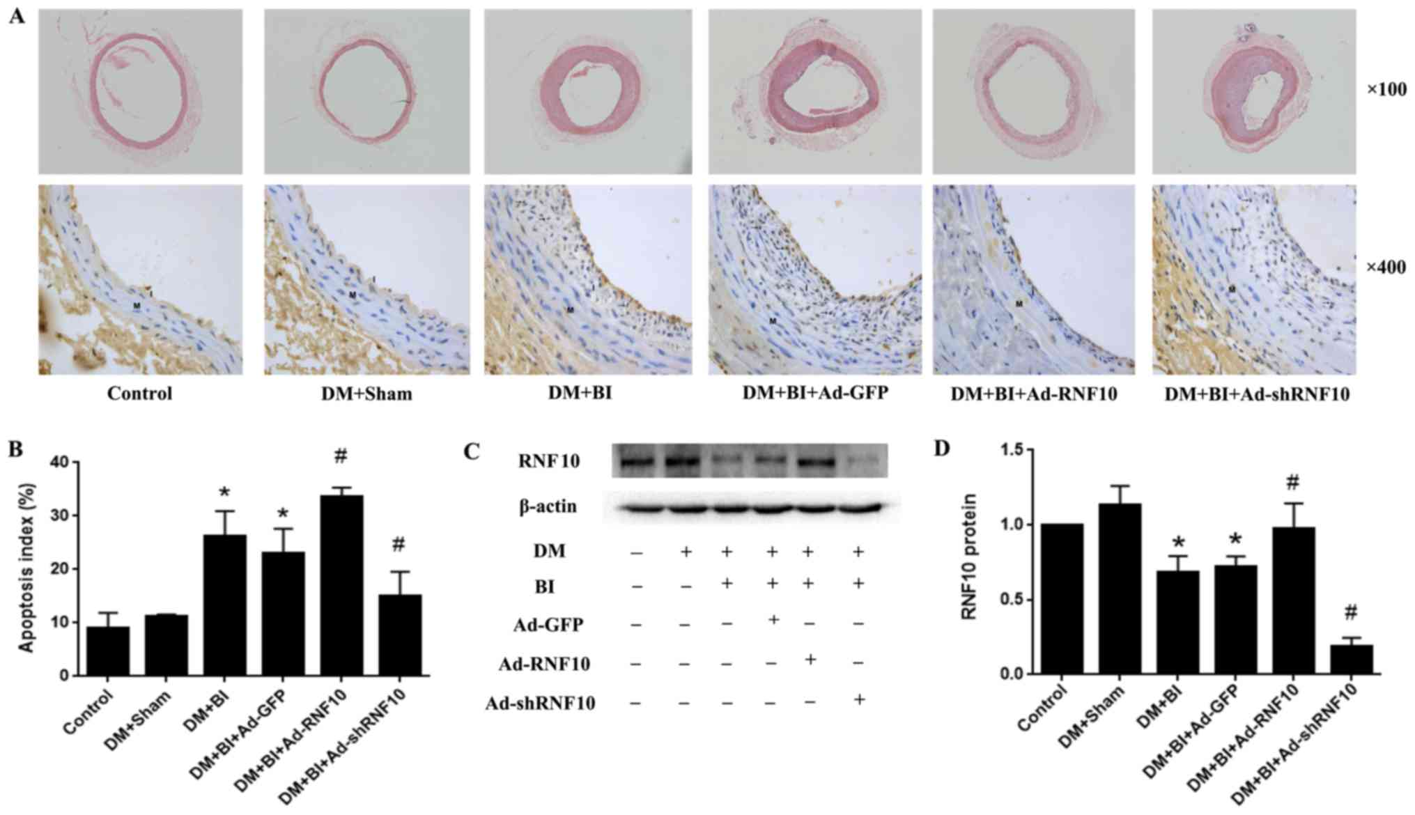 | Figure 1.Effects of RNF10 on apoptosis in the
carotid artery. (A) H&E staining (original magnification, ×100)
(top panels) and TUNEL staining (original magnification, ×400)
(bottom panels) on cross-sections of the carotid arteries. (B)
Quantification of the apoptosis index, defined as the ratio of
TUNEL-positive cell neointimal numbers to the total neointimal
numbers (n≥3). (C and D) Representative western blot analysis of
RNF10 protein and quantitative analysis of RNF10 protein level
(n≥3). *P<0.05 vs. the DM+Sham group; #P<0.05 vs. the
DM+BI+Ad-GFP group. DM, diabetes mellitus; RNF10, RING finger
protein 10; shRNF10, short hairpin RNF10; BI, balloon injury; Ad,
adenovirus; GFP, green fluorescent protein; TUNEL, terminal
deoxynucleotidyl transferase dUTP nick end labeling; H&E,
haematoxylin and eosin. |
To assess whether the balloon-injured arteries were
successfully transduced with the corresponding RNF10 gene, western
blotting was performed to detect RNF10 protein expression. Compared
with that of the arteries in the DM+Sham group, the RNF10 protein
expression of the balloon-injured arteries was significantly
decreased (P<0.001). Among the rats with balloon-injured
arteries that were infected with different adenoviruses, RNF10
protein expression was significantly increased in the group
receiving Ad-RNF10 (P<0.05) and was markedly decreased in the
Ad-shRNF10 group (P<0.001) (Fig. 1C
and D).
To examine whether RNF10 affected the apoptotic
response in the model of diabetic vascular remodeling, we used
TUNEL staining to determine the proportion of cells that were
apoptotic in the neointima of the carotid artery. Very few
apoptosis-positive cells were observed in the control group and the
DM+Sham group, but the apoptotic index was significantly higher in
the balloon-injured groups than in the DM+Sham groups (24.34±5.15
vs. 10.10±5.22%, P<0.001). Compared with that in the Ad-GFP
group, the proportion of apoptotic cells was significantly
increased in the Ad-RNF10 group (32.52±3.77 vs. 24.18±5.56%,
P<0.05), but the apoptosis index was clearly decreased in the
group infected with Ad-shRNF10 (14.59±4.29 vs. 24.18±5.56%,
P<0.01) (Fig. 1A and B).
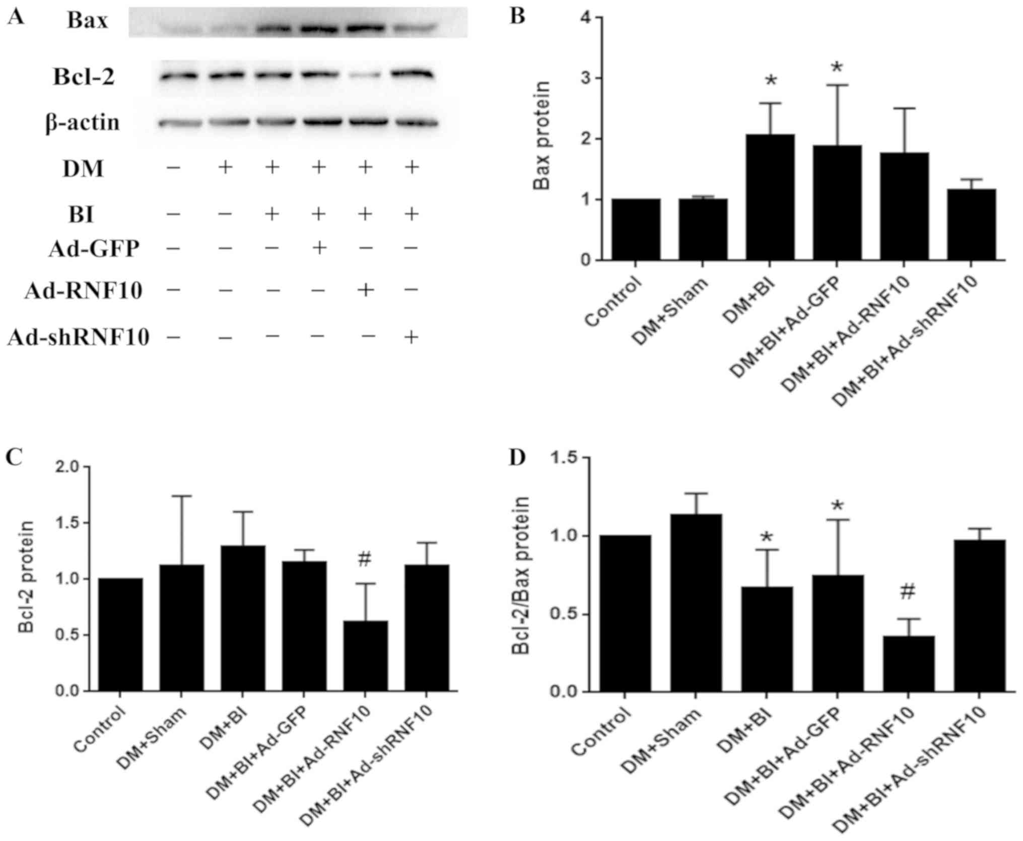
Overexpression of RNF10 attenuates
apoptosis by decreasing the expression of marker protein Bcl-2
To verify the expression of apoptotic proteins in
carotid vessels, western blotting was performed to detect the
levels of the Bax and Bcl-2 proteins. Bax protein expression was
significantly increased in the DM+BI group compared to the DM+Sham
group (P<0.05), but there were no significant differences due to
infection with Ad-GFP, Ad-RNF10 or Ad-shRNF10 (P>0.05) (Fig. 2A and B). Compared with that in the
arteries of the Ad-GFP-transduced rats, the Bcl-2 protein
expression in the arteries of the group receiving Ad-RNF10 group
was significantly reduced (P<0.05), while the expression was not
significantly changed in the group infected with Ad-shRNF10. In
addition, the ratio of Bcl-2 to Bax was significantly decreased in
the Ad-RNF10 group (P<0.05) and showed a trend towards a slight
increase in the group infected with Ad-shRNF10, but there was no
significant difference (P>0.05) (Fig. 2A, C and D).
Overexpression of RNF10 suppresses
VSMC inflammation levels by inhibiting the marker protein NF-κB in
diabetic rats
To determine whether the RNF10 gene was successfully
transduced into carotid arteries after balloon injury, qPCR was
used to detect RNF10 mRNA expression. Initially, the transcription
level of RNF10 in the DM +Sham group was higher than that in the
control group (P<0.001). After balloon injury, this elevation in
RNF10 mRNA was clearly reversed (P<0.001). Compared with that in
the Ad-GFP group, the expression of RNF10 mRNA in the group
receiving Ad-RNF10 was significantly enhanced (P<0.05), and the
expression in the group receiving Ad-shRNF10 was markedly reduced
(P<0.001) (Fig. 3A).
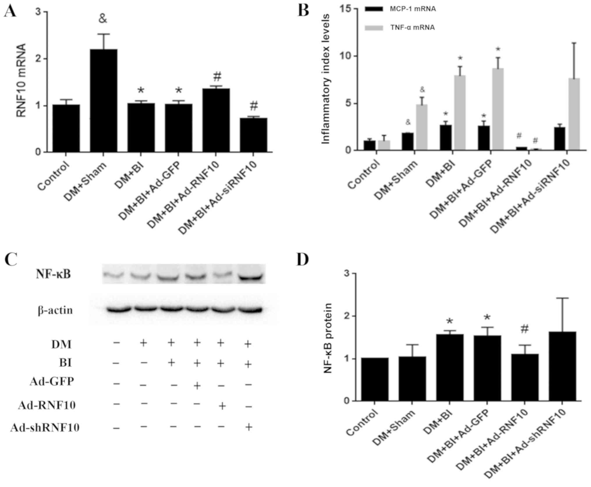 | Figure 3.Effects of RNF10 on the inflammatory
responses in carotid artery injury. (A) RNF10, (B) MCP-1 and TNF-α
mRNA expression levels were analyzed with qPCR (n≥3). (C)
Representative western blot of NF-κB protein. (D) Quantification of
NF-κB protein level as determined by western blot analysis (n≥3).
&P<0.05 vs. the corresponding control group; *P<0.05 vs.
the corresponding DM+Sham group; #P<0.05 vs. the corresponding
DM+BI+Ad-GFP group. DM, diabetes mellitus; RNF10, RING finger
protein 10; shRNF10, RNF10 short hairpin RNA; BI, balloon injury;
Ad, adenovirus; GFP, green fluorescent protein; qPCR, quantitative
polymerase chain reaction; TNF-α, tumor necrosis factor α; MCP-1,
monocyte chemotactic protein 1; NF-κB, nuclear factor-κ-gene
binding. |
To detect whether the RNF10 gene was involved in the
inflammatory response of vascular remodeling in diabetic rats, we
evaluated the mRNA levels of the inflammatory indicators MCP-1 and
TNF-α, and analysed the expression of the inflammatory marker
protein NF-κB. Compared with those in the control group, the levels
of MCP-1 and TNF-α mRNA in the DM+Sham group were markedly
increased (P<0.05). In addition, the expression of these
inflammatory indicators in the DM+BI group was higher than that in
the DM+Sham group (P<0.05). Importantly, the MCP-1 and TNF-α
transcription levels were decreased in the group receiving Ad-RNF10
compared with the group receiving Ad-GFP (P<0.001). Concerning
the inflammatory marker protein NF-κB, NF-κB levels were
significantly higher in groups with balloon injury than in the
DM+Sham group (P<0.05). Furthermore, balloon injured arteries
infected with Ad-RNF10 displayed a significant reduction in NF-κB
protein compared with carotid arteries infected with Ad-GFP
(P<0.05). However, the mRNA expression of the inflammatory
indicators MCP-1 and TNF-α mRNA, and the protein expression of
NF-κB were not significantly changed by Ad-shRNF10 infection
(P>0.05) (Fig. 3B-D).
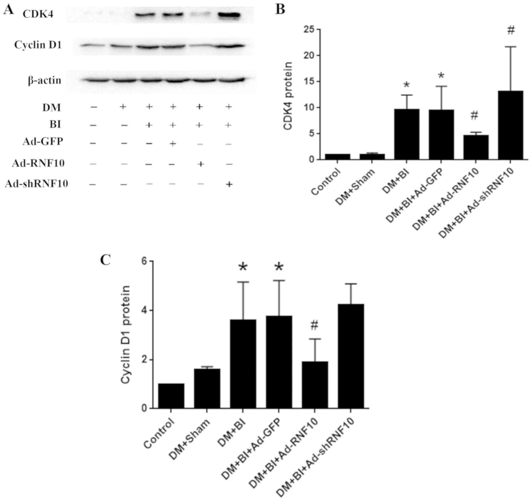
Differentially expressed RNF10
regulates vascular hyperproliferation in diabetic rats
To assess whether RNF10 regulated the
hyperproliferation of VSMCs in diabetic rats after balloon injury,
we detected the protein levels of cyclin D1 and CDK4 in the carotid
tissue. After balloon injury in DM rats, the protein levels of
cyclin D1 and CDK4 were markedly higher than those in the DM+Sham
group (P<0.05). In comparison to the Ad-GFP group, the Ad-RNF10
group showed significantly inhibited cyclin D1 protein expression
(P<0.05). While shRNF10 slightly promoted cyclin D1 expression,
there was no significant difference between the Ad-RNF10-treated
group and the Ad-GFP-treated group (P>0.05). In addition, CDK4
protein expression was significantly decreased in the Ad-RNF10
group and significantly increased in the Ad-shRNF10 group compared
with the Ad-GFP group (P<0.05) (Fig. 4A-C).
Discussion
In the present study, we found that overexpression
of RING finger protein 10 (RNF10) promoted an apoptotic reaction,
inhibited the inflammatory response and repressed vascular smooth
muscle cell (VSMC) hyperproliferation in carotid arteries, all of
which showed that RNF10 exerts a protective influence in diabetic
vascular remodeling. In addition, shRNF10 had an opposite impact on
hyperproliferation and apoptosis, further destroying arterial
function and structure in diabetes. Taken together, the results of
this study demonstrated that changes in RNF10 expression affected
vascular remodeling, indicating that the RNF10 gene plays a central
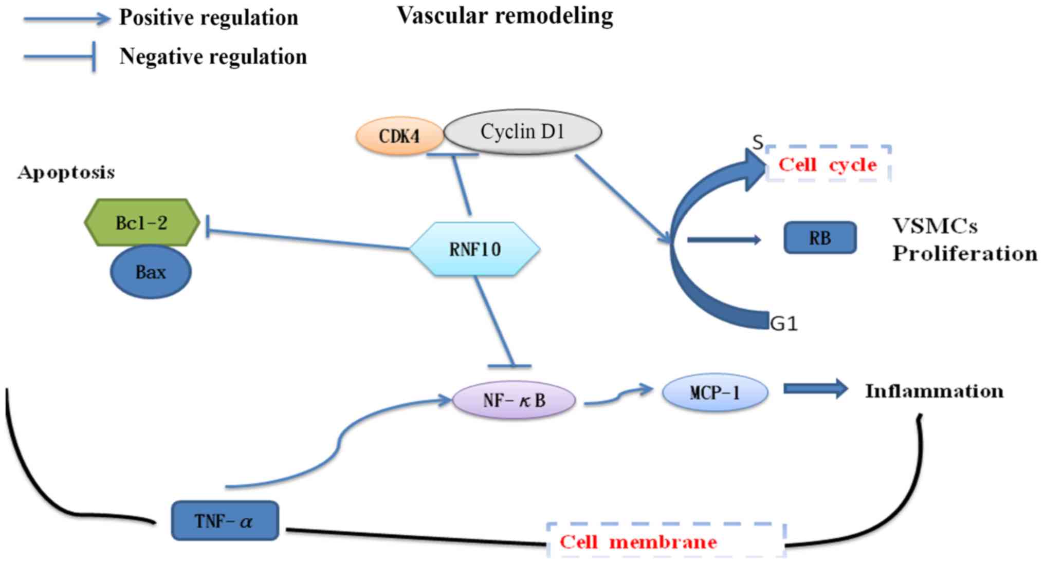
role in diabetic rats with vascular complications (Fig. 5).
A series of studies have revealed the relationship
between the ubiquitin-proteasome system (UPS) and vascular
remodeling, demonstrating that ubiquitin protease can inhibit
vascular intimal proliferation in a dose-dependent manner,
attenuate VSMC inflammation and promote apoptosis by decreasing the
activity of the NF-κB signalling pathway (9,20).
RNF10, an E3 ubiquitin ligase, is widely expressed in mammalian
tissues and actively participates in the UPS. Previous studies have
shown that RNF10 not only affects the signal transduction of
insulin, but also regulates nearly all physiological functions,
including apoptosis and the cell cycle (11,21).
Consistent with the results described above, our present study
found that the level of RNF10 mRNA was elevated in the DM group,
but this phenomenon was reversed following the balloon injury of
carotid arteries. This finding indicated that RNF10 may be
inhibited after balloon injury and may function in diabetic
vascular remodeling. Overall, this study further investigated the
comprehensive effects of RNF10 on vascular remodeling in a diabetic
model, including the role of RNF10 in anti-inflammation,
anti-proliferation and pro-apoptosis reactions.
Many clinical trials have demonstrated that chronic
hyperglycaemia is a pathological condition that accelerates the
development of multiple cardiovascular diseases and is clearly
correlated with microvascular and macrovascular complications in
diabetes (22). In diabetic
patients, hyperglycaemia-induced activation of the NF-κB signalling
pathway causes the overproduction of a large number of cytokines,
which is a crucial aspect of the process of diabetic vascular
remodeling. These activated cytokines, such as TNF-α and MCP-1,
trigger the aggregation and infiltration of inflammatory cells,
affecting the permeability and structure of blood vessels, and
contributing to the development of diabetic vascular disease
(23–26). Our study confirmed that
overexpression of RNF10 might inhibit the mRNA expression of the
inflammatory markers TNF-α and MCP-1 via the NF-κB signalling
pathway. However, shRNF10 could not change the levels of these
inflammatory indicators, probably due to compensatory mechanisms in
the organisms. Therefore, RNF10 might be a crucial target to
regulate inflammation reactions in diabetic vascular
remodeling.
VSMC hyperproliferation is also an important part of
the vascular remodeling process and is affected by the cell cycle
regulatory proteins cyclin D1 and CDK4. The regulation of the cell
cycle depends on complexes of cyclin and CDK, which are core
materials driving the smooth progression of the cell cycle
(27,28). A previous study demonstrated that
RNF10 could cause a 1.95-fold activation of the P21 promoter. P21,
an important cell cycle regulatory protein, effectively inhibits
pRb phosphorylation mediated by the cyclin D1-CDK4 complex, thereby
slowing the cell cycle transition from the G1 phase to the S phase
(21,29). Based on these findings, we suggest
that the inhibition of VSMC hyperproliferation may be ascribed to
the modulation of P21 expression and the cyclin D1-CDK4 complex.
Furthermore, shRNF10 also significantly aggravated
hyperproliferation by increasing CDK4 protein levels. However, as a
result of insufficient sample size, the levels of cyclin D1 were
not significantly altered. Therefore, RNF10 might be a central
factor that regulates VSMC hyperproliferation in diabetes.
Furthermore, the apoptosis reaction is also involved
in diabetic rats with microvascular and macrovascular
complications. The inhibition of apoptosis in VSMCs exposed to high
glucose determines the proliferation level in vascular remodeling
and is also regulated by the anti-apoptosis gene Bcl-2 and the
pro-apoptosis gene Bax (5). Both
Bcl-2 and Bax are crucial members of the Bcl-2 family that widely
regulate apoptosis, and their ratio always reflects the apoptosis
level in cells (30). In our
previous study, we observed that RNF10 promoted VSMC apoptosis and
downregulated the anti-apoptotic Bcl-2 protein to prevent
neointimal hyperplasia in metabolic syndrome (14). Although the Bcl-2/Bax protein ratio
was slightly increased without a significant difference, the
increase may have resulted from insufficient sample size. In this
study, overexpression of RNF10 was found to promote apoptosis to
prevent vascular remodeling by decreasing the Bcl-2/Bax ratio, and
shRNF10 was also found to inhibit the apoptosis response to
aggravate neointimal formation in diabetic rats. Therefore, RNF10
may be considered a key factor in regulating apoptosis in vascular
remodeling in diabetic rats.
In summary, RNF10 may be the central target for the
regulation of diabetic vascular remodeling due to its
pro-apoptosis, anti-inflammation and anti-hyperproliferation
activity (Fig. 5). However, the
specific mechanism of the signalling pathway remains unclear and
will be investigated in future studies.
Acknowledgements
The authors would like to thank The First Affiliated
Hospital of Chongqing Medical University for the experimental
platform assistance.
Funding
This study was sponsored by the National Natural
Science Foundation of China (grant no. 31501097).
Availability of data and materials
The datasets used and/or analysed during the current
study are available from the corresponding author on reasonable
request.
Authors' contributions
SL, GY, WH and RW performed the histological
examination and other animal experiments. SL and MC analyzed and
interpreted all the data. PP was a major contributor in designing
the study and writing the manuscript. All authors read and approved
the final manuscript and agree to be accountable for all aspects of
the research in ensuring that the accuracy or integrity of any part
of the work are appropriately investigated and resolved.
Ethics approval and consent to
participate
All animal experiments were approved by the Ethics
Committee of Chongqing Medical University (Chongqing, China) and
were performed according to the ARRIVE guidelines and the UK
Animals (Science Procedures) Act, 1986 (https://assets.publishing.service.gov.uk/government/uploads/system/uploads/attachment_data/file/116843/aspa-draft-guidance.pdf),
and its associated guidelines.
Patient consent for publication
Not applicable.
Competing interests
The authors declare that there have no competing
interests.
Glossary
Abbreviations
Abbreviations:
|
DM
|
diabetes mellitus
|
|
RNF10
|
RING finger protein 10
|
|
shRNF10
|
short hairpin RNF10
|
|
VSMCs
|
vascular smooth muscle cells
|
|
UPS
|
ubiquitin-proteasome system
|
|
HF
|
high fat
|
|
IPGTT
|
intraperitoneal glucose tolerance
test
|
|
BI
|
balloon injury
|
|
PBS
|
phosphate-buffered saline
|
|
Ad
|
adenovirus
|
|
GFP
|
green fluorescent protein
|
|
qPCR
|
quantitative polymerase chain
reaction
|
|
TNF-α
|
tumor necrosis factor α
|
|
MCP-1
|
monocyte chemotactic protein 1
|
|
NF-κB
|
nuclear factor-κ-gene binding
|
|
TUNEL
|
terminal deoxynucleotidyl transferase
dUTP nick end labeling
|
|
H&E
|
haematoxylin and eosin
|
|
Bax
|
B-cell CLL/lymphoma 2 associated X
protein
|
|
Bcl-2
|
B-cell CLL/lymphoma 2
|
|
CDK4
|
cyclin dependent kinase 4
|
References
|
1
|
Guariguata L, Whiting DR, Hambleton I,
Beagley J, Linnenkamp U and Shaw JE: Global estimates of diabetes
prevalence for 2013 and projections for 2035. Diabetes Res Clin
Pract. 103:137–149. 2014. View Article : Google Scholar : PubMed/NCBI
|
|
2
|
Doran AC, Meller N and McNamara CA: Role
of smooth muscle cells in the initiation and early progression of
atherosclerosis. Arterioscler Thromb Vasc Biol. 28:812–819. 2008.
View Article : Google Scholar : PubMed/NCBI
|
|
3
|
Huang CN, Chan KC, Lin WT, Su SL and Peng
CH: Hibiscus sabdariffa inhibits vascular smooth muscle cell
proliferation and migration induced by high glucose-a mechanism
involves connective tissue growth factor signals. J Agric Food
Chem. 57:3073–3079. 2009. View Article : Google Scholar : PubMed/NCBI
|
|
4
|
Hall JL, Matter CM, Wang X and Gibbons GH:
Hyperglycemia inhibits vascular smooth muscle cell apoptosis
through a protein kinase C-dependent pathway. Circ Res. 87:574–580.
2000. View Article : Google Scholar : PubMed/NCBI
|
|
5
|
Li H, Télémaque S, Miller RE and Marsh JD:
High glucose inhibits apoptosis induced by serum deprivation in
vascular smooth muscle cells via upregulation of Bcl-2 and Bcl-xl.
Diabetes. 54:540–545. 2005. View Article : Google Scholar : PubMed/NCBI
|
|
6
|
Balasubramanyam M, Sampathkumar R and
Mohan V: Is insulin signaling molecules misguided in diabetes for
ubiquitin-proteasome mediated degradation? Mol Cell Biochem.
275:117–125. 2005. View Article : Google Scholar : PubMed/NCBI
|
|
7
|
Patterson C: Search and destroy: The role
of protein quality control in maintaining cardiac function. J Mol
Cell Cardio. 40:438–441. 2006. View Article : Google Scholar
|
|
8
|
Drews O and Taegtmeyer H: Targeting the
ubiquitin-proteasome system in heart disease: The basis for new
therapeutic strategies. Antioxid Redox Signal. 21:2322–2343. 2014.
View Article : Google Scholar : PubMed/NCBI
|
|
9
|
Meiners S, Laule M, Rother W, Guenther C,
Prauka I, Muschick P, Baumann G, Kloetzel PM and Stangl K:
Ubiquitin-proteasome pathway as a new target for the prevention of
restenosis. Circulation. 105:483–489. 2002. View Article : Google Scholar : PubMed/NCBI
|
|
10
|
Herrmann J, Ciechanover A, Lerman LO and
Lerman A: The ubiquitin-proteasome system in cardiovascular
diseases-a hypothesis extended. Cardiovasc Res. 61:11–21. 2004.
View Article : Google Scholar : PubMed/NCBI
|
|
11
|
Seki N, Hattori A, Sugano S, Muramatsu M
and Saito T: cDNA cloning, expression profile, and genomic
structure of human and mouse RNF10/Rnf 10 genes, encoding a novel
RING finger protein. J Hum Genet. 45:38–42. 2000. View Article : Google Scholar : PubMed/NCBI
|
|
12
|
Saurin AJ, Borden KL, Boddy MN and
Freemont PS: Does this have a familiar RING? Trends Biochem Sci.
21:208–214. 1996. View Article : Google Scholar : PubMed/NCBI
|
|
13
|
Huang K, Nair AK, Muller YL, Piaggi P,
Bian L, Del Rosario M, Knowler WC, Kobes S, Hanson RL, Bogardus C
and Baier LJ: Whole exome sequencing identifies variation in CYB5A
and RNF10 associated with adiposity and type 2 diabetes. Obesity
(Silver Spring). 22:984–988. 2014. View Article : Google Scholar : PubMed/NCBI
|
|
14
|
Yu G, Chen J, Li S, Pu P, Huang W, Zhao Y,
Wang R and Lei H: RING finger protein 10 prevents neointimal
hyperplasia by promoting apoptosis in vitro and in vivo. Life Sci.
208:325–332. 2018. View Article : Google Scholar : PubMed/NCBI
|
|
15
|
Yang J, Fan Z, Yang J, Ding J, Yang C and
Chen L: MicroRNA-24 attenuates neointimal hyperplasia in the
diabetic rat carotid artery injury model by inhibiting Wnt4
signaling pathway. Int J Mol Sci. 17(pii): E7652016. View Article : Google Scholar : PubMed/NCBI
|
|
16
|
Yuan X, Zhang T, Yao F, Liao Y, Liu F, Ren
Z, Han L, Diao L, Li Y, Zhou B, et al: THO complex-dependent
posttranscriptional control contributes to vascular smooth muscle
cell fate decision. Circ Res. 123:538–549. 2018. View Article : Google Scholar : PubMed/NCBI
|
|
17
|
Zhang Y, Xia G, Zhang Y, Liu J, Liu X, Li
W, Lv Y, Wei S, Liu J and Quan J: Palmitate induces VSMC apoptosis
via toll like receptor (TLR)4/ROS/p53 pathway. Atherosclerosis.
263:74–81. 2017. View Article : Google Scholar : PubMed/NCBI
|
|
18
|
Yang D, Sun C, Zhang J, Lin S, Zhao L,
Wang L, Lin R, Lv J and Xin S: Proliferation of vascular smooth
muscle cells under inflammation is regulated by NF-κB
p65/microRNA-17/RB pathway activation. Int J Mol Med. 41:43–50.
2018.PubMed/NCBI
|
|
19
|
Pan CH, Li PC, Chien YC, Yeh WT, Liaw CC,
Sheu MJ and Wu CH: Suppressive activities and mechanisms of ugonin
J on vascular smooth muscle cells and balloon angioplasty-induced
neointimal hyperplasia. Phytother Res. 32:312–320. 2018. View Article : Google Scholar : PubMed/NCBI
|
|
20
|
Lecker SH, Solomon V, Price SR, Kwon YT,
Mitch WE and Goldberg AL: Ubiquitin conjugation by the N-end rule
pathway and mRNAs for its components increase in muscles of
diabetic rats. J Clin Invest. 104:1411–1420. 1999. View Article : Google Scholar : PubMed/NCBI
|
|
21
|
Lin J, Friesen MT, Bocangel P, Cheung D,
Rawszer K and Wigle JT: Characterization of mesenchyme homeobox 2
(MEOX2) transcription factor binding to RING finger protein 10. Mol
Cell Biochem. 275:75–84. 2005. View Article : Google Scholar : PubMed/NCBI
|
|
22
|
Paolisso G, Rizzo MR, Barbieri M, Manzella
D, Ragno E and Maugeri D: Cardiovascular risk in type 2 diabetics
and pharmacological regulation of mealtime glucose excursions.
Diabetes Metab. 29:335–340. 2003. View Article : Google Scholar : PubMed/NCBI
|
|
23
|
Navarro-González Juan F and Mora-Fernández
C: The role of inflammatory cytokines in diabetic nephropathy. J Am
Soc Nephrol. 19:433–442. 2008. View Article : Google Scholar : PubMed/NCBI
|
|
24
|
Winkler G, Salamon F, Harmos G, Salamon D,
Speer G, Szekers O, Hajós P, Kovács M, Simon K and Cseh K: Elevated
serum tumor necrosis factor-alpha concentrations and bioactivity in
type 2 diabetics and patients with android type obesity. Diabetes
Res Clin Pract. 42:169–174. 1998. View Article : Google Scholar : PubMed/NCBI
|
|
25
|
Bayón Y, Alonso A, Hernández M, Nieto ML
and Sánchez Crespo M: Mechanisms of cell signaling in
immune-mediated inflammation. Cytokines Cell Mol Ther. 4:275–286.
1998.PubMed/NCBI
|
|
26
|
Brownlee M: The pathobiology of diabetic
complications: A unifying mechanism. Diabetes. 54:1615–1625. 2005.
View Article : Google Scholar : PubMed/NCBI
|
|
27
|
Sherr CJ and Roberts JM: CDK inhibitors:
Positive and negative regulators of G1-phase progression. Genes
Dev. 13:1501–1512. 1999. View Article : Google Scholar : PubMed/NCBI
|
|
28
|
Sylvester AM, Chen D, Krasinski K and
Andrés V: Role of c-fos and E2F in the induction of cyclin A
transcription and vascular smooth muscle cell proliferation. J Clin
Invest. 101:940–948. 1998. View
Article : Google Scholar : PubMed/NCBI
|
|
29
|
Lu Z and Hunter T: Ubiquitylation and
proteasomal degradation of the p21(Cip1), p27(Kip1) and p57(Kip2)
CDK inhibitors. Cell Cycle. 9:2342–2352. 2010. View Article : Google Scholar : PubMed/NCBI
|
|
30
|
Korsmeyer SJ: BCL-2 gene family and the
regulation of programmed cell death. Cancer Res. 59 (Suppl
7):S1693–S1700. 1999.
|