Introduction
Vascular dementia (VD) is the second most common
type of dementia following Alzheimer's disease and is characterized
by a gradual decline in learning and memory ability (1,2). VD
may affect more individuals in the future as the population ages
and survival after cardiovascular disease and stroke increases
(3). The diagnosis of VD includes
a decline in cognitive abilities, resulting in impaired functional
abilities. To date, there is no effective treatment for VD, and the
exact mechanisms of VD remain unclear. Therefore, it is important
to study the pathogenesis and treatment of VD.
Resveratrol is a natural phenolic substance found in
the epidermis of red grapes and red wine, and it is abundant in
Polygonum cuspidatum (4).
Numerous studies have shown that resveratrol has many biological
properties (5–7), including anti-oxidative, antitumor
and anti-inflammatory properties. Resveratrol has been shown to
improve the histopathological and behavioral outcomes of various
types of acute central nerve injury, including stroke, traumatic
brain injury, spinal cord injury and subarachnoid hemorrhage
(8–11). Resveratrol also exhibits
neuroprotective effects in Alzheimer's disease (12,13),
showing cognitive-enhancing effects. However, there is a lack of
data concerning the role of resveratrol in VD. The present study
used bilateral common carotid artery occlusion (BCCAO) to establish
a rat model of VD, and the effects of resveratrol on cognitive
function in rats with VD and the related mechanisms of action of
resveratrol were explored by detecting the change in cognitive
function of the rats using a water maze test.
Materials and methods
Animals
Forty-five healthy male Sprague-Dawley rats aged 2
months and weighing 200–230 g were obtained from the Animal Center
of Hebei Medical University. The animals were housed in a room with
a constant temperature of 23±2°C on a 12-h light-dark cycle (lights
on at 8:00 a.m.). All rats were allowed free access to food and
water. All experiments were carried out in accordance with the
regulations of the Ethics Committee of Taizhou Central Hospital
(Taizhou University Hospital, Taizhou, Zhejiang).
Bilateral common carotid artery
occlusion (BCCAO)
The VD rats (the VD group and the Res group) were
generated by bilateral common carotid artery occlusion. The rats
were fasted 12 h before the operation, and the rats were
anesthetized with chloral hydrate (10%, 0.3 ml/100 g) by
intraperitoneal injection. After the rats were anesthetized, the
skin was cut along the midline of the neck to expose the bilateral
common carotid arteries. Then, the surrounding tissues were
carefully separated. The bilateral common carotid arteries were
tied with silk sutures (approximately 1 cm inferior to the origin
of the external carotid artery). The rats in the control group were
subjected to the same surgical procedure without the occlusion of
the bilateral common carotid arteries. After the operation, the
skin was stitched together and sterilized, and the animals were
placed in cages at room temperature and allowed to wake up
naturally and drink water normally.
Resveratrol administration
Forty-five SD rats were randomly divided into 3
groups: the control group (Con group, n=15), the model group (VD
group, n=15) and the resveratrol-treated VD group (the Res group,
n=15). We used bilateral common carotid artery occlusion to
establish VD in the rats (the VD group and the Res group). The rats
in the Res group received daily treatment with resveratrol
(Sigma-Aldrich; Merck KGaA; 20 and 10 ml/kg) intraperitoneally for
4 weeks. The rats in the other groups received saline
intraperitoneally.
Morris water maze (MWM) test
Following 4 weeks of resveratrol treatment, the
cognitive ability of the rats was tested by MWM, which measures
spatial learning and memory ability. The animals were subjected to
a daily session of four training trials for 5 consecutive days. In
each training session, the rats were placed into the pool at four
different starting points (different quadrants). The experimental
scheme is shown in Table I. The
rats were then permitted to find the platform within a maximum time
period of 120 sec, and the rats were allowed to remain on the
platform for an additional 20 sec. The time each rat took to find
the platform was recorded as the escape latency. If a rat was
unable to find the platform within the 120-sec time period, the rat
was then guided onto the platform by the experimenter and kept
there for 20 sec, and the escape latency was recorded as 120 sec.
On day 6 of the probe test, the platform was removed, and the rats
were placed into water and allowed to swim freely to find the
removed platform for 120 sed. The activity of each rat was recorded
by a video tracking system. The escape latency and the number of
times the platform location was crossed (crossing frequency) were
recorded.
 | Table I.The experimental scheme for the
Morris water maze (MWM) test. |
Table I.
The experimental scheme for the
Morris water maze (MWM) test.
| Day | Trial 1 | Trial 2 | Trial 3 | Trial 4 |
|---|
| 1 | N | E | SE | NW |
| 2 | SE | N | NW | E |
| 3 | NW | SE | E | N |
| 4 | E | NW | N | SE |
| 5 | N | SE | E | NW |
| 6 (Probe) | NE |
|
|
|
Superoxide dismutase (SOD) and
malondialdehyde (MDA) measurements
Following the MWM test, 5 rats from each group were
randomly selected, anaesthetized (sodium pentobarbital; 60 mg/kg;
Sinopharm Chemical Reagent Co., Ltd.), and immediately sacrificed
by cervical location, and the brain tissues were quickly isolated.
The hippocampus was separated for further use. The expression of
SOD was detected using the method described by Winterbourn et
al (14). For the assay, 0.067
M potassium phosphate buffer (pH 7.8) was added to 0.1 M EDTA
containing 0.3 mM sodium cyanide, 1.5 mM NBT and 0.1 ml of the
sample. Then, 0.12 mM riboflavin was added to each sample to
initiate the reaction, and the samples were incubated for 12 min.
The absorbance of the sample was read at 560 nm on a Genesys 10 UV
(Thermo Fisher Scientific, Inc.) spectrophotometer for 5 min. The
amount of enzyme required to produce 50% inhibition was 1 U, and
the results are expressed as U/mg protein (14). MDA was detected using the method
described by Riahi et al (15). The homogenized solution (0.1 ml)
was deproteinized by the addition of 0.2 ml of a zinc sulfate
solution and then centrifuged at 4,000 × g and 4°C for 20 min to
separate the supernatant for nitrate determination. The supernatant
of each sample (0.1 ml), pure water (as a blank) or sodium nitrite
(as a standard) was mixed with 0.1 ml of vanadium chloride III to
reduce the nitrite to nitrate. Then, 0.05 ml of sulfonamide (0.01%)
and 0.05 ml of N-[1-naphthyl] ethylenediamine dihydrochloride (NED,
0.01%) were incubated for 30 min at 37°C in the dark. Thereafter,
the absorbance of the solution was measured at a wavelength of 540
nm. The nitrate concentration was estimated from the standard curve
produced by the absorbance of each sodium nitrate solution. The
nitrate level is expressed as mmol/mg protein. The antibodies were
as follows: SOD (CAS: A001-1-1, Nanjing Jiancheng Bioengineering
Institute, China) and MDA (CAS: A003-1-1, Nanjing Jiancheng
Bioengineering Institute, China).
TUNEL assay
Apoptosis of hippocampal neurons was detected by a
TUNEL assay. The brain tissues were fixed in 4% PFA and embedded in
paraffin wax. An In Situ Cell Death Detection Kit (Roche,
Switzerland) was used to detect apoptosis according to the
manufacturer's instructions. Apoptotic changes were measured via
fluorescence microscopy (Olympus, Tokyo, Japan). The images were
captured using a light microscope (magnification, ×400; Olympus
Corporation). The apoptosis rate was analyzed as described in a
previous study (16).
Western blot analysis
The protein expression of Bax, Bcl-2 and caspase-3
was detected by western blot analysis. Five rats from each group
were randomly selected and anaesthetized, and the brain tissues
were quickly isolated. The hippocampus was rapidly separated on ice
and then quickly frozen and stored at −80°C for further use. The
hippocampus was homogenized with radioimmunoprecipitation assay
lysis buffer. The proteins were then separated by 10% SDS-PAGE and
transferred onto PVDF membranes. At room temperature, the membranes
were blocked with 5% skim milk for 2 h and then incubated with
primary antibodies overnight at 4°C. The antibodies were as
follows: caspase-3 (1:2,000, Abcam, ab13847), Bcl-2 (1:1,000,
ab196495, Abcam), Bax (1:2,000, Abcam, ab32503), and β-actin
(1:2,000, Abcam, ab179467). At room temperature, the membranes were
incubated with horseradish peroxidase-conjugated secondary
antibodies (1:3,000 diluted, Abcam, ab7097) for 1 h after several
washes. We detected the proteins using a chemiluminescence system.
Quantity One software (https://download.csdn.net/download/sinat_29367613/8846211)
v4.62 was used to quantify the protein bands. All relative optical
densities were normalized to β-actin.
Statistical analysis
Statistical analysis was performed using SPSS 17.0
software (SPSS, Inc., Chicago, IL, USA). All data are presented as
the mean ± SD. Differences among three or more groups were compared
by one-way analysis of variance (ANOVA) followed by Bonferroni
tests to explain the exact difference between the means. P-values
of 0.05 or less were regarded as indicative of a statistically
significant difference.
Results
Resveratrol improves cognitive
function in VD rats
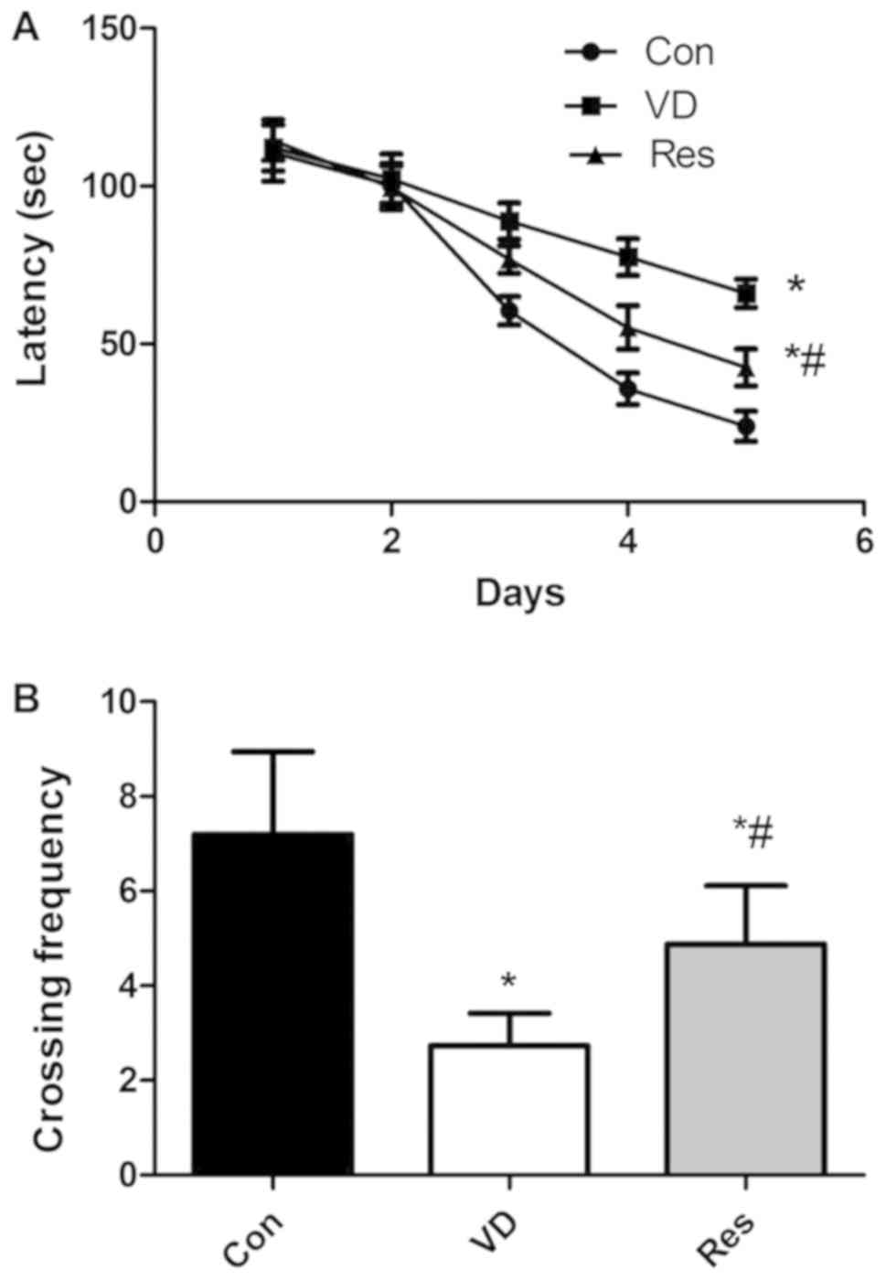
We used the MWM test to evaluate the learning and
memory ability of the different groups to determine whether
resveratrol improves cognitive function in VD rats (Fig. 1). Compared to that in the control
group, the escape latency in the VD group was significantly
increased. However, after 4 weeks of resveratrol administration,
the escape latency was significantly decreased (Fig. 1A). In the probe test, compared to
that in the control group, the crossing frequency in the VD group
was significantly decreased. Compared to that in the VD group, the
crossing frequency in the Res group was significantly increased
(Fig. 1B).
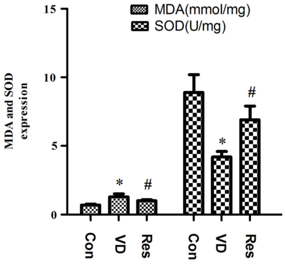
Resveratrol reduces the concentration
of MDA and increases the concentration of SOD in VD rats
Compared with that in the Con group, the hippocampal
content of MDA in the VD group was significantly increased, and the
hippocampal expression of SOD was significantly reduced. Compared
with that in the VD group, the hippocampal content of MDA in the
Res group was significantly reduced, and the expression of
hippocampal SOD was significantly increased. The results indicated
that the ability of the rats in the VD group to recover from
oxidative stress was impaired and that the ability of the VD rats
to recover from oxidative stress was improved after resveratrol
treatment but did not reach the level of the Con group. The
hippocampal expression of SOD and MDA is shown in Fig. 2.
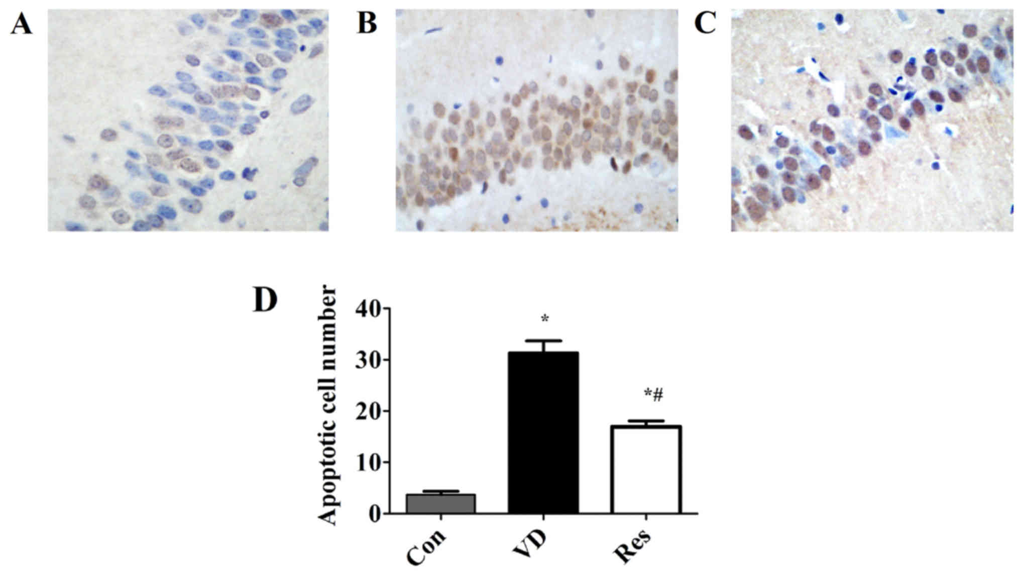
Resveratrol prevents the apoptosis of
hippocampal neurons in the CA1 region
The data were produced using TUNEL assay. As shown
in Fig. 3, the number of apoptotic
neurons in the CA1 region of the model group was significantly
increased. However, resveratrol significantly reduced the number of
apoptotic neurons in the CA1 region.
Resveratrol decreases hippocampal
apoptosis in VD rats
The western blot analysis results (Fig. 4), showed that compared with that in
the Con group, the expression of Bcl-2 in the VD group was
significantly decreased. Compared with that in the VD group, the
expression of Bcl-2 in the Res group was significantly increased.
Compared with that in the Con group, the expression of hippocampal
Bax and caspase-3 in the VD group was increased. However,
hippocampal Bax and caspase-3 protein expression was significantly
decreased after 4 weeks of resveratrol treatment. These results
revealed that cerebral hypoperfusion inhibited Bcl-2 expression and
enhanced hippocampal Bax and caspase-3 expression. Furthermore,
resveratrol treatment enhanced the expression of Bcl-2 and
inhibited Bax and caspase-3 expression in the hippocampus of VD
rats.
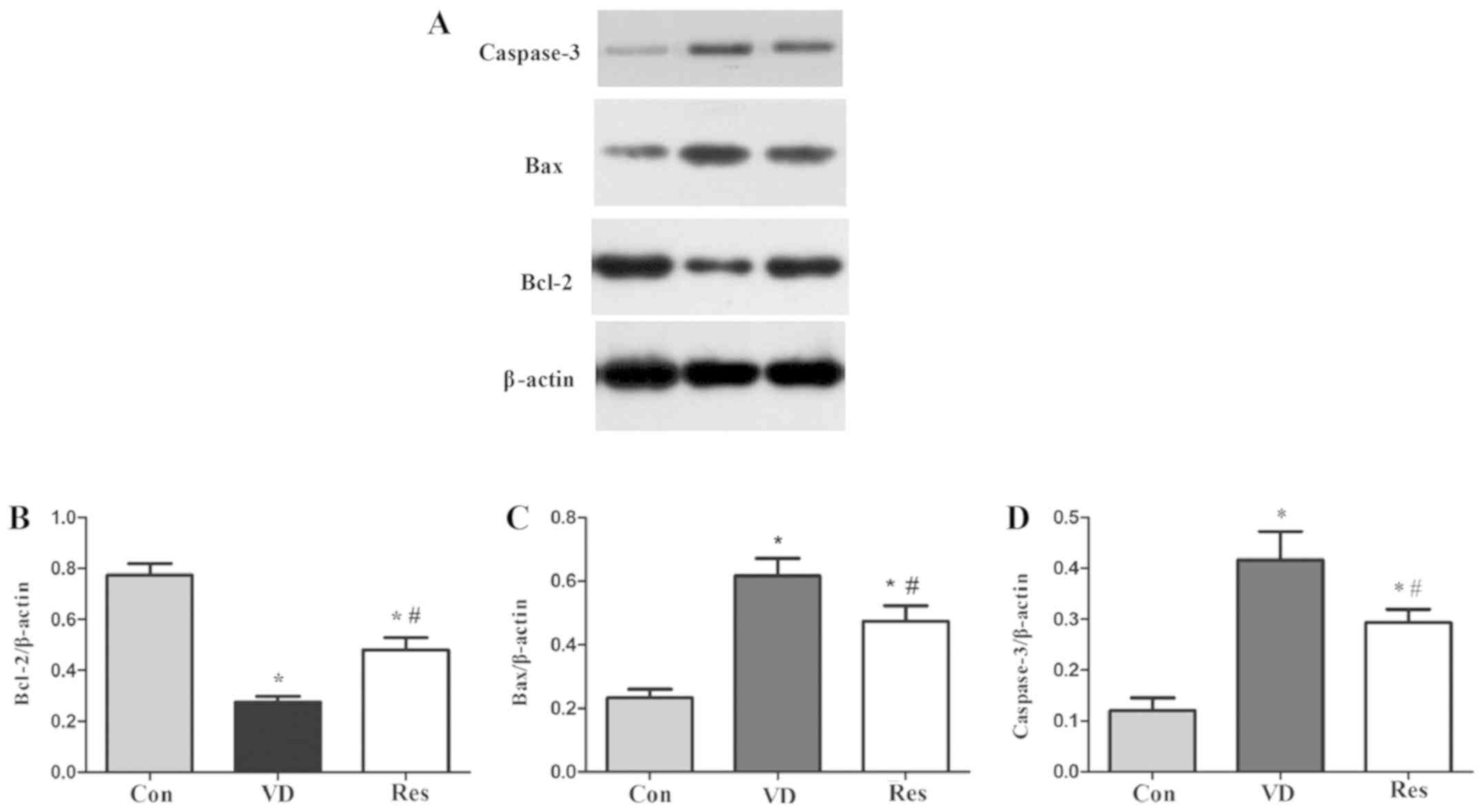 | Figure 4.Western blot analysis of Bcl-2, Bax
and caspase-3 expression. (A) Bcl-2, Bax, caspase-3 and β-actin
protein bands. (B) The relative protein expression of Bcl-2.
Compared with that in the Con group, the expression of Bcl-2 in the
VD group was significantly decreased. Compared with that in the VD
group, the expression of Bcl-2 in the Res group was significantly
increased. (C) The relative protein expression of Bax. Compared
with that in the Con group, the expression of Bax in the VD group
was significantly increased. However, hippocampal Bax protein
expression was significantly decreased after 4 weeks of resveratrol
treatment. (D) The relative protein expression of caspase-3.
Compared with that in the Con group, the expression of caspase-3 in
the VD group was significantly increased. However, hippocampal
caspase-3 protein expression was significantly decreased after 4
weeks of resveratrol treatment. *P<0.05 vs. the Con group;
#P<0.05 vs. the VD group. VD, vascular dementia; Con,
control; Res, resveratrol; Bcl-2, B-cell lymphoma 2; Bax,
Bcl-2-like protein 4. |
Discussion
Dementia is an acquired mental deficiency that
causes severe social or occupational impairment. Vascular dementia
(VD) is considered the second most common type of dementia,
including dementia resulting from all types of vascular disease
(17). When the brain's blood
supply is blocked or the diseased blood vessel system is impaired,
VD can occur leading to a gradual decline in memory and cognitive
function (18). Bilateral carotid
artery occlusion (BCCAO) can lead to the chronic hypoperfusion of
the brain in rats, resulting in severe leukodystrophy and learning
and memory impairment (19).
Therefore, BCCAO is useful for studying the mechanism of chronic
cerebral hypoperfusion and searching for drugs for VD in rat
models. Resveratrol is a natural phenolic substance found in the
epidermis of red grapes and red wine, and it is abundant in
Polygonum cuspidatum. Numerous studies have shown that
resveratrol has many biological properties, including
antioxidative, antitumor and anti-inflammatory properties. Although
many studies have reported a close link between resveratrol and
cognitive ability, the underlying mechanism remains unclear.
Therefore, we investigated the effect of resveratrol on the
cognitive ability of rats with VD. Our results showed that the
cognitive ability of VD rats decreased and that resveratrol
improved the cognitive function of VD rats. The results showed that
resveratrol is a potential neuroprotective drug that can counter
the cognitive impairment of BCCAO-induced VD in rat models.
The pathogenesis of VD includes oxidative stress,
the cytotoxicity of reactive oxygen, mitochondrial dysfunction and
apoptosis (20,21). Oxidative stress is caused by
changes in the body's oxidation and antioxidant balance, which is
more conducive to producing free radicals or reactive oxygen free
radicals rather than other antioxidant defense mechanisms (22). Oxidative stress is a regulator of
neurological injury and cognitive dysfunction after stroke, and a
large number of studies have found oxidative DNA damage around
cerebral infarctions in rats (23–25).
Oxidative stress is not only associated with VD but is also
associated with all risk factors for VD. Malondialdehyde (MDA) and
superoxide dismutase (SOD) are widely used in many studies to
represent the level of oxidative stress (26,27).
MDA, a common marker for oxidative stress injury, is the product of
lipid peroxidation in oxidative stress and is positively correlated
with oxidative stress and lipid peroxidation (26). Patients with VD have been found to
have high levels of MDA, and MDA levels in these patients are
higher than in patients with Alzheimer's disease (28). Clinical and experimental studies on
cerebrovascular diseases have found that the peroxidative damage of
free radicals is a likely mechanism of VD. SOD is an important
antioxidant enzyme in the body. It is involved in the most
important antioxidant system in the body and can directly scavenge
free radicals and eliminate peroxidative damage. Free radicals
obviously damage learning and memory. Clinical trials have found
that oxidative stress plays a role in brain damage in VD patients,
which may be related to the decreased SOD activity in red blood
cells of VD patients, especially in patients with cerebrovascular
lesions (29). The results of the
present study showed that MDA expression in VD rats was increased
and that the activity of SOD in VD rats was decreased, indicating
that bilateral carotid artery occlusion resulted in chronic
ischemia and further led to oxidative stress injury in the VD rat
model. Four weeks of resveratrol administration reversed the
expression of MDA and SOD, confirming that resveratrol treatment
improved the spatial learning and memory function of VD rats and
may be closely related to anti-oxidative stress injury.
Apoptosis is a process of programmed cell death, and
the term is derived from a Greek word meaning the falling of leaves
from a tree. In many different mechanisms of cell death, cell
apoptosis has been proposed to explain the cell loss observed in
many neurological disorders, such as Alzheimer's disease, VD,
Parkinson's disease, amyotrophic lateral sclerosis muscular
atrophy, and stroke (30). Bcl-2
and Bax are the two main genes responsible for regulating cell
apoptosis. Bcl-2 is a key member of the anti-apoptotic Bcl-2 family
and is essential for regulating cell apoptosis mediated by
mitochondria (31,32). The overexpression of Bcl-2 has been
shown to protect nerve cells from damage by neurotoxins (33). Bax, which promotes the release of
cytochrome c by transferring it to the mitochondrial
membrane, thereby promoting downstream cell apoptosis, is a member
of the survivin subfamily (34).
Caspase, which is an important medium for apoptosis is a member of
the cysteine protease family. Caspases mainly participate in the
process of apoptosis through two different pathways: the death
receptor pathway and the mitochondrial pathway (35). Caspase-3 plays the role of the
apoptotic executor, regardless of the pathway involved. Caspase-3
is a type of pro-apoptotic enzyme that activates the DNA
fragmentation factor and then activates endonucleases to split
nuclear DNA, eventually leading to apoptosis (36,37).
Research has confirmed that apoptosis is an important molecular
biological mechanism that is closely related to VD (38). In the present study, the protein
expression of Bax, Bcl-2 and caspase-3 was detected by western blot
analysis. The results showed that the hippocampal expression of Bax
and caspase-3 was significantly increased and that the expression
of hippocampal Bcl-2 was significantly reduced, confirming the
activation of the hippocampal apoptosis pathway in VD rats. In the
Res group, the hippocampal expression of Bax and caspase-3 was
decreased, and the hippocampal expression of Bcl-2 was increased,
indicating that resveratrol has obvious anti-apoptotic properties
in VD rats.
In conclusion, resveratrol significantly improved
cognitive impairment, decreased MDA levels, increased SOD activity
and altered the expression of apoptotic proteins in VD rats. The
mechanism of its neuroprotective effects may be closely related to
the inhibition of the apoptosis pathway and oxidative stress
injury. The results of this study confirmed the neuroprotective
effects of resveratrol treatment in VD rats, providing a
theoretical basis for the basic and clinical treatment of VD and
other types of dementia. Nevertheless, our experiments found that
resveratrol treatment did not improve cognitive function in VD rats
completely, showing the limitations of this treatment, and the
molecular mechanisms remain to be further explored in future
experiments. In addition, in future research, we will design a
positive control trial and determine whether resveratrol is
superior to other drugs. The rats had only mild cognitive
impairment, thus we used short-term intervention. If rats develop
severe cognitive impairment or even dementia, the damage is
irreversible, and even long-term intervention cannot reverse
dementia. Of course, in future studies, we will compare whether
short-term and long-term interventions have an impact on treatment
outcomes. Moreover, β-amyloid and tau, markers for VD, will be
tested in the future to strengthen our research findings.
Acknowledgements
Not applicable.
Funding
This study was funded by grants from the Taizhou
Science and Technology Planning Project (2018, 1801ky33).
Availability of data and materials
All data generated or analyzed during this study are
included in this published article.
Authors' contributions
YZ and JW made substantial contributions to the
conception and design of the study. YL made substantial
contributions to the conception and design of the study, and
drafted and revised the manuscript. YW, LM and DZ made
contributions to the acquisition of data. GW made contributions to
the analysis and intepretation of data. All authors read and
approved the manuscript and agree to be accountable for all aspects
of the research in ensuring that the accuracy or integrity of any
part of the work are appropriately investigated and resolved.
Ethics approval and consent to
participate
All experiments were carried out in accordance with
the regulations of the Ethics Committee of Taizhou Central Hospital
(Taizhou University Hospital, Taizhou, Zhejiang).
Patient consent for publication
Not applicable.
Competing interests
The authors declare that they have no competing
interests.
Glossary
Abbreviations
Abbreviations:
|
VD
|
vascular dementia
|
|
BCCAO
|
bilateral common carotid artery
occlusion
|
|
MWM
|
Morris water maze
|
References
|
1
|
Roman GC: Facts, myths, and controversies
in vascular dementia. J Neurol Sci. 226:49–52. 2004. View Article : Google Scholar : PubMed/NCBI
|
|
2
|
Rockwood K, Wentzel C, Hachinski V, Hogan
DB, MacKnight C and McDowell I: Prevalence and outcomes of vascular
cognitive impairment. Vascular Cognitive Impairment Investigators
of the Canadian Study of Health and Aging. Neurology. 54:447–451.
2000. View Article : Google Scholar : PubMed/NCBI
|
|
3
|
Levine DA and Langa KM: Vascular cognitive
impairment: disease mechanisms and therapeutic implications.
Neurotherapeutics. 8:361–373. 2011. View Article : Google Scholar : PubMed/NCBI
|
|
4
|
Villaflores OB, Chen YJ, Chen CP, Yeh JM
and Wu TY: Curcuminoids and resveratrol as anti-Alzheimer agents.
Taiwan J Obstet Gynecol. 51:515–525. 2012. View Article : Google Scholar : PubMed/NCBI
|
|
5
|
Wood LG, Wark PA and Garg ML: Antioxidant
and anti-inflammatory effects of resveratrol in airway disease.
Antioxid Redox Signal. 13:1535–1548. 2010. View Article : Google Scholar : PubMed/NCBI
|
|
6
|
Chang CC, Chang CY, Huang JP and Hung LM:
Effect of resveratrol on oxidative and inflammatory stress in liver
and spleen of streptozotocin-induced type 1 diabetic rats. Chin J
Physiol. 55:192–201. 2012. View Article : Google Scholar : PubMed/NCBI
|
|
7
|
Richard T, Pawlus AD, Iglésias ML, Pedrot
E, Waffo-Teguo P, Mérillon JM and Monti JP: Neuroprotective
properties of resveratrol and derivatives. Ann N Y Acad Sci.
1215:103–108. 2011. View Article : Google Scholar : PubMed/NCBI
|
|
8
|
Simão F, Matté A, Breier AC, Kreutz F,
Trindade VM, Netto CA and Salbego CG: Resveratrol prevents global
cerebral ischemia-induced decrease in lipid content. Neurol Res.
35:59–64. 2013. View Article : Google Scholar : PubMed/NCBI
|
|
9
|
Girbovan C, Morin L and Plamondon H:
Repeated resveratrol administration confers lasting protection
against neuronal damage but induces dose-related alterations of
behavioral impairments after global ischemia. Behav Pharmacol.
23:1–13. 2012. View Article : Google Scholar : PubMed/NCBI
|
|
10
|
Shao AW, Wu HJ, Chen S, Ammar AB, Zhang JM
and Hong Y: Resveratrol attenuates early brain injury after
subarachnoid hemorrhage through inhibition of NF-kappaB-dependent
inflammatory/MMP-9 pathway. CNS Neurosci Ther. 20:182–185. 2014.
View Article : Google Scholar : PubMed/NCBI
|
|
11
|
Ates O, Cayli S, Altinoz E, Gurses I,
Yucel N, Kocak A, Yologlu S and Turkoz Y: Effects of resveratrol
and methylprednisolone on biochemical, neurobehavioral and
histopathological recovery after experimental spinal cord injury.
Acta Pharmacol Sin. 27:1317–1325. 2006. View Article : Google Scholar : PubMed/NCBI
|
|
12
|
Li F, Gong Q, Dong H and Shi J:
Resveratrol, a neuroprotective supplement for Alzheimer's disease.
Cur Des. 18:27–33. 2012.
|
|
13
|
Ma T, Tan MS, Yu JT and Tan L: Resveratrol
as a Therapeutic Agent for Alzheimer's Disease. Biomed Res Int.
2014:3505162014. View Article : Google Scholar : PubMed/NCBI
|
|
14
|
Winterbourn CC, Peskin AV and Parsons-Mair
HN: Thiol oxidase activity of copper, zinc superoxide dismutase. J
Biol Chem. 277:1906–1911. 2002. View Article : Google Scholar : PubMed/NCBI
|
|
15
|
Riahi S, Mohammadi MT, Sobhani V and
Soleimany M: Chronic effects of aerobic exercise on gene expression
of LOX-1 receptor in the heart of rats fed with high fat diet. Iran
J Basic Med Sci. 18:805–812. 2015.PubMed/NCBI
|
|
16
|
Wang G, Fang H, Zhen Y, Xu G, Tian J,
Zhang Y, Zhang D, Zhang G, Xu J, Zhang Z, et al: Sulforaphane
prevents neuronal apoptosis and memory impairment in diabetic rats.
Cell Physiol Biochem. 39:901–907. 2016. View Article : Google Scholar : PubMed/NCBI
|
|
17
|
Román GC: Vascular dementia may be the
most common form of dementia in the elderly. J Neurol Sci.
203-204:7–10. 2002. View Article : Google Scholar : PubMed/NCBI
|
|
18
|
Jellinger KA: The enigma of vascular
cognitive disorder and vascular dementia. Acta Neuropathol.
113:349–388. 2007. View Article : Google Scholar : PubMed/NCBI
|
|
19
|
Ni J, Ohta H, Matsumoto K and Watanabe H:
Progressive cognitive impairment following chronic cerebral
hypoperfusion induced by permanent occlusion of bilateral carotid
arteries in rats. Brain Res. 653:231–236. 1994. View Article : Google Scholar : PubMed/NCBI
|
|
20
|
Wang J, Zhang HY and Tang XC: Cholinergic
deficiency involved in vascular dementia: Possible mechanism and
strategy of treatment. Acta Pharmacol Sin. 30:879–888. 2009.
View Article : Google Scholar : PubMed/NCBI
|
|
21
|
Bennett S, Grant MM and Aldred S:
Oxidative stress in vascular dementia and Alzheimer's disease: A
common pathology. J Alzheimers Dis. 17:245–257. 2009. View Article : Google Scholar : PubMed/NCBI
|
|
22
|
Bhatti JS, Kumar S, Vijayan M, Bhatti GK
and Reddy PH: Therapeutic strategies for mitochondrial dysfunction
and oxidative stress in age-related metabolic disorders. Prog Mol
Biol Transl Sci. 146:13–46. 2017. View Article : Google Scholar : PubMed/NCBI
|
|
23
|
Nagayama T, Lan J, Henshall DC, Chen D,
O'Horo C, Simon RP and Chen J: Induction of oxidative DNA damage in
the peri-infarct region after permanent focal cerebral ischemia. J
Neurochem. 75:1716–1728. 2000. View Article : Google Scholar : PubMed/NCBI
|
|
24
|
Nanetti L, Taffi R, Vignini A, Moroni C,
Raffaelli F, Bacchetti T, Silvestrini M, Provinciali L and Mazzanti
L: Reactive oxygen species plasmatic levels in ischemic stroke. Mol
Cell Biochem. 303:19–25. 2007. View Article : Google Scholar : PubMed/NCBI
|
|
25
|
Raz L, Zhang QG, Zhou CF, Han D, Gulati P,
Yang LC, Yang F, Wang RM and Brann DW: Role of Rac1 GTPase in NADPH
oxidase activation and cognitive impairment following cerebral
ischemia in the rat. PLoS One. 5:e126062010. View Article : Google Scholar : PubMed/NCBI
|
|
26
|
Liu H, Zhao M, Yang S, Gong DR, Chen DZ
and Du DY: (2R,3S)-Pinobanksin-3-cinnamate improves cognition and
reduces oxidative stress in rats with vascular dementia. J Nat Med.
69:358–365. 2015. View Article : Google Scholar : PubMed/NCBI
|
|
27
|
Ghorbanzadeh V, Mohammadi M, Mohaddes G,
Dariushnejad H, Chodari L and Mohammadi S: Protective effect of
crocin and voluntary exercise against oxidative stress in the heart
of high-fat diet-induced type 2 diabetic rats. Physiol Int.
103:459–468. 2016. View Article : Google Scholar : PubMed/NCBI
|
|
28
|
Gustaw-Rothenberg K, Kowalczuk K and
Stryjecka-Zimmer M: Lipids' peroxidation markers in Alzheimer's
disease and vascular dementia. Geriatr Gerontol Int. 10:161–166.
2010.PubMed/NCBI
|
|
29
|
Wallin A, Kapaki E, Boban M, Engelborghs
S, Hermann DM, Huisa B, Jonsson M, Kramberger MG, Lossi L, Malojcic
B and et a1: Biochemical markers in vascular cognitive impairment
associated with subcortical small vesseldisease-A consensus report.
BMC Neurol. 17:1022017. View Article : Google Scholar : PubMed/NCBI
|
|
30
|
Favaloro B, Allocati N, Graziano V, Di
Ilio C and De Laurenzi V: Role of apoptosis in disease. Aging
(Albany NY). 4:330–349. 2012. View Article : Google Scholar : PubMed/NCBI
|
|
31
|
Garner TP, Lopez A, Reyna DE, Spitz AZ and
Gavathiotis E: Progress in targeting the BCL-2 family of proteins.
Curr Opin Chem Biol. 39:133–142. 2017. View Article : Google Scholar : PubMed/NCBI
|
|
32
|
Brown LM, Hanna DT, Khaw SL and Ekert PG:
Dysregulation of BCL-2 family proteins by leukemia fusion genes. J
Biol Chem. 292:14325–14333. 2017. View Article : Google Scholar : PubMed/NCBI
|
|
33
|
Gustafsson AB and Gottlieb RA: Bcl-2
family members and apoptosis, taken to heart. Am J Physiol Cell
Physiol. 292:C45–C51. 2007. View Article : Google Scholar : PubMed/NCBI
|
|
34
|
Wolter KG, Hsu YT, Smith CL, Nechushtan A,
Xi XG and Youle RJ: Movement of Bax from the cytosol to
mitochondria during apoptosis. J Cell Biol. 139:1281–1292. 1997.
View Article : Google Scholar : PubMed/NCBI
|
|
35
|
Grütter MG: Caspases: Key players in
programmed cell death. Curr Opin Struct Biol. 10:649–655. 2000.
View Article : Google Scholar : PubMed/NCBI
|
|
36
|
Sun ZK, Ma XR, Jia YJ, Liu YR, Zhang JW
and Zhang BA: Effects of resveratrol on apoptosis in a rat model of
vascular dementia. Exp Ther Med. 7:843–848. 2014. View Article : Google Scholar : PubMed/NCBI
|
|
37
|
Chang CT, Korivi M, Huang HC, Thiyagarajan
V, Lin KY, Huang PJ, Liu JY, Hseu YC and Yang HL: Inhibition of ROS
production, autophagy or apoptosis signaling reversed the
anticancer properties of Antrodia salmonea in triple-negative
breast cancer (MDA-MB-231) cells. Food Chem Toxicol. 103:1–17.
2017. View Article : Google Scholar : PubMed/NCBI
|
|
38
|
Zhang Y, Wang LL, Wu Y, Wang N, Wang SM,
Zhang B, Shi CG and Zhang SC: Paeoniflorin attenuates hippocampal
damage in a rat model of vascular dementia. Exp Ther Med.
12:3729–3734. 2016. View Article : Google Scholar : PubMed/NCBI
|