Introduction
Toll-like receptors (TLRs) are well-conserved
pattern-recognition receptors that are primarily expressed in human
epithelial and immune cells (1,2). The
main function of TLRs is to promote the synthesis and release of
inflammatory cytokines and chemokines, thus triggering the
inflammatory response (3,4). To date, a total of 10 TLRs, namely
TLR1-TLR10, have been identified in humans. Previous studies have
demonstrated that TLRs are also expressed in numerous tumor cells,
and serve key roles in tumorigenesis, development and metastasis
(5,6). Research on how to effectively inhibit
the expression and activation of TLRs in order to reduce the
production and release of the corresponding inflammatory factors,
and ultimately inhibit the proliferation of cancer cells has been
receiving increasing attention.
Breast cancer is one of the most common malignant
tumors worldwide, and is the most prevalent type of cancer and
second leading cause of cancer-associated mortality in women
(7). In China, breast cancer is
the second most common type of cancer, second only to lung cancer
(8). Breast cancer is an
intrinsically heterogeneous disease with different biological
characteristics and clinical outcomes. Common immunohistochemistry
markers, such as estrogen receptor (ER), progesterone receptor (PR)
and human epidermal growth factor receptor 2 (HER2), together with
traditional clinicopathological features, including tumor size,
tumor grade and nodal involvement, are used to predict the outcome
and treatment response in breast cancer (9). Sørlie et al (10) have proposed five subgroups of
breast cancer based on gene expression profiling using DNA
microarrays, including Luminal A, Luminal B, HER2 overexpressing,
basal-like and normal-like breast cancer. Luminal type breast
cancer (both A and B, also known as Lum A/B) is the most common
type, accounting for ~70% of patients with breast cancer.
Basal-like type breast cancer lacks ER, PR and HER2 receptors
(triple-negative breast cancer), and has the worst prognosis
(11,12).
Although the association between TLRs and breast
cancer has not been thoroughly investigated, previous studies
suggested the presence of an important link between TLRs and breast
cancer. It has been reported that the expression of TLR2 was
~10-fold lower in the more malignant MDA-MB-231 cells as compared
with that in the less malignant MCF7 cells. Activation of TLR2
results in the activation of nuclear factor-κB (NF-κB) and
upregulation of interleukin (IL)-6, transforming growth factor-β,
vascular endothelial growth factor and matrix metalloproteinase 9
(13). Salaun et al
(14) reported that, among 194
cases of patients with breast cancer, 36–45% of breast cancer cells
expressed TLR3, while treatment with poly(A:U) reduced the risk of
recurrence and metastasis in patients with TLR3-positive breast
cancer. In addition, Haricharan and Brown (15) observed that TLR4 activation in
TP53-mutant breast cancer regulated the proliferation of cancer
cells by promoting the secretion of pro-growth cytokines. TLR4
expression has also been demonstrated to be strongly associated
with clinical indicators in metastatic ductal carcinoma (16). Cai et al (17) further reported that 80% of 75 cases
of breast cancer expressed TLR5, the majority of which were
high-grade ductal carcinomas. TLR5 was also overexpressed in 256
breast carcinomas specimens, and was correlated with lymph node
metastasis and cancer grade (18).
Furthermore, Berger et al (19) studied frozen breast specimens from
124 female patients with breast cancer and found that TLR9 mRNA
expression was positively correlated with tumor grade, suggesting
that TLR9 may be a molecular marker for poorly differentiated
breast cancer. Another study reported that activation of TLR9 in
MDA-MB-231 cells by CpG-ODN increased in vitro invasion
(20), suggesting that TLR9 is
involved in tumor progression and metastasis. These previous
studies collectively suggest that TLR signaling may serve an
important role in regulating the growth, metastasis and apoptosis
of breast cancer cells.
In the current study, mRNA expression and clinical
data of 1,215 patients with breast cancer were obtained from The
Cancer Genome Atlas (TCGA) database to analyze the expression
patterns of TLRs in different sample types, tumor subtypes and
tumor stages. Furthermore, cytokines downstream of the TLR
signaling pathway were studied, and survival analysis was performed
to investigate the effect of TLR expression on the outcome and
prognosis of patients with breast cancer. By investigating the role
of TLRs in the development of breast cancer, the present study
provides new insights for the diagnosis and treatment of breast
cancer.
Materials and methods
Patients and TCGA data retrieval
The data of patients with breast cancer, including
mRNA expression and corresponding clinical information, were
retrieved from TCGA database (https://www.cancer.gov/about-nci/organization/ccg/research/structural-genomics/tcga),
which is an open access, publicly available database. Clinical
characteristics of the patients included in the current study are
listed in Table I. The TCGA gene
expression profile was measured using the Illumina HiSeq 2000 RNA
Sequencing System (HiSeqV2; Illumina, Inc., San Diego, CA, USA).
The RSEM (also known as RNA-Seq by Expectation-Maximization)
normalized count was used as the gene level expression estimates in
the present study. Intrinsic tumor subtype, tumor stage information
and overall survival time were also extracted from TCGA data
portal. Patients without detailed clinicopathological data, such as
age, gender, race, histological subtype, tumor-node- metastasis
(TNM) stage or overall survival were excluded from the present
study. The study met the ethics and policies provided by TCGA
(https://www.cancer.gov/about-nci/organization/ccg/research/structural-genomics/tcga/history/policies).
 | Table I.Clinical characteristics of breast
cancer patients included in The Cancer Genome Atlas database. |
Table I.
Clinical characteristics of breast
cancer patients included in The Cancer Genome Atlas database.
| Characteristic | Value | Percentage (%) |
|---|
| Age at diagnosis
(years) | 57.98 | − |
| Follow up
(years)a | 1.65 | − |
| Ethnicity |
|
|
|
Caucasian | 757 | 60.99 |
| Black
or African American | 183 | 14.75 |
|
Asian | 61 |
4.92 |
|
American Indian or Alaska
native | 1 |
0.08 |
| NA | 239 | 19.26 |
| Survival |
|
|
| Alive
at last follow-up or succumbed to unrelated cause | 816 | 65.75 |
|
Disease-specific
mortality | 135 | 10.88 |
| NA | 290 | 23.37 |
| Tumor grade
(Nottingham) (37) |
|
|
| I | 133 | 10.72 |
| II | 446 | 35.94 |
|
III | 175 | 14.10 |
| IV | 15 |
1.21 |
| X | 10 |
0.81 |
| NA | 462 | 37.23 |
| Subtype |
|
|
|
Normal | 113 |
9.11 |
| Luminal
A | 434 | 34.97 |
| Luminal
B | 194 | 15.63 |
|
HER2-enriched | 67 |
5.40 |
|
Basal-like | 142 | 11.44 |
| Not
classified | 291 | 23.45 |
Statistical analysis
The analysis focused on 1,215 breast cancer cases
included in TCGA database. The data were expressed as the mean ±
standard deviation. Paired or unpaired Student's t-test, and
one-way analysis of variance functions of GraphPad Prism software
(version 6; GraphPad Software, Inc., San Diego, CA, USA) were used
to evaluate the significance of differential expression levels of
candidate genes among different samples, intrinsic tumor subtypes
and tumor stages. In addition, Spearman correlation and linear
regression analysis were used to examine the association of TLR
expression with various cytokines [IL-1β, IL-6, IL-8, necrosis
factor α (TNF-α), interferon (IFN)-α, IFN-β and C-X-C motif
chemokine 10 (CXCL10)], with 0<r<1 indicating that two
variables were changing in the same direction in a correlative
manner. Survival analysis was conducted using SPSS software
(version 22.0; IBM Corp., Armonk, NY, USA). Patients were grouped
based on the mRNA expression of TLRs, with the upper 50% and the
lower 50% representing the high and low expression groups,
respectively. Survival curves were estimated by the Kaplan-Meier
method, and the log-rank test was used to compare the overall
survival curves between groups. Only mortality cases as a result of
breast cancer were considered in the analysis. P<0.05 was
considered to indicate a statistically significant difference.
Results
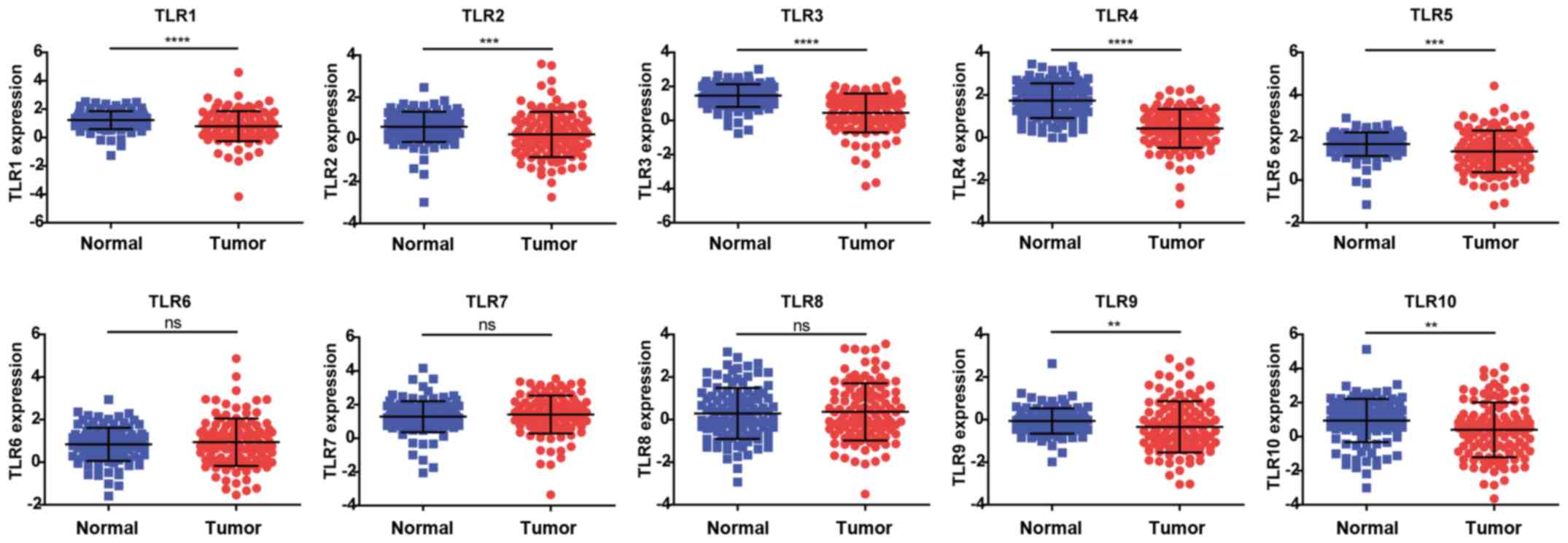
Expression levels of TLRs in breast
cancer cases included in TCGA database
The publicly available TCGA database, composed of
2.5 petabytes of data describing tumor tissues and matched normal
tissues from >11,000 patients, was used in the present study.
The expression levels of TLR1-TLR10 were initially analyzed using
TCGA breast cancer database in order to compare normal and tumor
tissues. Compared with the normal control tissues, the expression
levels of TLR1, TLR2, TLR3, TLR4, TLR5, TLR9 and TLR10 were
significantly decreased in breast cancer tissues. In addition, the
expression levels of TLR6, TLR7 and TLR8 were slightly increased in
breast cancer tissues, although statistical significance was not
reached (Fig. 1).
Expression levels of TLRs in various
subtypes and stages of breast cancer
The expression of TLRs in different subtypes of
breast cancer was subsequently analyzed. TLR1, TLR2 and TLR6
displayed higher expression levels in the triple-negative breast
cancer subtype as compared with those in Lum A/B and HER2-enriched
subtypes. The expression of TLR3 exhibited a gradient decline among
normal tissues and all the three subtypes; however, the difference
between the HER2-enriched and triple-negative subtypes was not
statistically significant. The TLR4 and TLR7 expression levels were
lower in the triple-negative subtype as compared with the other two
subtypes. TLR5 was the only gene that exhibited a statistically
significant difference in terms of expression among all the three
subtypes, with the lowest expression observed in the HER2-enriched
subtype, followed by the Lum A/B and triple-negative subtypes.
Furthermore, the expression of TLR8 was reduced in the Lum A/B
subtype compared with that exhibited in the other two subtypes,
although this difference was not significant. TLR9 expression
increased gradually among the three subtypes; however, similarly to
TLR3, there was no statistically significant difference between the
HER2-enriched and triple-negative subtypes. By contrast, TLR10
expression did not differ among the three subtypes of breast cancer
(Fig. 2A). These results suggest
that TLR3 and TLR9 may be useful biomarkers of ER-/PR-negative
breast cancer (HER2-enriched and triple-negative subtypes), while
TLR5 may serve an important role in HER2-enriched breast
cancer.
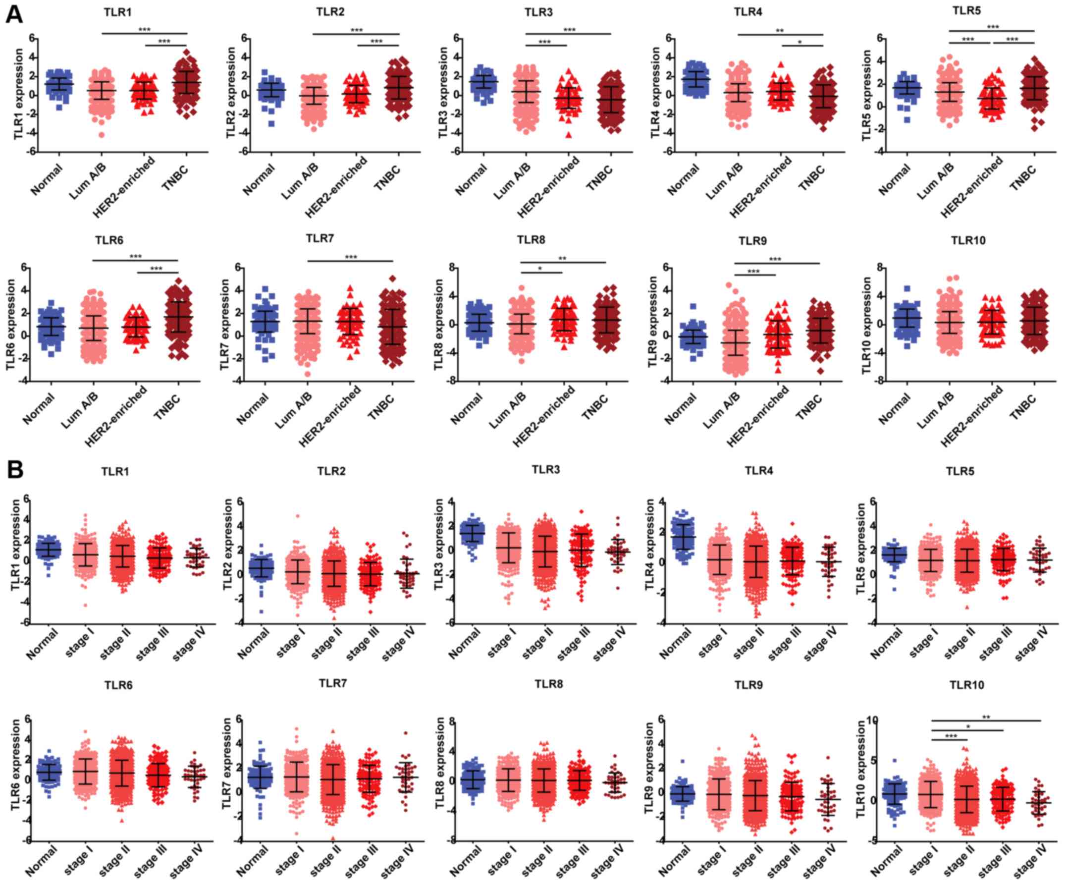 | Figure 2.TLR expression among different
subtypes and stages of breast cancer. (A) Subtype information was
exported from TCGA breast cancer database, including 113, 628, 67
and 142 samples from normal tissues, Luminal A/B, HER2-enriched and
triple-negative subtypes, respectively. (B) TNM stage information
was exported from TCGA breast cancer database, including 113, 279,
622, 131 and 40 samples from normal tissues, stage I, II, III and
IV, respectively. Data are expressed as the mean ± standard
deviation. One-way analysis of variance was used to evaluate the
statistical significance of differential mRNA expression levels of
candidate genes among groups. *P<0.05, **P<0.01 and
***P<0.001. TLR, Toll-like receptor; TCGA, The Cancer Genome
Atlas; ns, no significant difference. |
Next, the expression levels of TLRs in various
stages of cancer progression were analyzed based on the data
obtained from TCGA database. Breast cancer cases were staged
according to the TNM staging system, which uses the size and
extension of the primary tumor, its lymphatic involvement and the
presence of metastases to classify the progression of all solid
tumors (https://www.cancer.gov/types/breast/patient/adult/breast-treatment-pdq).
It was observed that the expression levels of TLRs varied in
different stages of breast cancer (Fig. 2B). Consistent with our earlier
results (Fig. 1), the expression
levels of TLR1, TLR3 and TLR4 were significantly reduced in all
stages relative to normal tissues, whereas TLR2, TLR5 and TLR10
exhibited lower expression in certain stages compared with that in
normal tissues. TLR10 was the only gene whose expression varied
significantly between stage I and II–IV breast cancer, with its
expression decreasing as the cancer stage advanced. These results
suggest that TLR10 may serve as a biomarker of breast cancer
progression. By contrast, the expression levels of TLR6, TLR7, TLR8
and TLR9 did not differ among different disease stages or in
comparison with those found in normal tissues.
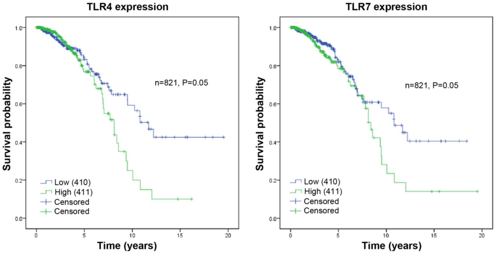
Survival analysis
The current study used overall survival data of
breast cancer patients (OS ≥90 days) from TCGA database to evaluate
the effect of TLR expression on patient prognosis. The patient
survival status was described as 0 or 1, where 0 represented
mortality and 1 represented censoring (patients who were lost, or
succumbed to other causes, or were alive at the end of follow-up).
The samples were divided into two groups based on TLR expression
levels, including the low and high expression groups. Fig. 3 illustrates the association between
patient survival and the expression of TLR4 or TLR7. The mean
survival time following the initial diagnosis of patients with low
TLR4 expression was 12.38 years, while that of patients with high
expression was 8.07 years (P=0.05). Furthermore, the mean survival
time of patients with low TLR7 expression was 11.66 years, while
that of patients with high expression was 8.96 years (P=0.05). The
remaining TLRs exhibited no significant association between
expression levels and survival (Table
II). Overall, high TLR4 and TLR7 expression may be associated
with poor prognosis.
| Table II.Analysis of overall survival in
relation to TLR expression. |
Table II.
Analysis of overall survival in
relation to TLR expression.
|
|
|
| 95% confidence
interval |
|
|
|---|
|
|
|
|
|
|
|
|---|
| Gene | Expression | Mean survival
(years) | Lower | Upper | χ2 | P-value |
|---|
| TLR1 | Low | 11.25 | 9.65 | 12.85 | 0.15 | 0.70 |
|
| High | 9.62 | 7.91 | 11.33 |
|
|
| TLR2 | Low | 10.59 | 9.04 | 12.14 | 0.41 | 0.52 |
|
| High | 10.64 | 8.83 | 12.46 |
|
|
| TLR3 | Low | 10.70 | 9.00 | 12.39 | 0.63 | 0.43 |
|
| High | 10.66 | 8.95 | 12.36 |
|
|
| TLR4 | Low | 12.38 | 10.71 | 14.06 | 4.00 | 0.05 |
|
| High | 8.07 | 6.94 | 9.20 |
|
|
| TLR5 | Low | 10.17 | 8.33 | 12.00 | 0.00 | 0.99 |
|
| High | 10.68 | 9.25 | 12.11 |
|
|
| TLR6 | Low | 11.20 | 9.64 | 12.76 | 1.98 | 0.16 |
|
| High | 9.93 | 8.08 | 11.77 |
|
|
| TLR7 | Low | 11.66 | 7.32 | 10.60 | 3.92 | 0.05 |
|
| High | 8.96 | 10.15 | 13.18 |
|
|
| TLR8 | Low | 11.77 | 10.15 | 13.40 | 1.90 | 0.17 |
|
| High | 8.44 | 7.18 | 9.70 |
|
|
| TLR9 | Low | 11.05 | 9.19 | 12.90 | 0.02 | 0.89 |
|
| High | 10.22 | 8.78 | 11.67 |
|
|
| TLR10 | Low | 11.19 | 9.48 | 12.90 | 0.02 | 0.89 |
|
| High | 9.62 | 8.30 | 10.93 |
|
|
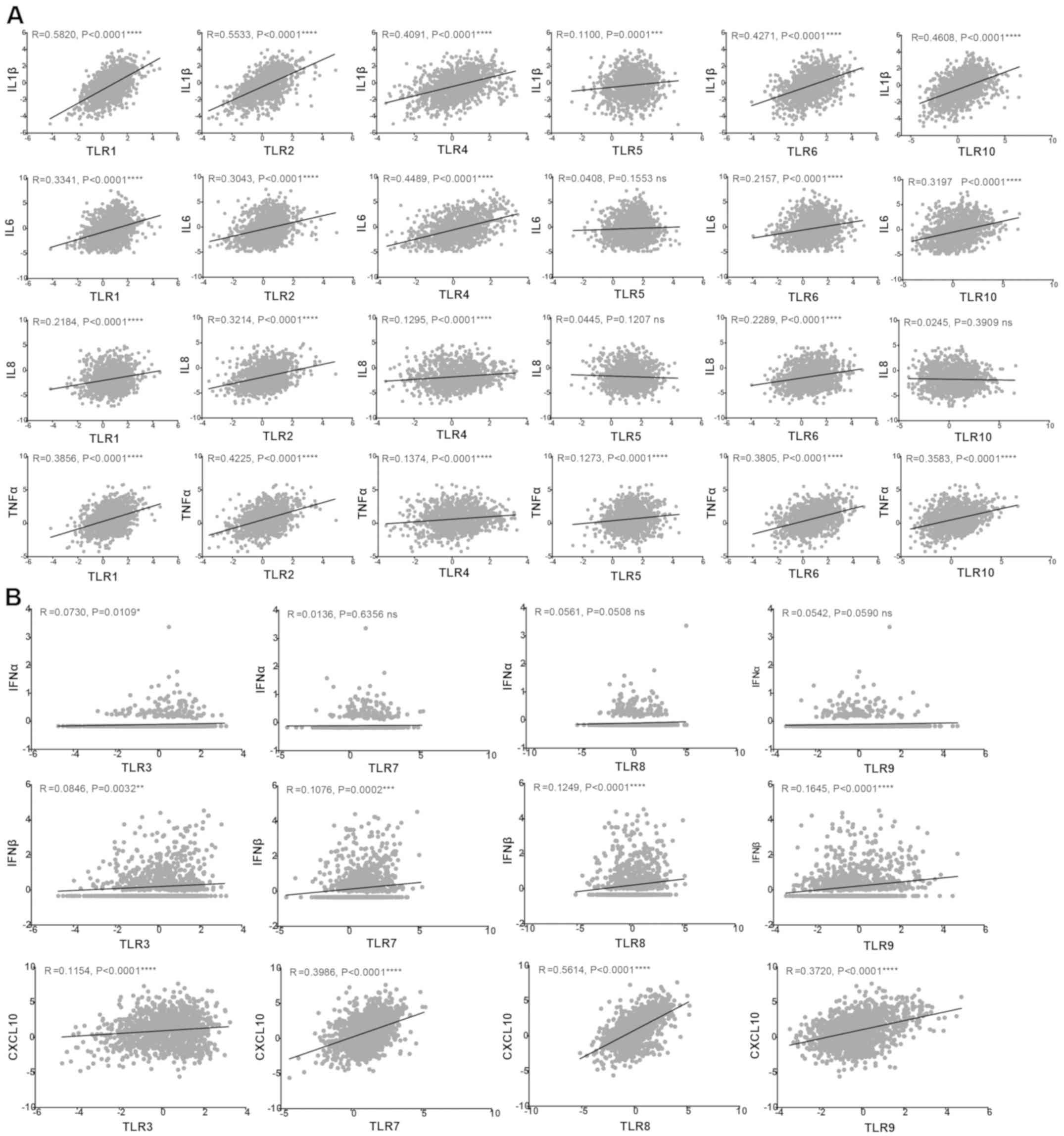
Correlation of inflammatory cytokines
and chemokines with TLRs
TLR expression in the tumor microenvironment has
been reported to be associated with cancer progression and to be
involved in inflammation (21).
The activation of TLRs expressed in tumor cells initiates signaling
cascades that mediate the release of cytokines, chemokines,
pro-angiogenic mediators and growth factors, leading to tumor
survival and progression (22).
Due to the different locations of TLRs in cancer cells, different
downstream signaling pathways are activated, resulting in the
production of different cytokines. In the present study, the TLR
family was divided into two groups: The first group included TLR1,
TLR2, TLR4, TLR5, TLR6 and TLR10, which are expressed on the cell
surface, while the second group comprised TLR3, TLR7, TLR8 and
TLR9, which are present in intracellular vesicles (23). A total of seven representative
cytokines and chemokines downstream of TLR signaling pathways were
then selected to analyze their correlation with TLR expression.
Fig. 4A shows the
correlation of inflammatory cytokines IL-1β, IL-6, IL-8 and tumor
necrosis factor α (TNF-α) with the cell surface receptors TLR1,
TLR2, TLR4, TLR5, TLR6 and TLR10. The expression levels of TLR1,
TLR2, TLR4 and TLR6 were found to be directly correlated with these
four cytokines. By contrast, the expression of TLR5 was only
correlated with IL-1β and TNF-α, while TLR10 was correlated with
IL-1β, IL-6 and TNF-α. As shown in Fig. 4B, the expression levels of the four
intracellular receptors (namely TLR3, TLR7, TLR8 and TLR9) was
positively correlated with IFN-β and CXCL10. TLR3 was also
correlated with IFN-α, whereas TLR7, TLR8 and TLR9 did not exhibit
significant correlations with IFN-α.
Discussion
Breast cancer is a major public health issue for
women worldwide. The occurrence and development of breast cancer
are affected by complex environmental and genetic factors. Numerous
studies have suggested that TLRs, which are normally associated
with immunity and inflammation, may be involved in the progression
and prognosis of breast cancer. TLRs are highly expressed in breast
cancer cells, and activation of these receptors can induce
aggressive tumor behavior, cell proliferation, cell invasion, cell
migration and metastasis (24).
The ‘cross-talk’ between TLRs and various other signaling pathways
in breast cancer constitutes a markedly complicated signaling
network system to promote secretion of inflammatory cytokines and
chemokines. These inflammatory mediators, in turn, can promote
tumor proliferation and apoptosis resistance, thus participating in
the development and progression of breast cancer (25). Therefore, TLRs may be potential
drug targets for breast cancer treatment.
The current study demonstrated that the expression
levels of the majority of TLRs (except for TLR6-TLR9) were
downregulated compared with those in normal tissues; these results
differ from those reported in previous studies (13,16,17,26–28).
This discrepancy may be due to the source of the model system from
which the data were derived. Previous studies have mainly focused
on the expression of TLRs in animal models or breast cancer cell
lines, whereas the present study utilized clinical data from TCGA
database. In the present study, although TLRs were found to be
generally downregulated in tumor tissues, certain tumor subtypes
exhibited higher expression compared with normal tissues. For
instance, TLR6 and TLR9 expression was significantly increased in
triple-negative subtype in comparison with that in normal tissues.
Thus, it is speculated that the possible reasons for the
discrepancy of the results between the present and previous studies
may be the size and source of samples, or the selection of tumor
subtypes. The present study involved a large number of samples
(1,215 cases), and different sample sources, subtypes, races,
genders and ages were included.
It has previously been reported that TLR3 sequence
variants may reduce 82% of breast cancer risk among
African-American women (29), and
the activation of TLR3 was associated with a significant decrease
in the risk of metastatic relapse in 194 patients with breast
cancer, suggesting an association between clinical outcome and TLR3
expression (26). This is
consistent with the results of the present study, which revealed
lower levels of TLR3 expression in more malignant subtypes. It was
also demonstrated that high level of TLR9 was strongly associated
with ER-negative subtypes (HER2-enriched and triple-negative),
which was also verified by previous studies. For instance, Berger
et al (19) reported that
TLR9 expression promoted cell migration, cell invasion and
aggressive tumor behavior in an ER-negative breast cancer cell
line. In addition, Jukkola-Vuorinen et al (30) demonstrated increased TLR9
expression in ER-negative versus ER-positive breast cancer samples
via immunofluorescence. The current study also revealed that low
expression of TLR5 was significantly associated with HER2-enriched
breast cancer, although further research is required to confirm
this association.
Multiple studies have focused on the effect of TLRs
on breast cancer prognosis. González-Reyes et al (27) revealed that high expression of TLR4
was associated with a large tumor size, distant metastasis and
recurrence upon investigating tumors from 74 patients with breast
cancer. In addition, TLR4 was expressed in a functional form in
patients with ER-/PR-negative breast cancer and was correlated with
a decreased survival (31). Lu
et al (32) demonstrated
that activation of TLR7 was associated with a significant
regression of spontaneous breast cancer in mice, suggesting a
better prognosis. In the present study, high expression levels of
TLR4 and TLR7 were revealed to have a close association with
shortened survival and worse prognosis. However, no significant
differences in survival were observed between high and low
expression levels of other TLRs, which may be due to the short
follow-up period of patients included in TCGA database (<5
years).
TLR signaling pathways promote survival,
proliferation and apoptosis of cancer cells, as well as IFN,
cytokine and chemokine production. For instance, TLR1 and TLR2
mediated by the PI3K-Akt pathway, or TLR4, TLR5 and TLR6 mediated
by the NF-κB pathway lead to increased levels of several
pro-inflammatory cytokines (including TNF-α, IL-1β, IL-6 and IL-12)
and chemokines [including IL-8, C-C motif chemokine ligand 3
(CCL3), CCL4 and CCL5], thus causing inflammation. TLR3, TLR7, TLR8
and TLR9 mediated by MyD88-dependent/independent pathways induce
the production of co-stimulatory molecules [such as cluster of
differentiation 40 (CD40), CD80 and CD86], inflammatory cytokines
(including IFN-α and IFN-β) and chemokines (including CXCL9, CXCL10
and CXCL11), thus promoting antibacterial and antiviral effects
(33–36). In line with this, the present study
revealed positive correlations between TLRs and the downstream
signaling molecules IL-1β, IL-6, IL-8, TNF-α, IFN-α, IFN-1β and
CXCL10 in breast cancer. These results suggest that TLR-mediated
signaling serves an important role in the regulation of
tumorigenesis. It may be useful to investigate how various TLR
pathways affect the different breast tumor cell types and their
potential roles in breast cancer development in future studies.
In conclusion, the current study analyzed the
expression profile of TLRs and identified that most TLRs were
downregulated in breast cancer. TLR3, TLR5 and TLR9 were associated
with specific subtypes, and TLR10 was related to tumor stages,
suggesting that TLRs have profound effects on breast cancer
incidence and progression. TLRs and their downstream inflammatory
cytokines serve important roles in innate immunity and are also key
regulatory factors in tumor development. Silencing or activating
TLRs may be an effective therapeutic target of breast cancer.
Therefore, studying the expression patterns of TLRs and the
interaction of their signaling pathways in breast cancer will help
to explore the clinical applications of anti-tumor therapy and to
provide new insights for breast cancer treatment.
Acknowledgements
Not applicable.
Funding
The present study was supported by the Shandong
Provincial Natural Science Foundation of China (grant nos.
ZR2011HZ004 and ZR2017BC073) and the Project of Shandong Province
Higher Educational Science and Technology Program (grant nos.
J10LC21 and J16LE07).
Availability of data and materials
The datasets used and analyzed during the current
study are available from TCGA database (http://cancergenome.nih.gov/).
Authors' contributions
SS and CX analyzed the data and drafted the
manuscript. XF, YZ and HL contributed to the interpretation of data
and revised the manuscript. GY and WW conceived and designed the
study. All authors have reviewed and approved the final version of
the manuscript.
Ethics approval and consent to
participate
Not applicable.
Patient consent for publication
Not applicable.
Competing interests
The authors declare that they have no competing
interests.
References
|
1
|
Mifsud EJ, Tan AC and Jackson DC: TLR
Agonists as modulators of the innate immune response and their
potential as agents against infectious disease. Front Immunol.
5:792014. View Article : Google Scholar : PubMed/NCBI
|
|
2
|
Brubaker SW, Bonham KS, Zanoni I and Kagan
JC: Innate immune pattern recognition: A cell biological
perspective. Annu Rev Immunol. 33:257–290. 2015. View Article : Google Scholar : PubMed/NCBI
|
|
3
|
Palm E, Demirel I, Bengtsson T and Khalaf
H: The role of Toll-like and protease-activated receptors in the
expression of cytokines by gingival fibroblasts stimulated with the
periodontal pathogen Porphyromonas gingivalis. Cytokine.
76:424–432. 2015. View Article : Google Scholar : PubMed/NCBI
|
|
4
|
Johnston DG and Corr SC: Toll-like
receptor signalling and the control of intestinal barrier function.
Methods Mol Biol. 1390:287–300. 2016. View Article : Google Scholar : PubMed/NCBI
|
|
5
|
Rakoff-Nahoum S and Medzhitov R: Toll-like
receptors and cancer. Nat Rev Cancer. 9:57–63. 2009. View Article : Google Scholar : PubMed/NCBI
|
|
6
|
Yu L and Chen S: Toll-like receptors
expressed in tumor cells: Targets for therapy. Cancer Immunol
Immunother. 57:1271–1278. 2008. View Article : Google Scholar : PubMed/NCBI
|
|
7
|
Siegel RL, Miller KD and Jemal A: Cancer
statistics, 2018. CA Cancer J Clin. 68:7–30. 2018. View Article : Google Scholar : PubMed/NCBI
|
|
8
|
Chen W, Zheng R, Baade PD, Zhang S, Zeng
H, Bray F, Jemal A, Yu XQ and He J: Cancer statistics in China,
2015. CA Cancer J Clin. 66:115–132. 2016. View Article : Google Scholar : PubMed/NCBI
|
|
9
|
Dai X, Li T, Bai Z, Yang Y, Liu X, Zhan J
and Shi B: Breast cancer intrinsic subtype classification, clinical
use and future trends. Am J Cancer Res. 5:2929–2243.
2015.PubMed/NCBI
|
|
10
|
Sørlie T, Perou CM, Tibshirani R, Aas T,
Geisler S, Johnsen H, Hastie T, Eisen MB, van de Rijn M, Jeffrey
SS, et al: Gene expression patterns of breast carcinomas
distinguish tumor subclasses with clinical implications. Proc Natl
Acad Sci USA. 98:10869–10874. 2001. View Article : Google Scholar : PubMed/NCBI
|
|
11
|
Perou CM, Sørlie T, Eisen MB, van de Rijn
M, Jeffrey SS, Rees CA, Pollack JR, Ross DT, Johnsen H, Akslen LA,
et al: Molecular portraits of human breast tumours. Nature.
406:747–752. 2000. View
Article : Google Scholar : PubMed/NCBI
|
|
12
|
Yadav BS, Chanana P and Jhamb S:
Biomarkers in triple negative breast cancer: A review. World J Clin
Oncol. 6:252–263. 2015. View Article : Google Scholar : PubMed/NCBI
|
|
13
|
Xie W, Wang Y, Huang Y, Yang H, Wang J and
Hu Z: Toll-like receptor 2 mediates invasion via activating
NF-kappaB in MDA-MB-231 breast cancer cells. Biochem Biophys Res
Commun. 379:1027–1032. 2009. View Article : Google Scholar : PubMed/NCBI
|
|
14
|
Salaun B, Zitvogel L, Asselin-Paturel C,
Morel Y, Chemin K, Dubois C, Massacrier C, Conforti R, Chenard MP,
Sabourin JC, et al: TLR3 as a biomarker for the therapeutic
efficacy of double-stranded RNA in breast cancer. Cancer Res.
71:1607–1614. 2011. View Article : Google Scholar : PubMed/NCBI
|
|
15
|
Haricharan S and Brown P: TLR4 has a
TP53-dependent dual role in regulating breast cancer cell growth.
Proc Natl Acad Sci USA. 112:E3216–E3225. 2015. View Article : Google Scholar : PubMed/NCBI
|
|
16
|
Ehsan N, Murad S, Ashiq T, Mansoor MU, Gul
S, Khalid S and Younas M: Significant correlation of TLR4
expression with the clinicopathological features of invasive ductal
carcinoma of the breast. Tumour Biol. 34:1053–1059. 2013.
View Article : Google Scholar : PubMed/NCBI
|
|
17
|
Cai Z, Sanchez A, Shi Z, Zhang T, Liu M
and Zhang D: Activation of Toll-like receptor 5 on breast cancer
cells by flagellin suppresses cell proliferation and tumor growth.
Cancer Res. 71:2466–2475. 2011. View Article : Google Scholar : PubMed/NCBI
|
|
18
|
Shuang C, Weiguang Y, Zhenkun F, Yike H,
Jiankun Y, Jing X, Xinghan L, Yue L and Dalin L: Toll-like receptor
5 gene polymorphism is associated with breast cancer
susceptibility. Oncotarget. 8:88622–88629. 2017. View Article : Google Scholar : PubMed/NCBI
|
|
19
|
Berger R, Fiegl H, Goebel G, Obexer P,
Ausserlechner M, Doppler W, Hauser-Kronberger C, Reitsamer R, Egle
D, Reimer D, et al: Toll-like receptor 9 expression in breast and
ovarian cancer is associated with poorly differentiated tumors.
Cancer Sci. 101:1059–1066. 2010. View Article : Google Scholar : PubMed/NCBI
|
|
20
|
Merrell MA, Ilvesaro JM, Lehtonen N, Sorsa
T, Gehrs B, Rosenthal E, Chen D, Shackley B, Harris KW and Selander
KS: Toll-like receptor 9 agonists promote cellular invasion by
increasing matrix metalloproteinase activity. Mol Cancer Res.
4:437–447. 2006. View Article : Google Scholar : PubMed/NCBI
|
|
21
|
Ridnour LA, Cheng RY, Switzer CH, Heinecke
JL, Ambs S, Glynn S, Young HA, Trinchieri G and Wink DA: Molecular
pathways: Toll-like receptors in the tumor microenvironment-poor
prognosis or new therapeutic opportunity. Clin Cancer Res.
19:1340–1346. 2013. View Article : Google Scholar : PubMed/NCBI
|
|
22
|
Sato Y, Goto Y, Narita N and Hoon DS:
Cancer cells expressing Toll-like receptors and the tumor
microenvironment. Cancer Microenviron. 2 (Suppl 1):S205–S214. 2009.
View Article : Google Scholar
|
|
23
|
Wang X, Smith C and Yin H: Targeting
Toll-like receptors with small molecule agents. Chem Soc Rev.
42:4859–4866. 2013. View Article : Google Scholar : PubMed/NCBI
|
|
24
|
Kidd LC, Rogers EN, Yeyeodu ST, Jones DZ
and Kimbro KS: Contribution of Toll-like receptor signaling
pathways to breast tumorigenesis and treatment. Breast Cancer (Dove
Med Press). 5:43–51. 2013.PubMed/NCBI
|
|
25
|
Green TL, Santos MF, Ejaeidi AA, Craft BS,
Lewis RE and Cruse JM: Toll-like receptor (TLR) expression of
immune system cells from metastatic breast cancer patients with
circulating tumor cells. Exp Mol Pathol. 97:44–48. 2014. View Article : Google Scholar : PubMed/NCBI
|
|
26
|
Amarante MK, de Oliveira KB, Guembarovski
RL, da Silva do Amaral Herrera AC, Guembarovski AL, Sobrinho WJ,
Voltarelli JC and Watanabe MA: Toll-like receptor 3: implications
for proinflammatory microenvironment in human breast cancer. Mol
Biol Rep. 39:11087–11092. 2012. View Article : Google Scholar : PubMed/NCBI
|
|
27
|
González-Reyes S, Marín L, González L,
González LO, del Casar JM, Lamelas ML, González-Quintana JM and
Vizoso FJ: Study of TLR3, TLR4 and TLR9 in breast carcinomas and
their association with metastasis. BMC Cancer. 10:6652010.
View Article : Google Scholar : PubMed/NCBI
|
|
28
|
Yang H, Zhou H, Feng P, Zhou X, Wen H, Xie
X, Shen H and Zhu X: Reduced expression of Toll-like receptor 4
inhibits human breast cancer cells proliferation and inflammatory
cytokines secretion. J Exp Clin Cancer Res. 29:922010. View Article : Google Scholar : PubMed/NCBI
|
|
29
|
Yeyeodu ST, Kidd LR, Oprea-Ilies GM, Burns
BG, Vancleave TT, Shim JY and Kimbro KS: IRAK4 and TLR3 sequence
variants may alter breast cancer risk among African-American women.
Front Immunol. 4:3382013. View Article : Google Scholar : PubMed/NCBI
|
|
30
|
Jukkola-Vuorinen A, Rahko E, Vuopala KS,
Desmond R, Lehenkari PP, Harris KW and Selander KS: Toll-like
receptor-9 expression is inversely correlated with estrogen
receptor status in breast cancer. J Innate Immun. 1:59–68. 2009.
View Article : Google Scholar : PubMed/NCBI
|
|
31
|
Mehmeti M, Allaoui R, Bergenfelz C, Saal
LH, Ethier SP, Johansson ME, Jirström K and Leandersson K:
Expression of functional toll like receptor 4 in estrogen
receptor/progesterone receptor-negative breast cancer. Breast
Cancer Res. 17:1302015. View Article : Google Scholar : PubMed/NCBI
|
|
32
|
Lu H, Wagner WM, Gad E, Yang Y, Duan H,
Amon LM, Van Denend N, Larson ER, Chang A, Tufvesson H and Disis
ML: Treatment failure of a TLR-7 agonist occurs due to
self-regulation of acute inflammation and can be overcome by IL-10
blockade. J Immunol. 184:5360–5367. 2010. View Article : Google Scholar : PubMed/NCBI
|
|
33
|
Bhatelia K, Singh K and Singh R: TLRs:
Linking inflammation and breast cancer. Cell Signal. 26:2350–2357.
2014. View Article : Google Scholar : PubMed/NCBI
|
|
34
|
Motshwene PG, Moncrieffe MC, Grossmann JG,
Kao C, Ayaluru M, Sandercock AM, Robinson CV, Latz E and Gay NJ: An
oligomeric signaling platform formed by the Toll-like receptor
signal transducers MyD88 and IRAK-4. J Biol Chem. 284:25404–25411.
2009. View Article : Google Scholar : PubMed/NCBI
|
|
35
|
Lin SC, Lo YC and Wu H: Helical assembly
in the MyD88-IRAK4-IRAK2 complex in TLR/IL-1R signalling. Nature.
465:885–890. 2010. View Article : Google Scholar : PubMed/NCBI
|
|
36
|
Gay NJ, Gangloff M and O'Neill LA: What
the Myddosome structure tells us about the initiation of innate
immunity. Trends Immunol. 32:104–109. 2011. View Article : Google Scholar : PubMed/NCBI
|
|
37
|
Rakha EA, Reis-Filho JS, Baehner F, Dabbs
DJ, Decker T, Eusebi V, Fox SB, Ichihara S, Jacquemier J, Lakhani
SR, et al: Breast cancer prognostic classification in the molecular
era: The role of histological grade. Breast Cancer Res. 12:2072010.
View Article : Google Scholar : PubMed/NCBI
|