Introduction
Ovarian cancer is a major health concern in women.
It has been reported to be the leading cause of cancer-associated
mortality in females worldwide and in the Chinese population.
Approximately 251,000 (239,000–266,000) cases and 161,000
(157,000–167,000) mortalities caused by ovarian cancer were
reported in China in 2015 (1,2).
Efficient therapeutic regimens partly suppress the growth of
tumors; however, chemotherapy resistance significantly reduces
treatment efficacy (3,4). Current research has primarily focused
on increasing the effectiveness of chemotherapy. Cisplatin (DDP)
and its analogues currently serve as the first line chemotherapy
for the treatment of ovarian cancer (5). DDP exerts cytotoxic effects and
triggers apoptosis in cancer cells by forming DNA-protein
cross-links, which leads to breakage of DNA strands (6). However, chemoresistance to DDP has
been proven to limit successful treatment outcomes for ovarian
cancer (7).
In the tumor microenvironment, extracellular
molecules for transducing signals and establishing connections
between cancer cells and stromal cells have gained increasing
attention in research (8).
Transforming growth factor-β (TGF-β) functions as an extracellular
signaling ligand by binding to transmembrane type I and II
serine/threonine kinase receptors (9), and has been extensively investigated
due to its role in tumorigenesis and cancer progression (9,10).
TGF-β has been reported to serve as a tumor suppressor in
premalignant samples, and is additionally known to serve as a tumor
promoter during the advanced stages of cancer development (10). Furthermore, TGF-β has been
demonstrated to be involved in cancer cell chemoresistance by
inducing epithelial-mesenchymal transition (EMT) (11–13).
However, there is limited knowledge on the function of bone
morphogenetic proteins (BMPs), which are members of TGF-β
superfamily, in tumor progression and treatment (9).
Previous studies have reported the effects of BMPs
on cancer progression, and BMP signaling has gained increasing
attention in research because of its dual role as a tumor
suppressor and promoter (14–17).
Furthermore, overexpression of BMPs has been detected in a number
of tumor types, including non-small cell lung carcinoma, prostate,
ovarian and gastric cancer (18).
Notably, BMP signaling has been demonstrated to be crucial to the
development and function of normal ovarian cells (19). In addition, BMPs were demonstrated
to exert proliferative effects on ovarian cancer cells (20,21).
BMP9, additionally termed growth differentiation
factor 2, belongs to the BMP family of proteins and is involved in
glucose homeostasis, angiogenesis and tumor progression (22–24).
During ovarian cancer progression, BMP9 has been proposed to exert
dual functions as a tumor promoter and suppressor (25–27).
In a recent study, BMP9 was reported to promote cell growth in
ovarian cancer cells (20).
However, how the proliferative or other effects of BMP9 affect the
efficacy of DDP chemotherapy during the treatment of ovarian cancer
remains unknown. In the present study, the role of BMP9 in the
treatment of ovarian cancer with DDP and the mechanisms underlying
the effects of BMP9 were investigated.
Materials and methods
Cell culture
Human ovarian cancer cell lines HO8910 (National
Infrastructure of Cell Line Resource, Beijing, China) and SKOV3
(National Science & Technology Infrastructure) were cultured in
Dulbecco's modified Eagle's medium (Corning Inc., Corning, NY, USA)
containing 10% fetal bovine serum (Thermo Fisher Scientific, Inc.,
Waltham, MA, USA) and 1% penicillin-streptomycin (cat. no.
PS2004HY; Tianjin HaoYang Biological Manufacture Co., Ltd.,
Tianjin, China), at 37°C in a humidified atmosphere containing 5%
CO2. Trypsin-EDTA (0.25%; cat. no. TE2004Y; Tianjin
HaoYang Biological Manufacture Co., Ltd.) was used to detach the
cells.
MTT assay
HO8910 (1×10⁴ cells/ml) and SKOV3
(2×10⁴/ml) cells in 100 µl culture medium were seeded
into 96-well plates and treated with BMP9 (0, 1, 3, 5 and 10 ng/ml;
cat. no. 120-07; PeproTech, Inc., Rocky Hill, NJ, USA) and DDP (0,
1.25, 2.5, 5, 10 and 20 µg/ml; Jiangsu Hansoh Pharmaceutical Co.,
Ltd., Jiangsu, China) for 24, 48 and 72 h, or pretreated with BMP9
(0, 1, 3, 5 and 10 ng/ml) for 72 h and subsequently treated with
DDP (0, 1.25, 2.5, 5, 10 and 20 µg/ml). Following this, 10 µl MTT
reagent (5 mg/ml in PBS) was incubated with cells for 4 h. The
supernatant was subsequently removed and 100 µl dimethyl sulfoxide
was added to dissolve the formazan product. Finally, the absorbance
was measured at 490 nm using a microplate reader and the optical
density (OD) values were analyzed. The inhibitory effects of DDP
were calculated as: OD value (without treatment of DDP)-OD value
(DDP treatment)/OD value (without treatment of DDP) in the MTT
assay. Each experiment was performed three times.
Flow cytometry
For analysis of cell apoptosis, HO8910 and SKOV3
cells were seeded into six-well plates at a density of
5×10⁴ cells/well. Cells were incubated with BMP9 (5
ng/ml) for 3 h and subsequently treated with DDP for a further 72
h. Cells were subsequently collected, washed with cold PBS,
suspended in 1X binding buffer, and incubated with fluorescent dyes
according to the staining protocol provided in the Annexin V-FITC
(7-AAD) apoptosis analysis kit (cat. no. AO2001-02A; Tianjin
Sungene Biotech Co., Ltd., Tianjin, China). Finally, the cells were
subjected to a fluorescence-activated cell sorting assay and
analyzed using FlowJo software (version 7.6; Tree Star, Inc.,
Ashland, OR, USA).
Alkaline comet assay
Cells were subjected to an Alkaline comet assay
using a comet assay kit (cat. no. 4250-050-K; Trevigen, Inc.;
Gaithersburg, MD, USA) according to the manufacturer's protocol. In
total, 12 µl 104 cells/ml were mixed with 120 µl
low-melting agarose at a ratio of 1:10. Subsequently, 50 µl of the
resulting mixture was immediately spread on a
CometSlide™, provided in the comet assay kit; the slides
were incubated at 4°C in a dark and humid environment. After 40
min, the slides were immersed in 4°C lysis buffer, additionally
provided in the kit, for 30 min. Subsequently, the slides were
coated with mixture of cells, and gels were immersed in DNA
unwinding solution (mixture of 0.4 g NaOH, 250 µl 200 mM EDTA and
49.75 ml distilled water) at room temperature for 30 min.
Subsequently, the slides were resolved via electrophoresis at 21 V
and 4°C for 30 min, and fixed in 70% ethanol at room temperature
for 5 min. Samples were subsequently dried at 37°C for 1 h and
stained with SYBR-Green I (Beijing Dingguo Changsheng Biotechnology
Co., Ltd., Beijing, China) in the dark at room temperature for 30
min. At least three images in each slide were captured via
fluorescence microscopy (Olympus Corporation, Tokyo, Japan;
magnification, ×200), and analyzed using Comet Score software
(version 1.5; TriTek Solutions, Inc., Rancho Santa Margarita, CA,
USA). According to the manufacturer's protocol, the extent of DNA
damage is proportional to the amount and length of the DNA
fragments in the comet tail. Tail moment is a damage measure that
combines the amount of DNA in the comet tail with the distance of
migration. The tail moment and the percent tail DNA in the comet
tail (% tail DNA) represented the degree of DNA damage.
Western blot analysis
A total of 5×10⁵ cells were seeded in 100
mm dishes and treated with BMP9 (0, 1, 3, 5 and 10 ng/ml) for 72 h.
Cells were subsequently lysed in radioimmunoprecipitation assay
buffer (cat. no. P0013B; Beyotime Institute of Biotechnology,
Shanghai, China) for isolation of protein. Total proteins (80
µg/lane), which were determined by a bicinchoninic acid assay (cat.
no. P0011; Beyotime Institute of Biotechnology), were separated by
8 or 12% SDS-PAGE and transferred to polyvinylidene fluoride
membranes (PVDF; EMD Millipore, Billerica, MA, USA). Following
this, PVDF membranes were blocked with 5% non-fat milk at room
temperature for 1 h and incubated with the following primary
antibodies overnight at 4°C: Anti-E-cadherin (dilution, 1:1,000;
cat. no. 208741-1-AP; ProteinTech Group, Inc., Chicago, IL, USA),
anti-N-cadherin (dilution, 1:1,000; cat. no. 610920; BD
Biosciences, Franklin Lakes, NJ, USA), anti-zinc finger protein
SNAI1 (Snail; dilution, 1:1,000; cat. no. 3895s; Cell Signaling
Technology, Inc., Danvers, MA, USA), anti-zinc finger protein SNAI2
(Slug; dilution, 1:500; cat. no. WL01508; Wanleibio Co., Ltd.,
Shanghai, China), anti-twist-related protein 1 (Twist; dilution,
1:500; cat. no. WL0109; Wanleibio Co., Ltd.) and anti-β-actin
(dilution, 1:5,000; cat. no. HC201-01; Beijing TransGen Biotech
Co., Ltd., Beijing, China). Samples were subsequently incubated
with goat anti-mouse or goat anti-rabbit secondary antibody
conjugated with horseradish peroxidase (dilution, 1:2,000; cat.
nos. 7076 and 7077; Cell Signaling Technology, Inc.) at room
temperature for 1 h. Protein expression levels were evaluated on a
chemiluminescent imaging system (LAS4010; GE Healthcare
Bio-Sciences, Pittsburgh, PA, USA) following exposure to
electrochemiluminescence reagent (Beijing TransGen Biotech Co.,
Ltd.). Gray values of protein bands were analyzed using ImageJ
software (version Java 1.6.0_20; National Institutes of Health,
Bethesda, MD, USA).
Immunofluorescence assay
A total of 5,000 cells/well were cultured in 8-well
chamber slides and treated with BMP9 (0, 1, 3, 5 and 10 ng/ml) for
72 h. Following this, samples were fixed in 4% paraformaldehyde for
5 min at room temperature and incubated with 5% bovine serum
albumin (cat. no. A8020; Beijing Solarbio Science & Technology
Co., Ltd.; Shanghai, China). Subsequently, the samples were
incubated with anti-E-cadherin (dilution, 1:1,000) and mouse
anti-N-cadherin (dilution, 1:1,000) primary antibodies overnight at
4°C. Samples were subsequently incubated with Alexa Fluor
594-conjugated goat anti-rabbit (dilution, 1:200; cat. no. A-11005;
Thermo Fisher Scientific, Inc.), and Alexa Fluor 488-conjugated
goat anti-mouse (dilution, 1:200; cat. no. A-11008; Thermo Fisher
Scientific, Inc.) secondary antibodies at room temperature for 1 h.
ProLong™ Gold Antifade Mountant with DAPI was used to
counterstain the nuclei (cat. no. P36931; Invitrogen; Thermo Fisher
Scientific, Inc.). Images of stained cells were acquired using a
fluorescent microscope (Axio Imager M2; Zeiss GmbH, Jena, Germany;
magnification, ×200).
Statistical analysis
JMP software (version 11; SAS Institute, Inc., Cary,
NC, USA) was used for statistical analysis. For comparing normally
distributed data among multiple groups, one-way analysis of
variance followed by the Tukey-Kramer method were used to analyze
differences between groups. The data are presented as the mean ±
standard deviation. Each experiment was conducted at least three
times. P<0.05 was considered to indicate a statistically
significant difference.
Results
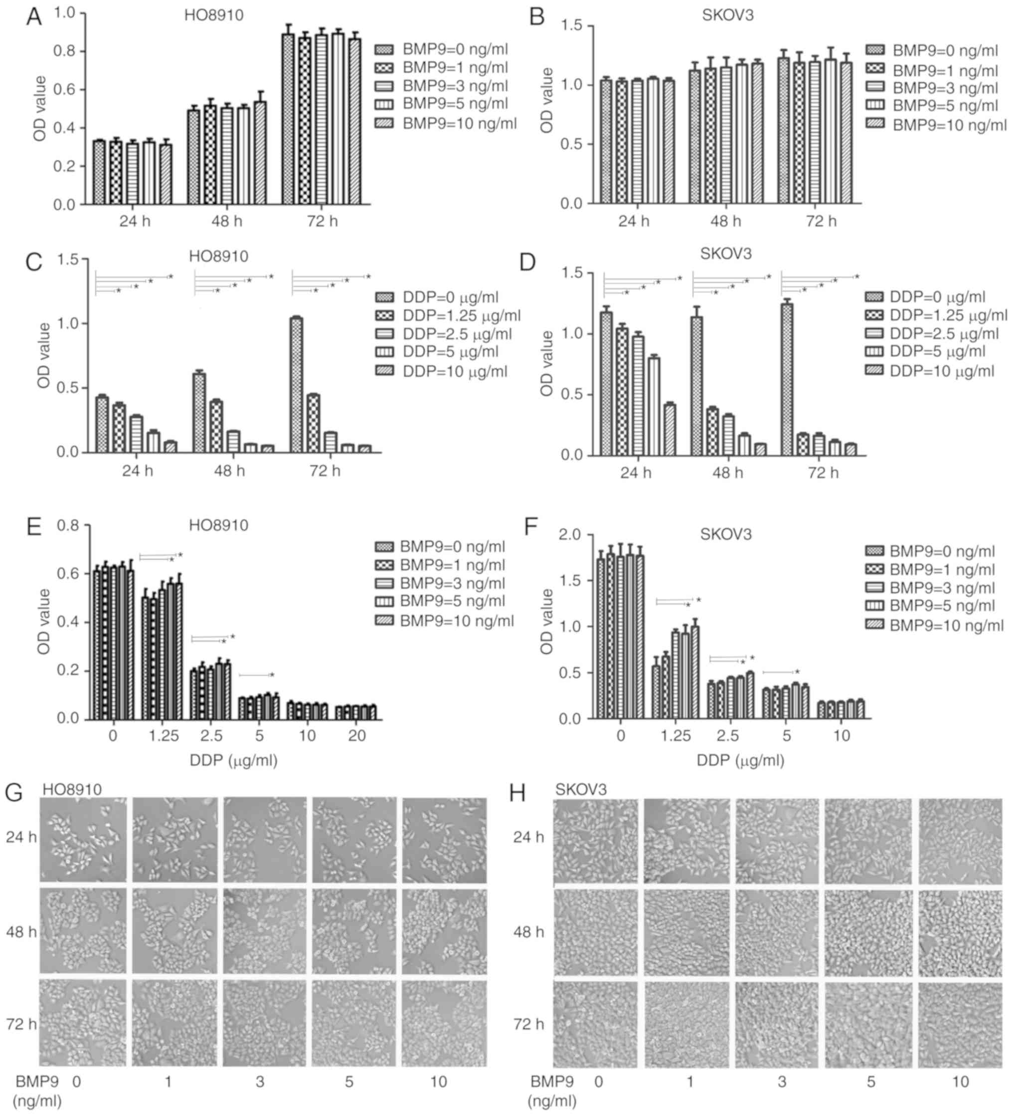
BMP9 enhances the chemoresistance of
ovarian cancer cells to DDP
To verify the effects of BMP9 on the efficacy of DDP
in ovarian cancer treatment, ovarian cancer cell lines (SKOV3 and
HO8910) were separately treated with BMP9 (Fig. 1A and B) or DDP (Fig. 1C and D), and in combination
(Fig. 1E and F). Cell morphology
was not notably altered following incubation with BMP9 (Fig. 1G and H). Results of the MTT assay
revealed no statistically significant differences among the OD
values of ovarian cancer cells treated with BMP9 (0, 1, 3, 5, and
10 ng/ml) for 24, 48 and 72 h, which indicated that BMP9 did not
significantly affect the ovarian cancer cell viability (Fig. 1A and B). However, the OD values of
ovarian cancer cells pretreated with BMP9 (5 and 10 ng/ml) for 72 h
and subsequently treated with DDP for a further 72 h were higher
than those of non-pretreated cells. On the other hand, BMP9 did not
blunt the cytotoxicity of DDP at concentrations of >5 µg/ml
(Fig. 1C and D). At a
concentration of 10 µg/ml, the inhibitory effects of treatment with
DDP for 72 h were significant, with inhibition ratios of 0.95±0.003
in H08910 cells and 0.93±0.01 in SKOV3 cells (Fig. 1C and D). These results indicated
that BMP9 partially counteracted the effects of DDP in ovarian
cancer cells, and that these effects were not caused by increased
cell viability.
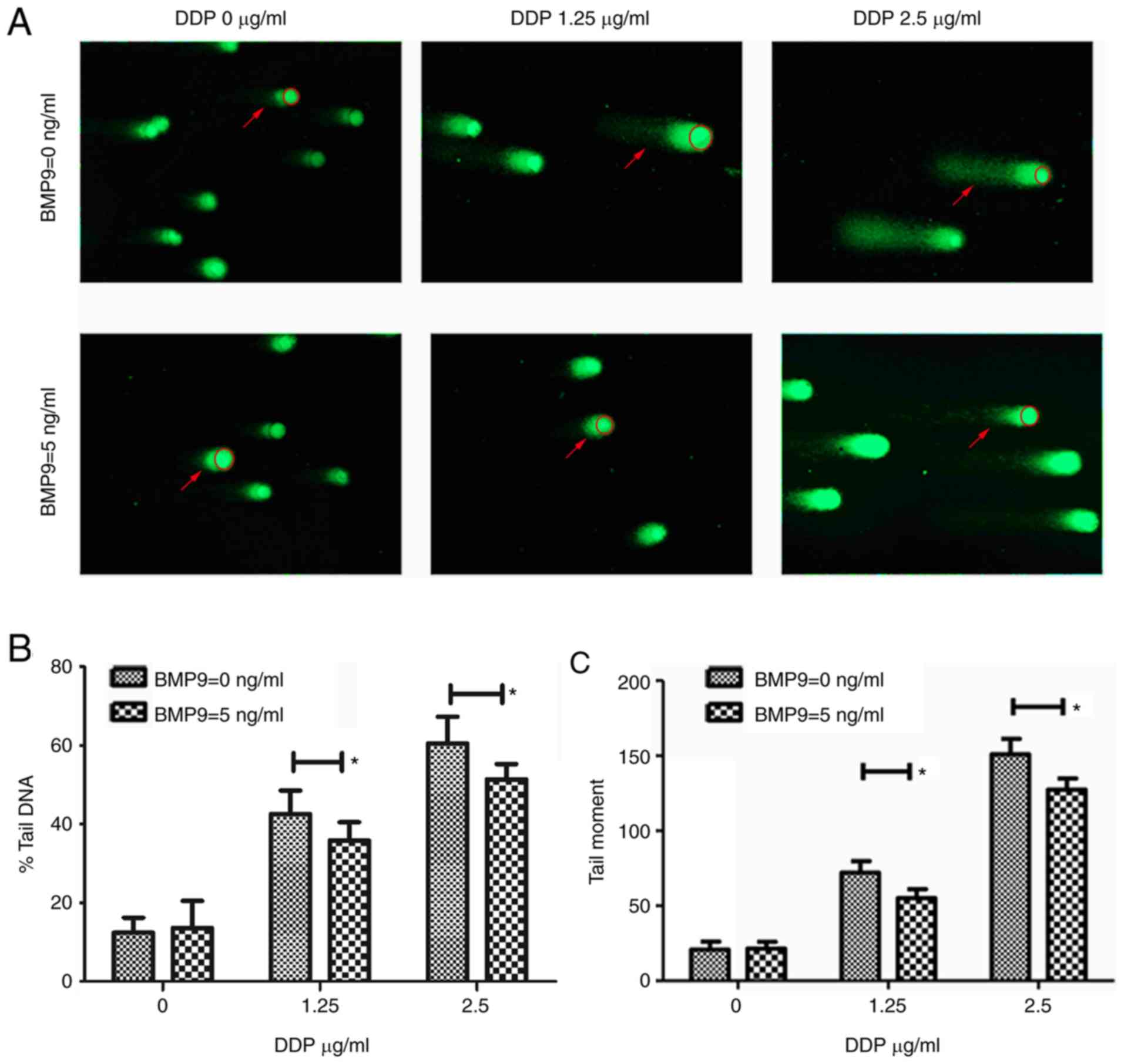
BMP9 reduces DDP efficacy by
attenuating DNA damage
DDP triggers apoptosis in cancer cells by forming
cross-links within DNA double strands, thereby leading to DNA
breakage and the generation of cleaved DNA fragments (28). The cytotoxic effect of DDP on SKOV3
was greater compared with HO8910. Upon treatment with 1.25 µg/ml
DDP for 72 h, the inhibitory effect of DDP on HO8910 was 0.57±0.01
(Fig. 1C), and the inhibitory
effect of DDP on SKOV3 was 0.86±0.01 (Fig. 1D), which was considered too severe.
Therefore, the HO8910 cell line was selected to evaluate the
effects of BMP9 on DNA damage. An alkaline comet assay was
conducted to evaluate the effects of BMP9 (5 ng/ml) on DDP-induced
DNA damage in HO8910 cells (1.25 and 2.5 µg/ml). Untreated cells
were used as the control group. The results demonstrated that BMP9
treatment reduced DNA quantity in the tail moment, and the % tail
DNA caused by DDP in HO8910 cells (Fig. 2A-C). Without considering the
distance of migration, % tail DNA is a normalized measure of the
percent of total cell DNA found in the tail. The above results
indicated that exposure to BMP9 prior to DDP treatment enhanced
ovarian cancer cell chemoresistance.
The apoptosis assay further confirmed the influence
of BMP9 on the apoptotic effects of DDP in HO8910 cells. Consistent
with the results of the comet assay, BMP9 (5 ng/ml) decreased the
apoptotic rate of HO8910 cells treated with DDP at 1.25 and 2.5
µg/ml (Fig. 2D). Therefore, it was
concluded that BMP9 treatment reduced DDP-induced DNA damage and
subsequently inhibited apoptosis in ovarian cancer cells.
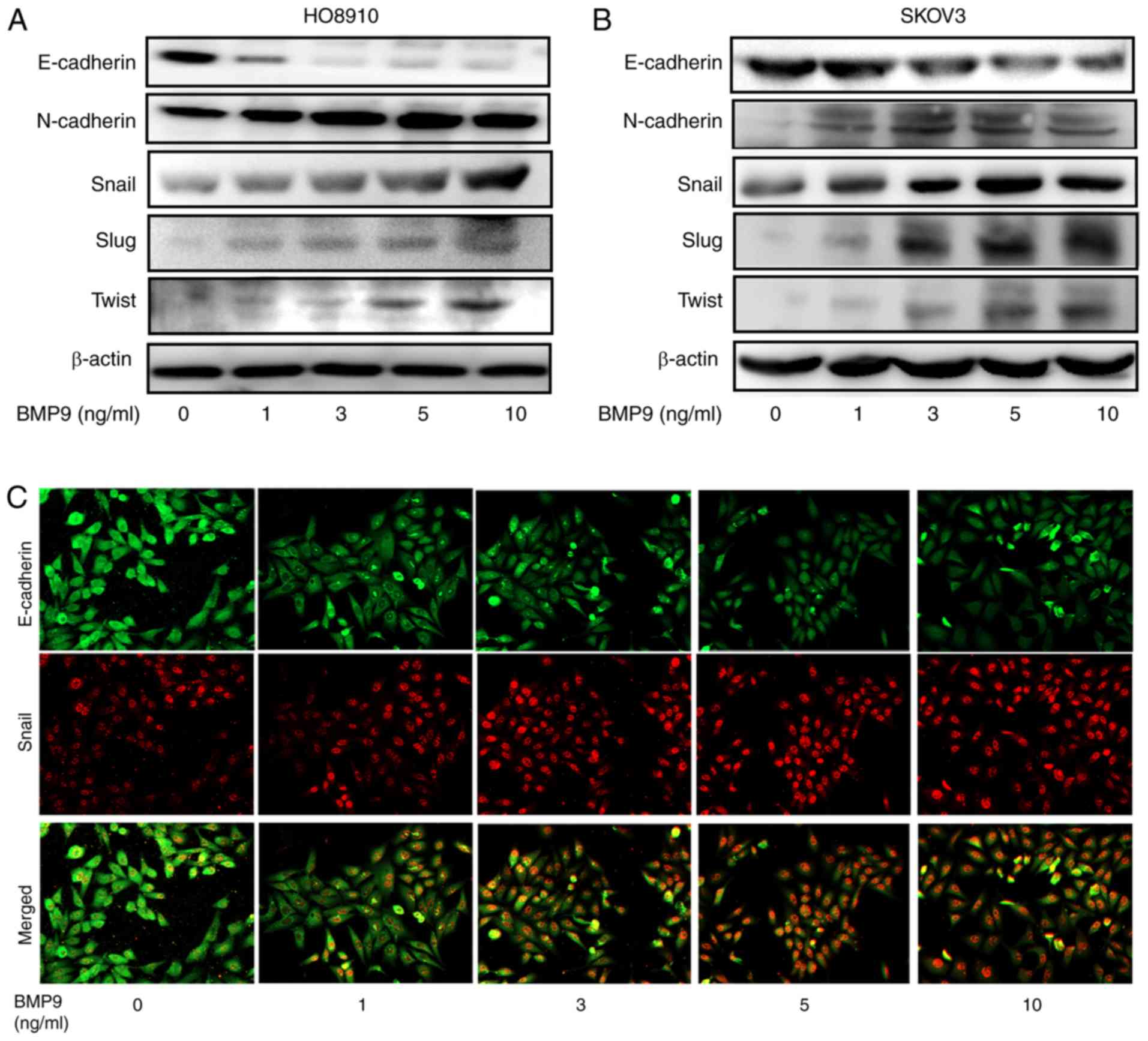
BMP9 induces EMT in ovarian cancer
cells
EMT has been demonstrated to act as the primary
mechanism responsible for chemoresistance during cancer treatment
(11). Therefore, whether
BMP9-induced resistance to DDP was associated with EMT was
investigated. Following BMP9 treatment (0, 1, 3, 5 and 10 ng/ml),
morphological alterations were not notable in HO8910 and SKOV3
cells. In addition, the protein expression levels of EMT markers,
including E-cadherin, N-cadherin, and Snail were detected via
western blotting and immunofluorescence analysis in HO8910 and
SKOV3 cells (Fig. 3). The results
revealed that BMP9 treatment (0, 1, 3, 5 and 10 ng/ml)
downregulated the expression of epithelial marker E-cadherin and
upregulated the expression of mesenchymal markers N-cadherin,
Snail, Slug and Twist in a dose-dependent manner. The above
findings demonstrated that BMP9 may promote EMT in ovarian cancer
cells, which may partially explain BMP9-induced DDP
chemoresistance.
Discussion
DDP is an important drug that is widely used for
chemotherapy in ovarian cancer (5). However, the efficiency of DDP is
significantly limited by the development of resistance during
therapy. BMP ligands are extracellular molecules that are secreted
by cancer and stromal cells into the tumor microenvironment, that
exert their effects on ovarian cancer cells by binding to
transmembrane receptors (20). In
the present study, BMP9 was demonstrated to reduce the cytotoxic
and apoptotic effects of DDP on ovarian cancer cells. In addition,
BMP9 treatment reduced DDP-induced DNA damage. These results
demonstrated that BMP9 enhanced the resistance of ovarian cancer
cells to DDP.
The association between BMPs and drug resistance
during chemotherapy has been previously reported in esophageal
carcinoma (29). Treatment with
TGF-β and BMP signaling pathway inhibitors has been reported to
enhance the antitumor effects of DDP in cancer cells (30,31).
Additionally, other members of the BMP family, such as BMP6, have
been implicated as negative chemoresistance-associated factors
(32). However, the effects of
BMP6 were evaluated based on protein expression in cancer cells
(32), and not as a ligand. The
results indicated that BMP ligands function independently of BMP
expression in cancer cells. Therefore, the effects of BMPs during
cancer treatment are versatile and require further examination.
Previous studies have demonstrated that BMPs,
including BMP2 and BMP9, promote the growth of ovarian cancer cells
(20,33). Therefore, the present study aimed
to investigate whether the proliferative effects of BMP9 influenced
the response of ovarian cancer cells to DDP. Notably, the results
indicated that BMP9-mediated DDP resistance was independent of the
proliferative effects of BMP9. Ovarian cancer cell viability
remained unchanged following exposure to BMP9 when compared to the
negative controls, concomitant with decreased sensitivity to DDP.
The above results indicated that the drug resistance mechanisms in
ovarian cancer cells were largely caused by an indirect effect on
other malignant phenotypes, excluding proliferation. The observed
non-proliferative effects of BMP9 appeared to be inconsistent with
the results of a previous study indicating that BMP9 serves as a
proliferative factor in ovarian cancer cells; the ovarian cancer
cells were exposed to serum-free medium containing BMP (33). This suggested that BMP9-induced
proliferation may be masked by physiologically relevant
concentrations of serum-derived BMP9 (33). In the present study, ovarian cancer
cells were cultured in medium containing serum that promoted the
proliferative environment to mimic the growth of ovarian cancer
in vivo. Therefore, the proliferative effect of BMP9 cannot
be excluded as an antagonistic mechanism against DDP in the
treatment of ovarian cancer cells. However, the results of the
present study indicated that BMP9-induced EMT serves a more
important role in chemoresistance.
BMP9 has been demonstrated to induce EMT, promote
the migration and increase the growth of cancer cells in
hepatocellular and renal carcinoma (26,34).
Furthermore, EMT has been associated with resistance to
platinum-based chemotherapy (11).
Thus, it was hypothesized that BMP9 may also induce EMT in ovarian
cancer cells, thus leading to chemoresistance to DDP. Alterations
in the expression of epithelial and mesenchymal markers in
BMP9-treated ovarian cancer cells were examined. The results
demonstrated that BMP9 downregulated the expression of E-cadherin
and upregulated the expression of N-cadherin, Snail, Slug and
Twist. These results indicated that BMP9 induced EMT in ovarian
cancer cells. Additionally, in the present study, cells treated
with BMP9 exhibited both upregulated mesenchymal-specific markers
and downregulated epithelial markers, but cell morphology had not
been altered. Such cells without spindle-shaped morphology are
likely to represent the intermediate stage of EMT, when epithelial
markers continue to be expressed but new mesenchymal markers have
already been acquired (35).
Although BMPs are dependent of the canonical mothers against
decapentaplegic homolog (SMAD) signaling pathway, they also
interact with non-SMAD signaling pathways, including the
Ras/RAF/mitogen-activated protein kinase and phosphoinositide
3-kinase/protein kinase B pathways, which may be induced by gene
alterations, including BRAF, KRAS or PTEN, and are involved in
triggering EMT (36,37). However, the cross talk between
mutation-activated signaling with EMT and BMP9 signaling pathways
remains to be elucidated.
EMT is known to promote the aggressiveness of
ovarian cancer cells (38) and
also to contribute to chemoresistance during treatment (39). In addition, patients with ovarian
cancer subjected to platinum-based chemotherapy were reported
exhibit and increase in EMT-like circulating tumor cells (40). Recently, EMT has been suggested to
cause an increased in the cancer stem cell-like properties of
cancer cells, which in turn further increases resistance to
chemotherapy (41). In addition,
Snail confers resistance to cell death induced by pro-apoptotic
signals (42). Previous studies
have successfully restored the cytotoxic effects of chemotherapy by
inhibiting EMT (43). Taken
together, these previous studies and the findings of the present
study indicated that BMP9-induced EMT contributes to DPP resistance
in ovarian cancer cells, and BMP9 antagonism may enhance the
sensitivity of ovarian cancer cells to DDP, which would potentially
benefit patients who have developed resistance to DDP
treatment.
In addition, EMT may lead to chemoresistance against
many drugs (41); the present
study only evaluated the effects of BMP-induced EMT on DDP
resistance. Subsequent studies should be extended to other drugs,
including carboplatin, paclitaxel and docetaxel, which are involved
in routine clinical adjuvant treatment. Taken together, the
findings of the current study indicated that BMP9 may be useful in
reducing acquired resistance to DDP during chemotherapy in ovarian
cancer. These findings have important implications for preventing
the development of chemoresistance during treatment against ovarian
cancer.
Acknowledgements
The authors would like to thank Dr Yuanyuan Wang
(Department of Oncology, The First Affiliated Hospital of Jinzhou
Medical University, Jinzhou, China) for analyzing the flow
cytometry results.
Funding
The present study was supported by Scientific
Research Starting Foundation of the Affiliated Hospital of Jinzhou
Medical University (grant no. FYK201202; Jinzhou, China).
Availability of data and materials
The datasets used and/or analysed during the current
study are available from the corresponding author on reasonable
request.
Authors' contributions
YW and BY contributed to the conception and design
of the study, acquired and analyzed the data, and drafted the
manuscript. JZ, XY, XL, LZ, YZ and XLL contributed to the design of
the study, acquired and analyzed the data, and revised the
manuscript. ZZ contributed to the conception and design of the
study, acquired and analyzed the data, and revised the article
critically for important intellectual content. All authors read and
approved the final manuscript.
Ethics approval and consent to
participate
Not applicable.
Patient consent for publication
Not applicable.
Competing interests
The authors declare that they have no competing
interests.
Glossary
Abbreviations
Abbreviations:
|
EMT
|
epithelial-mesenchymal transition
|
|
BMPs
|
bone morphogenetic proteins
|
|
TGF-β
|
transforming growth factor-β
|
|
DDP
|
cisplatin
|
References
|
1
|
Chen W, Zheng R, Baade PD, Zhang S, Zeng
H, Bray F, Jemal A, Yu XQ and He J: Cancer statistics in China,
2015. CA Cancer J Clin. 66:115–132. 2016. View Article : Google Scholar : PubMed/NCBI
|
|
2
|
Global Burden of Disease Cancer
Collaboration, ; Fitzmaurice C, Allen C, Barber RM, Barregard L,
Bhutta ZA, Brenner H, Dicker DJ, Chimed-Orchir O, Dandona R, et al:
Global, regional, and national cancer incidence, mortality, years
of life lost, years lived with disability, and disability-adjusted
life-years for 32 cancer groups, 1990 to 2015: A systematic
analysis for the global burden of disease study. JAMA Oncol.
3:524–548. 2017. View Article : Google Scholar : PubMed/NCBI
|
|
3
|
Zhao H, Wei W, Sun Y, Gao J, Wang Q and
Zheng J: Interference with the expression of β-catenin reverses
cisplatin resistance in A2780/DDP cells and inhibits the
progression of ovarian cancer in mouse model. DNA Cell Biol.
34:55–62. 2015. View Article : Google Scholar : PubMed/NCBI
|
|
4
|
Morgan SL, Medina JE, Taylor MM and
Dinulescu DM: Targeting platinum resistant disease in ovarian
cancer. Curr Med Chem. 21:3009–3020. 2014. View Article : Google Scholar : PubMed/NCBI
|
|
5
|
Ye H, Karim AA and Loh XJ: Current
treatment options and drug delivery systems as potential
therapeutic agents for ovarian cancer: A review. Mater Sci Eng C
Mater Biol Appl. 45:609–619. 2014. View Article : Google Scholar : PubMed/NCBI
|
|
6
|
McKeage MJ: New-generation platinum drugs
in the treatment of cisplatin-resistant cancers. Expert Opin
Investig Drugs. 14:1033–1046. 2005. View Article : Google Scholar : PubMed/NCBI
|
|
7
|
Yu X, Chen Y, Tian R, Li J, Li H, Lv T and
Yao Q: miRNA-21 enhances chemoresistance to cisplatin in epithelial
ovarian cancer by negatively regulating PTEN. Oncol Lett.
14:1807–1810. 2017. View Article : Google Scholar : PubMed/NCBI
|
|
8
|
Quail DF and Joyce JA: Microenvironmental
regulation of tumor progression and metastasis. Nat Med.
19:1423–1437. 2013. View
Article : Google Scholar : PubMed/NCBI
|
|
9
|
Pickup M, Novitskiy S and Moses HL: The
roles of TGFβ in the tumour microenvironment. Nat Rev Cancer.
13:788–799. 2013. View
Article : Google Scholar : PubMed/NCBI
|
|
10
|
Wakefield LM and Hill CS: Beyond TGFβ:
Roles of other TGFbeta superfamily members in cancer. Nat Rev
Cancer. 13:328–341. 2013. View
Article : Google Scholar : PubMed/NCBI
|
|
11
|
Marchini S, Fruscio R, Clivio L, Beltrame
L, Porcu L, Fuso Nerini I, Cavalieri D, Chiorino G, Cattoretti G,
Mangioni C, et al: Resistance to platinum-based chemotherapy is
associated with epithelial to mesenchymal transition in epithelial
ovarian cancer. Eur J Cancer. 49:520–530. 2013. View Article : Google Scholar : PubMed/NCBI
|
|
12
|
Liang S, Marti TM, Dorn P, Froment L, Hall
S, Berezowska S, Kocher G, Schmid RA and Peng R: 18P -
Epithelial-to-mesenchymal transition (EMT) is required for
resistance to anti-folate chemotherapy in lung cancer. J Thor Onc.
11:S632016. View Article : Google Scholar
|
|
13
|
Funaki S, Shintani Y, Kawamura T, Kanzaki
R, Minami M and Okumura M: Chemotherapy enhances programmed cell
death 1/ligand 1 expression via TGF-β induced epithelial
mesenchymal transition in non-small cell lung cancer. Oncol Rep.
38:2277–2284. 2017. View Article : Google Scholar : PubMed/NCBI
|
|
14
|
Ma W, Ma J, Xu J, Qiao C, Branscum A,
Cardenas A, Baron AT, Schwartz P, Maihle NJ and Huang Y: Lin28
regulates BMP4 and functions with Oct4 to affect ovarian tumor
microenvironment. Cell Cycle. 12:88–97. 2013. View Article : Google Scholar : PubMed/NCBI
|
|
15
|
Ehata S, Yokoyama Y, Takahashi K and
Miyazono K: Bi-directional roles of bone morphogenetic proteins in
cancer: Another molecular Jekyll and Hyde? Pathol Int. 63:287–296.
2013. View Article : Google Scholar : PubMed/NCBI
|
|
16
|
Wang K, Feng H, Ren W, Sun X, Luo J, Tang
M, Zhou L, Weng Y, He TC and Zhang Y: BMP9 inhibits the
proliferation and invasiveness of breast cancer cells MDA-MB-231. J
Cancer Res Clin Oncol. 137:1687–1696. 2011. View Article : Google Scholar : PubMed/NCBI
|
|
17
|
Wang J, Weng Y, Zhang M, Li Y, Fan M, Guo
Y, Sun Y, Li W and Shi Q: BMP9 inhibits the growth and migration of
lung adenocarcinoma A549 cells in a bone marrow stromal cellderived
microenvironment through the MAPK/ERK and NF-κB pathways. Oncol
Rep. 36:410–418. 2016. View Article : Google Scholar : PubMed/NCBI
|
|
18
|
Thawani JP, Wang AC, Than KD, Lin CY, La
Marca F and Park P: Bone morphogenetic proteins and cancer: Review
of the literature. Neurosurgery. 66:233–246; discussion 246. 2010.
View Article : Google Scholar : PubMed/NCBI
|
|
19
|
Shepherd TG and Nachtigal MW:
Identification of a putative autocrine bone morphogenetic
protein-signaling pathway in human ovarian surface epithelium and
ovarian cancer cells. Endocrinology. 144:3306–3314. 2003.
View Article : Google Scholar : PubMed/NCBI
|
|
20
|
Peng J, Yoshioka Y, Mandai M, Matsumura N,
Baba T, Yamaguchi K, Hamanishi J, Kharma B, Murakami R, Abiko K, et
al: The BMP signaling pathway leads to enhanced proliferation in
serous ovarian cancer-A potential therapeutic target. Mol Carcinog.
55:335–345. 2016. View
Article : Google Scholar : PubMed/NCBI
|
|
21
|
Peart TM, Correa RJ, Valdes YR, Dimattia
GE and Shepherd TG: BMP signalling controls the malignant potential
of ascites-derived human epithelial ovarian cancer spheroids via
AKT kinase activation. Clin Exp Metastasis. 29:293–313. 2012.
View Article : Google Scholar : PubMed/NCBI
|
|
22
|
Chen C, Grzegorzewski KJ, Barash S, Zhao
Q, Schneider H, Wang Q, Singh M, Pukac L, Bell AC, Duan R, et al:
An integrated functional genomics screening program reveals a role
for BMP-9 in glucose homeostasis. Nat Biotechnol. 21:294–301. 2003.
View Article : Google Scholar : PubMed/NCBI
|
|
23
|
David L, Mallet C, Keramidas M, Lamandé N,
Gasc JM, Dupuis-Girod S, Plauchu H, Feige JJ and Bailly S: Bone
morphogenetic protein-9 is a circulating vascular quiescence
factor. Circ Res. 102:914–922. 2008. View Article : Google Scholar : PubMed/NCBI
|
|
24
|
Herrera B, Dooley S and Breitkopf-Heinlein
K: Potential roles of bone morphogenetic protein (BMP)-9 in human
liver diseases. Int J Mol Sci. 15:5199–5220. 2014. View Article : Google Scholar : PubMed/NCBI
|
|
25
|
Ren W, Sun X, Wang K, Feng H, Liu Y, Fei
C, Wan S, Wang W, Luo J, Shi Q, et al: BMP9 inhibits the bone
metastasis of breast cancer cells by downregulating CCN2
(connective tissue growth factor, CTGF) expression. Mol Biol Rep.
41:1373–1383. 2014. View Article : Google Scholar : PubMed/NCBI
|
|
26
|
Li Q, Gu X, Weng H, Ghafoory S, Liu Y,
Feng T, Dzieran J, Li L, Ilkavets I, Kruithof-de Julio M, et al:
Bone morphogenetic protein-9 induces epithelial to mesenchymal
transition in hepatocellular carcinoma cells. Cancer Sci.
104:398–408. 2013. View Article : Google Scholar : PubMed/NCBI
|
|
27
|
Jung JW, Yoon SM, Kim S, Jeon YH, Yoon BH,
Yang SG, Kim MK, Choe S and Kuo MM: Bone morphogenetic protein-9 is
a potent growth inhibitor of hepatocellular carcinoma and reduces
the liver cancer stem cells population. Oncotarget. 7:73754–73768.
2016. View Article : Google Scholar : PubMed/NCBI
|
|
28
|
Sherma S and Lippard S: Structural aspects
of platinum anticancer durg interaction with DNA. Chem Rev.
87:1153–1157. 1987. View Article : Google Scholar
|
|
29
|
Zhou K, Shi X, Huo J, Liu W, Yang D, Yang
T, Qin T and Wang C: Bone morphogenetic protein 4 is overexpressed
in and promotes migration and invasion of drug-resistant cancer
cells. Int J Biol Macromol. 101:427–437. 2017. View Article : Google Scholar : PubMed/NCBI
|
|
30
|
Gao Y, Shan N, Zhao C, Wang Y, Xu F, Li J,
Yu X, Gao L and Yi Z: LY2109761 enhances cisplatin antitumor
activity in ovarian cancer cells. Int J Clin Exp Pathol.
8:4923–4932. 2015.PubMed/NCBI
|
|
31
|
Hover LD, Young CD, Bhola NE, Wilson AJ,
Khabele D, Hong CC, Moses HL and Owens P: Small molecule inhibitor
of the bone morphogenetic protein pathway DMH1 reduces ovarian
cancer cell growth. Cancer Lett. 368:79–87. 2015. View Article : Google Scholar : PubMed/NCBI
|
|
32
|
Lian WJ, Liu G, Liu YJ, Zhao ZW, Yi T and
Zhou HY: Downregulation of BMP6 enhances cell proliferation and
chemoresistance via activation of the ERK signaling pathway in
breast cancer. Oncol Rep. 30:193–200. 2013. View Article : Google Scholar : PubMed/NCBI
|
|
33
|
Herrera B, van Dinther M, Ten Dijke P and
Inman GJ: Autocrine bone morphogenetic protein-9 signals through
activin receptor-like kinase-2/Smad1/Smad4 to promote ovarian
cancer cell proliferation. Cancer Res. 69:9254–9262. 2009.
View Article : Google Scholar : PubMed/NCBI
|
|
34
|
Brand V, Lehamann C, Umkehrer C, Thier M,
de Wouters M, Raemsch R, Jucknischke U, Haas A and Breuer S: Impact
of selective anti-BMP9 treatment on tumor cells and tumor
angiogenesis. Mol Oncol. 10:1603–1620. 2016. View Article : Google Scholar : PubMed/NCBI
|
|
35
|
Kalluri R and Weinberg RA: The basics of
epithelial-mesenchymal transition. J Clin Invest. 119:1420–1428.
2009. View Article : Google Scholar : PubMed/NCBI
|
|
36
|
Zhang L, Ye Y, Long X, Xiao P, Ren X and
Yu J: BMP signaling and its paradoxical effects in tumorigenesis
and dissemination. Oncotarget. 7:78206–78218. 2016.PubMed/NCBI
|
|
37
|
Marcucci F, Stassi G and De Maria R:
Epithelial-mesenchymal transition: A new target in anticancer drug
discovery. Nat Rev Drug Discov. 15:311–325. 2016. View Article : Google Scholar : PubMed/NCBI
|
|
38
|
Rosso M, Majem B, Devis L, Lapyckyj L,
Besso MJ, Llauradó M, Abascal MF, Matos ML, Lanau L, Castellví J,
et al: E-cadherin: A determinant molecule associated with ovarian
cancer progression, dissemination and aggressiveness. PLoS One.
12:e01844392017. View Article : Google Scholar : PubMed/NCBI
|
|
39
|
Miow QH, Tan TZ, Ye J, Lau JA, Yokomizo T,
Thiery JP and Mori S: Epithelial-mesenchymal status renders
differential responses to cisplatin in ovarian cancer. Oncogene.
34:1899–1907. 2015. View Article : Google Scholar : PubMed/NCBI
|
|
40
|
Chebouti I, Kasimir-Bauer S, Buderath P,
Wimberger P, Hauch S, Kimmig R and Kuhlmann JD: EMT-like
circulating tumor cells in ovarian cancer patients are enriched by
platinum-based chemotherapy. Oncotarget. 8:48820–48831. 2017.
View Article : Google Scholar : PubMed/NCBI
|
|
41
|
Shibue T and Weinberg RA: EMT, CSCs, and
drug resistance: The mechanistic link and clinical implications.
Nat Rev Clin Oncol. 14:611–629. 2017. View Article : Google Scholar : PubMed/NCBI
|
|
42
|
Vega S, Morales AV, Ocaña OH, Valdés F,
Fabregat I and Nieto MA: Snail blocks the cell cycle and confers
resistance to cell death. Genes Dev. 18:1131–1143. 2004. View Article : Google Scholar : PubMed/NCBI
|
|
43
|
Liang SQ, Marti TM, Dorn P, Froment L,
Hall SR, Berezowska S, Kocher G, Schmid RA and Peng RW: Blocking
the epithelial-to-mesenchymal transition pathway abrogates
resistance to anti-folate chemotherapy in lung cancer. Cell Death
Dis. 6:e18242015. View Article : Google Scholar : PubMed/NCBI
|