|
1
|
Lisle R, Boekelaar M, Stannage K and
Whitewood C: Delayed diagnosis of developmental dislocation of the
hip: The Western Australian experience. ANZ J Surg. 82:612–615.
2012. View Article : Google Scholar : PubMed/NCBI
|
|
2
|
Shi D, Dai J, Zhu P, Qin J, Zhu L, Zhu H,
Zhao B, Qiu X, Xu Z, Chen D, et al: Association of the D repeat
polymorphism in the ASPN gene with developmental dysplasia of the
hip: A case-control study in Han Chinese. Arthritis Res Ther.
13:R272011. View
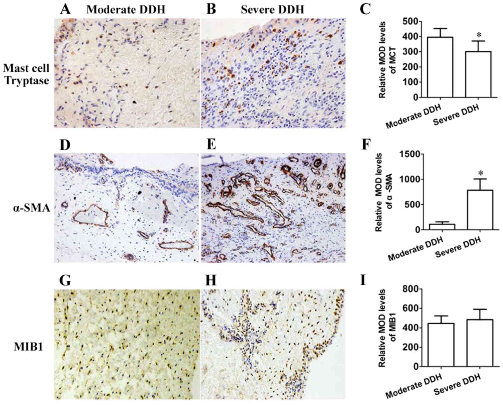
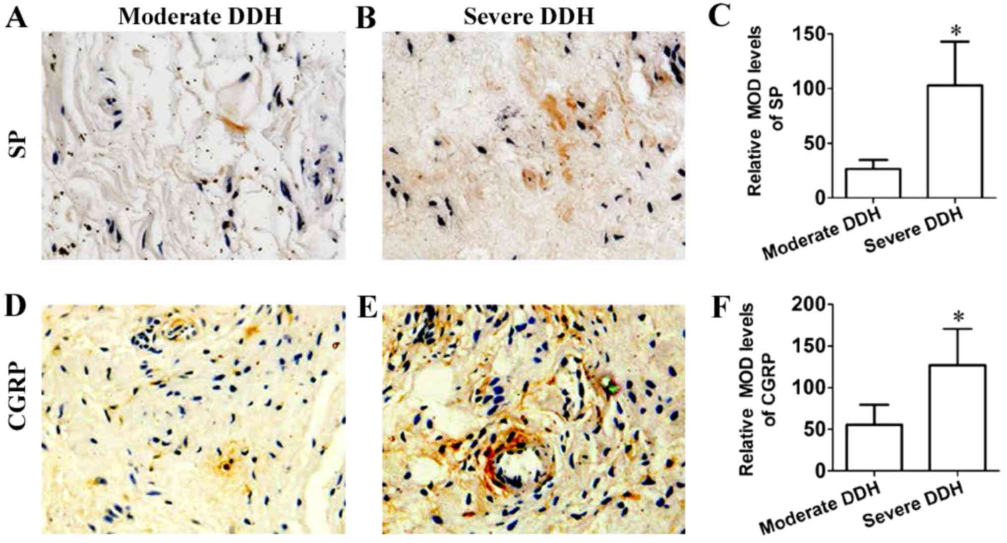
Article : Google Scholar : PubMed/NCBI
|
|
3
|
Sangal RB, Waryasz GR and Schiller JR:
Femoroacetabular impingement: A review of current concepts. R I Med
J (2013). 97:33–38. 2014.PubMed/NCBI
|
|
4
|
Alzaharani A, Bali K, Gudena R, Railton P,
Ponjevic D, Matyas JR and Powell JN: The innervation of the human
acetabular labrum and hip joint: An anatomic study. BMC
Musculoskelet Disord. 15:412014. View Article : Google Scholar : PubMed/NCBI
|
|
5
|
Henak CR, Abraham CL, Anderson AE, Maas
SA, Ellis BJ, Peters CL and Weiss JA: Patient-specific analysis of
cartilage and labrum mechanics in human hips with acetabular
dysplasia. Osteoarthritis Cartilage. 22:210–217. 2014. View Article : Google Scholar : PubMed/NCBI
|
|
6
|
Haversath M, Hanke J, Landgraeber S,
Herten M, Zilkens C, Krauspe R and Jäger M: The distribution of
nociceptive innervation in the painful hip: A histological
investigation. Bone Joint J 95-B. 770–776. 2013. View Article : Google Scholar
|
|
7
|
Nakajima T, Ohtori S, Inoue G, Koshi T,
Yamamoto S, Nakamura J, Takahashi K and Harada Y: The
characteristics of dorsal-root ganglia and sensory innervation of
the hip in rats. J Bone Joint Surg Br. 90:254–257. 2008. View Article : Google Scholar : PubMed/NCBI
|
|
8
|
Henrotin Y, Lambert C and Richette P:
Importance of synovitis in osteoarthritis: Evidence for the use of
glycosaminoglycans against synovial inflammation. Semin Arthritis
Rheum. 43:579–587. 2014. View Article : Google Scholar : PubMed/NCBI
|
|
9
|
Henrotin Y, Pesesse L and Lambert C:
Targeting the synovial angiogenesis as a novel treatment approach
to osteoarthritis. Ther Adv Musculoskelet Dis. 6:20–34. 2014.
View Article : Google Scholar : PubMed/NCBI
|
|
10
|
Shirai C, Ohtori S, Kishida S, Harada Y
and Moriya H: The pattern of distribution of PGP 9.5 and TNF-alpha
immunoreactive sensory nerve fibers in the labrum and synovium of
the human hip joint. Neurosci Lett. 450:18–22. 2009. View Article : Google Scholar : PubMed/NCBI
|
|
11
|
Takeshita M, Nakamura J, Ohtori S, Inoue
G, Orita S, Miyagi M, Ishikawa T and Takahashi K: Sensory
innervation and inflammatory cytokines in hypertrophic synovia
associated with pain transmission in osteoarthritis of the hip: A
case-control study. Rheumatology (Oxford). 51:1790–1795. 2012.
View Article : Google Scholar : PubMed/NCBI
|
|
12
|
Beckmann J, Schubert J, Morhenn HG, Grau
V, Schnettler R and Lips KS: Expression of choline and
acetylcholine transporters in synovial tissue and cartilage of
patients with rheumatoid arthritis and osteoarthritis. Cell Tissue
Res. 359:465–477. 2015. View Article : Google Scholar : PubMed/NCBI
|
|
13
|
de Lange-Brokaar BJ, Ioan-Facsinay A, van
Osch GJ, Zuurmond AM, Schoones J, Toes RE, Huizinga TW and
Kloppenburg M: Synovial inflammation, immune cells and their
cytokines in osteoarthritis: A review. Osteoarthritis Cartilage.
20:1484–1499. 2012. View Article : Google Scholar : PubMed/NCBI
|
|
14
|
Kim YS, Kim JM, Lee YG, Hong OK, Kwon HS
and Ji JH: Intercellular adhesion molecule-1 (ICAM-1, CD54) is
increased in adhesive capsulitis. J Bone Joint Surg Am.
95:e181–e188. 2013. View Article : Google Scholar : PubMed/NCBI
|
|
15
|
Hand GC, Athanasou NA, Matthews T and Carr
AJ: The pathology of frozen shoulder. J Bone Joint Surg Br.
89:928–932. 2007. View Article : Google Scholar : PubMed/NCBI
|
|
16
|
Wang H, Zhang X, He JY, Zheng XF, Li D, Li
Z, Zhu JF, Shen C, Cai GQ and Chen XD: Increasing expression of
substance P and calcitonin gene-related peptide in synovial tissue
and fluid contribute to the progress of arthritis in developmental
dysplasia of the hip. Arthritis Res Ther. 17:42015. View Article : Google Scholar : PubMed/NCBI
|
|
17
|
Crowe JF, Mani VJ and Ranawat CS: Total
hip replacement in congenital dislocation and dysplasia of the hip.
J Bone Joint Surg Am. 61:15–23. 1979. View Article : Google Scholar : PubMed/NCBI
|
|
18
|
Hart DJ and Spector TD: The classification
and assessment of osteoarthritis. Baillieres Clin Rheumatol.
9:407–432. 1995. View Article : Google Scholar : PubMed/NCBI
|
|
19
|
Cashman JP, Round J, Taylor G and Clarke
NM: The natural history of developmental dysplasia of the hip after
early supervised treatment in the Pavlik harness. A prospective,
longitudinal follow-up. J Bone Joint Surg Br. 84:418–425. 2002.
View Article : Google Scholar : PubMed/NCBI
|
|
20
|
Nakamura S, Ninomiya S and Nakamura T:
Primary osteoarthritis of the hip joint in Japan. Clin Orthop Relat
Res. 190–196. 1989.PubMed/NCBI
|
|
21
|
Rondelet B, Kerbaul F, Motte S, van
Beneden R, Remmelink M, Brimioulle S, McEntee K, Wauthy P, Salmon
I, Ketelslegers JM and Naeije R: Bosentan for the prevention of
overcirculation-induced experimental pulmonary arterial
hypertension. Circulation. 107:1329–1335. 2003. View Article : Google Scholar : PubMed/NCBI
|
|
22
|
O'Neill TW, Parkes MJ, Maricar N,
Marjanovic EJ, Hodgson R, Gait AD, Cootes TF, Hutchinson CE and
Felson DT: Synovial tissue volume: A treatment target in knee
osteoarthritis (OA). Ann Rheum Dis. 75:84–90. 2016. View Article : Google Scholar : PubMed/NCBI
|
|
23
|
Liu-Bryan R: Synovium and the innate
inflammatory network in osteoarthritis progression. Curr Rheumatol
Rep. 15:3232013. View Article : Google Scholar : PubMed/NCBI
|
|
24
|
Adães S, Mendonça M, Santos TN,
Castro-Lopes JM, Ferreira-Gomes J and Neto FL: Intra-articular
injection of collagenase in the knee of rats as an alternative
model to study nociception associated with osteoarthritis.
Arthritis Res Ther. 16:R102014. View
Article : Google Scholar : PubMed/NCBI
|
|
25
|
Daghestani HN, Pieper CF and Kraus VB:
Soluble macrophage biomarkers indicate inflammatory phenotypes in
patients with knee osteoarthritis. Arthritis Rheumatol. 67:956–965.
2015. View Article : Google Scholar : PubMed/NCBI
|
|
26
|
Zhang RX, Ren K and Dubner R:
Osteoarthritis pain mechanisms: Basic studies in animal models.
Osteoarthritis Cartilage. 21:1308–1315. 2013. View Article : Google Scholar : PubMed/NCBI
|
|
27
|
Wakamatsu K, Nanki T, Miyasaka N, Umezawa
K and Kubota T: Effect of a small molecule inhibitor of nuclear
factor-kappaB nuclear translocation in a murine model of arthritis
and cultured human synovial cells. Arthritis Res Ther.
7:R1348–R1359. 2005. View
Article : Google Scholar : PubMed/NCBI
|
|
28
|
Laragione T and Gulko PS: Liver X receptor
regulates rheumatoid arthritis fibroblast-like synoviocyte
invasiveness, matrix metalloproteinase 2 activation, interleukin-6
and CXCL10. Mol Med. 18:1009–1017. 2012. View Article : Google Scholar : PubMed/NCBI
|
|
29
|
Mifková A, Kodet O, Szabo P, Kučera J,
Dvořánková B, André S, Koripelly G, Gabius HJ, Lehn JM and Smetana
K Jr: Synthetic polyamine BPA-C8 inhibits TGF-β1-mediated
conversion of human dermal fibroblast to myofibroblasts and
establishment of galectin-1-rich extracellular matrix in vitro.
Chembiochem. 15:1465–1470. 2014. View Article : Google Scholar : PubMed/NCBI
|
|
30
|
Minton K: Extracellular matrix:
Preconditioning the ECM for fibrosis. Nat Rev Mol Cell Biol.
15:766–767. 2014. View Article : Google Scholar : PubMed/NCBI
|
|
31
|
Shinde AV and Frangogiannis NG:
Fibroblasts in myocardial infarction: A role in inflammation and
repair. J Mol Cell Cardiol. 70:74–82. 2014. View Article : Google Scholar : PubMed/NCBI
|
|
32
|
Leech MT and Morand EF: Fibroblasts and
synovial immunity. Curr Opin Pharmacol. 13:565–569. 2013.
View Article : Google Scholar : PubMed/NCBI
|
|
33
|
Sobel K, Tham M, Stark HJ, Stammer H,
Prätzel-Wunder S, Bickenbach JR and Boukamp P: Wnt-3a-activated
human fibroblasts promote human keratinocyte proliferation and
matrix destruction. Int J Cancer. 136:2786–2798. 2015. View Article : Google Scholar : PubMed/NCBI
|
|
34
|
Kägebein D, Gutjahr M, Große C, Vogel AB,
Rödel J and Knittler MR: Chlamydia trachomatis-infected epithelial
cells and fibroblasts retain the ability to express
surface-presented major histocompatibility complex class I
molecules. Infect Immun. 82:993–1006. 2014. View Article : Google Scholar : PubMed/NCBI
|
|
35
|
Scanzello CR and Goldring SR: The role of
synovitis in osteoarthritis pathogenesis. Bone. 51:249–257. 2012.
View Article : Google Scholar : PubMed/NCBI
|
|
36
|
Nigrovic PA and Lee DM: Synovial mast
cells: Role in acute and chronic arthritis. Immunol Rev. 217:19–37.
2007. View Article : Google Scholar : PubMed/NCBI
|
|
37
|
da Silva EZ, Jamur MC and Oliver C: Mast
cell function: A new vision of an old cell. J Histochem Cytochem.
62:698–738. 2014. View Article : Google Scholar : PubMed/NCBI
|