Introduction
Osteosarcoma (OS) is the most common primary
malignant bone tumor found in children and adolescents, which
mostly occurs in the metaphyseal part of tubular bones such as the
distal femur, proximal humerus and proximal tibia (1,2). A
previous study revealed that the introduction of neoadjuvant
chemotherapy and advanced surgical techniques have significantly
increased the overall survival rate from 20 to 70% (3). However, the overall survival rate of
patients with lung metastases is only 10–30% (4). Further studies are necessary to
understand the underlying mechanisms of OS metastasis and thereby
identify novel therapeutic targets.
Tumor metastasis is a complex, multistage process.
An increasing body of evidence has suggested that EMT plays an
important role in tumor progression and metastasis (5–7). EMT
is a physiological process whereby epithelial cells lose their
epithelial characteristics and convert to a mesenchymal phenotype
(8). At the molecular level,
E-cadherin expression is repressed, while mesenchymal marker
expression (including N-cadherin and vimentin) is increased
(9). Many signaling pathways
(TGF-β, Notch, Wnt/β-catenin, Hedgehog and Hippo) induce and
modulate the EMT process (10–14).
However, complex molecular networks that influence EMT in OS have
yet to be clarified.
Receptor interacting protein kinase 4 (RIPK4), a
member of the RIP kinase family, is comprised of a homologous
N-terminal serine/threonine kinase domain, an intermediate domain
and a C-terminal region containing ankyrin repeat domain 11
(15,16). Early RIPK4 studies focused mainly
on keratinocyte proliferation and differentiation (17). Recent studies have demonstrated
that RIPK4 was overexpressed in various types of human cancers,
including cervical, skin, colorectal and ovarian cancers (18,19).
Furthermore, RIPK4 overexpression has been associated with
progression and poor prognosis. However, RIPK4 acted as a tumor
suppressor in other types of cancers, such as tongue cancer and
hepatocellular carcinoma (20,21).
The biological function of RIPK4 in OS is largely unknown. The
present study detected the expression levels of RIPK4 in OS tissues
and cell lines. Furthermore, its relationship with
clinicopathological parameters and prognosis in OS patients were
analyzed, and its effects on EMT and the underlying molecular
mechanisms were investigated.
Materials and methods
Tissue specimens
A total of 36 OS tissues and 15 normal bone tissues
were obtained from The Second Hospital of Lanzhou University
(Gansu, China) from January 2010 to December 2015, and the
clinicopathological parameters of the 36 OS patients were recorded
(Table I). The 36 OS patients were
followed up by telephone, outpatient service and letters from the
date of operation. The deadline was May 2016. The follow-up period
was 5–72 months. The median follow-up time was 27 months.
Furthermore, 4 fresh specimens of OS and normal bone tissue were
collected and stored at −80°C for western blotting. None of the
patients were undergoing chemotherapy or radiotherapy treatment.
The present study was approved by the Medical Ethics Committee of
Lanzhou University and informed consent forms were signed by the
patients.
 | Table I.Relationship between expression of
RIPK4 and clinicopathologic parameters, RIPK4, receptor interacting
protein kinase 4. |
Table I.
Relationship between expression of
RIPK4 and clinicopathologic parameters, RIPK4, receptor interacting
protein kinase 4.
|
|
| RIPK4 expression
level |
|
|
|---|
|
|
|
|
|
|
|---|
| Clinicopathological
parameters | Total n | High | Low | χ2 | P-value |
|---|
| Sex |
|
|
|
|
|
| Male | 20 | 13 | 7 | 0.024 | 0.877 |
|
Female | 16 | 10 | 6 |
|
|
| Age/year |
|
|
|
|
|
|
>14 | 31 | 19 | 12 | 0.653 | 0.419 |
|
≤14 | 5 | 4 | 1 |
|
|
| Tumor size, cm |
|
|
|
|
|
|
>8 | 17 | 14 | 3 | 4.760 | 0.029a |
| ≤8 | 19 | 9 | 10 |
|
|
| Tumor site |
|
|
|
|
|
|
Femur | 17 | 9 | 8 | 1.798 | 0.407 |
| Tibia
and Fibula | 9 | 7 | 2 |
|
|
|
Other | 10 | 7 | 3 |
|
|
| Histological
type |
|
|
|
|
|
|
Osteoblastic | 21 | 12 | 9 | 3.739 | 0.291 |
|
Chondroblastic | 6 | 3 | 3 |
|
|
|
Fibroblastic | 5 | 4 | 1 |
|
|
|
Mixed | 4 | 4 | 0 |
|
|
| Enneking stage |
|
|
|
|
|
|
I/IIA | 9 | 2 | 7 | 9.030 | 0.003b |
|
IB/III | 27 | 21 | 6 |
|
|
| Metastasis |
|
|
|
|
|
|
Yes | 13 | 10 | 3 | 1.498 | 0.221 |
| No | 23 | 13 | 10 |
|
|
Cell culture and transfection
The human OS cell lines MG-63 and U2OS, and the
normal osteoblastic cell line hFOB 1.19 were obtained from the
Chinese Academy of Medical Sciences. MG-63 cells were cultured in
Minimum Essential Medium, U2OS in McCoy's 5A medium, hFOB 1.19 in
Dulbecco's modified Eagle's medium/F-12 medium (all from Hyclone;
GE Healthcare Life Sciences), and supplemented with 10% fetal
bovine serum (FBS; PAN-Biotech GmbH) and 100 U/ml
penicillin/streptomycin, at 37°C in a humidified atmosphere
containing 5% CO2. For RIPK4 expression silencing, the 6
µl RIPK4 small interfering (si)-RNA and 6 µl negative control siRNA
products (Santa Cruz Biotechnology, Inc.) were transfected into 80%
confluence MG-63 and U2OS cells using a siRNA reagent system (Santa
Cruz Biotechnology, Inc.) according to the manufacturer's
specifications. RIPK4-siRNA sense sequences:
5′-CACACGCAGUAUGAAGAUA-3′; antisense sequences:
5′-UAUCUUCAUACUGCGUGUG-3′; NC-siRNA sense sequences:
5′-UUGUGGCCUGUUAGCUUCA-3′; antisense sequences:
5′-UGAAGCUAACAGGCCACAA-3′. Following incubation for 48 h, RIPK4
expression levels were detected by western blot analysis.
Western blot analysis
Tissues and cells were lysed with RIPA buffer
(Beyotime Institute of Biotechnology) on ice containing a protease
inhibitor supplemented with 1 mmol/l phenylmethylsulfonyl fluoride.
Protein concentration was detected by a BCA protein assay kit
(Beyotime Institute of Biotechnology). Then equal amounts of
protein samples (40 µg) were separated by 8 or 10% SDS-PAGE and
transferred onto PVDF membranes. The membranes were blocked with 5%
non-fat dry milk for 2 h at room temperature, and subsequently
incubated overnight at 4°C with primary antibodies, namely:
Anti-RIPK4 (1:200, cat no. ab124649), anti-E-cadherin (1:500, cat
no. ab152102), anti-N-cadherin (1:1,000, cat no. ab76011),
anti-vimentin (1:500, cat no. ab8978), anti-β-catenin (1:500, cat
no. ab16051; all Abcam), anti-β-actin (1:5,000, cat no. 1978,
Sigma-Aldrich; Merck KGaA) and anti-Lamin B1 (1:5,000, cat no.
bsm-33442M, BIOSS). After washing three times with Tris-buffered
saline and 0.05% Tween (pH 7.4), the membranes were incubated with
the horseradish peroxidase (HRP)-conjugated anti-rabbit or
anti-mouse immunoglobulin (Ig)-G secondary antibodies (1:5,000,
catalog no. A0208; Beyotime Institute of Biotechnology) for 2 h at
room temperature. The protein bands were then detected using an ECL
system (EMD Millipore) and imaged using a ImageQuant TL A600
imaging system (GE Healthcare Biosciences). The density of each
band was analyzed with Pro-Plus 6.0 Software (Media Cybernetics,
Inc.).
Cell proliferation assay
The cell proliferation assay was evaluated with a
Cell Counting Kit-8 (CCK-8; Dojindo Molecular Technologies, Inc.).
At 48 h after the transfection incubation period, cells were seeded
in a 96-well plate at 3,000 cells/well with 100 µl culture medium.
At 0, 24, 48 and 72 h, 10 µl CCK-8 reagent solution was added to
each well and incubated for 4 h at 37°C. Absorbance was measured
using a microplate reader set at a 450 nm wavelength.
Migration and invasion assay
For the migration and invasion assays, 24-well
plates containing 8.0 µm pore size Transwell chambers (Corning
Incorporated) were used according to the manufacturer's
specifications. For the migration assay, ~1×10⁵
cells/well (48 h post transfection incubation period) were seeded
in the upper chambers with 100 µl of serum-free medium. Thereafter,
500 µl medium supplemented with 10% FBS as a chemoattractant was
added to the lower chambers of the wells. After 24 h of incubation
at 37°C, non-migrating cells on the upper surface of the membrane
were removed with cotton swabs, while migrating cells on the lower
surface of the membrane were fixed with 4% paraformaldehyde for 30
min, stained with 0.1% crystal violet for 10 min at 37°C and
photographed under an inverted microscope (magnification, ×200).
The invasion assay was performed in a similar manner, except 100 µl
of 1:10 serum-free medium-diluted Matrigel (BD Biosciences) was
also added to each well before the cells were seeded in the
membrane.
Wound healing assay
Following 48 h of transfection, the cells were
trypsinized by 0.25% trypsin, and 1×10⁵ cells seeded
into 6-well plates. When cell confluency reached 95%, a sterile 200
µl pipette tip was used to make a scratch. After washing with PBS,
cells were then incubated in serum-free medium for 24 h at 37°C.
The width of the scratch was measured under an inverted microscope
at 0 and 24 h, respectively.
Immunofluorescence staining
Cells (1×10⁵) were cultured on glass
coverslips in a 35 mm diameter dish. After 24 h, the cells were
fixed with 4% paraformaldehyde for 30 min at 37°C, permeabilized
with 0.1% Triton X-100 in PBS for 15 min at 37°C, blocked with 5%
bovine serum albumin (BSA, Beyotime Institute of Biotechnology) in
PBS for 1 h, and incubated with primary antibodies against
E-cadherin (1:100, cat. no. ab152102), N-cadherin (1:100, cat. no.
ab76011), vimentin (1:100, cat. no. ab8978) and β-catenin (1:100,
cat. no. ab16051; all from Abcam) at 4°C overnight. The cells were
then washed three times with PBS and incubated with fluorescein
isothiocyanate-conjugated goat anti-mouse or anti-rabbit secondary
antibodies (1:5,000, cat. nos. A0412 and A0423; Beyotime Institute
of Biotechnology) for 2 h at 37°C. Subsequently the nuclei were
counterstained with 4′,6-diamidino-2-phenylindole for 10 min at
37°C, and images were captured using a fluorescence microscope
(Olympus Corporation).
Immunohistochemical staining
Tissue samples were fixed with 10% formalin for 30
min at 37°C, embedded in paraffin, and cut into 4 µm sections. The
sections were dewaxed in xylene and rehydrated in gradients of high
ethanol concentration to distilled water. Endogenous peroxidase
activity was quenched by 3% H2O2 for 15 min
at 37°C. The sections were heated for 30 min at 95°C in 10 nM, pH
6.0 sodium citrate buffer for antigen retrieval, blocked with 5%
BSA (Beyotime Institute of Biotechnology) at 37°C for 1 h and
incubated overnight at 4°C with primary antibody against RIPK4
(1:200, cat. no. ab124649, Abcam). After rinsing with PBS, the
sections were incubated 1 h at room temperature with HRP-conjugated
goat anti-mouse IgG antibody (1:5,000, cat. no. ZB-2301, Beijing
Zhongshan Golden Bridge Biotechnology Co., Ltd.). Then the sections
were stained at 37°C for 3 min using 3,3′-diaminobenzidine,
counterstained for 1 min with Gill's hematoxylin and observed under
a light microscope (magnification, ×200). The stained results were
scored independently by two pathologists. The percentage of
positive staining cells were scored as follows: 0, <5%; 1,
5–25%; 2, 26–50%; 3, 51–75%; and 4, >75%. Staining intensity was
graded as follows: 0, no staining; 1, light brown; 2, brown; and 3,
dark brown. The final immunoreactive score was calculated by
multiplying the percentage of positive staining cells with the
staining intensity. Scores of 0–3 and 4–12 were designated as low
and high expression, respectively.
Statistical analysis
All statistical analyses was performed using SPSS
version 19.0 software (IBM Corp.). All data were presented as mean
± standard deviation. Continuous variables were analyzed by a
two-tailed Student's t-test or one-way analysis of variance
followed by Tukey's post hoc test. The association between RIPK4
expression levels and the clinicopathological parameters of the
patients was assessed using the Chi-squared test. Overall survival
was evaluated using the Kaplan-Meier method and a log-rank test.
P<0.05 was considered to indicate a statistically significant
difference. All experiments were independently repeated at least
three times.
Results
RIPK4 expression is upregulated in
human OS tissues and is associated with poor prognosis
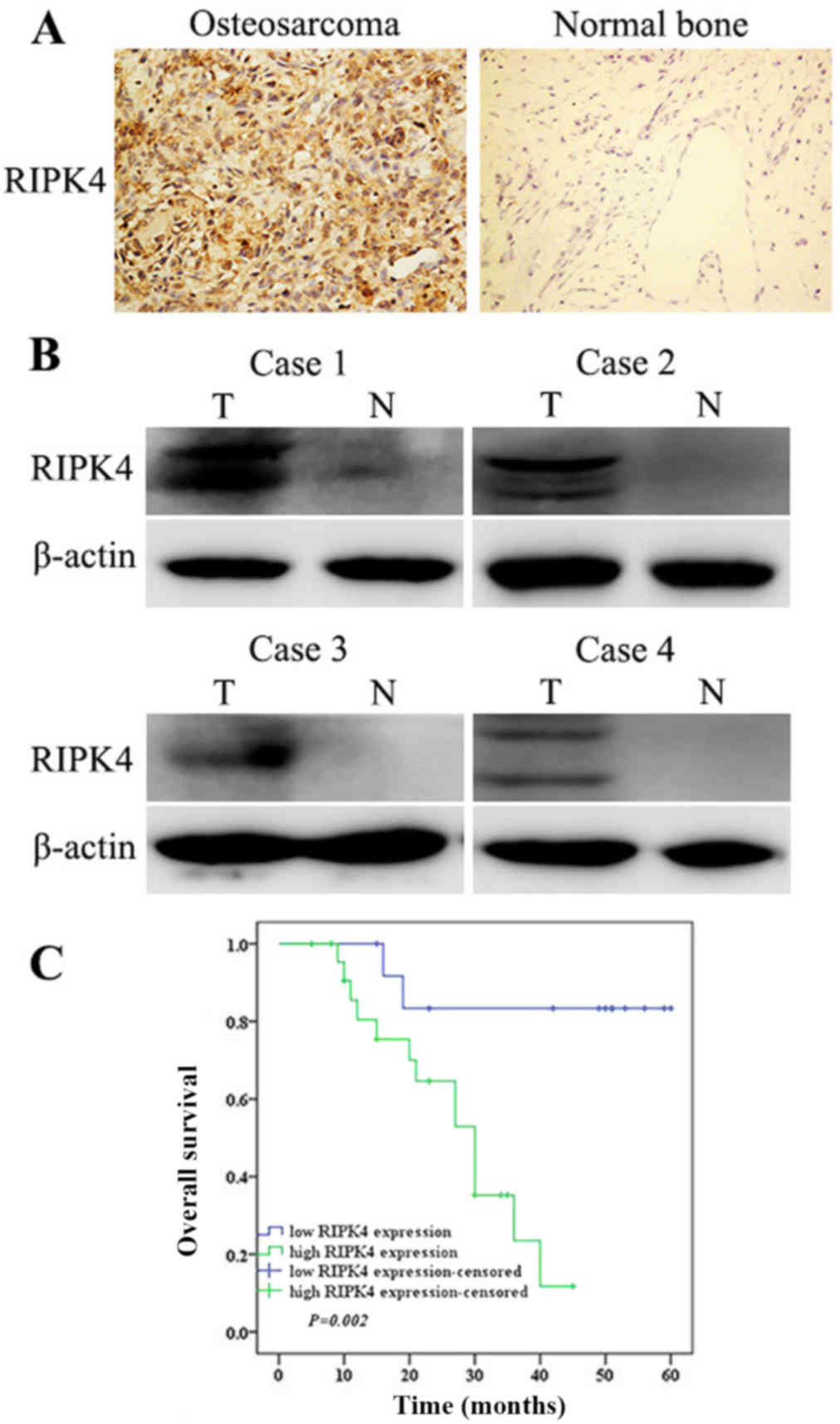
To investigate RIPK4 expression in OS, 36 OS tissues
and 15 normal bone tissues were analyzed via immunohistochemistry.
The results revealed that high RIPK4 expression was detected in
63.9% (23/36) of OS tissues, which was significantly higher than
that observed in normal bone tissues (0/15; Fig. 1A). Additionally, western blotting
was conducted to analyze the protein levels of RIPK4 in 4 fresh OS
and normal bone tissues. Consistent with the immunochemistry
results, RIPK4 protein levels were upregulated in OS tissues when
compared with those in normal bone tissues (Fig. 1B). The present study also analyzed
the relationship between RIPK4 expression and clinicopathological
parameters in OS. High RIPK4 expression was significantly
associated with larger tumor sizes and advanced Enneking stage.
There were no significant associations between high RIPK4
expression and other clinicopathological parameters, including sex,
age, tumor site, histological type and metastasis (Table I).
All OS patients had follow-up consultations from
their surgery date until they succumbed to the disease or the last
follow-up of the present study (range, 5–72 months; median
follow-up, 27 months). Kaplan-Meier survival analysis was used to
evaluate the association between RIPK4 expression and the prognosis
survival of patients with OS. Patients with high RIPK4 expression
had a significantly shorter survival time (median survival time, 21
months) as opposed to those with low RIPK4 expression (median
survival time, 42 months; Fig.
1C). Taken together, these results suggest that RIPK4 is
associated with OS progression and prognosis.
Silencing of RIPK4 inhibits cell
proliferation, migration and invasion in OS cells
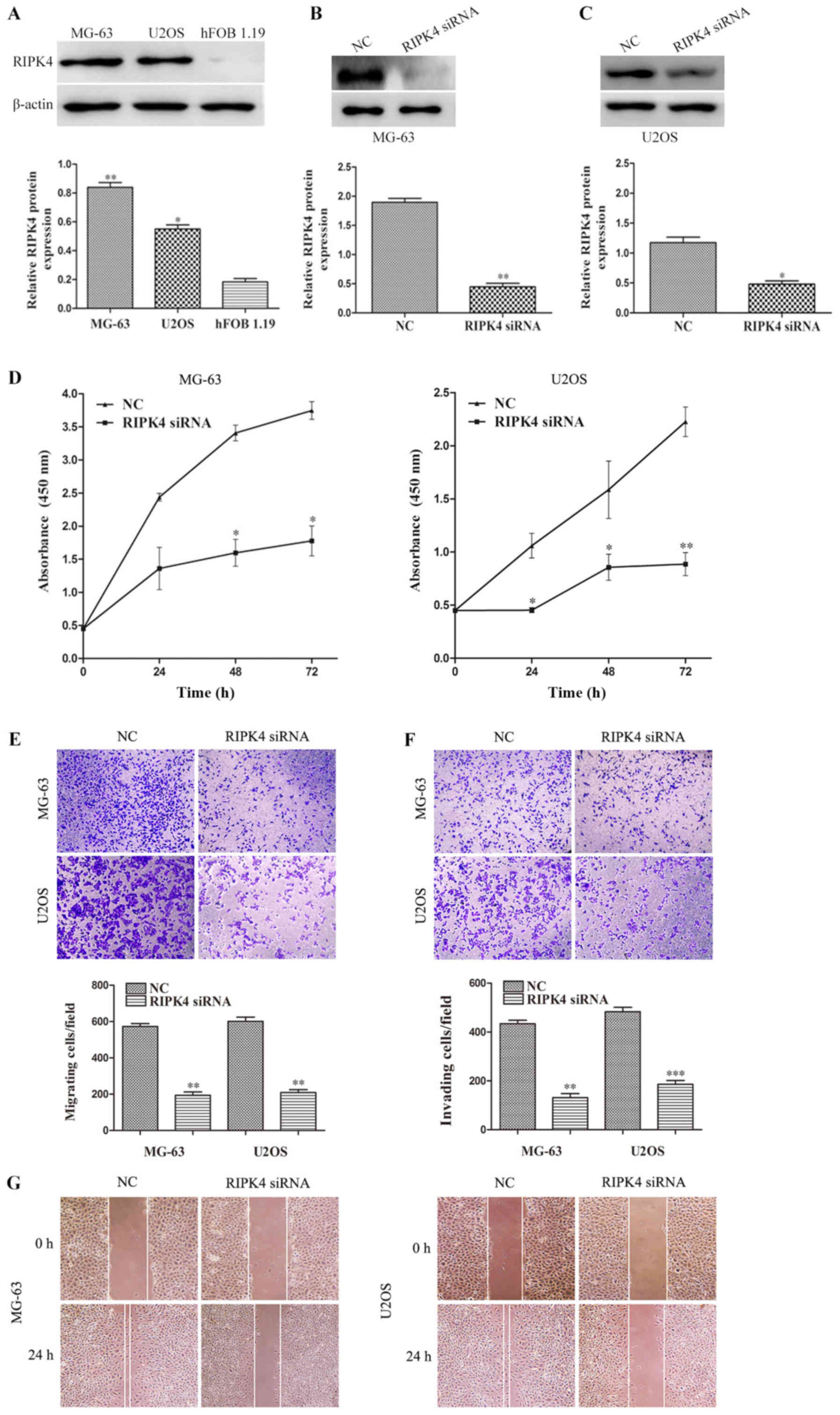
In the present study, RIPK4 expression was examined
in two human OS cell lines (MG-63 and U2OS) and a normal human
osteoblastic cell line (hFOB 1.19). Compared with hFOB 1.19 cells,
RIPK4 expression was upregulated in OS MG-63 and U2OS cells
(Fig. 2A). To further investigate
the role of RIPK4 in OS, the specific siRNA targeting the RIPK4
gene (RIPK4-siRNA) was transfected by liposome into MG-63 and U-2OS
cells. The silencing efficiency was analyzed by western blotting.
The results demonstrated that RIPK4 expression was significantly
decreased after transfection with RIPK4-siRNA, indicating that the
stable RIPK4 gene knockdown MG-63 and U-2OS cell lines were
successfully established (Fig. 2B and
C). Subsequently, the effect of RIPK4 knockdown on cell
proliferation, migration and invasion was examined. The CCK-8 assay
revealed that the proliferation of MG-63 at 48 and 72 h, and U2OS
cells at 24, 48 and 72 h, was significantly suppressed after RIPK4
knockdown (Fig. 2D). The wound
healing and migration assays indicated that RIPK4 knockdown
markedly inhibited cell migration in MG-63 and U2OS cells (Fig. 2E and G). The invasion assay results
showed that RIPK4 knockdown markedly reduced the invasion potential
of MG-63 and U2OS cells (Fig. 2F).
These results suggest that RIPK4 silencing inhibits the
proliferation, migration and invasion abilities of OS cells.
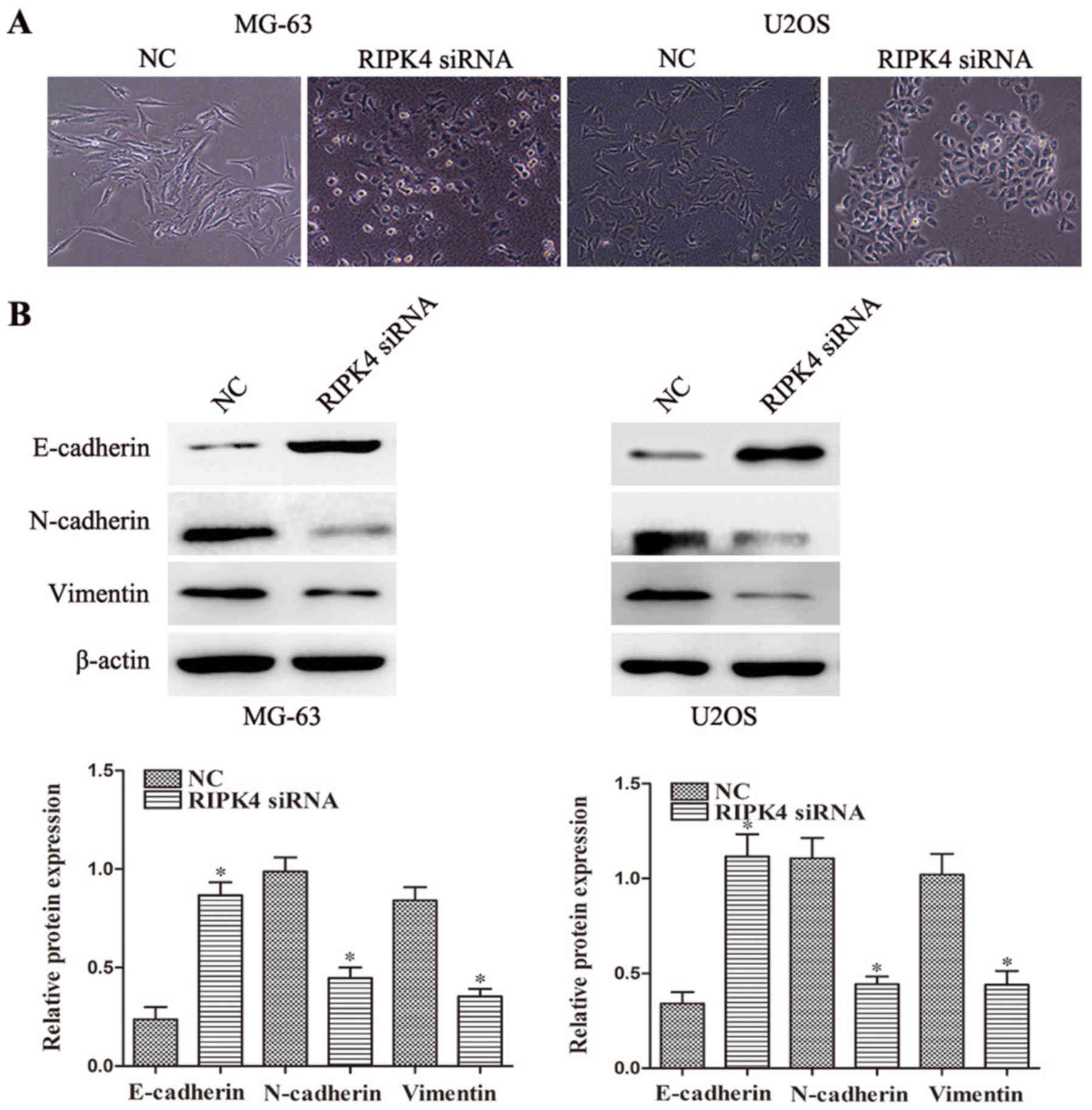
Silencing of RIPK4 suppresses EMT in
OS cells
EMT is an essential process in tumor migration and
invasion (5), therefore the
present study further investigated whether RIPK4 silencing inhibits
EMT in OS cells. RIPK4 knockdown led to morphological changes in
the MG-63 and U2OS cells at 24 h, as characterized by a
cobblestone-shaped appearance (Fig.
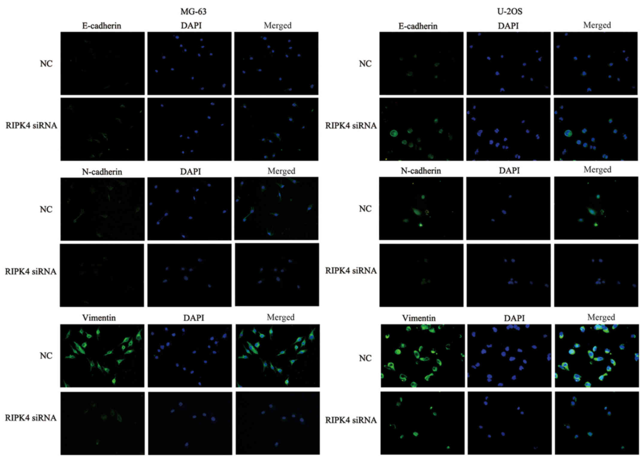
3A). The expression levels of the epithelial marker E-cadherin
and the mesenchymal markers, N-cadherin and vimentin, were examined
by western blotting and immunofluorescence staining. Significantly
increased E-cadherin expression levels and decreased N-cadherin and
vimentin expression levels were observed after RIPK4 silencing in
MG-63 cells. Similar results were observed in U2OS cells (Fig. 3B). The immunofluorescence staining
also supported these findings (Fig.
4). These results indicate that RIPK4 knockdown suppresses the
EMT process in OS cells.
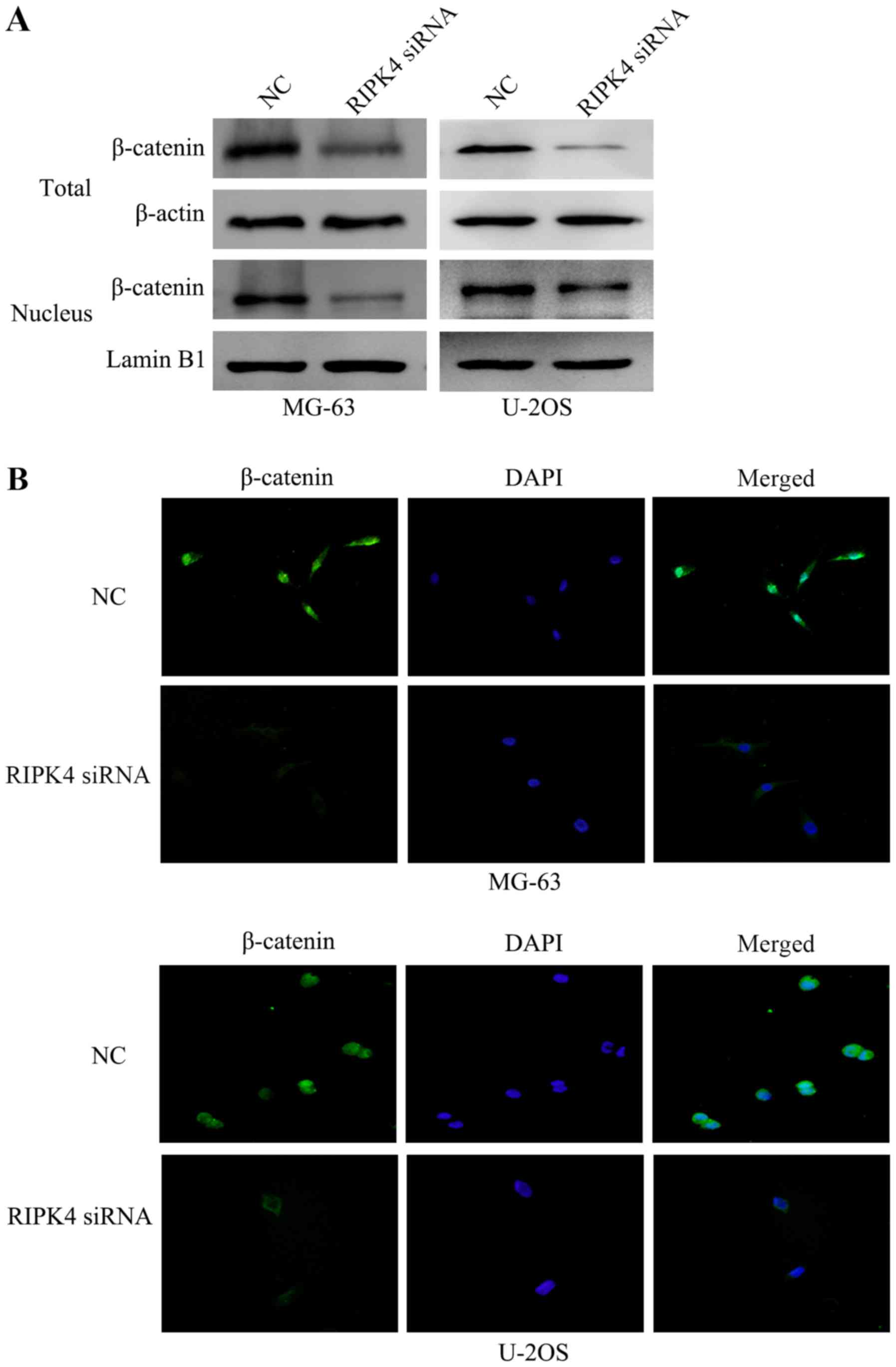
Silencing of RIPK4 suppresses EMT by
inactivating the Wnt/β-catenin signaling pathway
A previous study has reported that the Wnt/β-catenin
signaling pathway regulates cell EMT (12). To investigate the underlying
molecular mechanism of the EMT suppression induced by RIPK4
silencing, the present study examined the expression and
localization of the key molecule of the Wnt/β-catenin signaling
pathway, β-catenin, by western blotting. The results revealed that
RIPK4 silencing inhibited the protein expression of total and
nuclear β-catenin (Fig. 5A). In
addition, immunofluorescence analysis confirmed that the levels of
β-catenin were reduced following RIPK4 knockdown, in both the
cytoplasm and the cell nucleus (Fig.
5B). These results suggest that RIPK4 silencing inhibits the
EMT of OS cells by negatively regulating the Wnt/β-catenin
signaling pathway.
Discussion
Similar to other types of tumors, OS has a high
propensity for metastasis. It has been reported that ~20% of OS
patients with metastases were detected at the time of diagnosis,
while 80% of patients would develop metastases at a later stage
(22,23). Metastasis is the leading cause of
death in patients with OS (24).
Understanding the underlying mechanism of OS metastasis is an
essential prognostic factor.
Recent studies have demonstrated that RIPK4 is
important in the development and progression of many types of
malignant tumors. Liu et al (18) reported that RIPK4 expression was
significantly upregulated in cervical squamous cell carcinoma and
this increased expression was associated with invasive and
metastatic characteristics, including International Federation of
Gynecology and Obstetrics stage, tumor size and distant metastasis.
By contrast, other studies have shown that RIPK4 expression was
significantly downregulated and acted as a tumor suppressor
(20). Therefore, RIPK4 may
trigger different carcinogenic mechanisms in different types of
tumors. Little is known about the function of RIPK4 in OS. In the
present study, RIPK4 expression was significantly upregulated in OS
tissues and cell lines when compared with normal bone tissues and
osteoblastic cell lines. In addition, RIPK4 overexpression was
closely associated with a larger tumor size, advanced Enneking
stage and poor prognosis of OS patients. These results indicated
that RIPK4 might play a role the progression of OS.
EMT is a multistage process in which epithelial
cells lose polarity and acquire migratory and invasive properties;
it is considered to be a critical factor in invasion and metastasis
(25). An increasing body of
evidence has indicated that many sarcomas can undergo EMT-related
processes, which may be associated with an aggressive clinical
behavior. These processes may be particularly applicable to certain
sarcoma subtypes, such as carcinosarcomas exhibiting a dual
phenotype with mesenchymal and epithelial tumor characteristics
(26). A previous study reported a
depletion in RIPK4 expression using siRNA could inhibit a cervical
cancer cell (18). In the present
study, the results revealed that RIPK4 knockdown by siRNA
suppressed tumor cell migration and invasion in OS. Furthermore,
the mechanisms of the RIPK4-mediated suppression of migration and
invasion were investigated. The results showed that RIPK4 knockdown
significantly increased the expression of the epithelial marker
E-cadherin and decreased the expression of the mesenchymal markers,
N-cadherin and vimentin; it also induced morphological changes in
OS cell lines, from spindle-shaped fibroblast to cobblestone-shaped
epithelial-like morphology. These data suggested that the silencing
of RIPK4 might prevent tumor cell migration and invasion by
interfering with the EMT process in OS.
Further evidence has suggested the Wnt/β-catenin
signaling pathway is involved in embryogenesis and tumor
development (27,28). The aberrant activation of the
Wnt/β-catenin pathway signaling could promote EMT progression in
tumor cells, including OS cells (29). Huang et al (19) demonstrated that ectopic RIPK4
expression could induce cytosolic β-catenin accumulation and
upregulate canonical Wnt target genes including Cyclin D1, lymphoid
enhancer binding factor 1, Jun protooncogene AP-1 transcription
factor subunit, Myc and transcription factor 7 in A2780 and COV434
ovarian cancer cells, thereby implying that RIPK4 regulates the
Wnt/β-catenin signaling pathway. However, the association between
RIPK4 and Wnt/β-catenin signaling in OS is still unclear. In the
present study, the results showed that a significant decrease in
total and nuclear β-catenin levels was induced by endogenous RIPK4
knockdown in OS cells. The translocation of β-catenin is an
important molecular event in tumor formation (30). Consistent with these findings,
immunofluorescence analysis confirmed that the levels of β-catenin
were reduced following RIPK-4 knockdown, in both the cytoplasm and
the cell nucleus. All of the above results suggest that RIPK4
knockdown could suppress cell EMT by deactivating the Wnt/β-catenin
signaling pathway.
In conclusion, the present study revealed that RIPK4
was significantly upregulated in OS tissues and cell lines, and its
high expression was associated with larger tumor sizes, advanced
Enneking stage and poor prognosis. Furthermore, RIPK4 knockdown
inhibited cell migration and invasion by interfering with the EMT
process, which was mediated by the inactivation the Wnt/β-catenin
signaling pathway. This may provide a novel therapeutic target for
preventing OS cell metastasis. However, the precise regulatory
mechanisms need to be investigated further.
Acknowledgements
Not applicable.
Funding
The present study was supported by Basic Research
Innovative Group Project of Gansu Province (grant no.
1308RJIA004).
Availability of data and materials
The analyzed datasets used and/or analyzed during
the study are available from the corresponding author on reasonable
request.
Authors' contributions
ZY and PD conceived and designed the experiments.
ZY, YP and RG performed the experiments. YC, XR and WL analyzed the
data. WL, ZY, YP, RG and PD wrote the manuscript. All authors have
read and approved the final manuscript.
Ethics approval and consent to
participate
Written informed consent was obtained from all of
the patients and the research protocols were approved by the Ethics
Committee of Second hospital of Lanzhou University (Ganzu,
China).
Patient consent for publication
All patients within the present study provided
consent for the publication of their data.
Competing interests
The authors declare that they have no competing
interests.
References
|
1
|
Moore DD and Luu HH: Osteosarcoma. Cancer
Treat Res. 162:65–92. 2014. View Article : Google Scholar : PubMed/NCBI
|
|
2
|
Arndt CA, Rose PS, Folpe AL and Laack NN:
Common musculoskeletal tumors of childhood and adolescence. Mayo
Clin Proc. 87:475–487. 2012. View Article : Google Scholar : PubMed/NCBI
|
|
3
|
Daw NC, Billups CA, Rodriguez-Galindo C,
McCarville MB, Rao BN, Cain AM, Jenkins JJ, Neel MD and Meyer WH:
Metastatic osteosarcoma. Cancer. 106:403–412. 2006. View Article : Google Scholar : PubMed/NCBI
|
|
4
|
Farfalli GL, Albergo JI, Lobos PA, Smith
DE, Streitenberger PD, Pallotta Rodríguez MG and Aponte-Tinao LA:
Osteosarcoma lung metastases. Survival after chemotherapy and
surgery. Medicina (B Aires) (Article in Spanish). 75:87–90.
2015.
|
|
5
|
Diepenbruck M and Christofori G:
Epithelial-mesenchymal transition (EMT) and metastasis: Yes, no,
maybe. Curr Opin Cell Biol. 43:7–13. 2016. View Article : Google Scholar : PubMed/NCBI
|
|
6
|
Zheng X, Carstens JL, Kim J, Scheible M,
Kaye J, Sugimoto H, Wu CC, LeBleu VS and Kalluri R:
Epithelial-to-mesenchymal transition is dispensable for metastasis
but induces chemoresistance in pancreatic cancer. Nature.
527:525–530. 2015. View Article : Google Scholar : PubMed/NCBI
|
|
7
|
Mittal V: Epithelial mesenchymal
transition in aggressive lung cancers. Adv Exp Med Biol. 890:37–56.
2016. View Article : Google Scholar : PubMed/NCBI
|
|
8
|
Nieto MA, Huang RY, Jackson RA and Thiery
JP: EMT: 2016. Cell. 166:21–45. 2016. View Article : Google Scholar : PubMed/NCBI
|
|
9
|
Lamouille S, Xu J and Derynck R: Molecular
mechanisms of epithelial-mesenchymal transition. Nat Rev Mol Cell
Biol. 15:178–196. 2014. View
Article : Google Scholar : PubMed/NCBI
|
|
10
|
Moustakas A and Heldin CH: Mechanisms of
TGFβ-induced epithelial-mesenchymal transition. J Clin Med. 5(pii):
E632016. View Article : Google Scholar : PubMed/NCBI
|
|
11
|
Li Y, Ma J, Qian X, Wu Q, Xia J, Miele L,
Sarkar FH and Wang Z: Regulation of EMT by Notch signaling pathway
in tumor progression. Curr Cancer Drug Targets. 13:957–962. 2013.
View Article : Google Scholar : PubMed/NCBI
|
|
12
|
Gonzalez DM and Medici D: Signaling
mechanisms of the epithelial-mesenchymal transition. Sci Signal.
7:re82014. View Article : Google Scholar : PubMed/NCBI
|
|
13
|
Liu X, Yun F, Shi L, Li ZH, Luo NR and Jia
YF: Roles of signaling pathways in the epithelial-mesenchymal
transition in cancer. Asian Pac J Cancer Prev. 16:6201–6206. 2015.
View Article : Google Scholar : PubMed/NCBI
|
|
14
|
van Rensburg HJ and Yang X: The roles of
the Hippo pathway in cancer metastasis. Cell Signal. 28:1761–1772.
2016. View Article : Google Scholar : PubMed/NCBI
|
|
15
|
Meylan E and Tschopp J: The RIP kinases:
Crucial integrators of cellular stress. Trends Biochem Sci.
30:151–159. 2005. View Article : Google Scholar : PubMed/NCBI
|
|
16
|
Meylan E, Martinon F, Thome M, Gschwendt M
and Tschopp J: RIP4 (DIK/PKK), a novel member of the RIP kinase
family, activates NF-kappa B and is processed during apoptosis.
EMBO Rep. 3:1201–1208. 2002. View Article : Google Scholar : PubMed/NCBI
|
|
17
|
Adams S and Munz B: RIP4 is a target of
multiple signal transduction pathways in keratinocytes:
Implications for epidermal differentiation and cutaneous wound
repair. Exp Cell Res. 316:126–137. 2010. View Article : Google Scholar : PubMed/NCBI
|
|
18
|
Liu DQ, Li FF, Zhang JB, Zhou TJ, Xue WQ,
Zheng XH, Chen YB, Liao XY, Zhang L, Zhang SD, et al: Increased
RIPK4 expression is associated with progression and poor prognosis
in cervical squamous cell carcinoma patients. Sci Rep. 5:119552015.
View Article : Google Scholar : PubMed/NCBI
|
|
19
|
Huang X, McGann JC, Liu BY, Hannoush RN,
Lill JR, Pham V, Newton K, Kakunda M, Liu J, Yu C, et al:
Phosphorylation of dishevelled by protein kinase ripk4 regulates
wnt signaling. Science. 339:1441–1445. 2013. View Article : Google Scholar : PubMed/NCBI
|
|
20
|
Wang X, Zhu W, Zhou Y, Xu W and Wang H:
RIPK4 is downregulated in poorly differentiated tongue cancer and
is associated with migration/invasion and cisplatin-induced
apoptosis. Int J Biol Markers. 29:e150–159. 2014. View Article : Google Scholar : PubMed/NCBI
|
|
21
|
Heim D, Cornils K, Schulze K, Fehse B,
Lohse AW, Brümmendorf TH and Wege H: Retroviral insertional
mutagenesis in telomerase-immortalized hepatocytes identifies RIPK4
as novel tumor suppressor in human hepatocarcinogenesis. Oncogene.
34:364–372. 2015. View Article : Google Scholar : PubMed/NCBI
|
|
22
|
Bhattasali O, Vo AT, Roth M, Geller D,
Randall RL, Gorlick R and Gill J: Variability in the reported
management of pulmonary metastases in osteosarcoma. Cancer Med.
4:523–531. 2015. View
Article : Google Scholar : PubMed/NCBI
|
|
23
|
Hayden JB and Hoang BH: Osteosarcoma:
Basic science and clinical implications. Orthop Clin North Am.
37:1–7. 2006. View Article : Google Scholar : PubMed/NCBI
|
|
24
|
Ottaviani G and Jaffe N: The epidemiology
of osteosarcoma. Cancer Treat Res. 152:3–13. 2009. View Article : Google Scholar : PubMed/NCBI
|
|
25
|
Kotiyal S and Bhattacharya S: Events of
molecular changes in epithelial-mesenchymal transition. Crit Rev
Eukaryot Gene Expr. 26:163–171. 2016. View Article : Google Scholar : PubMed/NCBI
|
|
26
|
Sannino G, Marchetto A, Kirchner T and
Grünewald TGP: Epithelial-to-mesenchymal and
mesenchymal-to-epithelial transition in mesenchymal tumors: A
paradox in sarcomas. Cancer Res. 77:4556–4561. 2017. View Article : Google Scholar : PubMed/NCBI
|
|
27
|
Caronia-Brown G, Anderegg A and Awatramani
R: Expression and functional analysis of the Wnt/beta-catenin
induced mir-135a-2 locus in embryonic forebrain development. Neural
Dev. 11:92016. View Article : Google Scholar : PubMed/NCBI
|
|
28
|
Zhan T, Rindtorff N and Boutros M: Wnt
signaling in cancer. Oncogene. 36:1461–1473. 2017. View Article : Google Scholar : PubMed/NCBI
|
|
29
|
Yang G, Yuan J and Li K: EMT transcription
factors: Implication in osteosarcoma. Med Oncol. 30:6972013.
View Article : Google Scholar : PubMed/NCBI
|
|
30
|
Prakash S and Swaminathan U: β-catenin in
health: A review. J Oral Maxillofac Pathol. 19:230–238. 2015.
View Article : Google Scholar : PubMed/NCBI
|