Introduction
Irritable bowel syndrome (IBS) is a common
gastrointestinal disorder with unclear pathophysiology (1,2).
However, several factors are known to contribute to the
pathophysiology of this condition, including genetic factors, diet,
the intestinal microbiota, enteroendocrine cells and low-grade
inflammation (1–4).
Gastrointestinal endocrine cells produce >10
different hormones that regulate several functions of the
gastrointestinal tract, such as gastrointestinal motility, visceral
sensitivity, absorption, secretion, proliferation, local immune
defense and appetite (1,5). The gastrointestinal endocrine cells
interact and integrate with each other, and with the enteric,
autonomic and central nervous systems (6). In IBS, gastrointestinal motility and
secretion are abnormal and patients exhibit visceral
hypersensitivity (5). The density
of enteroendocrine cells in patients with IBS has been reported to
be lower than healthy subjects, which may contribute to the
pathophysiology of IBS (2,5,7–15).
Previous studies have suggested that the intestinal
density of Musashi-1- and neurogenin-3-positive cells were reduced
in patients with IBS (7,13,16).
Musashi-1 is a marker for intestinal stem cells and their early
progeny (17–21). Neurogenin-3 is expressed by an
early progenitor for endocrine cells (22,23).
It has been proposed that the low density of enteroendocrine cells
seen in patients with IBS could be caused by this abnormality in
stem cells and their differentiation into enteroendocrine cells
(6,7,9–13,16,24–26).
Furthermore, the role of genetic factors, diet, intestinal
microbiota, enteroendocrine cells and low-grade inflammation in the
pathophysiology of IBS is likely mediated through intestinal stem
cells (27).
The density of endocrine cells in the stomach corpus
is similar in patients with IBS and healthy subjects (28,29).
However, the density of gastrin-positive cells in the stomach
antrum has been reported to be higher, whereas the density of
somatostatin-positive cells has been reported to be lower in
patients with IBS, compared with healthy subjects (30). These differences may explain the
high incidence of erosive esophagitis observed in patients with IBS
(31), as gastrin stimulates acid
secretion and somatostatin inhibits acid secretion (6).
The aim of the present study was to investigate
whether the changes in the density of gastrin- and
somatostatin-positive in the stomach of patients with IBS were
associated with alterations in stem cells.
Materials and methods
Patients and controls
Patients with IBS (n=54), according to the Rome IV
criteria (32), were recruited
from an outpatient clinic at Stord Hospital (Stord, Norway). A
control group was formed from healthy subjects (n=51) without any
gastrointestinal complaints recruited through an announcement in
the local newspaper. The controls comprised 38 females and 13 males
with a mean age of 38 years (range, 20–67 years). The
characteristics of the patients with IBS are provided in Table I. Co-existence of other functional
gastrointestinal disorders was not recorded in the patients with
IBS included in the study. The present study was approved by The
Regional Committee for Medical and Health Research Ethics West
(approval no. 2017/1197/REK Vest). All participants gave oral and
written consent to participate.
 | Table I.Characteristics of the controls and
patients with IBS. |
Table I.
Characteristics of the controls and
patients with IBS.
| Clinicopathological
variable | Control group,
n=51 | IBS group,
n=54 |
|---|
| Age, mean (range)
years | 36 (20–64) | 32 (18–52) |
| Sex, n |
|
|
|
Male | 41 | 44 |
|
Female | 10 | 10 |
| IBS subtype, n |
|
|
|
IBS-D | 0 | 20 |
|
IBS-C | 0 | 21 |
|
IBS-M | 0 | 13 |
| IBS duration, years
(mean ± SD) | 0 | 15.5±8.0 |
| H. pylori
infection | 2 | 3 |
| Medication, n
(%) |
|
|
| PPI
medication | 2 (3.9) | 53 (98.2) |
| Birth
control medication | 32 (62.8) | 42 (77.8) |
|
Antimigraine medication | 0 (0.0) | 2 (3.7) |
|
Medication against
asthma/allergies | 0 (0.0) | 3 (5.6) |
|
Medication with
laevothyroxine | 0 (0.0) | 1 (1.9) |
|
Medication with heart/vascular
drugs | 1 (2.0) | 0 (0.0) |
Gastroscopy and histopathology
Both the patients and controls underwent standard
gastroscopy after an overnight fast. Three biopsy samples were
taken from the antrum and three from the corpus (major curvature)
of the stomach. Two additional biopsies were taken from the antrum
and analyzed in a rapid urease test for Helicobacter pylori
(HelicotecUT Plus, Strong Biotech Corp.). The two biopsy samples
were placed in polystyrene gel containing urea substrate for 1 h at
room temperature. In the presence of Helicobacter pylori
urease hydrolysis urea to produce ammonia and CO2, which
changes the pH and is detected as a change in the color of the gel
from yellow to red. The biopsy samples were fixed in 4% buffered
paraformaldehyde at room temperature overnight, embedded in
paraffin wax, then cut into 5-µm sections that were stained with
hematoxylin for 20 sec and eosin at room temperature for 3–4 min
and were examined under a light microscope (Olympus BX43, Olympus
Corporation).
Immunohistochemistry and computerized
image analysis
The sections were immunostained using the
avidin-biotin-complex (ABC) method with the Vectastain®
Elite ABC-HRP kit (cat. no. PK-6200, Vector Laboratories, Inc.).
The sections were incubated for 20 min with the blocking normal
horse serum then with the primary antibodies at room temperature
overnight. The sections were incubated with the secondary
biotinylated horse anti-rabbit IgG antibody for 30 min at room
temperature followed by the Avidin-bioytin complex for 30 min at
room temperature. The following polyclonal rabbit primary
antibodies were used: Anti-human gastrin-17 (cat. no. IR519; 1:500;
Dako; Agilent Technologies, Inc.); anti-synthetic cyclic
(amino-acid sequence 1–14) somatostatin (cat. no. A0566; 1:200;
Dako; Agilent Technologies, Inc.); and an antibody raised against a
synthetic peptide derived from human Musashi-1 (amino acids 1–100;
internal sequence) conjugated to keyhole limpet haemocyanin (cat.
no. ab21628; 1:100; Abcam).
The density of Musashi-1-positive cells was
semi-quantified on a computer linked to a BX43 light microscope
equipped with a DP26 digital camera using Olympus cellSens imaging
software (version 1.7; all from Olympus Corporation). The number of
Musashi-1-immunoreactive cells, and the number of gastric or
pyloric gland necks in each field were counted manually by pointing
and clicking the computer mouse in ten randomly chosen fields. A
×400 magnification was used and each field on the monitor
represented a tissue area of 0.14 mm2. The data from the
fields were tabulated, and the cell density was expressed as the
number of cells per gastric or pyloric gland neck. The number of
gastrin- or somatostatin-immunoreactive cells, and the area of
epithelial cells were measured using the same system in 10 randomly
chosen fields. The density of gastrin- and somatostatin-positive
cells were expressed as number of cells/mm2 epithelium.
Immunostained sections from the patients with IBS and controls were
coded and mixed, with measurements made by an investigator who was
blind to the identity of the sections.
Statistical analysis
The χ2 test was used to assess
differences between the patients and controls with regards to sex
and the incidence of H. pylori infection. Differences in
age, and in the densities of gastrin-, somatostatin-, and
Musashi-1-positive cells were tested using the Mann-Whitney
nonparametric test. The statistical analyses were performed using
GraphPad Prism software (version 8; GraphPad Software, Inc.).
P<0.05 was considered to indicate a statistically significant
difference.
Results
Patients and controls
The sex and age distributions did not differ
significantly between the patients with IBS and the controls (P=0.9
and 0.8, respectively). The urease test and histopathological
examination indicated H. pylori infection in three patients
and two controls, also with no significant difference between the
groups (P=0.6; Table I).
Gastroscopy and histopathology
Gastroscopy suggested that the esophagus, stomach
and duodenum were all macroscopically normal in both groups. A
histopathological examination of the corpus and antrum indicated
normal histology, with the exception of individuals with H.
pylori infection, in whom metaplasia was found in the
antrum.
Immunohistochemistry and computerized
image analysis
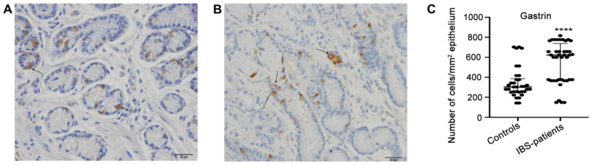
The gastrin-positive cell density in the controls
and patients with IBS were 303.0±560.0 and 623.0±668.0 (median ±
range; P<0.0001), respectively (Fig. 1). The corresponding figures for
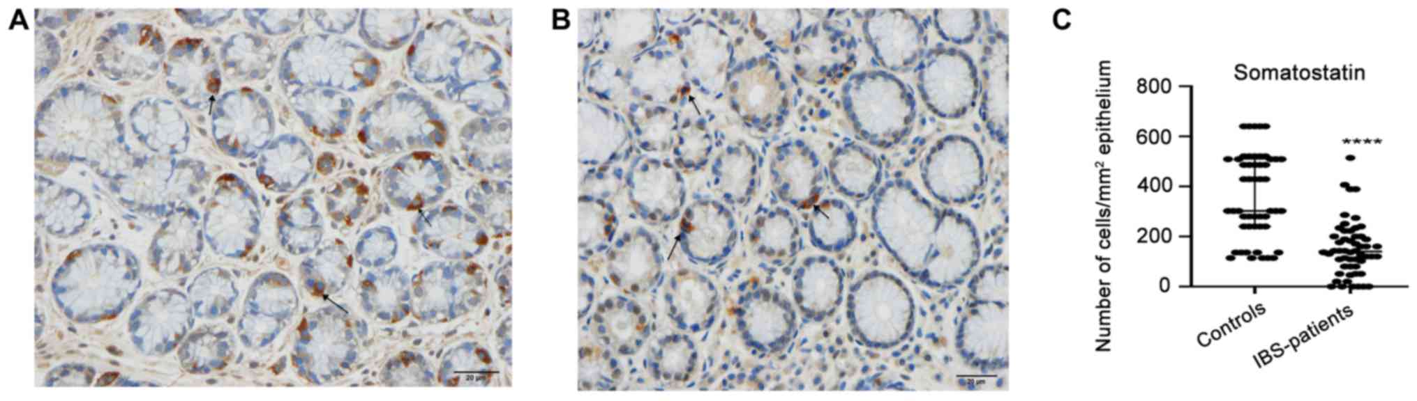
somatostatin-positive cell density were 302.0±526.0 and 140.0±514.0
(P<0.0001; Fig. 2).
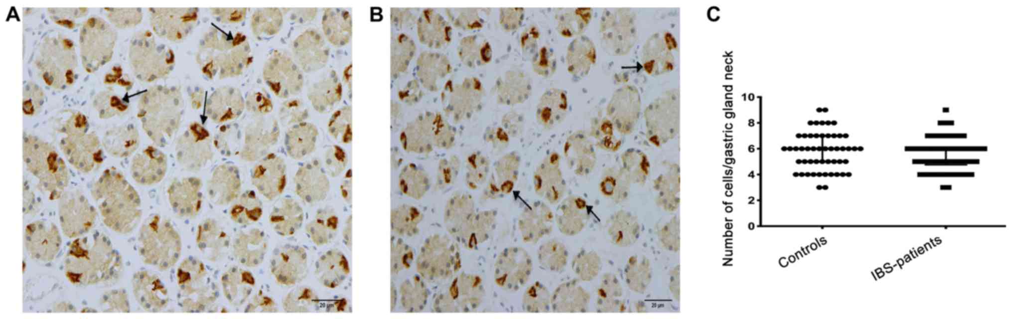
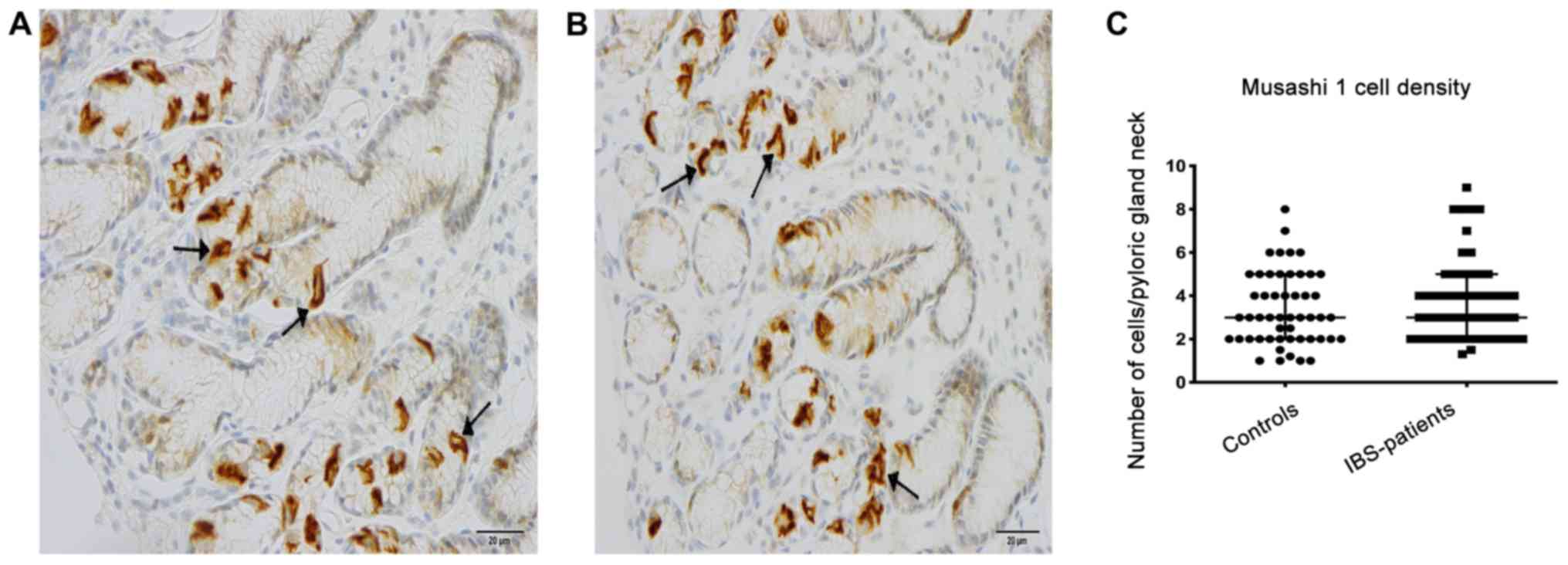
Musashi-1-immunoreactive cells were concentrated at
the necks of the gastric glands of the corpus and of the pyloric
glands of the antrum, where they were evident mostly in a
clonogenic proliferation or differentiation form (Fig. 3). The density of Musashi-1-positive
cells in the corpus was 6.0±6.0 in the controls and 6.0±6.0 in the
patients with IBS (P=0.4); the corresponding values in the antrum
were 3.0±7.0 and, 3.8±7.7 respectively (P=0.3; Fig. 4). A subgroup analysis based on the
phenotype of the disorder (diarrhea predominant, constipation
predominant or mixed type) was not performed, as the number of
patients in these subgroups was too small to allow reliable
analysis.
Discussion
The stomach is divided anatomically into the corpus
and antrum, each comprising different cells and functions (33). The gastric glands of the corpus
contain mucous neck cells, parietal cells, chief cells and
endocrine cells. In the antrum, endocrine cells and mucous cells
are the main cell types in the pyloric glands (33,34).
While serotonin- and somatostatin-expressing cells are present in
both the corpus and the antrum, ghrelin-positive cells are present
only in the oxyntic mucosa of the corpus, while gastrin-positive
cells are exclusive to the pyloric glands of the antrum (28,30).
Several attempts have been made to identify a specific marker for
gastric stem cells (33).
Musashi-1, which is considered a marker for intestinal stem cells
and their early progeny, is reportedly expressed by cells in the
corpus and antrum of the stomach (35,36).
The present study found that Musashi-1-expressing
cells were mostly present at the necks of the gastric glands of the
corpus and of the pyloric glands of the antrum, which is consistent
with previous observations (35,36).
The density of Musashi-1-positive cells in both the corpus and
antrum did not differ between patients with IBS and healthy
controls. These observations may explain why, in a previous study,
the total density of endocrine cells detected using chromogranin A
in the corpus and antrum was similar in patients with IBS and
healthy controls (29). In
addition, the densities of ghrelin-, serotonin- and
somatostatin-expressing cells in the oxyntic mucosa of the corpus
did not differ between patients with IBS and healthy controls in a
previous study (28). However, the
densities of gastrin-positive cells were higher and
somatostatin-positive cells were lower in the antrum of patients
with IBS, compared with healthy controls. These differences could
not be explained by abnormalities in stem cells like those
previously observed in the small and large intestines of patients
with IBS, where density of Musashi-1-positive cells were lower than
that of healthy controls (2,5,37).
The mechanisms underlying the changes in the
densities of antral gastrin- and somatostatin-positive cells in
patients with IBS remain unclear. These changes could represent a
response to the changes in the intestinal chromogranin A,
serotonin, secretin, cholecystokinin, gastric inhibitory peptide,
somatostatin, peptide YY and enteroglucagon (oxyntomodulin) cells
seen in these patients (2,5,7–15).
The increase in the number of gastrin-expressing cells and the
reduction in the number of somatostatin-expressing cells could
result from activation of gastrin synthesis and inhibition of
somatostatin synthesis, a phenomenon that has been observed in
gastrointestinal endocrine cells (38,39).
However, the present study was limited to measuring changes in
densities of gastrin-, somatostatin- and Musashi-1-expressing
cells. The expression or secretion of these active substances were
not evaluated.
In conclusion, the changes in the density of antral
gastrin- and somatostatin-expressing cells in patients with IBS
cannot be explained by a low density of stem cells; thus as the gut
endocrine cells integrate and interact with each other, the changes
in gastrin-and somatostatin-expressing cells in patients with IBS
may be caused by the direct changes in the intestinal endocrine
cells.
Acknowledgements
Not applicable.
Funding
The study was supported by grants from Helse Fonna
(grant no. 40415) and Helse Vest (grant no. 192234).
Availability of data and materials
The datasets used and/or analyzed during the present
study are available from the corresponding author on reasonable
request.
Authors' contributions
MES designed the study, obtained the funding,
recruited the patients, performed gastroscopies, collected,
analyzed and interpreted the data, and drafted the manuscript. TH
and JGH contributed to the design of the study, and to the analysis
and interpretation of the data, and critically revised the
manuscript for important intellectual content. All authors read and
approved the final the manuscript.
Ethics approval and consent to
participate
The present study was approved by The Regional
Committee for Medical and Health Research Ethics West, Bergen,
Norway (approval no. 2017/1197/REK Vest). Oral and written consent
was obtained from all subjects.
Patient consent for publication
Not applicable.
Competing interests
The authors declare that they have no competing
interests.
References
|
1
|
El-Salhy M: Irritable bowel syndrome:
Diagnosis and pathogenesis. World J Gastroenterol. 18:3135–5163.
2012. View Article : Google Scholar
|
|
2
|
El-Salhy M: Recent developments in the
pathophysiology of irritable bowel syndrome. World J Gastroenterol.
21:7621–7636. 2015. View Article : Google Scholar : PubMed/NCBI
|
|
3
|
El-Salhy M, Gundersen D, Hatlebakk JG and
Hausken T: Irritable bowel syndrome: Diagnosis, pathogenesis and
treatment options. Nova Science Publishers, Inc.; New York:
2012
|
|
4
|
El-Salhy M, Hatlebakk JG, Gilja OH and
Hausken T: Irritable bowel syndrome: Recent developments in
diagnosis, pathophysiology, and treatment. Expert Rev Gastroenterol
Hepatol. 8:435–443. 2014. View Article : Google Scholar : PubMed/NCBI
|
|
5
|
El-Salhy M, Gundersen D, Gilja OH,
Hatlebakk JG and Hausken T: Is irritable bowel syndrome an organic
disorder? World J Gastroenterol. 20:384–400. 2014. View Article : Google Scholar : PubMed/NCBI
|
|
6
|
El-Salhy M, Seim I, Chopin L, Gundersen D,
Hatlebakk JG and Hausken T: Irritable bowel syndrome: The role of
gut neuroendocrine peptides. Front Biosci (Elite Ed). 4:2783–2800.
2012.PubMed/NCBI
|
|
7
|
El-Salhy M and Gilja OH: Abnormalities in
ileal stem, neurogenin 3, and enteroendocrine cells in patients
with irritable bowel syndrome. BMC Gastroenterol. 17:902017.
View Article : Google Scholar : PubMed/NCBI
|
|
8
|
El-Salhy M, Gilja OH, Gundersen D,
Hatlebakk JG and Hausken T: Duodenal chromogranin a cell density as
a biomarker for the diagnosis of irritable bowel syndrome.
Gastroenterol Res Pract. 2014:4628562014. View Article : Google Scholar : PubMed/NCBI
|
|
9
|
El-Salhy M, Gilja OH, Gundersen D,
Hatlebakk JG and Hausken T: Endocrine cells in the ileum of
patients with irritable bowel syndrome. World J Gastroenterol.
20:2383–2391. 2014. View Article : Google Scholar : PubMed/NCBI
|
|
10
|
El-Salhy M, Gundersen D, Hatlebakk JG,
Gilja OH and Hausken T: Abnormal rectal endocrine cells in patients
with irritable bowel syndrome. Regul Pept. 188:60–65. 2014.
View Article : Google Scholar : PubMed/NCBI
|
|
11
|
El-Salhy M, Gundersen D, Ostgaard H,
Lomholt-Beck B, Hatlebakk JG and Hausken T: Low densities of
serotonin and peptide YY cells in the colon of patients with
irritable bowel syndrome. Dig Dis Sci. 57:873–878. 2012. View Article : Google Scholar : PubMed/NCBI
|
|
12
|
El-Salhy M, Patcharatrakul T, Hatlebakk
JG, Hausken T, Gilja OH and Gonlachanvit S: Chromogranin A cell
density in the large intestine of Asian and European patients with
irritable bowel syndrome. Scand J Gastroenterol. 52:691–697. 2017.
View Article : Google Scholar : PubMed/NCBI
|
|
13
|
El-Salhy M, Patcharatrakul T, Hatlebakk
JG, Hausken T, Gilja OH and Gonlachanvit S: Enteroendocrine,
Musashi 1 and neurogenin 3 cells in the large intestine of Thai and
Norwegian patients with irritable bowel syndrome. Scand J
Gastroenterol. 52:1331–1339. 2017. View Article : Google Scholar : PubMed/NCBI
|
|
14
|
Dizdar V, Spiller R, Singh G, Hanevik K,
Gilja OH, El-Salhy M and Hausken T: Relative importance of
abnormalities of CCK and 5-HT (serotonin) in Giardia-induced
post-infectious irritable bowel syndrome and functional dyspepsia.
Aliment Pharmacol Ther. 31:883–891. 2010.PubMed/NCBI
|
|
15
|
El-Salhy M, Vaali K, Dizdar V and Hausken
T: Abnormal small-intestinal endocrine cells in patients with
irritable bowel syndrome. Dig Dis Sci. 55:3508–3513. 2010.
View Article : Google Scholar : PubMed/NCBI
|
|
16
|
El-Salhy M, Hatlebakk JG and Hausken T:
Reduction in duodenal endocrine cells in irritable bowel syndrome
is associated with stem cell abnormalities. World J Gastroenterol.
21:9577–9587. 2015. View Article : Google Scholar : PubMed/NCBI
|
|
17
|
Montgomery RK and Breault DT: Small
intestinal stem cell markers. J Anat. 213:52–58. 2008. View Article : Google Scholar : PubMed/NCBI
|
|
18
|
Kayahara T, Sawada M, Takaishi S, Fukui H,
Seno H, Fukuzawa H, Suzuki K, Hiai H, Kageyama R, Okano H and Chiba
T: Candidate markers for stem and early progenitor cells, Musashi-1
and Hes1, are expressed in crypt base columnar cells of mouse small
intestine. FEBS Lett. 535:131–135. 2003. View Article : Google Scholar : PubMed/NCBI
|
|
19
|
Potten CS: Stem cells in gastrointestinal
epithelium: Numbers, characteristics and death. Philos Trans R Soc
Lond B Biol Sci. 353:821–830. 1998. View Article : Google Scholar : PubMed/NCBI
|
|
20
|
Barker N, van Oudenaarden A and Clevers H:
Identifying the stem cell of the intestinal crypt: Strategies and
pitfalls. Cell Stem Cell. 11:452–460. 2012. View Article : Google Scholar : PubMed/NCBI
|
|
21
|
Potten CS, Booth C and Pritchard DM: The
intestinal epithelial stem cell: The mucosal governor. Int J Exp
Pathol. 78:219–243. 1997. View Article : Google Scholar : PubMed/NCBI
|
|
22
|
Lee CS, Perreault N, Brestelli JE and
Kaestner KH: Neurogenin 3 is essential for the proper specification
of gastric enteroendocrine cells and the maintenance of gastric
epithelial cell identity. Genes Dev. 16:1488–1497. 2002. View Article : Google Scholar : PubMed/NCBI
|
|
23
|
Lee CS and Kaestner KH: Clinical
endocrinology and metabolism. Development of gut endocrine cells.
Best Pract Res Clin Endocrinol Metab. 18:453–462. 2004. View Article : Google Scholar : PubMed/NCBI
|
|
24
|
El-Salhy M, Mazzawi T, Gundersen D and
Hausken T: Chromogranin A cell density in the rectum of patients
with irritable bowel syndrome. Mol Med Rep. 6:1223–1225. 2012.
View Article : Google Scholar : PubMed/NCBI
|
|
25
|
El-Salhy M, Wendelbo I and Gundersen D:
Reduced chromogranin A cell density in the ileum of patients with
irritable bowel syndrome. Mol Med Rep. 7:1241–1244. 2013.
View Article : Google Scholar : PubMed/NCBI
|
|
26
|
El-Salhy M, Wendelbo I and Gundersen D:
Serotonin and serotonin transporter in the rectum of patients with
irritable bowel disease. Mol Med Rep. 8:451–455. 2013. View Article : Google Scholar : PubMed/NCBI
|
|
27
|
El-Salhy M: Possible role of intestinal
stem cells in the pathophysiology of irritable bowel syndrome.
World J Gastroenterol. 26:1427–1438. 2020. View Article : Google Scholar : PubMed/NCBI
|
|
28
|
El-Salhy M, Gilja OH, Gundersen D and
Hausken T: Endocrine cells in the oxyntic mucosa of the stomach in
patients with irritable bowel syndrome. World J Gastrointest
Endosc. 6:176–185. 2014. View Article : Google Scholar : PubMed/NCBI
|
|
29
|
El-Salhy M, Gilja OH and Hausken T:
Chromogranin A cells in the stomachs of patients with sporadic
irritable bowel syndrome. Mol Med Rep. 10:1753–1757. 2014.
View Article : Google Scholar : PubMed/NCBI
|
|
30
|
El-Salhy M, Gilja OH, Hatlebakk JG and
Hausken T: Stomach antral endocrine cells in patients with
irritable bowel syndrome. Int J Mol Med. 34:967–974. 2014.
View Article : Google Scholar : PubMed/NCBI
|
|
31
|
El-Salhy M, Gilja OH and Hatlebakk JG:
Overlapping of irritable bowel syndrome with erosive esophagitis
and the performance of Rome criteria in diagnosing IBS in a
clinical setting. Mol Med Rep. 20:787–794. 2019.PubMed/NCBI
|
|
32
|
Longstreth GF, Thompson WG, Chey WD,
Houghton LA, Mearin F and Spiller RC: Functional bowel disorders.
Gastroenterology. 130:1480–1491. 2006. View Article : Google Scholar : PubMed/NCBI
|
|
33
|
Han ME and Oh SO: Gastric stem cells and
gastric cancer stem cells. Anat Cell Biol. 46:8–18. 2013.
View Article : Google Scholar : PubMed/NCBI
|
|
34
|
Khurana S and Mills JC: The gastric mucosa
development and differentiation. Prog Mol Biol Transl Sci.
96:93–115. 2010. View Article : Google Scholar : PubMed/NCBI
|
|
35
|
Akasaka Y, Saikawa Y, Fujita K, Kubota T,
Ishikawa Y, Fujimoto A, Ishii T, Okano H and Kitajima M: Expression
of a candidate marker for progenitor cells, Musashi-1, in the
proliferative regions of human antrum and its decreased expression
in intestinal metaplasia. Histopathology. 47:348–356. 2005.
View Article : Google Scholar : PubMed/NCBI
|
|
36
|
Murata H, Tsuji S, Tsujii M, Nakamura T,
Fu HY, Eguchi H, Asahi K, Okano H, Kawano S and Hayashi N:
Helicobacter pylori infection induces candidate stem cell
marker Musashi-1 in the human gastric epithelium. Dig Dis Sci.
53:363–369. 2008. View Article : Google Scholar : PubMed/NCBI
|
|
37
|
El-Salhy M, Hausken T, Gilja OH and
Hatlebakk JG: The possible role of gastrointestinal endocrine cells
in the pathophysiology of irritable bowel syndrome. Expert Rev
Gastroenterol Hepatol. 11:139–148. 2017. View Article : Google Scholar : PubMed/NCBI
|
|
38
|
Habib AM, Richards P, Rogers GJ, Reimann F
and Gribble FM: Co-localisation and secretion of glucagon-like
peptide 1 and peptide YY from primary cultured human L cells.
Diabetologia. 56:1413–1416. 2013. View Article : Google Scholar : PubMed/NCBI
|
|
39
|
El-Salhy M, Hatlebakk JG and Hausken T:
Possible role of peptide YY (PYY) in the pathophysiology of
irritable bowel syndrome (IBS). Neuropeptides. 79:1019732020.
View Article : Google Scholar : PubMed/NCBI
|