Introduction
Ischemia/reperfusion (I/R) injury of the limb occurs
as a result of exposure to a large degree of blood flux in vascular
reperfusion during hypoxia. I/R usually occurs following various
types of trauma, including vascular injury, surgical procedures and
extreme pressure (1). Limb I/R can
damage the source limb, as well as remote organs. The lung is
highly sensitive to inflammatory reactions and prone to acute
injury during reperfusion (2).
Lung inflammation involves complex mechanisms, including
inflammatory reaction, oxidative stress and immune dysfunction
(3,4).
It is crucial to identify that endogenous proteins
that are produced after lung injury at the early phase of I/R
(5). Toll-like receptors (TLRs)
are a class of pattern recognition receptors expressed on immune
and non-immune cell surfaces (6).
In particular, TLR4 has been demonstrated to play a major role in
the development of lung injury (7,8).
Moreover, Activation of TLR4 stimulates downstream NF-κB, resulting
in an increased release of downstream inflammatory cytokines,
including interleukin 1 (IL)-1, IL-6 and tumor necrosis factor-α
(TNF-α) (9).
Acupuncture is a traditional therapeutic technique.
A variant of acupuncture, electroacupuncture (EA), has an
anti-inflammatory effect on lung injury caused by inflammatory
diseases (10). Our previous study
suggested that EA pre-treatment could reduce the inflammatory
reactions in patients with lung injury associated with limb I/R, as
well as the associated release of inflammatory cytokines (11). However, the underlying mechanism
remains unknown. Gong et al (12) demonstrated that EA attenuated limb
I/R-induced lung injury via the p38 mitogen-activated protein
kinase/nuclear factor erythroid 2-like 2/heme oxygenase 1 pathway.
However, whether EA also reduces lung injury induced by limb I/R
via the suppression the TLR4/NF-κB pathway has not yet been
elucidated. Therefore, in the present study, a rat model of limb
I/R-induced lung injury was used in order to assess whether EA
applied at Zusanli (ST36) and Sanyinjiao (SP6) can inhibit
inflammation and achieve a protective effect. In addition, the
hypothesis that EA exerts an anti-inflammatory effect via the
suppression of the TLR4/NF-κB pathway was also tested.
Materials and methods
Animals
Male Sprague-Dawley rats aged 7–8 weeks and weighing
250–300 g were acquired from The Animal Center of Wenzhou Medical
University. Rats were housed in a temperature-controlled chamber
with a 12-h light/dark cycle at 24°C and 50–60% relative humidity.
All rats were provided standard chow and tap water ad
libitum until they were made to fast for 12 h before
experiments. All animal experiments were approved by The Animal
Research Ethics Committee of Wenzhou Medical University and were
performed in accordance with the guidelines of The National
Institutes of Health for the Care and Use of Laboratory Animals
(13).
Establishment of the limb I/R injured
rat model
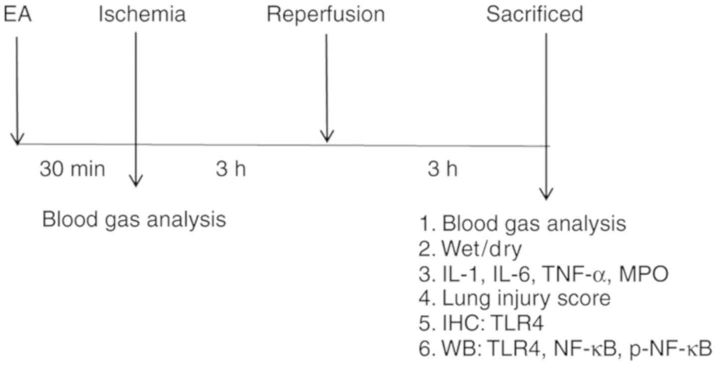
The rats (n=32) were randomly divided into four
groups of eight rats each. The lower limb I/R method was adapted
from a previous report (14). The
treatment schedule is presented in Fig. 1. All rats in the study survived the
treatment procedures. At the end of the experiment, the rats were
intraperitoneally injected with 1 g/kg urethane and sacrificed by
piercing of the heart and bleeding until cessation of breathing and
heartbeat. In the I/R group, A rubber band (size 13 mm) above the
greater trochanter was applied to interrupt the arterial blood for
3 h and then followed by 3 h of reperfusion; no drugs or EA
treatment were administered. The band was put in place without
being fastened in the sham group. In the sham EA (SEA) group,
acupuncture needles were inserted 2–3 mm deep at the ST36 and SP6
acupoints, without stimulation. In the EA group, the rats received
EA for 30 min before I/R, and the stimulation parameters comprised
dispersed waves of 2 and 15 Hz at 1 mA. The ST36 acupoint is
located 5 mm below the head of the fibula under the knee joint, and
2 mm lateral to the anterior tubercle of the tibia in the rat
(15). SP6 is located 10 mm above
the hindlimb medial malleolus front of the tibia and fibula
(15). EA was performed using a
LH202H Hans® Acupoint Nerve Stimulator (Hans Therapy,
Co., Ltd.).
Blood gas analysis
Left common carotid artery blood gas analysis
(I-STAT 300, Abbott Pharmaceutical Co. Ltd.) was performed to
measure the arterial partial pressure of oxygen (PaO2),
base excess and hemoglobin (Hb) content. The derived variables
included the arterial-alveolar oxygen tension ratio (a/A),
alveolar-arterial oxygen tension difference (A-aO2) and
respiratory index (RI).
Lung tissue wet/dry weight ratio
The right upper tissue of the lung in each rat was
removed and weighed immediately after removal (wet weight).
Sections were desiccated in an oven at 60°C for 2 days until a
stable dry weight was reached. The wet/dry weight ratio was then
calculated.
Histological analysis and lung injury
score
Lungs were perfused with cold saline, followed by 4%
paraformaldehyde. After fixation at 4°C for 24 h, the tissues were
embedded in paraffin, cut into 5-µm sections, then stained with
hematoxylin and eosin at room temperature for 5 min. The stained
lung sections were observed under a light microscope (Leica
Microsystems GmbH) at ×400 magnification by an experienced
pathologist blinded to the protocols of the present study. Lung
injury scores were assessed in a blinded manner and determined
based on four independent parameters: i) Alveolar edema; ii)
hemorrhage; iii) infiltration of inflammatory cells; and iv)
thickened alveolar septum (16).
Measurement of lung IL-1, IL-6, TNF-α
and myeloperoxidase (MPO) concentrations
The concentrations of IL-1, IL-6, TNF-α and MPO in
the rat lung were measured using ELISA kits according to the
manufacturer's instructions (Shanghai Boyun Biotechnology Co.,
Ltd.). The catalogue numbers for the ELISA kits were IL-1
(BP-E30419), IL-6 (BP-E30646), TNF-α (BP-E30635) and MPO
(BP-E31651). The absorbance value at 450 nm was determined with a
multifunctional microplate reader (Thermo Fisher Scientific,
Inc.).
Immunohistochemistry
TLR4 expression in lung tissues was evaluated using
immunohistochemistry. The lung samples were fixed in 4%
paraformaldehyde at room temperature for 24 h. The next day the
lungs were dehydrated in graded ethanol solution, embedded in
paraffin for three times an hour at 55°C and embedded in a small
stainless steel container. Sections were cut at 5 µm and were
heated in an oven for 1 h, deparaffinized in xylene, rehydrated in
graded ethanol solutions and microwaved at 100°C in sodium citrate
buffer for 20 min. The slides were cooled to room temperature and
incubated for 10 min in 3% H2O2. The sections
were blocked using 5% donkey serum (Beijing Solarbio Science &
Technology Co., Ltd.) at room temperature for 1 h, then incubated
at 4°C overnight with primary antibody against TLR4 (1:100; cat.
no. AF7017; Affinity Biosciences). The sections were subsequently
washed three times with PBS every 5 min and incubated with 100 µl
enzyme-labeled goat anti-rabbit IgG polymer (cat. no. PV-9001;
OriGene Technologies, Inc.) for 30 min at 37°C. The samples were
then incubated with 3,3′-diaminobenzidine substrate for 30 sec at
room temperature. After dehydration and drying, the sections were
mounted with neutral gum. Subsequently, the slides were examined
under a light microscope (magnification, ×200).
An immunohistochemical score (IHS) was
used to evaluate TLR4 expression (17)
The IHS accounts for the percentage of
immunoreactive cells (quantity score) and the staining intensity
(staining intensity score). The quantity score was assigned
follows: i) No staining was scored as 0; ii) 1–10% of cells stained
as 1; iii) 11–50% as 2; iv) 51–80% as 3; and v) 81–100% as 4.
Staining intensity was rated on a scale of 0–3, where: i) 0 was
negative; ii) 1 was weak; iii) 2 was moderate; and iv) 3 was
strong. When there was multifocal immunoreactivity and there were
significant differences in staining intensities between foci, the
average of the least intense and most intense staining was
recorded. The raw data were converted to the IHS by multiplying the
quantity and staining intensity scores.
Western blot analysis
The lung tissues were dissociated using RIPA buffer
(Beijing Solarbio Science & Technology Co., Ltd.) with protease
(1:100) and phosphatase inhibitors (1:50). Extracts were
homogenized, then centrifuged at 12,000 × g for 30 min at 4°C.
Protein concentration was measured in the supernatant using a
bicinchoninic acid protein assay kit (Thermo Fisher Scientific,
Inc.). Samples were boiled for 5 min at 95°C in 5X loading buffer.
Protein (50 µg) was then separated by SDS-PAGE on 10% gels and then
electro-transferred to a 0.22-µm PVDF membrane (Bio-Rad
Laboratories, Inc.) for 1.5 h. The membranes were subsequently
blocked with 5% bovine serum albumin (Biosharp Life Sciences) for 1
h at room temperature. Membranes were then incubated with primary
antibodies on a table concentrator overnight at 4°C. Primary
antibodies against β-actin (1:8,000; cat. no. AP0060; Bioworld
Technology, Inc.), TLR4 (1:1,000; cat. no. AF7017; Affinity
Biosciences), NF-κB (1:1,000; cat. no. AF5006; Affinity
Biosciences) and phosphorylated (p)-NF-κB (1:1,000; cat. no.
AF2006; Affinity Biosciences) were used. After washing the
membranes three times with Tris-buffered saline plus 0.1% Tween-20,
a horseradish peroxidase-conjugated secondary antibody (1:5,000;
cat. no. BL003A; Biosharp Life Sciences) was added for 1.5 h at
room temperature. The membranes were subsequently washed three
times with Tris-buffered saline plus 0.1% Tween-20 and were
visualized by electrochemiluminescence (cat. no. K-12045-D10;
Advansta, Inc.). Images were analyzed by Image Lab Analysis System
v6.0 (Bio-Rad Laboratories, Inc.).
Statistical analysis
Data are presented as the mean ± SD from ≥6
independent experiments. Statistical analysis was conducted using
one-way ANOVA, followed by Tukey's post hoc test. P<0.05 was
considered to indicate a statistically significant difference.
Analysis was performed using the SPSS v 19.0 software (IBM
Corp.).
Results
EA pre-treatment at ST36 and SP6
alleviates limb I/R-induced lung injury in rats
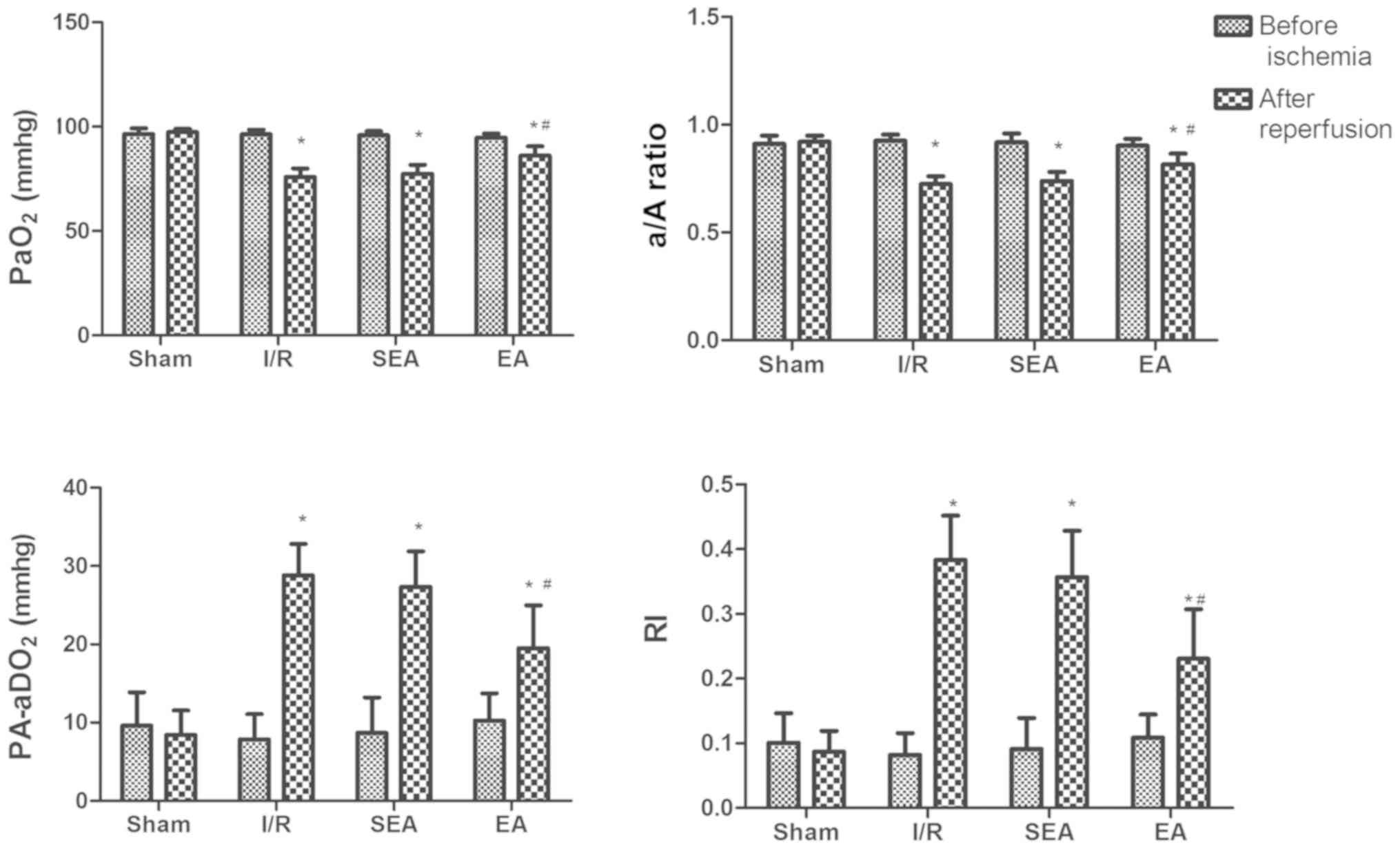
The protective effect of EA on lung injury induced
by limb I/R was examined by blood gas analysis (PaO2,
A-aO2, a/A ratio and RI). Before ischemia, the analyzed
parameters did not differ between the sham, I/R, SEA and EA groups.
Following 3 h of reperfusion, rats in the I/R, SEA and EA groups
displayed significant pulmonary dysfunction, compared with the sham
group, indicating that I/R-induced injury had been successfully
established in this model. However, in the EA group, pulmonary
oxygenation was significantly improved, compared with the I/R
group, demonstrating the protective effect of EA pre-treatment
against limb I/R-induced lung injury (Fig. 2).
EA pre-treatment alleviates pulmonary
injury in I/R-injured rats
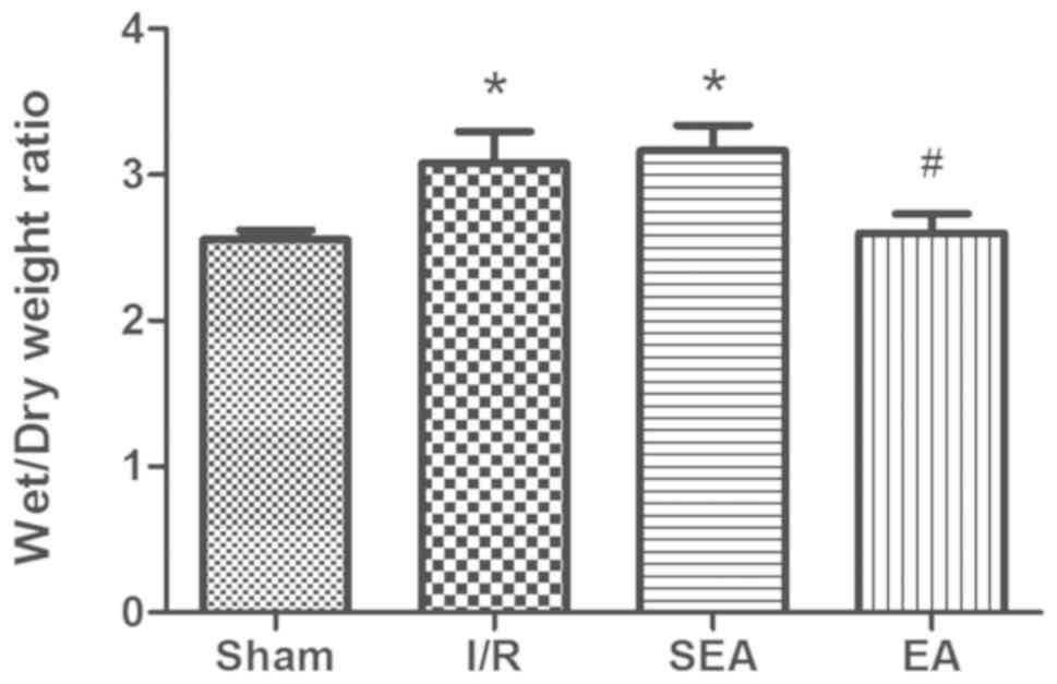
To investigate the effect of EA pre-treatment on
pulmonary inflammation induced by limb I/R injury, the wet/dry
weight ratio and lung injury scores were compared between the sham,
I/R, SEA and EA groups. The wet/dry weight ratio was significantly
increased in the I/R and SEA groups compared with sham group.
However, pre-treatment with E/A significantly reduced the wet/dry
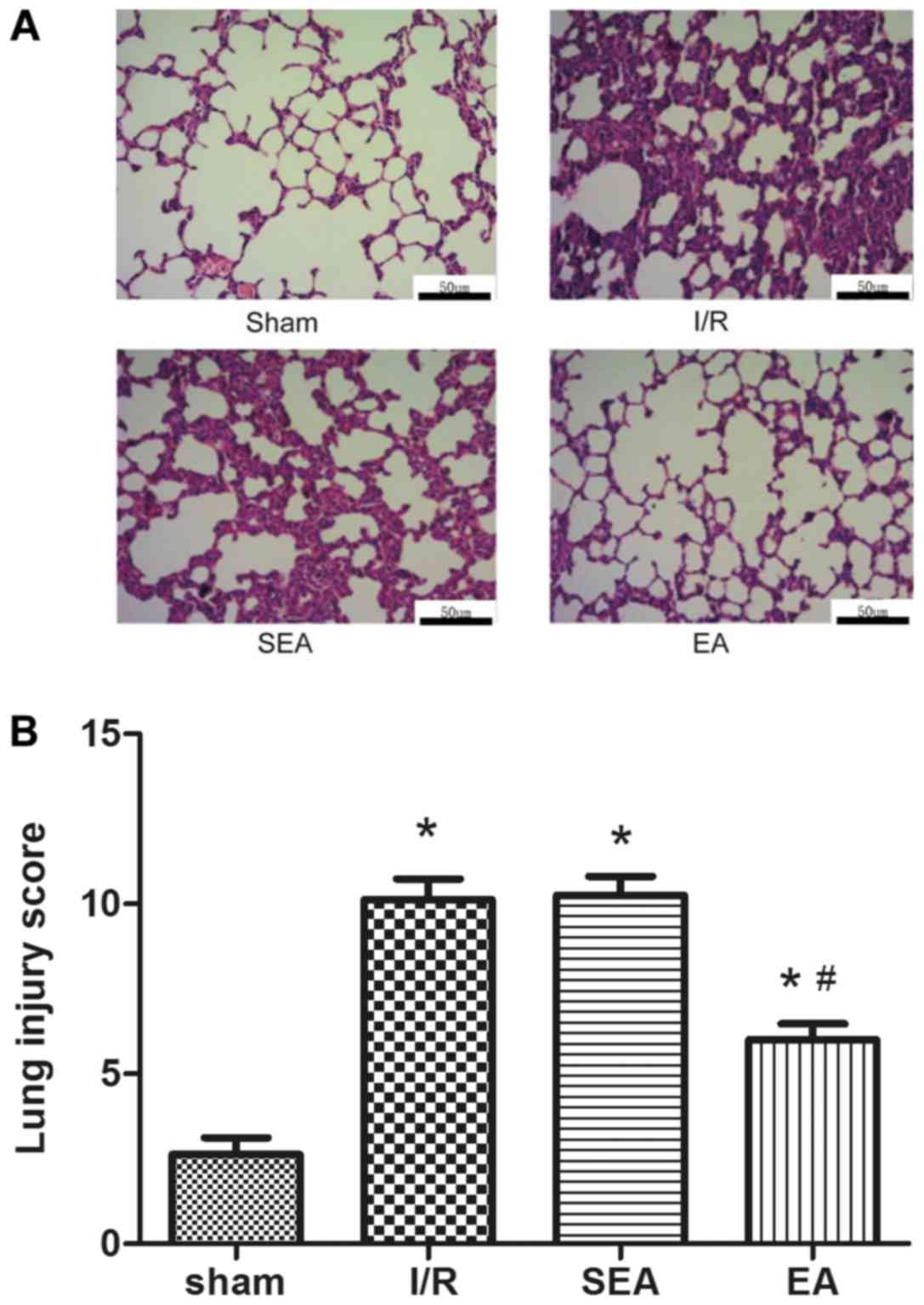
weight ratio compared with the I/R group (Fig. 3). Moreover, increased lung injury
scores, edema of the lung interstitium and destruction of the
alveolar architecture were observed in the I/R and SEA groups.
However, lung injury scores and histopathology were significantly
improved with EA pre-treatment, compared with the I/R group
(Fig. 4). These results
demonstrated that EA pre-treatment could exert an anti-inflammatory
effect on limb I/R mediated lung injury.
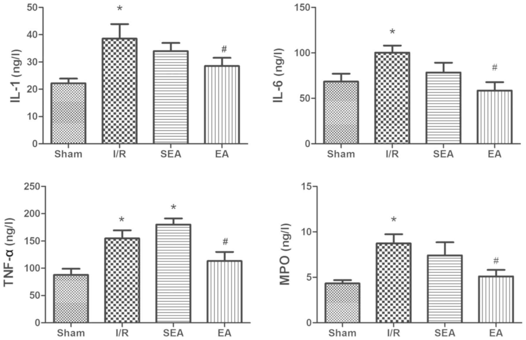
EA pre-treatment regulates secretion
of inflammatory cytokines in limb I/R-injured rats
To examine the effects of EA pre-treatment on lung
inflammation, the levels of inflammatory factors IL-1, IL-6, TNF-α
and MPO were measured in pulmonary tissue using ELISA (Fig. 5). The secretion of these factors
was significantly increased in the I/R and SEA groups compared with
the sham group. However, EA pre-treatment effectively inhibited
limb I/R-induced secretion of inflammatory cytokines IL-1, IL-6,
TNF-α and MPO.
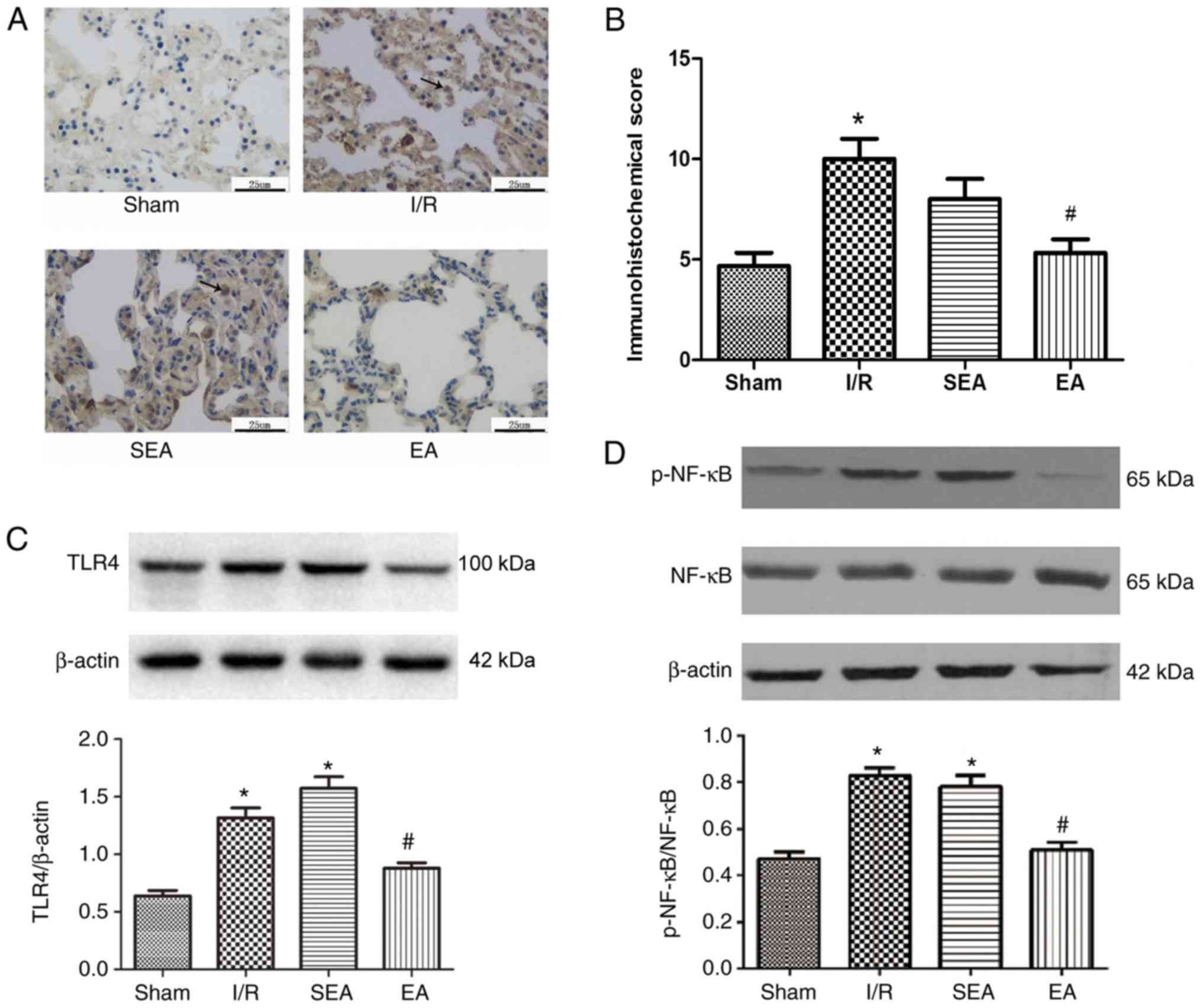
EA pre-treatment suppresses activation
of the TLR4/NF-κB pathway in limb I/R- injured rats
TLR4 plays a major role in the development of lung
inflammatory injury. Activated TLR4 stimulates downstream NF-κB,
resulting in increased release of inflammatory cytokines (18). To determine the mechanism
underlying the anti-inflammatory property of EA, the effect of EA
pre-treatment on the TLR4/NF-κB signaling pathway was examined in
the injured lung (Fig. 6).
Immunohistochemistry demonstrated that TLR4 expression was
increased in the lung tissue of rats in the I/R and SEA groups,
compared with the sham group. By contrast, the IHS was
significantly reduced in the EA group, compared with the I/R group.
Moreover, TLR4 and p-NF-κB expression levels were significantly
increased in the I/R and SEA groups compared with the sham group.
However, compared with the I/R group, the expression levels of TLR4
and p-NF-κB were significantly reduced in the EA group. These
findings suggested that pre-treatment with EA at the ST36 and SP6
acupoints could suppress the activation of the TLR4/NF-κB signaling
pathway, thus attenuating pulmonary inflammatory injury induced by
limb I/R in rats.
Discussion
In the present study, 30-min EA pre-conditioning
significantly reduced the inflammatory response and release of
inflammatory cytokines in the lungs of rats with limb I/R-induced
injury. Moreover, the mechanism underlying this protective effect
may be related to the TLR4/NF-κB signaling pathway.
Limb I/R can cause significant damage to distant
organs, including lung tissue. Limb I/R-induced lung injury can
occur following orthopedic surgery, major vascular injury, limb
thrombosis and other surgical procedures (19). Previous studies have reported that
I/R-induced lung injury resulted from the release of inflammatory
cytokines, enhancement of oxidative stress and increased apoptosis
during reperfusion (20).
Furthermore, limb I/R could induce the activation of neutrophils,
monocytes and macrophages, which can promote inflammation and
release a large number of inflammatory mediators, including
cytokines and chemokines (21).
Inflammatory factors in the ischemic area and the extensive release
of oxygen free radicals cause damage to distal organs and the lung
is particularly susceptible to acute injury (22). Previous studies on animal models
have demonstrated that the expression levels of inflammatory
cytokines, such as TNF-α, IL-1, IL-6 and IL-8, are significantly
increased after limb I/R (23–25).
The occurrence of acute lung injury is closely related to the
activation of various inflammatory factors; the extensive release
of inflammatory cytokines is one of the most important factors that
ultimately leads to the onset of systemic inflammatory response
syndrome (24).
TNF-α is a key mediator of acute lung injury, acute
respiratory distress syndrome and early lung tissue injury
(26). During the lung injury
process, TNF-α is primarily produced by alveolar macrophages, which
can mobilize polymorphonuclear leukocytes in the blood to
accumulate at lung injury sites, thus activating inflammatory cells
and endothelial cells, and supporting the formation of factors such
as endothelial cells and neutrophils that increase the expression
levels of IL-1, IL-6 and IL-8 (27). TNF-α can also promote
polymorphonuclear leukocyte degranulation and the release of
lysosomes, mediate alveolar-capillary membrane damage and increase
the function of the inflammatory cascade (28). IL-1 has a strong chemotactic
effect, inducing the expression of vascular endothelial cell
adhesion molecules, thus promoting leukocyte adhesion (29). Moreover, IL-1 primarily serves a
role in chemoattraction, macrophage stimulation and increase of
downstream cell and chemical factors. IL-6 is also involved in
systemic immune responses and inflammatory cascades, and can
activate neutrophils to exacerbate the production of inflammatory
mediators after trauma, effectively enhancing the severity of
tissue injury (30). MPO is
produced by polymorphonuclear leukocytes, and the levels of active
MPO reflect the activation of polymorphonuclear leukocytes in the
lungs (31).
In the present study, the expression levels of
proinflammatory cytokines, such as IL-1, IL-6 and TNF-α, were
significantly elevated following I/R. This was accompanied by a
reduction in intrapulmonary exchange function in the I/R group. The
incidence of lung injury was also significantly increased, together
with TLR4 and NF-κB expression levels. However, EA pre-treatment at
the ST36 and SP6 points could significantly reduce the extent of
lung injury, as well as the expression levels of inflammatory
cytokines, TLR4 and NF-κB, while significantly improving lung
exchange function. Therefore, these results suggested that EA
pre-treatment had an anti-inflammatory effect, as well as a
potential protective effect on the lungs.
EA, as a minimally invasive intervention, has been
reported to have a significant anti-inflammatory effect in numerous
diseases (32). ST36 and SP6 are
two main anti-inflammatory EA points. Our previous clinical studies
demonstrated that transcutaneous electrical acupoint stimulation at
the ST36 and SP6 acupoints could inhibit the inflammatory response
in the patients undergoing limb ischemia-reperfusion (33). Torres-Rosas et al (34) also revealed that EA pre-treatment
at these acupoints could inhibit inflammation in a murine model of
sepsis established by cecal ligation and puncture or
lipopolysaccharide.
The TLR4/NF-κB pathway is one of the pathways of EA
anti-inflammatory. Lan et al (35) suggested that EA at ST36 and Quchi
points decreased the levels of inflammatory factors in the brain
after reperfusion injury in a middle cerebral artery occlusion rat
model, by inhibiting the TLR4/NF-κB pathway. Chen et al
(36) also reported that EA
pre-conditioning could reduce the occurrence of cognitive
dysfunction caused by limb I/R, via the inhibition of microglial
activity. Previous animal and clinical studies on
inflammation-induced lung injury indicated that EA had an
anti-inflammatory effect that contribute to lung protection
(37,38). Similarly, the present study
suggested that EA pre-treatment at the ST36 and SP6 points had a
significant anti-inflammatory effect. Moreover, compared with the
control group, lung function and lung injury indexes were
signficantly improved, as were TLR4 and NF-κB expression levels.
Collectively, the present findings suggested that the
anti-inflammatory effect of EA could prevent lung injury and the
underlying mechanism may be related to the regulation of the
TLR4/NF-κB pathway in the lungs.
Previous studies have reported that the TLR4/NF-κB
pathway plays a key role in lung injury leading to several
inflammatory conditions, including septic, mechanical and I/R lung
injury (39,40). Furthermore, inhibition of the
TLR4/NF-κB pathway activity can greatly lower the incidence of lung
injury (41). In the present
study, limb I/R increased TLR4 and NF-κB expression levels in the
lungs and induced the release of inflammatory cytokines, indicating
that TLR4/NFκB was involved in the development of lung injury after
limb I/R. A previous study reported that EA could significantly
reduce TLR4 expression in cerebral I/R injury, thus inhibiting the
occurrence of an inflammatory response in the brain (35). In the present study, it was also
demonstrated that EA attenuated the expression levels of TLR4 and
NF-κB in the lungs. Therefore, activation of the TLR4/NF-κB
signaling pathway may be the primary mediator of lung injury after
limb I/R. Moreover, the anti-inflammatory effect of EA may be
mediated via the inhibition of the TLR4 signaling pathway, leading
to a protective effect on the lung.
However, the present study had a number of
limitations. TLR4 agonits were not included in the present study,
as these reagents could potentially cause lung damage. Therefore,
whether a TLR4 agonist could reverse the protective effects exerted
by EA pre-treatment was not evaluated. In addition, the experiments
described in the present study focused on TLR4 expression in the
lungs and did not locate TLR4 receptors in other tissues.
In conclusion, the present study demonstrated that
EA pre-treatment at the ST36 and SP6 acupoints improved lung
oxygenation and exerted an anti-inflammatory effect on lung injury
induced by limb I/R, which was via the inhibition of the TLR4/NF-κB
pathway. Thus, the present findings suggested that EA may be used
as a potentially effective therapy for lung injury induced by limb
I/R.
Acknowledgements
Not applicable.
Funding
The present study was supported by The National
Natural Science Foundation of China (grant nos. 81603685, 81573742,
81704180 and 81774109), The Zhejiang Province Natural Science
Foundation (grans nos. LY19H290008 and LY15H290006) and The Wenzhou
Municipal Science and Technology Bureau (grant no. 2018ZY003).
Availability of data and materials
The datasets used and/or analyzed during the current
study are available from the corresponding author on reasonable
request.
Authors' contributions
YM, JW and YL conceived and designed the study. QY,
KX, MFB, YT, GL and CZ performed the experiments, analyzed the
data, interpreted the experimental results, prepared the figures
and drafted the manuscript. QD, WG and YM were involved in
analyzing the data and revising the manuscript. All authors read
and approved the final manuscript.
Ethics approval and consent to
participate
All experimental procedures and animal care were
approved by The Animal Experimentation Ethics Committee of Wenzhou
Medical University and were conducted in accordance with the
Guidelines of The National Institutes of Health on the Care and Use
of Animals.
Patient consent for publication
Not applicable.
Competing interests
The authors declare that they have no competing
interests.
References
|
1
|
Xu YL, Zhang MH, Guo W, Xue Y, Du X, Zhang
T, Wu N and Wu Y: MicroRNA-19 restores vascular endothelial cell
function in lower limb ischemia-reperfusion injury through the
KLF10-dependent TGF-β1/Smad signaling pathway in rats. J Cell
Biochem. 119:3225–9315. 2018. View Article : Google Scholar
|
|
2
|
Tang B, Ma L, Yao X, Tan G, Han P, Yu T,
Liu B and Sun X: Hydrogen sulfide ameliorates acute lung injury
induced by infrarenal aortic cross-clamping by inhibiting
inflammation and angiopoietin 2 release. J Vasc Surg.
65:501–508.e1. 2017. View Article : Google Scholar : PubMed/NCBI
|
|
3
|
Campanholle G, Landgraf RG, Gonçalves GM,
Paiva VN, Martins JO, Wang PH, Monteiro RM, Silva RC, Cenedeze MA,
Teixeira VP, et al: Lung inflammation is induced by renal ischemia
and reperfusion injury as part of the systemic inflammatory
syndrome. Inflamm Res. 59:861–869. 2010. View Article : Google Scholar : PubMed/NCBI
|
|
4
|
Liao WI, Wu SY, Wu GC, Pao HP, Tang SE,
Huang KL and Chu SJ: Ac2-26, an Annexin A1 Peptide, Attenuates
Ischemia-Reperfusion-Induced Acute Lung Injury. Int J Mol Sci.
18:182017. View Article : Google Scholar
|
|
5
|
Xu MJ, Liu BJ, Wang CL, Wang GH, Tian Y,
Wang SH, Li J, Li PY, Zhang RH, Wei D, et al:
Epigallocatechin-3-gallate inhibits TLR4 signaling through the
67-kDa laminin receptor and effectively alleviates acute lung
injury induced by H9N2 swine influenza virus. Int Immunopharmacol.
52:24–33. 2017. View Article : Google Scholar : PubMed/NCBI
|
|
6
|
Areal H, Abrantes J and Esteves PJ:
Signatures of positive selection in Toll-like receptor (TLR) genes
in mammals. BMC Evol Biol. 11:3682011. View Article : Google Scholar : PubMed/NCBI
|
|
7
|
Sherif IO and Al-Shaalan NH: Vildagliptin
Attenuates Hepatic Ischemia/Reperfusion Injury via the TLR4/NF-κB
Signaling Pathway. Oxid Med Cell Longev. 2018:35090912018.
View Article : Google Scholar : PubMed/NCBI
|
|
8
|
Zhou Z, Zhu X, Chen J, Yang S, Sun R and
Yang G: The interaction between Toll-like receptor 4 signaling
pathway and hypoxia-inducible factor 1α in lung
ischemia-reperfusion injury. J Surg Res. 188:290–297. 2014.
View Article : Google Scholar : PubMed/NCBI
|
|
9
|
Arumugam TV, Okun E, Tang SC, Thundyil J,
Taylor SM and Woodruff TM: Toll-like receptors in
ischemia-reperfusion injury. Shock. 32:4–16. 2009. View Article : Google Scholar : PubMed/NCBI
|
|
10
|
Yunhe F, Bo L, Xiaosheng F, Fengyang L,
Dejie L, Zhicheng L, Depeng L, Yongguo C, Xichen Z, Naisheng Z, et
al: The effect of magnolol on the Toll-like receptor 4/nuclear
factor κB signaling pathway in lipopolysaccharide-induced acute
lung injury in mice. Eur J Pharmacol. 689:255–261. 2012. View Article : Google Scholar : PubMed/NCBI
|
|
11
|
Zijlstra FJ, van den Berg-de Lange I,
Huygen FJ and Klein J: Anti-inflammatory actions of acupuncture.
Mediators Inflamm. 12:59–69. 2003. View Article : Google Scholar : PubMed/NCBI
|
|
12
|
Gong LR, Kan YX, Lian Y, Dong SA, Zhao DH,
Shi J and Yu JB: Electroacupuncture Attenuates Limb Ischemia-
Reperfusion-Induced Lung Injury Via p38 Mitogen-Activated Protein
Kinase-Nuclear Factor Erythroid-2-Related Factor-2/Heme Oxygenase
Pathway. J Surg Res. 246:170–181. 2020. View Article : Google Scholar : PubMed/NCBI
|
|
13
|
Carbone L: Pain management standards in
the eighth edition of the Guide for the Care and Use of Laboratory
Animals. J Am Assoc Lab Anim Sci. 51:322–328. 2012.PubMed/NCBI
|
|
14
|
Yassin MM, Harkin DW, Barros D'Sa AA,
Halliday MI and Rowlands BJ: Lower limb ischemia-reperfusion injury
triggers a systemic inflammatory response and multiple organ
dysfunction. World J Surg. 26:115–121. 2002. View Article : Google Scholar : PubMed/NCBI
|
|
15
|
Santos EL, Dias BH, Andrade AC, Pascoal
AM, Vasconcelos Filho FE, Medeiros F and Guimarães SB: Effects of
acupuncture and electroacupuncture on estradiol-induced
inflammation and oxidative stress in health rodents. Acta Cir Bras.
28:582–588. 2013. View Article : Google Scholar : PubMed/NCBI
|
|
16
|
Chen CM, Wang LF, Su B and Hsu HH:
Methylprednisolone effects on oxygenation and histology in a rat
model of acute lung injury. Pulm Pharmacol Ther. 16:215–220. 2003.
View Article : Google Scholar : PubMed/NCBI
|
|
17
|
Soslow RA, Dannenberg AJ, Rush D, Woerner
BM, Khan KN, Masferrer J and Koki AT: COX-2 is expressed in human
pulmonary, colonic, and mammary tumors. Cancer. 89:2637–2645. 2000.
View Article : Google Scholar : PubMed/NCBI
|
|
18
|
Peng LY, Shi HT, Yuan M, Li JH, Song K,
Huang JN, Yi PF, Shen HQ and Fu BD: Madecassoside Protects Against
LPS-Induced Acute Lung Injury via Inhibiting TLR4/NF-κB Activation
and Blood-Air Barrier Permeability. Front Pharmacol. 11:8072020.
View Article : Google Scholar : PubMed/NCBI
|
|
19
|
Zhao YR, Wang D, Liu Y, Shan L and Zhou
JL: The PI3K/Akt, p38MAPK, and JAK2/STAT3 signaling pathways
mediate the protection of SO2 against acute lung injury
induced by limb ischemia/reperfusion in rats. J Physiol Sci.
66:229–239. 2016. View Article : Google Scholar : PubMed/NCBI
|
|
20
|
Men X, Han S, Gao J, Cao G, Zhang L, Yu H,
Lu H and Pu J: Taurine protects against lung damage following limb
ischemia reperfusion in the rat by attenuating endoplasmic
reticulum stress-induced apoptosis. Acta Orthop. 81:263–267. 2010.
View Article : Google Scholar : PubMed/NCBI
|
|
21
|
Chou WC, Kao MC, Yue CT, Tsai PS and Huang
CJ: Caffeine Mitigates Lung Inflammation Induced by
Ischemia-Reperfusion of Lower Limbs in Rats. Mediators Inflamm.
2015:3616382015. View Article : Google Scholar : PubMed/NCBI
|
|
22
|
Zou H and Sun X: Post-treatment curcumin
reduced ischemia-reperfusion-induced pulmonary injury via the
Notch2/Hes-1 pathway. J Int Med Res. 2019:3000605198924322019.
|
|
23
|
Dong X, Xing Q, Li Y, Han X and Sun L:
Dexmedetomidine protects against ischemia-reperfusion injury in rat
skeletal muscle. J Surg Res. 186:240–245. 2014. View Article : Google Scholar : PubMed/NCBI
|
|
24
|
Zhao YR, Lv WR and Zhou JL: Role of
carbonyl sulfide in acute lung injury following limb
ischemia/reperfusion in rats. Eur J Med Res. 22:122017. View Article : Google Scholar : PubMed/NCBI
|
|
25
|
Liu Y, Zhou C, Jiang J, Su Q and Ding X:
Blockade of HMGB1 preserves vascular homeostasis and improves blood
perfusion in rats of acute limb ischemia/reperfusion. Microvasc
Res. 112:37–40. 2017. View Article : Google Scholar : PubMed/NCBI
|
|
26
|
Yu S, Xie J, Xiang Y, Dai S, Yu D, Sun H,
Chen B and Zhou M: Downregulation of TNF-α/TNF-R1 Signals by
AT-Lipoxin A4 May Be a Significant Mechanism of Attenuation in
SAP-Associated Lung Injury. Mediators Inflamm. 2019:90194042019.
View Article : Google Scholar : PubMed/NCBI
|
|
27
|
De Campos T, Deree J and Coimbra R: From
acute pancreatitis to end-organ injury: Mechanisms of acute lung
injury. Surg Infect (Larchmt). 8:107–120. 2007. View Article : Google Scholar : PubMed/NCBI
|
|
28
|
Yang WS, Lee JM, Han NJ, Kim YJ, Chang JW
and Park SK: Mycophenolic acid attenuates tumor necrosis
factor-alpha-induced endothelin-1 production in human aortic
endothelial cells. Atherosclerosis. 211:48–54. 2010. View Article : Google Scholar : PubMed/NCBI
|
|
29
|
Wang J, Tan J, Liu Y, Song L, Li D and Cui
X: Amelioration of lung ischemia reperfusion injury by JNK and p38
small interfering RNAs in rat pulmonary microvascular endothelial
cells in an ischemia reperfusion injury lung transplantation model.
Mol Med Rep. 17:1228–1234. 2018.PubMed/NCBI
|
|
30
|
Pimenta MB, Aguilar-Nascimento JE, Martins
DC, Silva DR, Bacelo KL, Bocchese IC, Zaffani S, Zaffani E,
Silveira EA, Carmo AV and Ferreira SS: The intestinal tract as the
major source of interleukin 6 production during abdominal aortic
clamping and hind limb ischaemia-reperfusion injury. Acta Cir Bras.
22 (Suppl 1):34–39. 2007. View Article : Google Scholar : PubMed/NCBI
|
|
31
|
Schmekel B, Seveus L, Xu SY and Venge P:
Human neutrophil lipocalin (HNL) and myeloperoxidase (MPO). Studies
of lung lavage fluid and lung tissue. Respir Med. 94:564–568. 2000.
View Article : Google Scholar : PubMed/NCBI
|
|
32
|
Jin H, Guo J, Liu J, Lyu B, Foreman RD,
Yin J, Shi Z and Chen JDZ: Anti-inflammatory effects and mechanisms
of vagal nerve stimulation combined with electroacupuncture in a
rodent model of TNBS-induced colitis. Am J Physiol Gastrointest
Liver Physiol. 313:G192–G202. 2017. View Article : Google Scholar : PubMed/NCBI
|
|
33
|
Mo Y, Chen S, Yang L, Huang L, Jin D, Yu
Z, Wang L, Wang L, Luo S and Wang J: The Effect of Transcutaneous
Electrical Acupoint Stimulation on Inflammatory Response in
Patients Undergoing Limb Ischemia-Reperfusion. Mediators Inflamm.
2017:83697372017. View Article : Google Scholar : PubMed/NCBI
|
|
34
|
Torres-Rosas R, Yehia G, Peña G, Mishra P,
del Rocio Thompson-Bonilla M, Moreno-Eutimio MA, Arriaga-Pizano LA,
Isibasi A and Ulloa L: Dopamine mediates vagal modulation of the
immune system by electroacupuncture. Nat Med. 20:291–295. 2014.
View Article : Google Scholar : PubMed/NCBI
|
|
35
|
Lan L, Tao J, Chen A, Xie G, Huang J, Lin
J, Peng J and Chen L: Electroacupuncture exerts anti-inflammatory
effects in cerebral ischemia-reperfusion injured rats via
suppression of the TLR4/NF-κB pathway. Int J Mol Med. 31:75–80.
2013. View Article : Google Scholar : PubMed/NCBI
|
|
36
|
Chen Y, Zhou J, Li J, Yang SB, Mo LQ, Hu
JH and Yuan WL: Electroacupuncture pretreatment prevents cognitive
impairment induced by limb ischemia-reperfusion via inhibition of
microglial activation and attenuation of oxidative stress in rats.
Brain Res. 1432:36–45. 2012. View Article : Google Scholar : PubMed/NCBI
|
|
37
|
Geng WY, Liu ZB, Song NN, Geng WY, Zhang
GH, Jin WZ, Li L, Cao YX, Zhu DN and Shen LL: Effects of
electroacupuncture at Zusanli (ST36) on inflammatory cytokines in a
rat model of smoke-induced chronic obstructive pulmonary disease. J
Integr Med. 11:213–219. 2013. View Article : Google Scholar : PubMed/NCBI
|
|
38
|
Yu JB, Dong SA, Luo XQ, Gong LR, Zhang Y,
Wang M, Cao XS and Liu DQ: Role of HO-1 in protective effect of
electro-acupuncture against endotoxin shock-induced acute lung
injury in rabbits. Exp Biol Med (Maywood). 238:705–712. 2013.
View Article : Google Scholar : PubMed/NCBI
|
|
39
|
Zhao Q, Wu J, Lin Z, Hua Q, Zhang W, Ye L,
Wu G, Du J, Xia J, Chu M, et al: Resolvin D1 Alleviates the Lung
Ischemia Reperfusion Injury via Complement, Immunoglobulin, TLR4,
and Inflammatory Factors in Rats. Inflammation. 39:1319–1333. 2016.
View Article : Google Scholar : PubMed/NCBI
|
|
40
|
Ramani V, Madhusoodhanan R, Kosanke S and
Awasthi S: A TLR4-interacting SPA4 peptide inhibits LPS-induced
lung inflammation. Innate Immun. 19:596–610. 2013. View Article : Google Scholar : PubMed/NCBI
|
|
41
|
Standiford LR, Standiford TJ, Newstead MJ,
Zeng X, Ballinger MN, Kovach MA, Reka AK and Bhan U: TLR4-dependent
GM-CSF protects against lung injury in Gram-negative bacterial
pneumonia. Am J Physiol Lung Cell Mol Physiol. 302:L447–L454. 2012.
View Article : Google Scholar : PubMed/NCBI
|