Introduction
Cancer is the second most common cause of mortality
worldwide, and lead to an estimated 18.1 incident cases and 9.6
million deaths in 2018 (1).
Currently, surgery and cytotoxic chemotherapy are the primary
treatments for cancer. However, 70% of cancer mortality occurs in
low- or middle-income countries, which is due to delays in
diagnosis and cancer being at a late stage at first presentation
(2). Curative surgical resection
may no longer be an option in such cases, leaving chemotherapy as
the only treatment option (1–3). The
global disease burden attributed to cancer underscores the
importance of developing chemotherapeutic regimens with enhanced
safety, tolerability and efficacy (3).
Paclitaxel is a first-line chemotherapeutic agent
for solid tumors, but its use has been confounded by poor
solubility, toxicity and the emergence of resistance during therapy
(4,5). In order to improve efficacy and to
decrease the emergence of resistance, clinicians combine paclitaxel
with cisplatin and other antineoplastic drugs (6,7). As
nanotechnology has progressed, multiple types of paclitaxel
nanoparticles (NPs) have been developed to modulate drug release,
promote drug encapsulation, improve bioavailability and target
cancer cells by using frame materials of polyethylene glycol,
polylactic acid, polyglycolin acid or liposomes (8). Examples include abraxane, which was
synthesized by attaching six or seven paclitaxel molecules to
albumin NPs of 130-nm diameter, and is considered to be among the
most successful nanotherapeutics, as it has been approved as a
first-line treatment for non-small cell lung carcinoma by the US
Food and Drug Administration (8).
Folic acid (FA)-poly NPs have been prepared to target ovarian tumor
tissue, and NPs that carry rituximab and paclitaxel have been
designed to target CD20-positive B-cell lymphoma (9).
Gold NPs (AuNPs) have received increasing attention
as drug delivery vehicles for cancer therapeutics as they can be
engineered to obviate drug insolubility, carry nucleic acid
payloads for gene therapy, target specific tumor cell lines,
modulate drug release and amplify photothermal therapy (PTT)
(10,11). The development of gene therapy has
generated increasing interest in the potential of AuNPs to deliver
therapeutic nucleic acid payloads (10,11).
AuNPs have been designed to carry p53, vascular cell adhesion
molecule-1 and other mRNAs (10,11).
Furthermore, poly(thymine)-functionalized AuNPs have been
synthesized to target mRNA translation using a pcDNA6 vector
expressing a bovine growth hormone polyadenylation signal (12). Consequently, it is important to
investigate the potential of AuNPs to enhance paclitaxel drug
delivery and gene therapy (12).
In the current review, the role of paclitaxel in clinical oncology,
and previous research on AuNPs and the development of paclitaxel
AuNP-based drug delivery and its limitations are examined.
Mechanisms of paclitaxel action and drug
resistance
Paclitaxel, which is isolated from Pacific Yew tree
Taxus brevifolia, has been indicated to be a potent
antimitotic agent in solid tumor cell lines (13). In 1992, paclitaxel was approved for
ovarian cancer treatment by the US Food and Drug Administration
(14). According to the National
Comprehensive Cancer Network guidelines, paclitaxel is still a
first-line chemotherapeutic, is combined with cisplatin,
atezolizumab and other anti-neoplastic agents to treat lung,
breast, gastric and colorectal cancer, and is also incorporated
into pre- and post-operative chemoradiation protocols (15–18).
The antimitotic activity of paclitaxel is mediated
via its disruption of microtubular function (19). Paclitaxel promotes the assembly of
microtubules from tubulin dimers and stabilizes microtubules by
preventing depolymerization (20).
This inhibits the reorganization of the microtubule network, which
is essential for interphase and mitotic cellular function (20). The polymerization of tubulin dimers
decreases their concentration below that required for spindle
assembly, thus arresting the cell cycle at the M/G2 phase (21–23).
In addition, paclitaxel promotes tumor apoptosis by upregulating
death receptor signaling and mitochondrial apoptotic pathways and
increases beclin-1-mediated autophagy (21,24).
These mechanisms of action have contributed to the emergence of
paclitaxel as a first-line antineoplastic drug.
With repeated use, resistance commonly emerges
during cancer therapy, and there are three major resistance
mechanisms governing this resistance. The first, metabolic
inactivation via the cytochrome P450 (CYP) system, was identified
in 1994 (25). CYP3A and CYP2C
were revealed to serve major roles in paclitaxel metabolism
(26). It has also been
demonstrated that paclitaxel treatment induces overexpression of
CYP2C8 and CYP3A4/5, which is mediated by a nuclear receptor in
multiple types of tumor cells, such as pancreatic ductal
adenocarcinoma (PDAC), pancreatic adenocarcinoma 2 (PACO2) and
PACO7, which are two primary PDAC cells propagated from
immune-deficient
NOD.Cg-PrkdcscidIl2rgtm1Wjl
mice, leading to cell-autonomous detoxification (27). A second mechanism behind resistance
is the overexpression of ATP binding cassette (ABC) drug efflux
transporters. ABCB1, ABCC1, ABCC2 and ABCG2 have been implicated in
paclitaxel resistance in vivo and in vitro (28). ABCB1 and ABCC3 exhibit prominent
activity in breast cancer resistance (28). A third mechanism for resistance is
associated with drug transport that is mediated by solute carrier
proteins. Changes in expression levels of solute carrier proteins
may alter drug uptake and determine chemotherapeutic efficacy
(29). Single nucleotide
polymorphisms in genes that encode solute carriers (SLCs, including
SLC31A2, SLC43A1, SLC35A5, SLC41A2 and SLCO1B3) are
associated with paclitaxel sensitivity and resistance in
vitro (29). In addition,
tumor suppressor genes such as BRCA1, TP53, PTEN,
adenomatous polyposis coli, Cyclin Dependent Kinase Inhibitor
1A/2A, High in normal-1 and Bax also affect
paclitaxel resistance (30,31).
Furthermore, low solubility also limits the use of paclitaxel and
all of the aforementioned factors decrease its efficacy. Therefore,
it is essential to identify carriers to increase paclitaxel
delivery and overcome insolubility and resistance. AuNPs have a
number of advantages, including large surface area for drug and
nucleic acid binding, non-toxicity and a unique property of
amplifying PTT (10–12).
Development of AuNPs for antineoplastic drug
delivery
In the past decade, NPs have been evaluated as novel
drug delivery systems. The majority of these particles have a
feature size <200 nm, which helps to overcome the mucus barrier,
resulting in low reactogenicity and immunogenicity (32). A previous study demonstrated that
NPs >200 nm in size induce a mucosal immune response and are
taken up by dendritic cells, which secrete inflammation-associated
factors, resulting in an immune response (33). In addition, NP carriers exhibit
multiple pharmacokinetic advantages, including high loading
efficiencies of lipophilic drugs such as 5-fluorouracil, which have
low water solubility and intracytoplasmic bioavailability,
protection of drugs from degradation, increased drug uptake by
tumor cells, enhanced drug concentrations in tumor
microenvironments, prolonged drug release and targeted delivery
(34,35). The use of NPs for targeted drug
delivery allows lower systemic drug exposures and minimal side
effects (36). As cancer is the
second leading cause of mortality worldwide, the future development
and use of antineoplastic nanodrugs is essential (37,38).
Currently, there are four types of nanodelivery
systems used or in development in oncology: Polymeric, magnetic,
metallic and lipid NPs (36).
AuNPs are non-toxic, can be easily synthesized into different sizes
and shapes, have a large surface area to transport drugs, exhibit
good biocompatibility and absorb near-infrared light (NIR) that can
be converted to heat to enable focused thermal therapy (39). AuNPs are used in diagnostic
imaging, targeted delivery of drugs, radioisotopes and reactive
oxygen species-generating enzymes, and plasmonic PTT and
photodynamic therapy (40). Within
the past year, >100 studies of AuNPs for drug delivery were
identified. These studies are classified into 3 groups within the
current review: Simple drug-carrying AuNPs, simple nucleic
acid-carrying AuNPs and targeted (tissue directed) AuNPs.
Simple drug-carrying AuNPs
AuNPs were evaluated initially as antineoplastic
drug carriers in 2004 (41). AuNPs
with a diameter of 32 nm carrying tumor necrosis factor (TNF) have
been used to target MC-38 colon carcinoma tumors in vivo, as
the majority of TNF AuNPs target tumor cells with little
accumulation in the liver and other organs in mice (41). Furthermore, TNF-carrying AuNPs have
been indicated to be more effective against tumor cells compared
with naive TNF (41). Studies have
also been performed on AuNPs featuring optimization of either the
particle or the drug payload (42,43).
DM1, which is a maytansine analogue, was developed as a
microtubulin inhibitor with the same mechanism of action as
paclitaxel, but its toxicity and narrow therapeutic window limited
its clinical development. Conjugation of DM1 with ultra-small
(diameter, 2 nm) AuNPs prolonged the half-life of DM1, increased
its cytotoxicity in Bel7404 and HepG2 cells and also improved its
tolerability and efficacy in ectopic xenograft models of
hepatocellular carcinoma (42).
The conjugation of kaempferol, a flavonoid that damages DNA in
malignant cells, to 2-nm gold nanoclusters decreases toxicity to
normal cells, enhances toxicity to cancer cells and decreases
proliferation, colony formation and migration of A549 lung cancer
cells (43).
The amplification of PTT is a unique property of
AuNPs that facilitates the development of a novel therapeutic
modality, combined chemo-PTT (44). Gold nanoshell-coated wedelolactone
liposomes combined with NIR-induced hyperthermia release 97.34% of
wedelolactone in 8 h and exhibit an excellent antitumor efficacy
in vivo (44). In
conclusion, simple drug-carrying AuNPs are less toxic to normal
cells and more toxic to cancer cells.
Nucleic acid-carrying AuNPs
As AuNPs have positive-charged surfaces whereas
nucleic acids exhibit negative charges, AuNPs are easily adapted to
deliver nucleic acid payloads (45). Small interfering RNAs (siRNAs),
mRNAs and micro (mi)RNAs conjugated with AuNPs are used for
diagnosis and gene therapy (45,46).
Receptor tyrosine kinase-like orphan receptor 1 (ROR1) siRNA-HIV-1
Tat peptide-capped AuNPs have a hydrophilic arginine-rich section
that facilitates DNA binding; increases the stability of ROR1
siRNA; enhances cellular uptake and induces apoptosis and necrosis
in MDA-MB-231 cells (46).
Furthermore, surfactant-free AuNPs deliver plasmid
DNA for tumor treatment (10).
AuNPs capped with L-cystine methylester hydrochloride and loaded
with p53 plasmid DNA increase p53 expression levels and induce A549
apoptosis, thus inhibiting proliferation without inducing
cytotoxicity to normal lung cells (10). The conversion of amine-modified
siRNA duplexes into dithiocarbamate ligands followed by conjugation
with AuNPs increases siRNA stability and enhances targeted release
of siRNA (47). Additional
potential advantages of AuNP-based therapy have been demonstrated
by the development of an intracellular self-assembly system of DNA
and AuNPs (48). In a previous
study (48), mRNA of survivin,
which is an apoptosis inhibitor, and AuNPs in a complex with its
complementary DNA sequence were transfected into cancer cells
separately and spontaneously formed cytoplasmic aggregates.
Survivin mRNA and AuNPs entered cells easily due to their small
size, whereas the larger aggregates exhibited improved
intracellular retention. This resulted in enhanced apoptosis and
photothermal function, leading to cancer cell death rates of up to
93.3%, with minimal toxicity to normal cells (48).
17-N-allylamino-17-demethoxygeldanamycin (17-AAG) is
a heat shock protein 90 (HSP90) inhibitor that blocks ATP binding
and inhibits HER2(+) breast cancer cell proliferation (49). However, the anti-tumor effect of
17-AAG is decreased in the absence of Cullin-5 (Cul5), an E3
ubiquitin ligase that is required for HSP90 inhibitor function
(49). Consequently, the delivery
of 17-AAG and Cul5 DNA via AuNPs can sensitize 17-AAG-resistant
breast cancer cells (49).
Similarly, poly(sodium 4-styrenesulfonate) and
poly(-diallyldimethylammonium chloride) have been used to coat gold
nanoprisms carrying a siRNA targeting human program death-ligand 1
(hPD-L1), resulting in the downregulation of hPD-L1 expression and
enabling photoacoustic imaging and PTT of lung cancer cells and
cell-derived tumors (50).
Zhang et al (51) developed gold nanoshells that are
designed for the PTT-stimulated release of siRNA to downregulate
HER-2 expression levels and a genetic sequence to express the
immunological adjuvant cytosine-guanine motifs to activate
anti-tumor immune responses that are mediated by toll-like receptor
9 signaling. This integration of gene therapy, immunotherapy and
PTT has been demonstrated to perform well in the treatment of a
murine gastric cancer model (51).
Consequently, in the presence or absence PTT, AuNPs may be used to
carry tumor suppressor genes, oncogene-siRNA and miRNA to inhibit
tumor cell proliferation (51).
Targeted AuNPs
In addition to simple AuNPs that are used for drug
and nucleic acid delivery and PTT, multifunctional AuNPs have been
designed for targeted delivery mediated by pH, short peptides,
aptamers, antibodies and receptors (52–57).
Glutathione (GSH)-coated Au-Fe3O4 nanoshells
for doxorubicin (DOX) delivery feature a GSH layer on the nanoshell
surface that is attached with an Au-S native bond, which is stable
at physiological pH but rapidly broken down in acidic tumor
microenvironments, resulting in local DOX release and cytotoxicity
(52). AuNPs coated with
biocompatible marine carbohydrate carrageenan oligosaccharide
(CAO-AuNPs) have also been designed for pH-dependent drug release.
For example, epirubicin (EPI)-CAO-AuNPs release their payload in
acidic environments and more easily enter cells via endocytosis,
inducing more potent cytotoxicity and increasing apoptosis compared
with free EPI (53). A study by
Pedrosa et al (53) used
polyethylene glycol-coated AuNPs carrying a novel chemotherapeutic
candidate (ZnD) and the monoclonal antibody cetuximab that targets
epidermal growth factor receptor (EGFR) receptors that are
overexpressed in cancer cells, and demonstrated that the addition
of cetuximab facilitated both targeting and a second mechanism of
action (antagonism of receptor-dependent EFGR signal transduction,
resulting in interruption of the cell cycle, apoptosis and
inhibition of metastasis and angiogenesis). This candidate was also
demonstrated to be active against DOX-resistant tumors in a murine
model (54). Afatinib (Afb) is a
tyrosine kinase inhibitor that targets EGFR-positive lung cancer,
and has disadvantages, including low bioavailability and high
toxicity (54). In addition, the
native structure of Afb prevents its direct conjugation to AuNPs.
In order to mitigate these issues, an Afb analog containing a
pedant alkynyl has been synthesized (55). The pedant alkynyl is coupled to an
azide-functionalized lipoic acid moiety using a copper(I)-catalyzed
Huisgen 1,3-dipolar cycloaddition to improve attachment to the AuNP
surface with an alkylthiol-gold bond. Afb-AuNPs attenuate
pro-inflammatory cytokine release and promote Afb cellular uptake
and consequently, Afb-AuNPs exhibit a 3.7-fold increased potency in
inhibiting EGFR mutant lung cancer cells, including PC-9 cells
(55). Porous silicon NPs in a
complex with gold nanorods have been designed for delivery of a
three-drug combination of hydrophilic and hydrophobic drugs (Afb,
DOX and rapamycin), and the inclusion of Afb, which targets HER-2
and EGFR. This construct confers targeted drug delivery to
malignant cells, while gold nanorods facilitate PTT (56). This candidate has demonstrated
positive results in vitro and in vivo (56).
The use of short peptides, antibodies and receptors
in the design of targeted AuNPs is receiving increasing attention.
Paris et al (56) recently
investigated a novel strategy of targeting tumor angiogenesis
rather than disrupting malignant cells. A tumor vascular
endothelium-targeting nanoparticle was designed by incorporating an
isoAspGly-Arg peptide that targets αβ-integrin-overexpressing cells
(For example, tumor vascular endothelia), an anti-angiogenic drug
(DOX), a vascular disrupting agent (fosbretabulin) and a gold
nanorod. This enabled both drug delivery and PTT in a murine
fibrosarcoma xenograft model and the results indicated a decrease
in existing vasculature and the inhibition of neovascularization
within tumors (57). Furthermore,
delivery of interleukin-12 in RGD-peptide AuNPs may enhance
adoptive T-cell therapy and improve its therapeutic potential
(58). Neuropilin (Nrp)-1
receptor-specific short peptide-coated cucurbituril AuNPs deliver
and release curcumin in Nrp-1-overexpressing melanoma cells
(59). AuNPs coated with an
anti-HER2 monoclonal antibody (trastuzumab)-cytotoxic drug
[monomethyl auristatin E (MMAE)] conjugate and a cell-penetrating
peptide (HIV Tat) have been indicated to target HER-2-positive
cells, increase cellular uptake and enhance the antimitotic potency
and therapeutic index of free MMAE in vitro (60). Anti-EGFR-coated
5-fluorouracil-AuNPs have also been revealed to target colorectal
cancer cells that overexpress EGFR, increase their apoptosis rate
and improve their anti-tumor effects (9). DOX and anti-PD-L1 antibody-conjugated
AuNPs enable drug delivery and PTT to target and induce CT-26
cellular apoptosis and inhibit cell proliferation (9). Furthermore, the attachment of glucose
to polyethylene glycol (PEG) at the C6 position followed by
installation on sub-50-nm nanocarrier enhances the delivery of
polo-like kinase 1 siRNA to cancer stem-like cells, which
overexpress glucose transporter 1 on their surface (61).
By carrying drugs in combination with specific
nucleic acid sequences, short peptides, antibodies and receptors,
AuNPs can aggregate at target tissues and cells to modulate drug
release rate and improve efficacy.
Further innovations
Eco-friendly AuNPs
Protection of the environment has received
increasing attention in recent years, and the use of eco-friendly
materials has been investigated regarding the synthesis of AuNPs.
The flower bud extract of Tussilago farfara has been used as
a reducing agent to synthesize AuNPs that are suitable for
anticancer drug delivery (62).
Photosynthesized AuNPs from Catharanthus roseus (CR) leaf
extract have also been studied, as CR is a plant that is used in
traditional Chinese medicine (63). Without the addition of a
chemotherapeutic or siRNA therapeutic payload, these AuNPs inhibit
tumor proliferation by inducing mitochondrial-mediated apoptosis
via reactive oxygen species (Fig.
1) (63). In addition, leaf
extracts of Ziziphus zizyphus, Coleus aromaticus, Indigofera
tinctoria, Bauhinia purpurea and mulberry have been used
to synthesize environmentally friendly AuNPs (64–68).
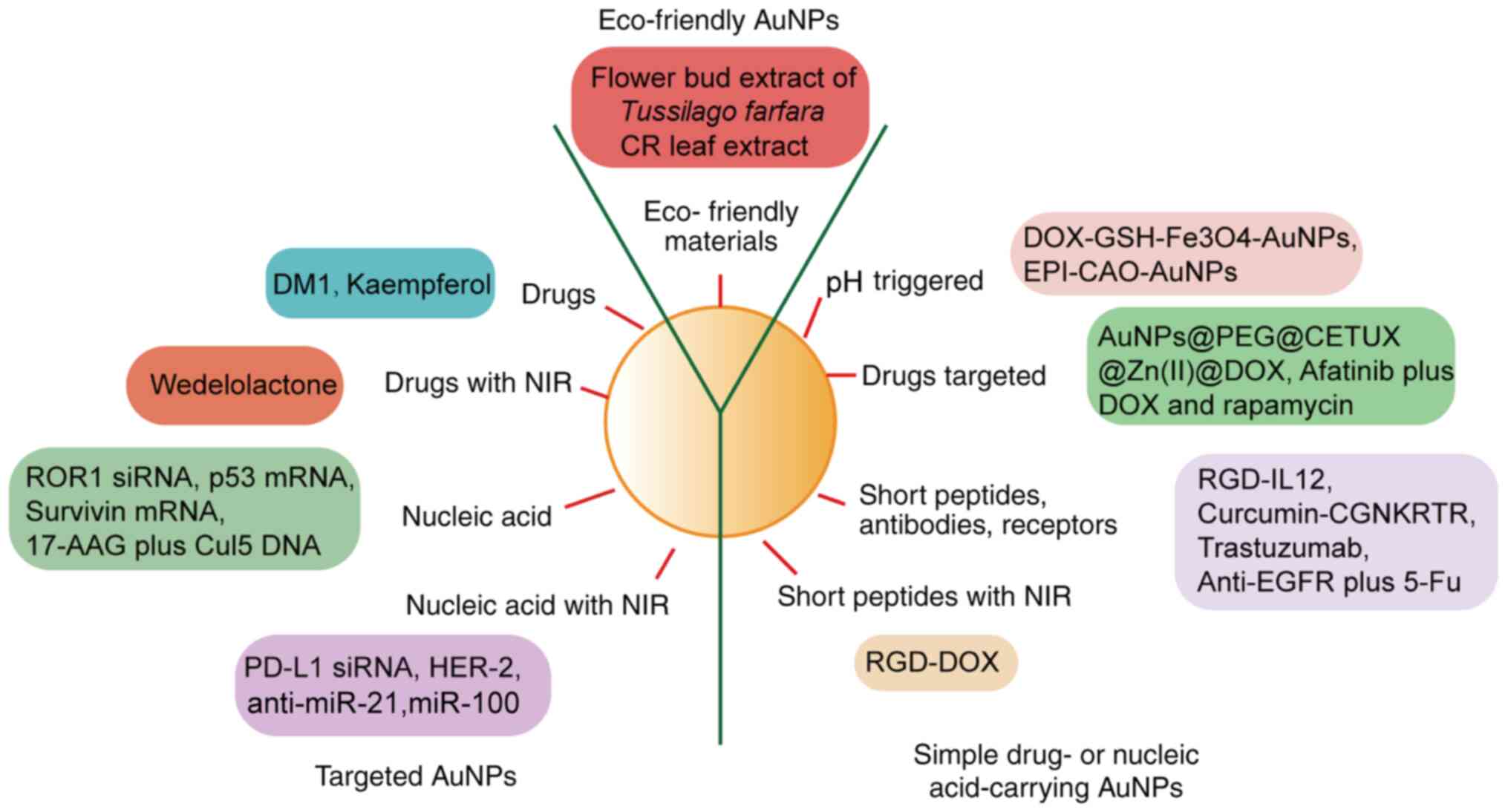 | Figure 1.AuNPs are classified into three
groups: Simple drug-carrying AuNPs, nucleic acid-carrying AuNPs and
multifunctional AuNPs. Eco-friendly AuNPs may be synthesized using
environmentally safe materials and production processes. AuNP, gold
nanoparticle; NIR, near-infrared; ROR1, receptor tyrosine
kinase-like orphan receptor 1; siRNA, small interfering RNA;
17-AAG, 17-N-allylamino-17-demethoxygeldanamycin; Cul5, Cullin5;
PD-L1, program death-ligand 1; miR, microRNA; DOX, doxorubicin;
GSH, glutathione; EPI, epirubicin; CETUX, cetuximab; RGD,
arginylglyclaspartic; IL, interleukin; EGFR, epidermal growth
factor receptor; 5-FU, 5-fluorouracil; PEG, polyethylene glycol;
CGNKRTR, Nrp-1 receptor-specific short peptide. |
Function of paclitaxel-carrying
AuNPs
As the first-line chemotherapeutic agent for lung
and ovarian cancer, paclitaxel has been evaluated as a payload for
multiple types of NPs, including FA/polylactic-co-glycolic acid
(PLGA) NPs and hyaluronic acid-coated paclitaxel-nanostructure
lipid carriers (37,69,70).
AuNPs are emerging as the paclitaxel carrier of choice because they
are easy to synthesize across a range of sizes, absorb NIR, are
highly biocompatible and are non-toxic. Table I outlines multiple paclitaxel-AuNP
constructs that include nanoshells and nanorods that are added to
frame materials, including PLGA, β-cyclodextrin (β-CD) and PEG, and
are modified by thiol, chitosan and FA. Multi-NPs exhibit higher
paclitaxel loading capability, enhanced cytotoxicity to cancer
cells, decreased toxicity to normal cells, longer circulation time
and improved targeting to tumor cells (69–76).
PEG and gold nanoparticles (GNP) hybrid systems may solve the
solubility and stability issues of AuNPs and increase their loading
capacity (69,70). Conjugating AuNPs and paclitaxel via
covalent bonding results in the attachment of ~70 molecules of
paclitaxel per NP, facilitating enhanced antiproliferative and
pro-apoptotic potency (71,72).
AuNP delivery of paclitaxel has been previously demonstrated to
prevent P-glycoprotein-mediated multi-drug efflux in H460 cells
that is induced by exposure to drugs without nanoparticle carriers
(73). A novel drug delivery
strategy has been demonstrated by the design of NPs containing
perfluorohexane (PFH), gold nanorods and paclitaxel, where FA was
added to target malignant cells that overexpressed FA receptors
(74). A cell culture study
indicated that upon laser irradiation, PFH is vaporized, resulting
in rapid intracellular drug release and apoptosis (74). Proof-of-concept was further
demonstrated in vivo (74).
Poly(e-caprolactonediol)-based
polyurethane/poly(N-isopropylacrylamide)-grafted chitosan
core-shell nanofibers exhibit pH/temperature dual-responsive
activity and have been indicated to be highly active against breast
cancer cells (75). Hybrid AuNPs
coated with PEG, biotin (a growth promoter targeting biotin
receptor-overexpressing cancer cells), paclitaxel and rhodamine
B-linked β-CD (to improve paclitaxel solubility) exhibit increased
cellular uptake and cytotoxicity in cancer cells, without toxicity
to normal cells (76). The
potential of AuNPs to deliver cytokine TNF has gained increasing
attention. In addition to promoting apoptosis, TNF disrupts tumor
vasculature and causes vascular leakage, which increases local
delivery of systemically administered chemotherapy and also
sensitizes the adjacent tumor to radiation and thermal-based
therapies (77). Furthermore,
AuNPs carrying TNF and paclitaxel analogs are more potent than free
paclitaxel in vivo (77).
CYT-21625, which is a PEG-Thiol gold NP carrying paclitaxel analog
5 and TNF, is stable in plasma and enhances drug delivery by a
reductive cleavage mechanism that releases native paclitaxel into
the tumor microenvironment (77).
In B16/F10 tumor-burdened mice, CYT-21625 improves paclitaxel
delivery to tumors and increases pharmacokinetic exposure compared
with free paclitaxel and paclitaxel analog 5 (77). The dose of free paclitaxel required
to exert a similar anti-tumor effect was demonstrated to be 16
fold-higher than CYT-21625 (77).
 | Table I.Overview of PTX-AuNPs. |
Table I.
Overview of PTX-AuNPs.
| Study (Author,
year) | Type of AuNP | Size, nm | Advantages | Targets | (Refs.) |
|---|
| Ding et al,
2013 | GNPs with thiol
capping | – | Prolonged
circulation, stability across pH levels | T47D cells | (70) |
| Banstola et
al, 2019 |
GNPs-pD-PTX-PLGA-Ms | 19.50±4.00 | PTT | Panc-1 cells | (37) |
| Bao et al,
2014 | PTX-PEG400@GNPs in
liposomes | 281.10±5.40 | Prolonged
circulation, targeted delivery (hepatocellular | Sprague-Dawley | (69) |
|
|
|
| carcinoma) | rats, ICR mice |
|
| Gibson et
al, 2007 | PTX-PEG@GNP | 155.00±24.20 | Prolonged
circulation, targeted intracellular release, improved tumor cell
killing | HepG2 cells, ICR
mice | (71) |
| Zhu et al,
2019 | TL-PC-HDL-PTX | – | Improved drug
release kinetics, enhanced long-term | A-549, PC-9
and | (82) |
|
|
|
| release | NCI-H358 cells |
|
| Farboudi et
al, 2019 |
PTX-PNIPAAm-grafted-chitosan-GNPs | – | Improved dose
precision, targeted delivery | T47D cells | (75) |
| Gupta et al
2012 | MS-HAuNS-PTX | 30.00–50.00 | Increased plasma
PTX levels and tumor necrosis | VX2 tumor | (81) |
|
|
|
| apoptotic index;
PTT | -bearing
rabbits |
|
| Heo et al,
2012 |
AuNPs-PTX-β-CD-biotin | – | Enhanced PTX
efficiency | HeLa, A549 and | (76) |
|
|
|
|
| MG63 cells |
|
| Liaskoni et
al, 2018 |
B33-AuMOA-FA-PTX | 174.56±37.59 | Enhanced
permeability and retention, accelerated release | A549 cells | (78) |
|
|
|
| at acidic pH
resembling tumor microenvironment and |
|
|
|
|
|
| acidic
intracellular compartments, induced apoptosis |
|
|
| Manivasagan et
al, 2016 | PTX-COS AuNPs | 61.86±3.01 | Sustained and
pH-dependent drug release, potent | MDA-MB-231 | (79) |
|
|
|
| cytotoxicity |
|
|
| Paciotti et
al, 2016 |
AuNPs-TNFα-PEG-Thiol-PTX analogs | ~27.00 | Targeted solid
tumor, induced vascular leakage, prolonged release, enhanced
potency | – | (77) |
| Pandey et
al, 2017 | AuNP-MSNPs-PTX | ~200.00 | Biocatalytic
activity and robust framework for PTX | – | (84) |
|
|
|
| loading |
|
|
| Peralta et
al, 2015 | PAC-AuNR-HSAPs | 299.00±6.00 | Increased loading
efficiency and cell death with | 4T1 cells | (80) |
|
|
|
| irradiation |
|
|
| Vemuri et
al, 2019 | AuNPs-Pacli | ~87.60 | Inhibited cell
proliferation, apoptosis, angiogenesis, colony formation and
spheroid formation | MCF-7 and MDA-MB
231 | (72) |
|
|
|
|
| cells |
|
| Wang et al,
2019 | PTX-PP@AuNPs | 147.00±1.16 | Controlled drug
release, blocked TRPV6 cation channel, enhanced cell cycle arrest,
elevated temperature and generated ROS | PC3 ×enograft tumor
mice | (85) |
| Yahyaei and
Pourali, 2019 | PLGA-GNP-PTX | – | Combined imaging
and therapy in a single procedure | MCF7 cells | (86) |
| Wu et al,
2016 |
GNR@HPMOs-PTX@MSCs | ~227.00 | High PTX loading
capability, PTT, improved dispersion and distribution in tumor
tissue | MCF-7 cells | (87) |
| You et al,
2013 | HAuNPs-PTX into
glycolipid-like | – | Increased drug
delivery and toxicity to tumor cells, rapid | SKOV3 and A549 | (88) |
|
| polymer micelles
with NIR- EphB4 |
| and repetitive drug
release, PTT | cells |
|
| Zhong et al,
2016 | PTX-PAnP-FA | ~184.70 | Rapid drug release,
targeted FA receptor over-expressed | HeLa cells | (74) |
|
|
|
| cells, combined
drug release, imaging and PTT |
|
|
| Zhu et al,
2019 |
PTX-TSL-siCOX-2(9R/DG-GNS) | – | Increased apoptosis
at elevated temperatures, inhibited | HUVECs and | (82) |
|
|
|
| drug
resistance | PTX-resistant |
|
|
|
|
|
| HepG2 cells |
|
| Li et al,
2016 | f-PGNPs | 27.00±5.30 | Potent cytotoxicity
in drug-resistant cancer cells |
Pgp-H460PTX and | (73) |
|
|
|
|
| H460 cells |
|
Targeted pH-triggered NPs have been synthesized by
combining FA and pH-sensitive
poly(2-vinylpyridine)-based-poly(ethylene oxide) with
mercaptooctanoic acid (MOA) (78).
These AuNPs are stable in the normal physiological environment,
efficiently penetrate targeted tumor cells and quickly release
their payload in the acidic tumor environment (78,79).
B33-AuMOA-FA-paclitaxel NPs exhibit enhanced permeability,
retention and prolonged circulation, and efficiently target tumor
cells and induce apoptosis in A549 cells (78). In addition, chitosan
oligosaccharide-coated AuNPs exhibit release rates that are higher
at pH 5.5 (96%), and lower at pH 6.8 and 7.4 (50–60%). This may be
associated with enhanced cytotoxicity in a more acidic
microenvironment (79).
Paclitaxel-carrying AuNPs have also been indicated
to amplify PTT (80). Combined
with PTT, it has been indicated that microsphere-hollow Au
nanosphere-paclitaxel results in enhanced tumor necrosis in a
rabbit liver tumor model (81).
The addition of gold nanorods to human serum albumin NPs (HSAPs)
carrying paclitaxel has also been revealed to enable PTT, and NIR
treatment of murine 4T1 breast cancer cells that had been treated
with paclitaxel/gold rod-loaded HSAPs was observed to increase cell
death from ~82 to ~94% (80).
Hybrid GNPs-Polydopamine-paclitaxel-PLGA-microspheres with NIR
irradiation generate more reactive oxygen species by downregulating
antioxidant enzyme expression levels, and enhancing cytotoxicity
(37).
Furthermore, PTT using paclitaxel-loaded liposomes,
which is modified with the addition of gold nanostars linking
cyclooxygenase (COX)-2 siRNA with a targeting ligand (2-DG) and the
transmembrane peptide 9-poly-D-arginine, has been identified to
exhibit activity against drug-resistant cells (82). As COX-2 serves important roles in
tumorigenesis, angiogenesis and the development of multi-drug
resistance, its selection as a therapeutic target may yield
substantial clinical benefits (82).
From 21 studies that examined paclitaxel AuNPs
(Table I) (83–88),
reports were identified that examined the synthesis of pH-sensitive
drug systems and conjugates of antibodies, nucleic acids and
receptors to target paclitaxel delivery into specific tissues and
to modulate paclitaxel release, permeability and retention.
Association between blood-brain
barrier (BBB) and AuNPs
AuNPs of varying sizes may have differential uptake
in different tissue types, and size may also influence BBB
penetration. The BBB allows neutral, lipophilic molecules and
compounds with molecular weights <400 Da to cross, while
preventing the entry of larger toxic molecules into the brain
(89). A murine study demonstrated
that AuNPs with diameters of 15 and 50 nm were delivered to the
brain and other tissues, whereas BBB penetration of 200-nm diameter
AuNPs was poor (90). Moreover,
AuNPs coated with exosome-derived membranes exhibit improved BBB
penetration as well as improved targeting of brain neurons
(91). In addition to penetrating
the BBB, AuNPs may also bypass the barrier via neuronal uptake and
retrograde axonal transport to the central nervous system (92,93).
Wheat germ agglutinin horseradish peroxidase-conjugated AuNPs
deliver drugs to rat spinal cord and brainstem by bypassing the BBB
via peripheral uptake and transport via the phrenic nerve (92). In a murine model of glioblastoma
multiforme, polymer-coated gold-iron oxide nanoparticles carrying
therapeutic miRNAs were successfully delivered to the central
nervous system via intranasal administration, bypassing the BBB and
augmenting the therapeutic effect of systemic temozolimide
(93). In summary, AuNPs may
provide alternative drug delivery platforms by either crossing or
bypassing the BBB.
Limitations
Although AuNPs offer multiple advantages as drug
carriers, safety is the foremost limitation to their widespread
application. The majority of reports have indicated that AuNPs are
non-toxic, however, additional studies have demonstrated toxicity
(94–97). Toxicity may be associated with
size, shape, conjugated materials and nucleic acids, dose and
biodegradability (94,95). Nanostars are less toxic than
nanospheres (96). In addition,
surface charges and ligand types may influence toxicity. For
example, cationic and polyelectrolyte-wrapped AuNPs are more toxic
than electronegative and anionic 3-mercaptopropionic acid- and
cationic3-mercaptopropylamine-wrapped AuNPs to Gram-negative and
-positive bacteria (Shewanella oneidensis and Bacillus
subtilis, respectively) (97).
Based on these controversial findings, more detailed standardized
criteria to evaluate AuNP toxicity are required.
Conclusion
Paclitaxel is a first-line anti-cancer drug for
ovarian and breast cancer, as well as other types of solid tumor.
However, poor solubility and resistance limit its widespread use.
Currently, multiple types of paclitaxel NPs have been synthesized
to increase efficacy, improve drug release kinetics and target
specific tissues (8,98,99).
Among NPs of various frame materials, AuNPs are receiving
increasing attention as drug delivery systems because they are not
immunogenic and are generally considered to be non-toxic.
Furthermore, their mass production is facilitated by their ease of
synthesis and controlled sizes. Of utmost importance are the
capacity of AuNPs to carry nucleic acid payloads and their unique
role in PTT (10,11). The current review divided AuNPs
into 3 groups: Simple drug-carrying AuNPs, simple nucleic
acid-carrying AuNPs and targeted AuNPs. The u use and functions of
the three types were then further examined. The synthesis of
paclitaxel AuNPs was subsequently discussed. Studies of paclitaxel
AuNPs are increasingly focused on hybrid particles that incorporate
multiple frame materials to improve solubility, prolong circulation
times and enhance targeted release. Because cancer is a multigenic
disease, nucleic acid-based therapy is a promising therapeutic
modality that can be expedited by AuNPs (12,45,46).
In addition, AuNPs enable the innovative modality of synergistic
chemo-PTT (44). Furthermore,
because the sizes of AuNPs are easily controlled, 15 and 50 nm
AuNPs cross the BBB and AuNPs may bypass the BBB via retrograde
axonal transport, AuNPs may offer novel drug delivery platforms for
the treatment of central nervous system disease (90,92,93).
However, the safety of AuNPs is still controversial. If accurate
criteria to ensure non-toxicity can be developed, the
pharmacokinetic and pharmacodynamic advances of AuNPs may enhance
the therapeutic indices of paclitaxel and other cytotoxic agents,
facilitate the development of gene therapy and PTT, and improve
chemotherapeutic efficacy.
Acknowledgements
Not applicable.
Funding
The present study was supported by Scientific
Technology Research and Development Program of Shaanxi Province,
China (grant no. 2011K12-23), and the National Science Foundation
for Young Scientists of China (grant no. 81801647).
Availability of data and materials
Not applicable.
Authors' contributions
ZGW, JYD, QJJZ and SSJ designed and wrote the
original manuscript. YY, LJZ, JRZ, SQZ, JJW and YZ acquired data,
generated the figure and table, and reviewed and edited the
manuscript. ZGW and JYD, SSJ revised the manuscript. All authors
read and approved the final manuscript.
Ethics approval and consent to
participate
Not applicable.
Patient consent for publication
Not applicable.
Competing interests
The authors declare that they have no competing
interests.
References
|
1
|
World Health Organization (WHO), . Cancer.
simplehttps://www.who.int/health-topics/cancer#tab=tab_1
|
|
2
|
Ferlay J, Colombet M, Soerjomataram I,
Mathers C, Parkin DM, Piñeros M, Znaor A and Bray F: Estimating the
global cancer incidence and mortality in 2018: GLOBOCAN sources and
methods. Int J Cancer. 144:1941–1953. 2019. View Article : Google Scholar : PubMed/NCBI
|
|
3
|
Fidler MM, Bray F and Soerjomataram I: The
global cancer burden and human development: A review. Scand J
Public Health. 46:27–36. 2018. View Article : Google Scholar : PubMed/NCBI
|
|
4
|
Du X, Khan AR, Fu M, Ji J, Yu A and Zhai
G: Current development in the formulations of non-injection
administration of paclitaxel. Int J Pharm. 542:242–252. 2018.
View Article : Google Scholar : PubMed/NCBI
|
|
5
|
Reshma PL, Unnikrishnan BS, Preethi GU,
Syama HP, Archana MG, Remya K, Shiji R, Sreekutty J and Sreelekha
TT: Overcoming drug-resistance in lung cancer cells by paclitaxel
loaded galactoxyloglucan nanoparticles. Int J Biol Macromol.
136:266–274. 2019. View Article : Google Scholar : PubMed/NCBI
|
|
6
|
Nakamura F, Seino M, Suzuki Y, Sakaki H,
Sudo T, Ohta T, Tsutsumi S and Nagase S: Successful management of
cutaneous lymphangitis carcinomatosa arising from cervical cancer
with paclitaxel-cisplatin and bevacizumab combination therapy: A
case report and review of the literature. J Med Case Rep.
13:3282019. View Article : Google Scholar : PubMed/NCBI
|
|
7
|
Lan YQ, Wu RP, Huang XB, Wang XL, Zhong
DT, Huang CY and Song JT: Paclitaxel, oxaliplatin, 5-fluorouracil
and leucovorin combination chemotherapy in patients with recurrent
or metastatic gastric cancer. Tumori. 104:22–29. 2018. View Article : Google Scholar : PubMed/NCBI
|
|
8
|
Sofias AM, Dunne M, Storm G and Allen C:
The battle of ‘nano’ paclitaxel. Adv Drug Deliv Rev. 122:20–30.
2017. View Article : Google Scholar : PubMed/NCBI
|
|
9
|
Emami F, Banstola A, Vatanara A, Lee S,
Kim JO, Jeong JH and Yook S: Doxorubicin and Anti-PD-L1 antibody
conjugated gold nanoparticles for colorectal cancer
photochemotherapy. Mol Pharm. 16:1184–1199. 2019. View Article : Google Scholar : PubMed/NCBI
|
|
10
|
Abdel-Rashid RS, Omar SM, Teiama MS,
Khairy A, Magdy M and Anis B: Fabrication of gold nanoparticles in
absence of surfactant as in vitro carrier of plasmid DNA. Int J
Nanomed. 14:8399–8408. 2019. View Article : Google Scholar
|
|
11
|
Uddin MI, Kilburn TC, Yang R, McCollum GW,
Wright DW and Penn JS: Targeted imaging of VCAM-1 mRNA in a mouse
model of laser-induced choroidal neovascularization using antisense
hairpin-DNA-functionalized gold-nanoparticles. Mol Pharm.
15:5514–5520. 2018. View Article : Google Scholar : PubMed/NCBI
|
|
12
|
Chan KP, Chao SH and Kah JCY: Universal
mRNA translation enhancement with gold nanoparticles conjugated to
oligonucleotides with a Poly(T) sequence. ACS Appl Mater
Interfaces. 10:5203–5212. 2018. View Article : Google Scholar : PubMed/NCBI
|
|
13
|
Cragg GM: Paclitaxel (Taxol): A success
story with valuable lessons for natural product drug discovery and
development. Med Res Rev. 18:315–331. 1998. View Article : Google Scholar : PubMed/NCBI
|
|
14
|
Khanna C, Rosenberg M and Vail DM: A
review of paclitaxel and novel formulations including those
suitable for use in dogs. J Vet Intern Med. 29:1006–1012. 2015.
View Article : Google Scholar : PubMed/NCBI
|
|
15
|
Ettinger DS, Wood DE, Aggarwal C, Aisner
DL, Akerley W, Bauman JR, Bharat A, Bruno DS, Chang JY, Chirieac
LR, et al: NCCN guidelines insights: Non-small cell lung cancer,
version 1.2020. J Natl Compr Canc Netw. 17:1464–1472. 2019.
View Article : Google Scholar : PubMed/NCBI
|
|
16
|
Goetz MP, Gradishar WJ, Anderson BO,
Abraham J, Aft R, Allison KH, Blair SL, Burstein HJ, Dang C, Elias
AD, et al: NCCN guidelines insights: Breast cancer, version 3.2018.
J Natl Compr Canc Netw. 17:118–126. 2019. View Article : Google Scholar : PubMed/NCBI
|
|
17
|
Benson AB, Venook AP, Al-Hawary MM,
Cederquist L, Chen YJ, Ciombor KK, Cohen S, Cooper HS, Deming D,
Engstrom PF, et al: NCCN guidelines insights: Colon cancer, version
2.2018. J Natl Compr Canc Netw. 16:359–369. 2018. View Article : Google Scholar : PubMed/NCBI
|
|
18
|
Ajani JA, D'Amico TA, Almhanna K, Bentrem
DJ, Chao J, Das P, Denlinger CS, Fanta P, Farjah F, Fuchs CS, et
al: Gastric Cancer, Version 3.2016, NCCN Clinical Practice
Guidelines in Oncology. J Natl Compr Canc Netw. 14:1286–1312. 2016.
View Article : Google Scholar : PubMed/NCBI
|
|
19
|
Zhu L and Chen L: Progress in research on
paclitaxel and tumor immunotherapy. Cell Mol Biol Lett. 24:402019.
View Article : Google Scholar : PubMed/NCBI
|
|
20
|
Spitz DR, Dornfeld KJ, Krishnan K and
Giusthe D: Oxidative stress in cancer biology and therapy. Humana
Press; 2012, View Article : Google Scholar
|
|
21
|
Hsiao JR, Leu SF and Huang BM: Apoptotic
mechanism of paclitaxel-induced cell death in human head and neck
tumor cell lines. J Oral Pathol Med. 38:188–197. 2009. View Article : Google Scholar : PubMed/NCBI
|
|
22
|
Honore S, Kamath K, Braguer D, Horwitz SB,
Wilson L, Briand C and Jordan MA: Synergistic suppression of
microtubule dynamics by discodermolide and paclitaxel in non-small
cell lung carcinoma cells. Cancer Res. 64:4957–4964. 2004.
View Article : Google Scholar : PubMed/NCBI
|
|
23
|
Abu Samaan TM, Samec M, Liskova A, Kubatka
P and Büsselberg D: Paclitaxel's mechanistic and clinical effects
on breast cancer. Biomolecules. 9:7892019. View Article : Google Scholar
|
|
24
|
Ai B, Bie Z, Zhang S and Li A: Paclitaxel
targets VEGF-mediated angiogenesis in ovarian cancer treatment. Am
J Cancer Res. 6:1624–1635. 2016.PubMed/NCBI
|
|
25
|
Nakajima T, Elovaara E, Gonzalez FJ,
Gelboin HV, Raunio H, Pelkonen O, Vainio H and Aoyama T: Styrene
metabolism by cDNA-expressed human hepatic and pulmonary
cytochromes P450. Chem Res Toxicol. 7:891–896. 1994. View Article : Google Scholar : PubMed/NCBI
|
|
26
|
van Eijk M, Boosman RJ, Schinkel AH,
Huitema ADR and Beijnen JH: Cytochrome P450 3A4, 3A5, and 2C8
expression in breast, prostate, lung, endometrial, and ovarian
tumors: Relevance for resistance to taxanes. Cancer Chemother
Pharmacol. 84:487–499. 2019. View Article : Google Scholar : PubMed/NCBI
|
|
27
|
Noll EM, Eisen C, Stenzinger A, Espinet E,
Muckenhuber A, Klein C, Vogel V, Klaus B, Nadler W, Rösli C, et al:
CYP3A5 mediates basal and acquired therapy resistance in different
subtypes of pancreatic ductal adenocarcinoma. Nat Med. 22:278–287.
2016. View Article : Google Scholar : PubMed/NCBI
|
|
28
|
Němcová-Fürstová V, Kopperová D,
Balušíková K, Ehrlichová M, Brynychová M, Václavíková R, Daniel P,
Souček P and Kovář J: Characterization of acquired paclitaxel
resistance of breast cancer cells and involvement of ABC
transporters. Toxicol Appl Pharmacol. 310:215–228. 2016. View Article : Google Scholar : PubMed/NCBI
|
|
29
|
Njiaju UO, Gamazon ER, Gorsic LK, Delaney
SM, Wheeler HE, Im HK and Dolan ME: Whole-genome studies identify
solute carrier transporters in cellular susceptibility to
paclitaxel. Pharmacogenet Genomics. 22:498–507. 2012. View Article : Google Scholar : PubMed/NCBI
|
|
30
|
Cao X, Hou J, An Q, Assaraf YG and Wang X:
Towards the overcoming of anticancer drug resistance mediated by
p53 mutations. Drug Resist Updat. 49:1006712019. View Article : Google Scholar : PubMed/NCBI
|
|
31
|
Xu JH, Hu SL, Shen GD and Shen G: Tumor
suppressor genes and their underlying interactions in paclitaxel
resistance in cancer therapy. Cancer Cell Int. 16:132016.
View Article : Google Scholar : PubMed/NCBI
|
|
32
|
Boraschi D, Italiani P, Palomba R, Decuzzi
P, Duschl A, Fadeel B and Moghimi SM: Nanoparticles and innate
immunity: New perspectives on host defence. Semin Immunol.
34:33–51. 2017. View Article : Google Scholar : PubMed/NCBI
|
|
33
|
Primard C, Rochereau N, Luciani E, Genin
C, Delair T, Paul S and Verrier B: Traffic of poly(lactic acid)
nanoparticulate vaccine vehicle from intestinal mucus to
sub-epithelial immune competent cells. Biomaterials. 31:6060–6068.
2010. View Article : Google Scholar : PubMed/NCBI
|
|
34
|
Ashour AE, Badran M, Kumar A, Hussain T,
Alsarra IA and Yassin AEB: Physical PEGylation enhances the
cytotoxicity of 5-fluorouracil-loaded PLGA and PCL nanoparticles.
Int J Nanomedicine. 14:9259–9273. 2019. View Article : Google Scholar : PubMed/NCBI
|
|
35
|
Yang S and Gao H: Nanoparticles for
modulating tumor microenvironment to improve drug delivery and
tumor therapy. Pharmacol Res. 126:97–108. 2017. View Article : Google Scholar : PubMed/NCBI
|
|
36
|
Wang Y, Xie Y, Li J, Peng ZH, Sheinin Y,
Zhou J and Oupický D: Tumor-penetrating nanoparticles for enhanced
anticancer activity of combined photodynamic and hypoxia-activated
therapy. ACS Nano. 11:2227–2238. 2017. View Article : Google Scholar : PubMed/NCBI
|
|
37
|
Banstola A, Pham TT, Jeong JH and Yook S:
Polydopamine-tailored paclitaxel-loaded polymeric microspheres with
adhered NIR-controllable gold nanoparticles for chemo-phototherapy
of pancreatic cancer. Drug Deliv. 26:629–640. 2019. View Article : Google Scholar : PubMed/NCBI
|
|
38
|
Zhao L, Bi D, Qi X, Guo Y, Yue F, Wang X
and Han M: Polydopamine-based surface modification of paclitaxel
nanoparticles for osteosarcoma targeted therapy. Nanotechnology.
30:2551012019. View Article : Google Scholar : PubMed/NCBI
|
|
39
|
Li W, Cao Z, Liu R, Liu L, Li H, Li X,
Chen Y, Lu C and Liu Y: AuNPs as an important inorganic
nanoparticle applied in drug carrier systems. Artif Cells Nanomed
Biotech. 47:4222–4233. 2019. View Article : Google Scholar
|
|
40
|
Das P, Fatehbasharzad P, Colombo M,
Fiandra L and Prosperi D: Multifunctional magnetic gold
nanomaterials for cancer. Trends Biotech. 37:995–1010. 2019.
View Article : Google Scholar
|
|
41
|
Paciotti GF, Myer L, Weinreich D, Goia D,
Pavel N, McLaughlin RE and Tamarkin L: Colloidal gold: A novel
nanoparticle vector for tumor directed drug delivery. Drug Deliv.
11:169–183. 2004. View Article : Google Scholar : PubMed/NCBI
|
|
42
|
Hale SJM, Perrins RD, Garci A CE, Pace A,
Peral U, Patel KR, Robinson A, Williams P, Ding Y, Saito G, et al:
DM1 loaded ultrasmall gold nanoparticles display significant
efficacy and improved tolerability in murine models of
hepatocellular carcinoma. Bioconjug Chem. 30:703–713. 2019.
View Article : Google Scholar : PubMed/NCBI
|
|
43
|
Govindaraju S, Roshini A, Lee MH and Yun
K: Kaempferol conjugated gold nanoclusters enabled efficient for
anticancer therapeutics to A549 lung cancer cells. Int J
Nanomedicine. 14:5147–5157. 2019. View Article : Google Scholar : PubMed/NCBI
|
|
44
|
Zhang X, Liu Y, Luo L, Li L, Xing S, Yin
T, Bian K, Zhu R and Gao D: A chemo-photothermal synergetic
antitumor drug delivery system: Gold nanoshell coated wedelolactone
liposome. Mater Sci Eng C Mater Biol Appl. 101:505–512. 2019.
View Article : Google Scholar : PubMed/NCBI
|
|
45
|
Tortiglione C and de la Fuente JM:
Synthesis of gold nanoparticles for gene silencing. Methods Mol
Biol. 1974:203–214. 2019. View Article : Google Scholar : PubMed/NCBI
|
|
46
|
Wang J, Thomas M, Lin P, Cheng JX, Matei
DE and Wei A: siRNA delivery using dithiocarbamate-anchored
oligonucleotides on gold nanorods. Bioconjug Chem. 30:443–453.
2019. View Article : Google Scholar : PubMed/NCBI
|
|
47
|
Ye W, Li H, Li X, Fan X, Jin Q and Ji J:
mRNA guided intracellular self-assembly of DNA-gold nanoparticle
conjugates as a precise trigger to up-regulate cell apoptosis and
activate photothermal therapy. Bioconjug Chem. 30:1763–1772. 2019.
View Article : Google Scholar : PubMed/NCBI
|
|
48
|
Talamantez-Lyburn S, Brown P,
Hondrogiannis N, Ratliff J, Wicks SL, Nana N, Zheng Z, Rosenzweig
Z, Hondrogiannis E, Devadas MS and Ehrlich ES: Gold nanoparticles
loaded with cullin-5 DNA increase sensitivity to 17-AAG in cullin-5
deficient breast cancer cells. Int J of Pharm. 564:281–292. 2019.
View Article : Google Scholar
|
|
49
|
Liu B, Cao W, Qiao G, Yao S, Pan S, Wang
L, Yue C, Ma L, Liu Y and Cui D: Effects of gold nanoprism-assisted
human PD-L1 siRNA on both gene down-regulation and photothermal
therapy on lung cancer. Acta Biomater. 99:307–319. 2019. View Article : Google Scholar : PubMed/NCBI
|
|
50
|
Sukumar UK, Bose RJC, Malhotra M, Babikir
HA, Afjei R, Robinson E, Zeng Y, Chang E, Habte F, Sinclair R, et
al: Intranasal delivery of targeted polyfunctional gold-iron oxide
nanoparticles loaded with therapeutic microRNAs for combined
theranostic multimodality imaging and presensitization of
glioblastoma to temozolomide. Biomaterials. 218:1193422019.
View Article : Google Scholar : PubMed/NCBI
|
|
51
|
Zhang J, Zhao T, Han F, Hu Y and Li Y:
Photothermal and gene therapy combined with immunotherapy to
gastric cancer by the gold nanoshell-based system. J
Nanobiotechnology. 17:802019. View Article : Google Scholar : PubMed/NCBI
|
|
52
|
Chen X, Han W, Zhao XAO, Tang W and Wang
F: Epirubicin-loaded marine carrageenan oligosaccharide capped gold
nanoparticle system for pH-triggered anticancer drug release. Sci
Rep. 9:67542019. View Article : Google Scholar : PubMed/NCBI
|
|
53
|
Pedrosa P, Corvo ML, Margarida FS, Martins
P, Carvalheiro MC, Costa PM, Martins C, Martins LMDRS, Baptista PV
and Fernandes AR: Targeting cancer resistance via multifunctional
gold nanoparticles. Int J Mol Sci. 20:55102019. View Article : Google Scholar
|
|
54
|
Cryer AM, Chan C, Eftychidou A, Maksoudian
C, Mahesh M, Tetley TD, Spivey AC and Thorley AJ: Tyrosine kinase
inhibitor gold nanoconjugates for the treatment of non-small cell
lung cancer. ACS App Mat Interfaces. 11:16336–16346. 2019.
View Article : Google Scholar
|
|
55
|
Zhang H, Cui W, Qu X, Wu H, Qu L, Zhang X,
Mäkilä E, Salonen J, Zhu Y, Yang Z, et al: Photothermal-responsive
nanosized hybrid polymersome as versatile therapeutics codelivery
nanovehicle for effective tumor suppression. Proc Nat Acad Sci USA.
116:7744–7749. 2019. View Article : Google Scholar : PubMed/NCBI
|
|
56
|
Paris JL, Villaverde G, Gómez-Graña S and
Vallet-Regí M: Nanoparticles for multimodal antivascular
therapeutics: Dual drug release, photothermal and photodynamic
therapy. Acta Biomater. 101:459–468. 2020. View Article : Google Scholar : PubMed/NCBI
|
|
57
|
Gasparri AM, Sacch A, Basso V, Cortesi F,
Freschi M, Rrapaj E, Bellone M, Casorati G, Dellabona P, Mondino A,
et al: Boosting interleukin-12 antitumor activity and synergism
with immunotherapy by targeted delivery with isoDGR-tagged
nanogold. Small. 15:e19034622019. View Article : Google Scholar : PubMed/NCBI
|
|
58
|
Barman S, Das G, Gupta V, Mondal P, Jana
B, Bhunia D, Khan J, Mukherjee D and Ghos S: Dual-arm nanocapsule
targets neuropilin-1 receptor and microtubule: A potential
nanomedicine platform. Mol Pharm. 16:2522–2531. 2019. View Article : Google Scholar : PubMed/NCBI
|
|
59
|
Cruz E and Kayser V: Synthesis and
enhanced cellular uptake in vitro of Anti-HER2 multifunctional gold
nanoparticles. Cancers (Basel). 11:8702019. View Article : Google Scholar
|
|
60
|
Groysbeck N, Stoessel A, Donzeau M, da
Silva EC, Lehmann M, Strub JM, Cianferani S, Dembélé K and Zuber G:
Synthesis and biological evaluation of 2.4 nm thiolate-protected
gold nanoparticles conjugated to Cetuximab for targeting
glioblastoma cancer cells via the EGFR. Nanotechnology. 30:184005.
2019. View Article : Google Scholar : PubMed/NCBI
|
|
61
|
Yi Y, Kim HJ, Zheng M, Mi P, Naito M, Kim
BS, Min HS, Hayash K, Perche F, Toh K, et al: Glucose-linked
sub-50-nm unimer polyion complex-assembled gold nanoparticles for
targeted siRNA delivery to glucose transporter 1-overexpressing
breast cancer stem-like cells. J Control Release. 295:268–277.
2019. View Article : Google Scholar : PubMed/NCBI
|
|
62
|
Lee YJ, Song K, Cha SH, Cho S, Kim YS and
Park Y: Sesquiterpenoids from Tussilago farfara flower bud
extract for the eco-friendly synthesis of silver and gold
nanoparticles possessing antibacterial and anticancer activities.
Nanomaterials (Basel). 9:8192019. View Article : Google Scholar
|
|
63
|
Boomi P, Ganesan RM, Poorani G,
Gurumallesh Prabu H, Ravikumar S and Jeyakanthan J: Biological
synergy of greener gold nanoparticles by using Coleus aromaticus
leaf extract. Mater Sci Eng C Mater Biol Appl. 99:202–210. 2019.
View Article : Google Scholar : PubMed/NCBI
|
|
64
|
Vijayan R, Joseph S and Mathew B:
Indigofera tinctoria leaf extract mediated green synthesis of
silver and gold nanoparticles and assessment of their anticancer,
antimicrobial, antioxidant and catalytic properties. Artif Cells
Nanomed Biotechnol. 46:861–871. 2018. View Article : Google Scholar : PubMed/NCBI
|
|
65
|
Aljabali AAA, Akkam Y, Al Zoubi MS,
Al-Batayneh KM, Al-Trad B, Abo Alrob O, Alkilany AM, Benamara M and
Evans DJ: Synthesis of Gold Nanoparticles Using Leaf Extract of
Ziziphus zizyphus and their Antimicrobial Activity. Nanomaterials
(Basel). 8:1742018. View Article : Google Scholar
|
|
66
|
Xu L, Li W, Shi Q, Li H, Yang Z, Liao D,
Li L, Yang X and Zhang J: Synthesis of mulberry leaf extract
mediated gold nanoparticles and their ameliorative effect on
Aluminium intoxicated and diabetic retinopathy in rats during
perinatal life. J Photochem Photobiol B. 196:1115022019. View Article : Google Scholar : PubMed/NCBI
|
|
67
|
Ke Y, Al Aboody MS, Alturaiki W, Alsagaby
SA, Alfaiz FA, Veeraraghavan VP and Mickymaray S: Photosynthesized
gold nanoparticles from Catharanthus roseus induces
caspase-mediated apoptosis in cervical cancer cells (HeLa). Artif
Cells Nanomed Biotechnol. 47:1938–1946. 2019. View Article : Google Scholar : PubMed/NCBI
|
|
68
|
Vijayan R, Joseph S and Mathew B:
Anticancer, antimicrobial, antioxidant, and catalytic activities of
green-synthesized silver and gold nanoparticles using Bauhinia
purpurea leaf extract. Bioprocess Biosyst Eng. 42:305–319. 2019.
View Article : Google Scholar : PubMed/NCBI
|
|
69
|
Bao QY, Zhang N, Geng DD, Xue GW, Merritt
M, Zhang C and Ding Y: The enhanced longevity and liver
targetability of Paclitaxel by hybrid liposomes encapsulating
paclitaxel-conjugated gold nanoparticles. Int J Pharm. 477:408–415.
2014. View Article : Google Scholar : PubMed/NCBI
|
|
70
|
Ding Y, Zhou YY, Chen H, Geng DD, Wu DY,
Hong J, Shen WB, Hang TG and Zhang C: The performance of
thiol-terminated PEG-paclitaxel-conjugated gold nanoparticles.
Biomaterials. 34:10217–10227. 2013. View Article : Google Scholar : PubMed/NCBI
|
|
71
|
Gibson JD, Khanal BP and Zubarev ER:
Paclitaxel-functionalized gold nanoparticles. J Am Chem Soc.
129:11653–11661. 2007. View Article : Google Scholar : PubMed/NCBI
|
|
72
|
Vemuri SK, Banala RR, Mukherjee S, Uppula
P, Gpv S, A V GR and T M: Novel biosynthesized gold nanoparticles
as anti-cancer agents against breast cancer: Synthesis, biological
evaluation, molecular modelling studies. Mater Sci Eng C Mater Biol
Appl. 99:417–429. 2019. View Article : Google Scholar : PubMed/NCBI
|
|
73
|
Li F, Zhou XF, Zhou HY, Jia JB, Li LW,
Zhai SM and Yan B: Reducing both Pgp overexpression and drug efflux
with anti-cancer gold-paclitaxel nanoconjugates. PLoS One.
11:e01600422016. View Article : Google Scholar : PubMed/NCBI
|
|
74
|
Zhong J, Yang S, Wen L and Xing D:
Imaging-guided photoacoustic drug release and synergistic
chemo-photoacoustic therapy with paclitaxel-containing
nanoparticles. J Control Release. 226:77–87. 2016. View Article : Google Scholar : PubMed/NCBI
|
|
75
|
Farboudi A, Nouri A, Shirinzad S, Sojoudi
P, Davaran S, Akrami M and Irani M: Synthesis of magnetic gold
coated poly (epsilon-caprolactonediol) based
polyurethane/poly(N-isopropylacrylamide)-grafted-chitosan
core-shell nanofibers for controlled release of paclitaxel and
5-FU. Int J Biol Macromol. 150:1130–1140. 2020. View Article : Google Scholar : PubMed/NCBI
|
|
76
|
Heo DN, Yang DH, Moon HJ, Lee JB, Bae MS,
Lee SC, Lee WJ, Sun IC and Kwon IK: Gold nanoparticles
surface-functionalized with paclitaxel drug and biotin receptor as
theranostic agents for cancer therapy. Biomaterial. 33:856–866.
2012. View Article : Google Scholar
|
|
77
|
Paciotti GF, Zhao JL, Cao SG, Brodie PG,
Tamarkin L, Huhta M, Myer LD, Friedman J and Kingston DG: Synthesis
and evaluation of paclitaxel-loaded gold nanoparticles for
tumor-targeted drug delivery. Bioconjug Chem. 27:2646–2657. 2016.
View Article : Google Scholar : PubMed/NCBI
|
|
78
|
Liaskoni A, Angelopoulou A, Voulgari E,
Popescu MT, Tsitsilianis C and Avgoustakis K: Paclitaxel controlled
delivery using a pH-responsive functional-AuNP/block-copolymer
vesicular nanocarrier composite system. Eur J Pharm Sci.
117:177–186. 2018. View Article : Google Scholar : PubMed/NCBI
|
|
79
|
Manivasagan P, Bharathiraja S, Bui NQ, Lim
IG and Oh JH: Paclitaxel-loaded chitosan oligosaccharide-stabilized
gold nanoparticles as novel agents for drug delivery and
photoacoustic imaging of cancer cells. Int J Pharm. 511:367–379.
2016. View Article : Google Scholar : PubMed/NCBI
|
|
80
|
Peralta DV, Heidari Z, Dash S and Tarr MA:
Hybrid paclitaxel and gold nanorod-loaded human serum albumin
nanoparticles for simultaneous chemotherapeutic and photothermal
therapy on 4T1 breast cancer cells. ACS Appl Mater Interfaces.
7:7101–7111. 2015. View Article : Google Scholar : PubMed/NCBI
|
|
81
|
Gupta S, Stafford RJ, Javadi S, Ozkan E,
Ensor JE, Wright KC, Elliot AM, Jian Y, Serda RE, Dixon KA, et al:
Effects of near-infrared laser irradiation of biodegradable
microspheres containing hollow gold nanospheres and paclitaxel
administered intraarterially in a rabbit liver tumor model. J Vasc
Interv Radiol. 23:553–561. 2012. View Article : Google Scholar : PubMed/NCBI
|
|
82
|
Zhu HY, Han WL, Gan Y, Li QF, Li XL, Shao
LL, Zhu D and Guo HW: Combined modality therapy based on hybrid
gold nanostars coated with temperature sensitive liposomes to
overcome paclitaxel-resistance in hepatic carcinoma. Pharmaceutics.
11:6832019. View Article : Google Scholar
|
|
83
|
England CG, Miller MC, Kuttan A, Trent JO
and Frieboes HB: Release kinetics of paclitaxel and cisplatin from
two and three layered gold nanoparticles. Eur J Pharm Biopharm.
92:120–129. 2015. View Article : Google Scholar : PubMed/NCBI
|
|
84
|
Pandey PC, Pandey G and Narayan RJ:
Polyethylenimine-mediated synthetic insertion of gold nanoparticles
into mesoporous silica nanoparticles for drug loading and
biocatalysis. Biointerphases. 12:0110052017. View Article : Google Scholar : PubMed/NCBI
|
|
85
|
Wang Q, Zhang X, Sun Y, Wang L, Ding L,
Zhu WH, Di W and Duan YR: Gold-caged copolymer nanoparticles as
multimodal synergistic photodynamic/photothermal/chemotherapy
platform against lethality androgen-resistant prostate cancer.
Biomaterials. 212:73–86. 2019. View Article : Google Scholar : PubMed/NCBI
|
|
86
|
Yahyaei B and Pourali P: One step
conjugation of some chemotherapeutic drugs to the biologically
produced gold nanoparticles and assessment of their anticancer
effects. Sci Rep. 9:102422019. View Article : Google Scholar : PubMed/NCBI
|
|
87
|
Wu J, Liu Y, Tang YX, Wang SJ, Wang CY, Li
YJ, Su XD, Tian JH, Tian Y, Pan J, et al: Synergistic
chemo-photothermal therapy of breast cancer by mesenchymal stem
cell-encapsulated yolk-shell GNR@HPMO-PTX nanospheres. ACS Appl
Mater Interfaces. 8:17927–17935. 2016. View Article : Google Scholar : PubMed/NCBI
|
|
88
|
You J, Wang Z, Du Y, Yuan H, Zhang P, Zhou
J, Liu F, Li C and Hu F: Specific tumor delivery of paclitaxel
using glycolipid-like polymer micelles containing gold nanospheres.
Biomaterials. 34:4510–4519. 2013. View Article : Google Scholar : PubMed/NCBI
|
|
89
|
Pardridge WM: The blood-brain barrier,
bottleneck in brain drug development. NeuroRx. 2:3–14. 2005.
View Article : Google Scholar : PubMed/NCBI
|
|
90
|
Sonavane G, Tomoda K and Makino K:
Biodistribution of colloidal gold nanoparticles after intravenous
administration, effect of particle size. Colloids Surf B
Biointerfaces. 66:274–280. 2008. View Article : Google Scholar : PubMed/NCBI
|
|
91
|
Khongkow M, Yata T, Boonrungsiman S,
Ruktanonchai UR, Graham D and Namdee K: Surface modification of
gold nanoparticles with neuron-targeted exosome for enhanced
blood-brain barrier penetration. Sci Rep. 9:82782019. View Article : Google Scholar : PubMed/NCBI
|
|
92
|
Zhang Y, Walker JB, Minic Z, Liu F,
Goshgarian H and Mao G: Transporter protein and drug-conjugated
gold nanoparticles capable of bypassing the blood-brain barrier.
Sci Rep. 6:257942016. View Article : Google Scholar : PubMed/NCBI
|
|
93
|
Singh N, Nayak J, Sahoo SK and Kumar R:
Glutathione conjugated superparamagnetic
Fe3O4-Au core shell nanoparticles for pH
controlled release of DOX. Mater Sci Eng C Mater Biol App.
100:453–465. 2019. View Article : Google Scholar
|
|
94
|
Gao W, Xu K, Ji L and Tang B: Effect of
gold nanoparticles on glutathione depletion-induced hydrogen
peroxide generation and apoptosis in HL7702 cells. Toxicol Lett.
205:86–95. 2011. View Article : Google Scholar : PubMed/NCBI
|
|
95
|
Coradeghini R, Gioria S, García CP, Nativo
P, Franchini F, Gilliland D, Ponti J and Rossi F: Size-dependent
toxicity and cell interaction mechanisms of gold nanoparticles on
mouse fibroblasts. Toxicol Lett. 217:205–216. 2013. View Article : Google Scholar : PubMed/NCBI
|
|
96
|
Favi PM, Gao M, Johana Sepúlveda Arango L,
Ospina SP, Morales M, Pavon JJ and Webster TJ: Shape and surface
effects on the cytotoxicity of nanoparticles: Gold nanospheres
versus gold nanostars. J Biomed Mater Res A. 103:3449–3462. 2015.
View Article : Google Scholar : PubMed/NCBI
|
|
97
|
Feng ZV, Gunsolus IL, Qiu TA, Hurley KR,
Nyberg LH, Frew H, Johnson KP, Vartanian AM, Jacob LM, Lohse SE, et
al: Impacts of gold nanoparticle charge and ligand type on surface
binding and toxicity to Gram-negative and Gram-positive bacteria.
Chem Sci. 6:5186–5196. 2015. View Article : Google Scholar : PubMed/NCBI
|
|
98
|
Gorain B, Choudhury H, Pandey M and
Kesharwani P: Paclitaxel loaded vitamin E-TPGS nanoparticles for
cancer therapy. Mater Sci Eng C Mater Biol Appl. 91:868–880. 2018.
View Article : Google Scholar : PubMed/NCBI
|
|
99
|
Kundranda MN and Niu J: Albumin-bound
paclitaxel in solid tumors: Clinical development and future
directions. Drug Des Devel Ther. 9:3767–3777. 2015. View Article : Google Scholar : PubMed/NCBI
|


