Introduction
Lung cancer is one of the most common cancers
leading to mortality, and its incidence is increasing on a yearly
basis (1,2). It is reported that there were >2.09
million new lung cancer cases in 2018 worldwide, ranking first
among all cancers (3). Lung cancer
is the second most common cancer in USA and the most common cancer
in China (4). In some developing
countries, due to the high smoking prevalence, lung cancer
incidence is still rising (5). In
relation to sex differences, lung cancer is more common and
prevalent in males globally (5).
According to epidemiological data, the incidence of lung cancer in
China was around 69% among men and 31% among women in 2015;
meanwhile, the incidence in United States was 52% in males and 48%
in females (6). Non-small cell lung
cancer (NSCLC) is a relatively common type of lung cancer. For
instance, >0.23 million new NSCLC cases, far more than breast
and colon cases, were reported in the USA in 2018 (7). Chemotherapy, which promotes tumor cell
apoptosis, is a commonly used method to treat NSCLC, however, NSCLC
cells exhibit high drug tolerance (8–11).
Therefore, exploring novel molecular targets that have the ability
to increase lung cancer drug-sensitivity is of great importance for
the treatment of lung cancer.
MicroRNAs (miRNAs), a class of endogenous non-coding
RNA molecules of ~20 nucleotides (nts) in length, have been
revealed to have an essential role in the drug resistance of
multiple types of cancer, such as colorectal cancer and epithelial
ovarian cancer (12–14). The role of miRNAs in a variety of
intracellular signaling processes, including those associated with
tumors, has been verified (15,16).
They are also involved in the regulation of gene expression at the
transcriptional and post-transcriptional levels (17), and in the regulation of the cell
cycle (18), cell proliferation
(19,20), cell differentiation (21) and apoptosis (22,23). A
previous study claimed that miRNAs are abnormally expressed in lung
cancer tissues, and are associated with tumor invasion, metastasis
and prognosis (24). miRNAs have
been suggested as potential biomarkers in lung cancer diagnosis and
adenocarcinoma (25,26). For instance, miR-139-5p was
significantly reduced in NSCLC tissues, acting as a tumor
suppressor gene in the occurrence and development of NSCLC
(27). Meanwhile, miR-139-5p was
also reported to suppress proliferation or induce apoptosis by
targeting tyrosine kinase receptor cellular-mesenchymal to
epithelial transition factor in lung cancer (28). In terms of drug resistance,
miR-139-5p was previously reported to be associated with cisplatin
(DDP) sensitivity in human nasopharyngeal carcinoma cells (29) and colorectal cancer cells (30). In addition, miR-139-5p was effective
in inhibiting NSCLC cell proliferation and invasion (31). However, the effect of miR-139-5p on
DDP sensitivity in NSCLC cells and the underlying mechanisms have
not yet been fully elucidated.
The HOX genes are master transcription factors that
are expressed in coordinated spatiotemporal patterns in order to
ensure normal development (32).
Moreover, HOX genes are reported to be involved in multiple
cellular processes, including cell differentiation, proliferation
and survival. Homeobox protein Hox-B2 (HOXB2) is a key HOX gene,
which also serves as an oncogene in several cancers, such as
bladder cancer (33) and
osteosarcoma (34). Moreover, HOXB2
was reported to regulate the PI3K/AKT signaling pathway to modulate
the pathogenesis of different tumors, such as osteosarcoma
(34) and acute myeloid leukemia
(35). In terms of lung cancer,
HOXB2 was reported to promote the invasion of lung cancer cells
(36), functioning as a novel
prognostic biomarker for lung adenocarcinomas (37). However, the association between
HOXB2 and miR-139-5p in the regulation of DDP sensitivity in NSCLC
still remains unclear.
Therefore, the present study primarily focused on
investigating the effect of miR-139-5p, combined with DDP, on cell
proliferation and apoptosis of NSCLC cells. The results obtained
could provide a novel approach in the reversal of DDP resistance
and increasing chemosensitivity in the treatment of NSCLC.
Materials and methods
Patients and tissues
Tissue samples were taken from a total of 60
patients (60–75 years, 69.5±5.2 years, 39 males and 21 females)
with NSCLC between July 2016 and July 2017 from Weifang Yidu
Central Hospital (Weifang, China). The inclusion criteria included:
i) Patients diagnosed with NSCLC via biopsy; ii) >18 years of
age; iii) histopathological stage confirmed as early or metastatic,
adenocarcinoma or squamous cell carcinoma. Exclusion criteria
included: i) Currently suffering from any other types of cancer;
and ii) currently taking medications or any adjuvant
treatments.
The tumor and paracarcinoma tissues (5 cm away from
the tumor tissues) were removed by surgical section, and
immediately placed in liquid nitrogen or 10% formalin. All samples
were confirmed by pathological examination, and no radiotherapy or
chemotherapy was performed prior to surgery. In addition, the
present study was approved by the Ethics Committee of Weifang Yidu
Central Hospital, and written informed consent was obtained from
each participant prior to the study.
Cell culture and cell
transfection
The A549 lung adenocarcinoma cell line was purchased
from Shanghai Institute of Biochemistry and Cell Biology (Shanghai,
China) and cultured in Hyclone™ Dulbecco's modified Eagle's medium
(DMEM) containing 10% fetal bovine serum (FBS), 80 U/ml penicillin
and 0.08 mg/ml streptomycin (all from Gibco, Thermo Fisher
Scientific, Inc.). Cells were cultured in an incubator at 37°C with
an atmosphere of 5% CO2. The medium was replaced every
other day. Cells in the exponential phase of growth were used for
subsequent experiments.
Cells were seeded in 6-well plates at a density of
2×10⁴ cells/well, and subsequently divided into three
groups: i) Control group (Control); ii) negative control group
(NC); and iii) miR-139-5p mimics group (mimics). The miR-139-5p
mimics and control plasmids were obtained from New England Biolabs,
Inc. The sequences used were as follows: miR-NC,
5′-GUGUAUUCUACAGUGCACGUGUCUCCAGUGU-3′; miR-139-5p mimics,
5′-GGCUCGGAGGCUGGAGACGCGGCCCUGUUGGAGUAAC-3′. Cells were transfected
with 500 ng plasmids or miRusing 2.5 µl Lipofectamine®
2000 (Invitrogen; Thermo Fisher Scientific, Inc.). Following cell
incubation for 6 h, the medium was replaced with fresh medium
containing 10% FBS. Experiments were performed in triplicate 48 h
post-transfection.
In a separate experiment, 1×10⁵
cells/well cells were seeded into 24-well plates and divided into
four groups: i) Control group; ii) DDP group; iii) DDP + miR-NC
mimics; and iv) mimics group (DDP + miR-139-5p mimics). Prior to
transfection, all cells were treated with 1.0 µM DDP (cat. no.
R00313; Rechemscience Co., Ltd.) and cultured for 2 h at 37°C, 5%
CO2, as described previously (38). Transfections were then performed at
37°C with 5% CO2 for 24 h.
Reverse transcription-quantitative PCR
(RT-qPCR)
RT-qPCR was used to determine the differences in
miR-139-5p and HOXB2 expression in tumor tissues and paracarcinoma
tissues, and to investigate the mRNA expression levels of PI3K, AKT
and caspase-3 in cells. Total RNA was extracted with
TRIzol® reagent (Invitrogen; Thermo Fisher Scientific,
Inc.), and the concentration of RNA was determined using a
NanoDrop™ 2000 spectrophotometer (Thermo Fisher Scientific, Inc.).
Subsequently, the RNA was reversed-transcribed into complementary
DNA using a PrimeScript™ RT reagent kit (Takara Bio, Inc.) at 42°C
for 10 min. To detect mRNA expression, RNA was reverse-transcribed
into cDNA using a Mir-X™ miRNA First Strand Synthesis kit, and
RT-qPCR was performed with a Mir-X™ miRNA RT-qPCR SYBR®
kit (both from Takara Bio, Inc.). SYBR-Green (10 µl), molecular
grade water (6 ml), 1 µl each of the forward and reverse primers
and 2 µl cDNA were mixed in each reaction on the PCR plate.
Amplification was performed using the following thermocycling
conditions: 10 min at 95°C, 50 cycles at 95°C for 15 sec, and 60°C
for 60 sec. Each reaction was performed three times. Primer
sequences used in the present study are shown in Table I. The comparative cycle
quantification (Cq) method was used to analyze the RT-qPCR data,
and 2−ΔΔCq values were chosen to reflect the mRNA
difference (39). The U6 gene was
selected as an internal control for miR-139-5p, and GAPDH was used
as an internal control for HOXB2, PI3K, AKT and caspase-3. All
experiments were performed in triplicate. All primers were
displayed in Table I.
 | Table I.Oligonucleotide primers used for
reverse transcription-quantitative PCR. |
Table I.
Oligonucleotide primers used for
reverse transcription-quantitative PCR.
| Gene | Primer sequences
(5→3) |
|---|
| miR-139-5p | F:
GCCTCTACAGTGCACGTGTCTC |
|
| R:
CGCTGTTCTCATCTGTCTCGC |
| U6 | F:
CTCGCTTCGGCAGCACA |
|
| R:
AACGCTTCACGAATTTGCGT |
| HOXB2 | F:
TCCTCCTTTCGAGCAAACCTTCC |
|
| R:
AGTGGAATTCCTTCTCCAGTTCC |
| PI3K | F:
TCAATGTCCATCTCCATTCTCCT |
|
| R:
GATTGCCTCCAGTTGCTTCC |
| AKT | F:
CATCCTCATGGAAGAGATCCGC |
|
| R:
GAGGAAGAACCTGTGCTCCATG |
| Caspase-3 | F:
CATGGAAGCGAATCAATGGACT |
|
| R:
CTGTACCAGACCGAGATGTCA |
| GAPDH | F:
GGAGCGAGATCCCTCCAAAAT |
|
| R:
GGCTGTTGTCATACTTCTCATGG |
Luciferase reporter assay
The binding sites of miR-139-5p and HOXB2 were
determined using the TargetScan online tool (40). To observe the binding of miR-139-5p
to HOXB2 mRNA, the 3′-untranslated region (3′-UTR) segment of HOXB2
mRNA was amplified by PCR as aforementioned and inserted into the
pGL3/luciferase vector (Promega Corporation). The wild-type 3′-UTR
of HOXB2 mRNA was obtained by PCR amplification as aforementioned
and cloned and inserted into pGL3/luciferase, as described for the
wild-type 3′-UTR plasmid. Co-transfections of the HOXB2 3′-UTR or
mutated HOXB2 3′-UTR plasmid with miR-139-5p mimics into the cells
were performed using Lipofectamine 2000 (Invitrogen; Thermo Fisher
Scientific, Inc.). Luciferase activity was measured 48 h after
transfection using the Dual-Luciferase Reporter assay system
(Promega Corporation) and normalized to Renilla luciferase.
Experiments were performed in triplicate.
Cell proliferation assay
Cell proliferation was measured using a Cell
Counting Kit-8 (CCK-8; ApexBio Technology LLC) assay according to
the manufacturer's protocol. Transfected cells were seeded into
96-well plates at a density of 5,000 cells/well, and the cells were
cultured for 6 h. Aliquots of 10 µl CCK-8 were added into each
well, and the cell mixture was incubated at 37°C for 4 h. Optical
density (OD) was detected at a wavelength of 450 nm.
Cell apoptosis assay
Apoptosis of transfected A549 cells (early and late
apoptosis) was measured by flow cytometry using Annexin V/propidium
iodide (PI) double staining. Cells were seeded into 600 mm-diameter
culture dishes at a density of 4×10⁴ cells/well. Cells
were harvested, and washed twice with ice-cold PBS. Subsequently,
the cells were resuspended in 300 ml binding buffer containing
Annexin V (3 µl) and PI (3 µl) for 15 min at room temperature in
the dark. Finally, cell apoptosis was quantified using an FACScan
flow cytometer (Becton-Dickinson and Company) and analyzed using
CellQuest software version 5.1 (BD Biosciences). During the course
of these experiments, at least 10,000 cells for each sample were
analyzed.
Western blot analysis
Total protein of the tissues and transfected cells
was extracted using a protein extraction kit (Beyotime Institute of
Biotechnology), and protein concentration was determined using a
bicinchoninic acid kit (Thermo Fisher Scientific, Inc.). Equal
amounts of protein (20 µg) were separated via SDS-PAGE on 10% gel,
and subsequently transferred to a polyvinylidene difluoride
membrane. After blocking for 2 h in 5% skimmed milk at room
temperature, the membranes were incubated with the following
primary antibodies overnight at 4°C: Anti-HOXB2 (cat. no. ab220390;
Abcam), anti-phosphorylated (p)-PI3K (cat. no. ab32089; Abcam)
anti-p-AKT (cat. no. 4058; Cell Signaling Technology, Inc.),
anti-caspase-3 (cat. no. 9662; Cell Signaling Technology, Inc.),
anti-cleaved-caspase-3 (cat. no. 9664; Cell Signaling Technology,
Inc.) and anti-GAPDH (cat. no. 5174; Cell Signaling Technology,
Inc.). The primary antibodies were used at 1:1,000 dilution. After
washing three times with TBS with 0.3% Tween-20, the membrane was
incubated with horseradish peroxidase-conjugated secondary antibody
(1:5,000; cat. no. 7074; Cell Signaling Technology, Inc.) at 37°C
for 30 min. Protein expression was visualized using ECL-Plus
reagent (Santa Cruz Biotechnology, Inc.) and analyzed with the
ChemiDoc™ XRS imaging system (Bio-Rad Laboratories, Inc.). GAPDH
was used as the loading control.
Statistical analysis
All data are expressed as the mean ± standard error
of the mean. Student's t-test was performed to test differences
between two groups, whereas one-way analysis of variance, followed
by Tukey's post hoc analysis, was used to examine differences among
multiple groups. P<0.05 was considered to indicate a
statistically significant difference.
Results
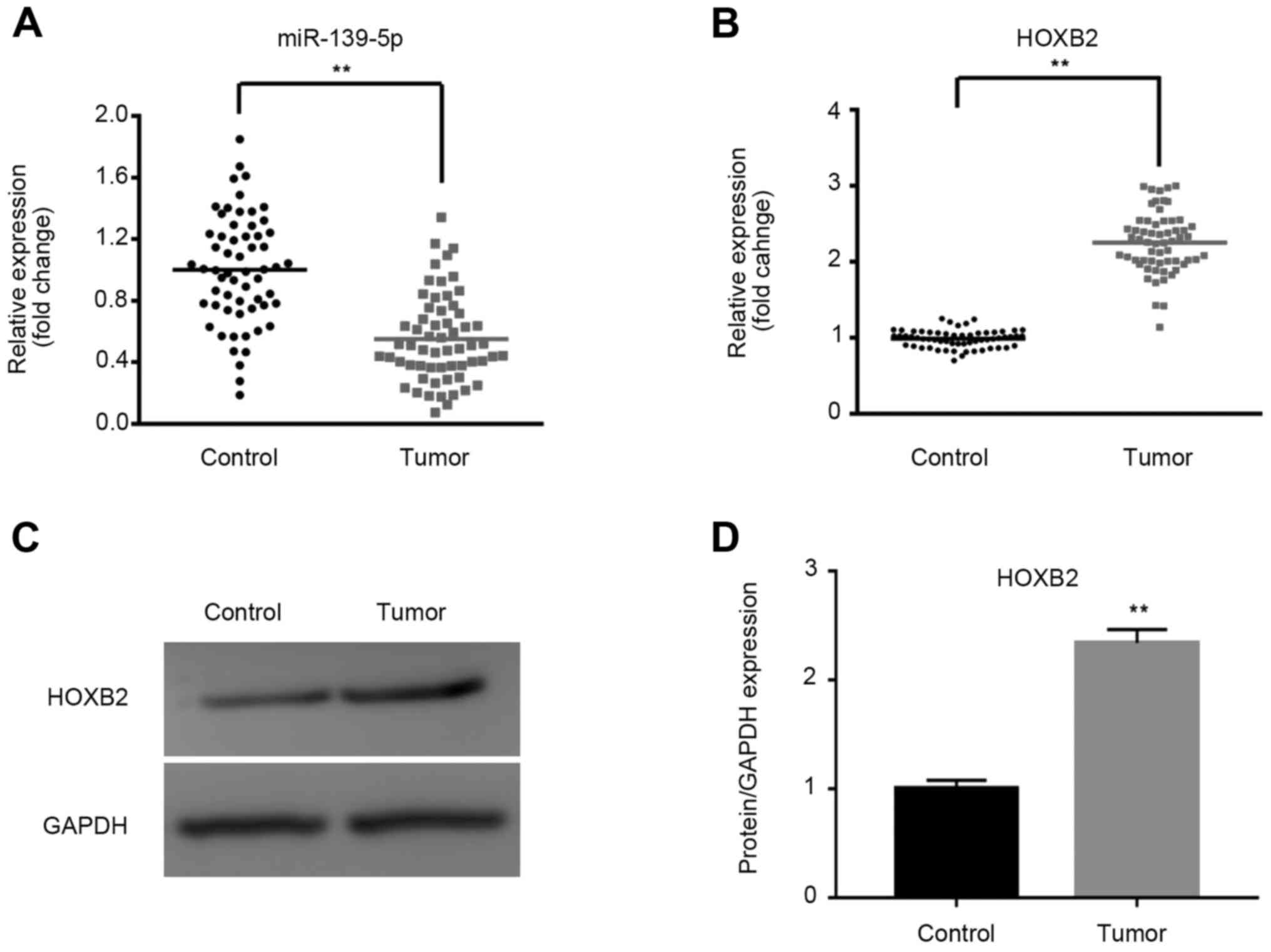
miR-139-5p expression is
downregulated, whereas HOXB2 expression is upregulated, in human
NSCLC tissues
The mRNA and protein expression levels of miR-139-5p
and HOXB2 in tissues were determined using RT-qPCR and western
blotting. As shown in Fig. 1A and
B, miR-139-5p was significantly downregulated, whereas HOXB2
mRNA was significantly upregulated, in NSCLC tissues compared with
paracarcinoma tissues (P<0.01). Furthermore, the expression of
HOXB2 protein was significantly upregulated in tumor tissues, as
shown in Fig. 1C and D.
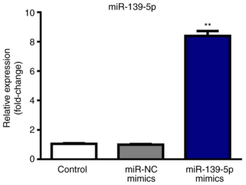
miR-139-5p expression in cells is
upregulated following transfection with mimics
As shown in Fig. 2,
miR-139-5p was upregulated in the mimics group compared with the
Control and NC groups (P<0.01) after transfection with
miR-139-5p mimics.
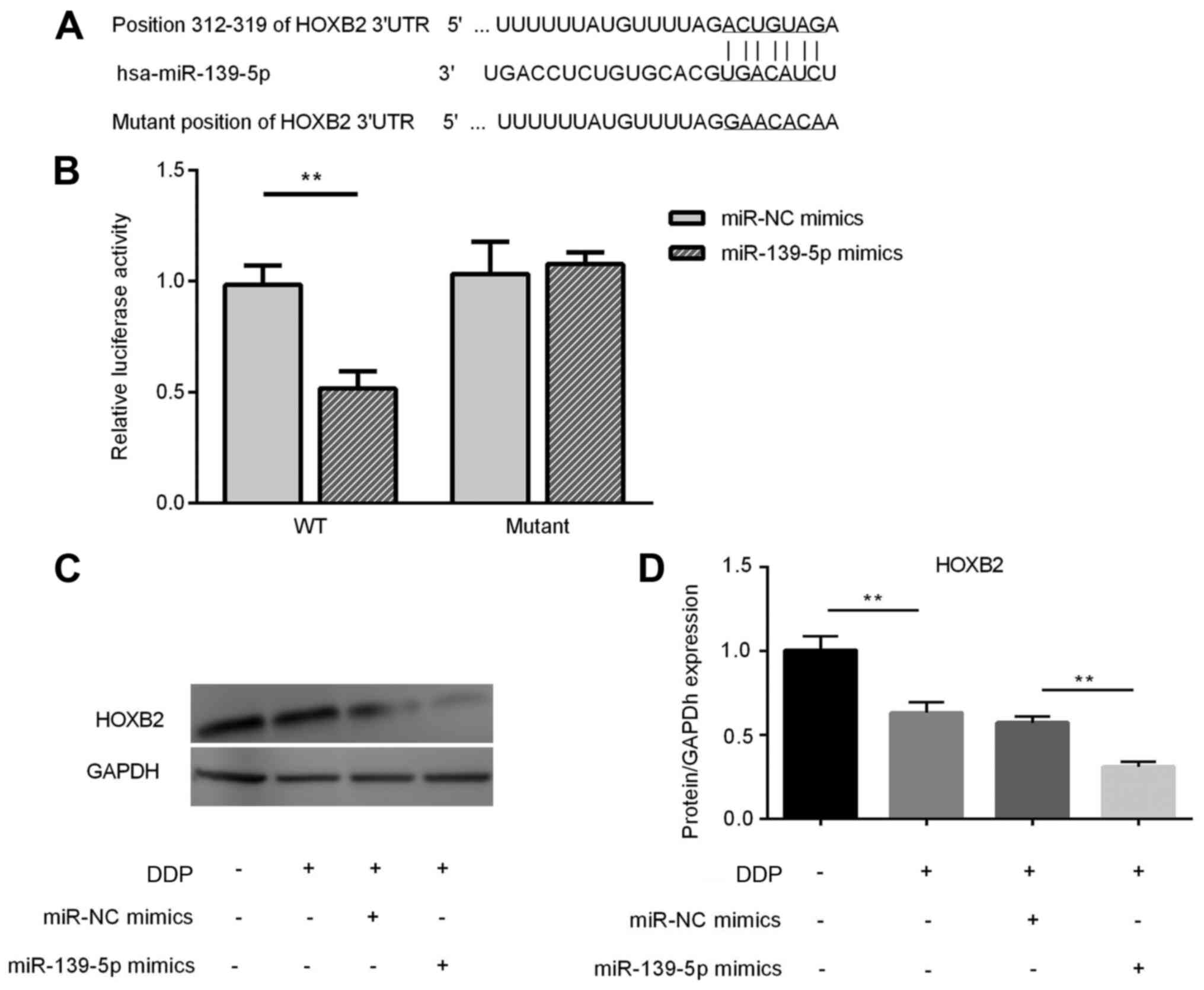
HOXB2 is a direct target of miR-139-5p
and its expression is downregulated in DDP-treated A549 cells
following miR-139-5p overexpression
Based on putative target sequences at nt positions
312–319 of the HOXB2 3′-UTR (Fig.
3A), HOXB2 was selected as a potential target of miR-139-5p. A
luciferase reporter assay was used to confirm HOXB2 as a direct
target of miR-139-5p. As shown in Fig.
3B, miR-139-5p led to a significant decrease in the luciferase
activity of the wild-type HOXB2 3′-UTR, but not that of the mutant
3′-UTR, in A549 cells. Compared with the Control group, the
expression of HOXB2 was significantly decreased in the cisplatin
group (P<0.01). The protein expression of HOXB2 in DDP-treated
A549 cells transfected with miR-139-5p mimics was significantly
downregulated compared with the DDP + miR-NC group (P<0.01)
(Fig. 3C and D).
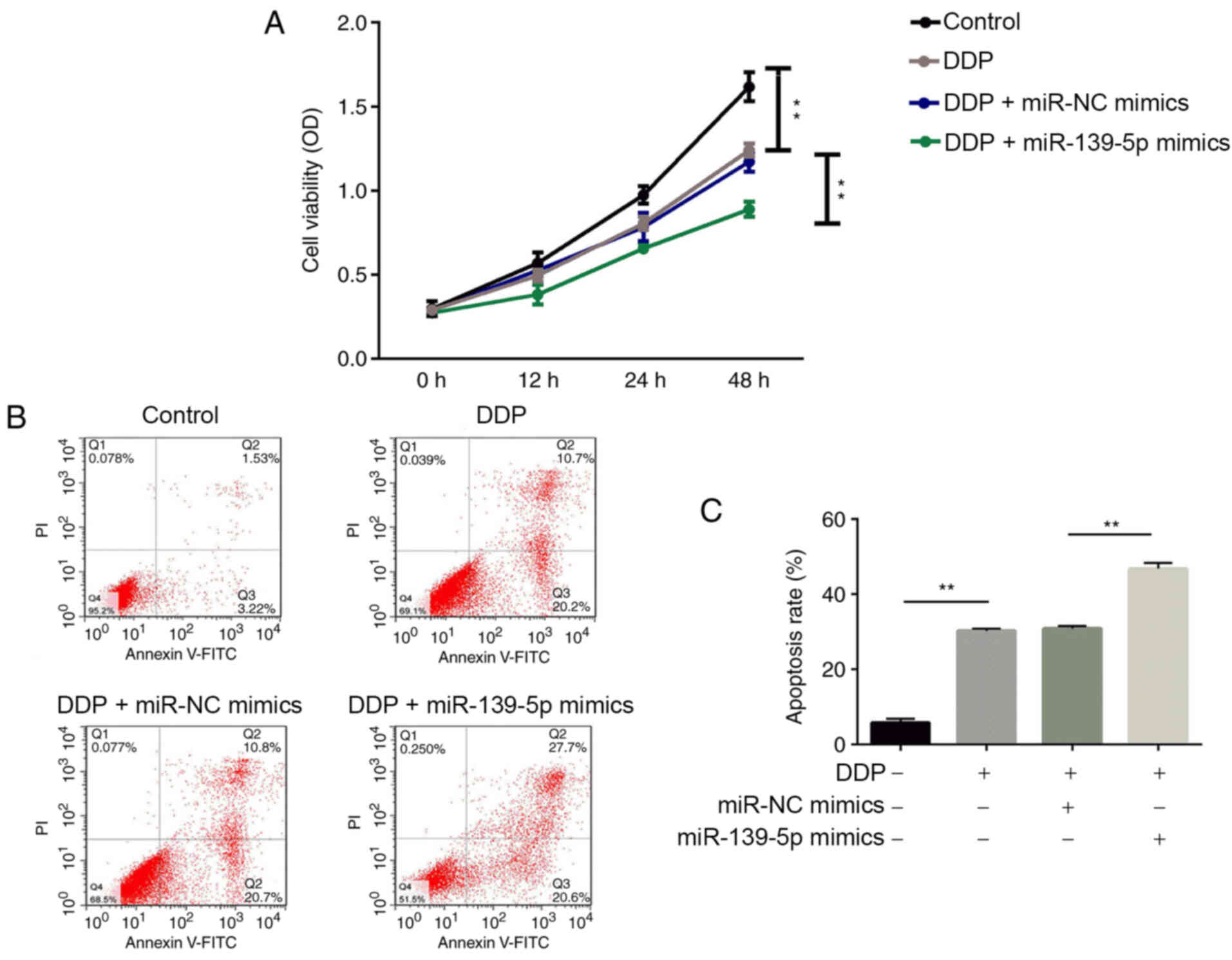
Transfection with miR-139-5p mimics in DDP-treated
A549 cells inhibits NSCLC cell proliferation and promotes
apoptosis. As shown in Fig. 4A,
cell proliferation was significantly suppressed by DDP treatment
compared with the Control group. Furthermore, cell proliferation in
the DDP + miR-139-5p group was significantly decreased compared
with the DDP + miR-NC mimics group (P<0.01).
The results of the flow cytometry analysis are shown
in Fig. 4B and C. Significantly
increased rates of cell apoptosis were observed in the DDP group
compared with the Control group. The apoptosis rate of A549 cells
treated with miR-139-5p mimics combined with DDP was significantly
elevated compared with the DDP + miR-NC mimics group
(P<0.01).
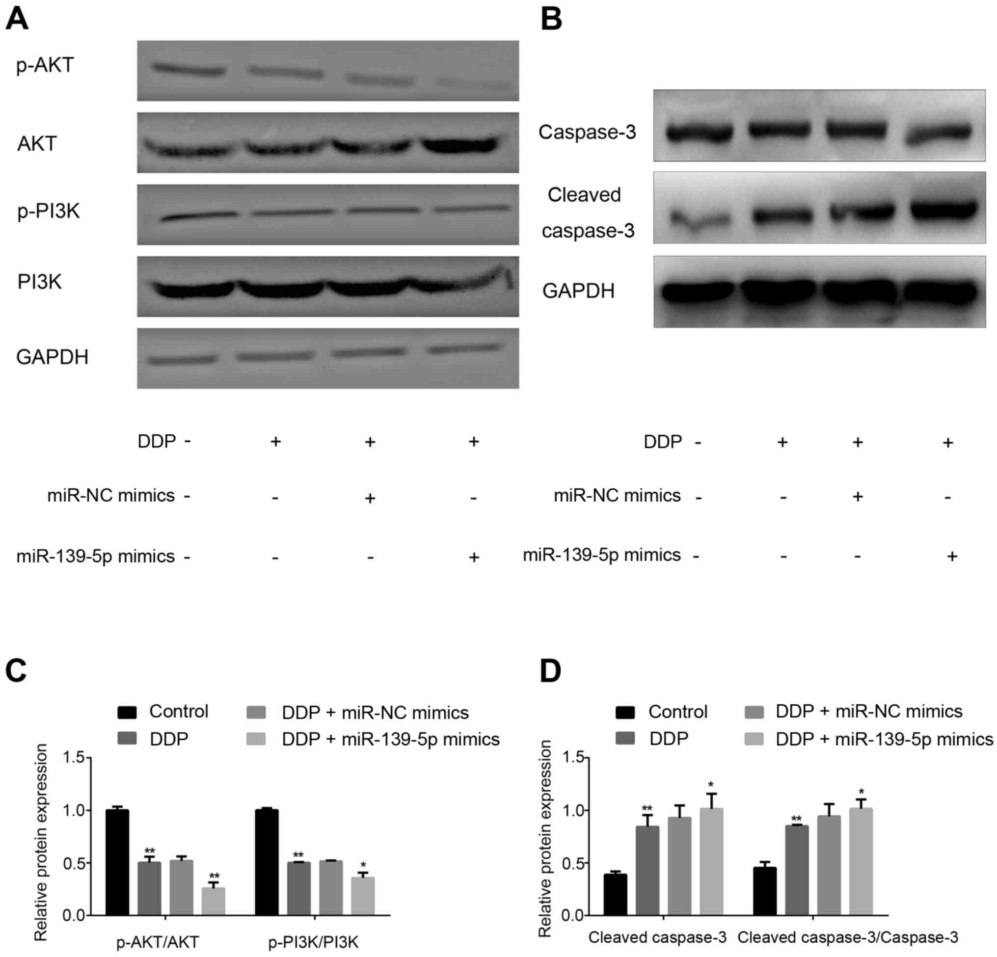
p-AKT and p-PI3K expression is
downregulated, whereas caspase-3 expression is upregulated, in
DDP-treated A549 cells following miR-139-5p overexpression
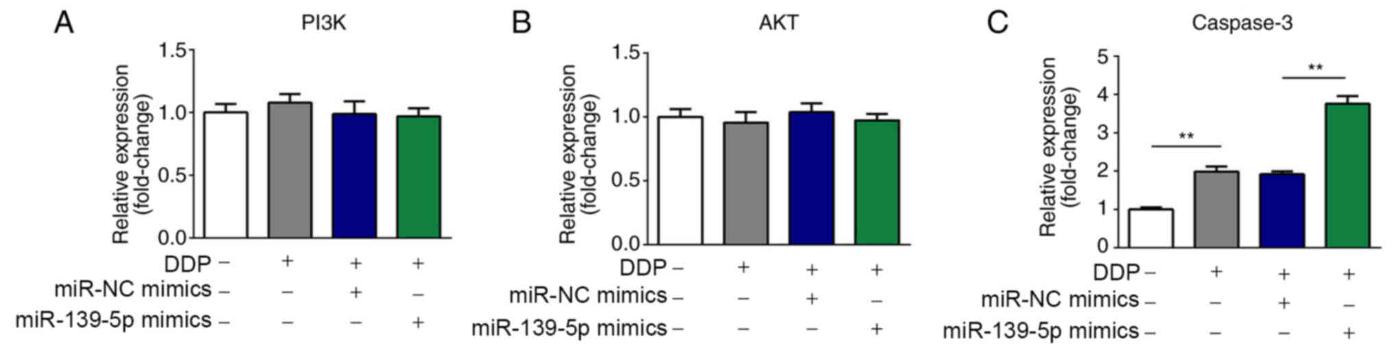
The relative expression levels of PI3K, AKT and
caspase-3 in cells were determined by RT-qPCR. As shown in Fig. 5A-C, the mRNA expression levels of
AKT and PI3K were not altered, whereas caspase-3 mRNA expression
was significantly upregulated in DDP-treated A549 cells following
miR-139-5p overexpression.
As shown in Fig.
6A-D, the expression levels of p-AKT and p-PI3K were
significantly decreased in the DDP group compared with the Control
group (P<0.01). In addition, cells treated with DDP + miR-139-5p
mimics revealed a significant decrease compared with the DDP +
miR-NC mimics group (P<0.01). Furthermore, increased expression
of cleaved-caspase-3 protein was also found. Therefore, miR-139-5p
may be able to inhibit the expression of p-AKT and p-PI3K, while
increasing cleaved-caspase-3 expression to enhance DDP
sensitivity.
Discussion
DDP is generally considered to be the most suitable
chemotherapeutic drug for the treatment of NSCLC (41). However, resistance to DDP is
becoming increasingly problematic, and this is turning into a major
impediment in the clinical treatment of patients with lung cancer
(42). Alterations in drug uptake,
drug metabolism and drug targets are the molecular mechanisms by
which a cancer cell acquires resistance to a specific treatment
(43).
miRNAs serve key roles in numerous biological
processes, and are frequently dysregulated in tumor cells. An
increasing number of miRNAs have been demonstrated to be involved
in the drug-sensitivity of cancer cells in different tumor types
via mechanisms such as inhibition of cell proliferation, apoptosis
induction and cell cycle arrest (44–47).
miR-139-5p, a potential biomarker, is often downregulated in
numerous types of cancer (48–50).
miR-139-5p suppresses cancer cell proliferation, migration,
invasion and metastasis, suggesting that it may be a potential
novel target for cancer treatment (51,52).
At the same time, miR-139-5p has been reported to exert positive
effects on drug resistance and radiotherapy resistance during the
process of cancer treatment (53,54).
The aim of the present study was to assess the effects of
miR-139-5p on DDP-treated NSCLC cells, and to uncover the
underlying mechanism. Tumor cell proliferation and dysregulation of
apoptosis are important processes in tumorigenesis and
development.
HOX genes have been implicated in numerous studies
investigating prognosis and tumorigenesis in patients with cancer
(55–57). The expression of Hox transcript
antisense intergenic RNA was demonstrated to be higher in a
DDP-resistant ovarian carcinoma cell line compared with the
associated DDP-sensitive cell line (56). A previous study revealed that HOX
gene homeobox protein BarH-like 2 could modulate DDP sensitivity in
human epithelial ovarian cancer (57). As a member of the HOX gene family,
HOXB2 was revealed to be overexpressed during cervical cancer
progression (58). miR-139-5p was
reported to target the HOXA10 transcript and to suppress
endometrial cancer cell growth and migration during human
endometrial cancer pathogenesis (59). In the present study, miR-139-5p was
identified to have relevant binding sites in HOXB2, and HOXB2 was
significantly upregulated in NSCLC tissues compared with
paracarcinoma tissues. Compared with the Control group, the protein
expression level of HOXB2 in A549 cells treated with DDP was
significantly downregulated. In addition, the protein expression of
HOXB2 in DDP-treated A549 cells transfected with miR-139-5p mimics
was significantly decreased compared with the DDP + miR-NC mimics
treatment group. These results indicated that miR-139-5p increased
cell sensitivity to DDP via downregulation of HOXB2.
The PI3K/AKT signaling pathway, a crucial and
extensively investigated intracellular signaling pathway, is
instrumental in regulating cellular apoptosis, stimulating cell
growth and increasing cell proliferation (60). caspase-3 is a member of the group of
proteolytic enzymes that mediate apoptosis, and is an important
effector molecule for apoptosis. miR-139-5p was demonstrated to
increase cell proliferation by activating the PI3K/AKT signaling
pathway in supratentorial pediatric low-grade gliomas (61). Inhibition of the PI3K/AKT/mTOR
signaling pathway enhanced the sensitivity of the SKOV3/DDP ovarian
cancer cell line to DDP in vitro (62). miRNAs were revealed to regulate
DDP-induced gastric cancer cell death by modulating the
PI3K/AKT/survivin pathway (63).
The tyrosine kinase inhibitor, dasatinib, enhanced DDP sensitivity
in human esophageal squamous cell carcinoma cells via suppression
of the PI3K/AKT and STAT3 pathways (64). In the present study, the mRNA
expression levels of PI3K and AKT in A549 cells were not affected
by DDP treatment, whereas the mRNA expression of caspase-3 was
increased upon treatment with DDP and DPP + miR-139-5p mimics.
Overexpression of miR-139-5p further enhanced the DDP-induced
decrease in the protein expression of p-AKT and p-PI3K, and further
upregulated the DDP-induced increase in the expression of
cleaved-caspase-3 protein. Therefore, miR-139-5p overexpression may
lead to increased DDP sensitivity by modulating the
PI3K/AKT/caspase-3 signaling pathway.
The present study aimed to investigate the effect of
miR-139-5p expression on the modulation of DDP sensitivity in lung
cancer, not the effect of DDP on the regulation of miR-139-5p
expression. Hence, the expression of miR-139-5p after cisplatin
treatment was not investigated. Moreover, the current study was
limited as it only investigated the role of miR-139-5p in DDP
sensitivity. Further studies will be conducted in the future to
investigate whether miR-139-5p has a role in the sensitivity of
cancer cells to other chemotherapy drugs.
In conclusion, the present study revealed that
miR-139-5p could increase the efficacy of DDP on cell proliferation
and apoptosis in vitro via modulation of the
PI3K/AKT/caspase-3 pathway. The present study also demonstrated
that miR-139-5p expression was significantly lower in NSCLC tissues
compared with that in paracarcinoma tissues. Furthermore, HOXB2 was
identified as a target of miR-139-5p, and miR-139-5p may function
as a DDP sensitizer by targeting HOXB2 in NSCLC. The regulation of
miR-139-5p expression could therefore provide a novel approach to
reverse DDP resistance and increase chemosensitivity in the
treatment of NSCLC.
Acknowledgements
Not applicable.
Funding
No funding was received.
Availability of data and materials
The datasets used and/or analyzed during the current
study are available from the corresponding author on reasonable
request.
Authors' contributions
HD, YB and CL collected, analyzed and interpreted
the patient data. HD, AZ and YN contributed to the manuscript
conception, experiment design, manuscript drafting and revising.
All authors read and approved the final manuscript.
Ethics approval and consent to
participate
The present study was approved by the Ethics
Committee of Weifang Yidu Central Hospital (approval no.
201601580), and written informed consent was obtained from each
participant prior to the study.
Patient consent for publication
Not applicable.
Competing interests
The authors declare that they have no competing
interests.
References
|
1
|
Zha L, Sobue T, Kitamura T, Kitamura Y,
Sawada N, Iwasaki M, Sasazuki S, Yamaji T, Shimazu T and Tsugane S:
Changes in smoking Status and mortality from all causes and lung
cancer: A longitudinal analysis of a population-based study in
Japan. J Epidemiol. 29:11–17. 2019. View Article : Google Scholar
|
|
2
|
Imbimbo M, Vitali M, Fabbri A, Ottaviano
M, Pasello G, Petrini I, Palmieri G, Berardi R, Zucali P,
Ganzinelli M, et al: Relevent trial: Phase II trial of ramucirumab,
carboplatin, and paclitaxel in previously untreated thymic
carcinoma/B3 thymoma with area of carcinoma. Clin Lung Cancer.
19:e811–e814. 2018. View Article : Google Scholar
|
|
3
|
Yang D, Liu Y, Bai C, Wang X and Powell
CA: Epidemiology of lung cancer and lung cancer screening programs
in China and the United States. Cancer Lett. 468:82–87. 2020.
View Article : Google Scholar
|
|
4
|
Cao M and Chen W: Epidemiology of lung
cancer in China. Thorac Cancer. 10:3–7. 2019. View Article : Google Scholar
|
|
5
|
Duma N, Santana-Davila R and Molina JR:
Non-small cell lung cancer: Epidemiology, screening, diagnosis, and
treatment. Mayo Clin Proc. 94:1623–1640. 2019. View Article : Google Scholar
|
|
6
|
Neal RD, Sun F, Emery JD and Callister ME:
Lung cancer. BMJ. 365:l17252019. View Article : Google Scholar
|
|
7
|
Siegel RL, Miller KD and Jemal A: Cancer
statistics, 2018. CA Cancer J Clin. 68:7–30. 2018. View Article : Google Scholar
|
|
8
|
Lee YG, Lee JH, Kim SH, Kim YJ, Lee H, Ahn
S, Jang JS, Lee JS and Kim JH: Comparative analysis between
combination and single-agent chemotherapy for elderly patients with
advanced non-small cell lung cancer: A nationwide population-based
outcome study. Lung Cancer. 122:88–93. 2018. View Article : Google Scholar
|
|
9
|
Lee JS, Lee KH, Cho EK, Kim DW, Kim SW,
Kim JH, Cho BC, Kang JH, Han JY, Min YJ, et al: Nivolumab in
advanced non-small-cell lung cancer patients who failed prior
platinum-based chemotherapy. Lung Cancer. 122:234–242. 2018.
View Article : Google Scholar
|
|
10
|
Yu X, Zhang L and Chen J: Effectiveness of
treatment with endostatin in combination with emcitabine,
carboplatin, and gemcitabine in patients with advanced non-small
cell lung cancer: A retrospective study. Open Med (Wars).
13:142–147. 2018. View Article : Google Scholar
|
|
11
|
Makino A, Miyazaki A, Tomoike A, Kimura H,
Arimitsu K, Hirata M, Ohmomo Y, Nishii R, Okazawa H, Kiyono Y, et
al: PET probe detecting non-small cell lung cancer susceptible to
epidermal growth factor receptor tyrosine kinase inhibitor therapy.
Bioorg Med Chem. 26:1609–1613. 2018. View Article : Google Scholar
|
|
12
|
To KK, Tong CW, Wu M and Cho WC: MicroRNAs
in the prognosis and therapy of colorectal cancer: From bench to
bedside. World J Gastroenterol. 24:2949–2973. 2018. View Article : Google Scholar
|
|
13
|
Migliore C and Giordano S: Resistance to
targeted therapies: A role for microRNAs? Trends Mol Med.
19:633–642. 2013. View Article : Google Scholar
|
|
14
|
Cao L, Wan Q, Li F and Tang CE: MiR-363
inhibits cisplatin chemoresistance of epithelial ovarian cancer by
regulating snail-induced epithelial-mesenchymal transition. BMB
Rep. 51:456–461. 2018. View Article : Google Scholar
|
|
15
|
Wang F, Zhao L, Zhang J, Meng Z, Zhou C,
Wang G, Liu Y, Li M, Xi J, Niu W, et al: Chemotherapy-induced
miR-141/MAP4K4 signaling suppresses progression of colorectal
cancer. Biosci Rep. 38:BSR201809782018. View Article : Google Scholar
|
|
16
|
Li Q, Li B, Li Q, Wei S, He Z, Huang X,
Wang L, Xia Y, Xu Z, Li Z, et al: Exosomal miR-21-5p derived from
gastric cancer promotes peritoneal metastasis via
mesothelial-to-mesenchymal transition. Cell Death Dis. 9:8542018.
View Article : Google Scholar
|
|
17
|
Aggarwal P, Challa KR, Rath M, Sunkara P
and Nath U: Generation of inducible transgenic lines of arabidopsis
transcription factors regulated by microRNAs. Methods Mol Biol.
1830:61–79. 2018. View Article : Google Scholar
|
|
18
|
Bueno MJ and Malumbres M: MicroRNAs and
the cell cycle. Biochim Biophys Acta. 1812:592–601. 2011.
View Article : Google Scholar
|
|
19
|
Schneider C, Setty M, Holmes AB, Maute RL,
Leslie CS, Mussolin L, Rosolen A, Dalla-Favera R and Basso K:
MicroRNA 28 controls cell proliferation and is down-regulated in
B-cell lymphomas. Proc Natl Acad Sci USA. 111:8185–8190. 2014.
View Article : Google Scholar
|
|
20
|
Bukhari SI, Vasquez-Rifo A, Gagné D,
Paquet ER, Zetka M, Robert C, Masson JY and Simard MJ: The microRNA
pathway controls germ cell proliferation and differentiation in C.
elegans. Cell Res. 22:1034–1045. 2012. View Article : Google Scholar
|
|
21
|
Zhang Z, Zhang C, Li F, Zhang B and Zhang
Y: Regulation of memory CD8+ T cell differentiation by
microRNAs. Cell Physiol Biochem. 47:2187–2198. 2018. View Article : Google Scholar
|
|
22
|
Fu X, He Y, Wang X, Peng D, Chen X, Li X
and Wan Q: MicroRNA-16 promotes ovarian granulosa cell
proliferation and suppresses apoptosis through targeting PDCD4 in
polycystic ovarian syndrome. Cell Physiol Biochem. 48:670–682.
2018. View Article : Google Scholar
|
|
23
|
Li J, Zhou Q, Liang Y, Pan W, Bei Y, Zhang
Y, Wang J and Jiao Z: miR-486 inhibits PM2.5-induced apoptosis and
oxidative stress in human lung alveolar epithelial A549 cells. Ann
Transl Med. 6:209–218. 2018. View Article : Google Scholar
|
|
24
|
Li H, Feng C and Shi S: miR-196b promotes
lung cancer cell migration and invasion through the targeting of
GATA6. Oncol Lett. 16:247–252. 2018.
|
|
25
|
Maemura K, Watanabe K, Ando T, Hiyama N,
Sakatani T, Amano Y, Kage H, Nakajima J, Yatomi Y, Nagase T, et al:
Altered editing level of microRNAs is a potential biomarker in lung
adenocarcinoma. Cancer Sci. 109:3326–3335. 2018. View Article : Google Scholar
|
|
26
|
Ulivi P and Zoli W: miRNAs as non-invasive
biomarkers for lung cancer diagnosis. Molecules. 19:8220–8237.
2014. View Article : Google Scholar
|
|
27
|
Yong-Hao Y, Xian-Guo W, Ming X and
Jin-Ping Z: Expression and clinical significance of miR-139-5p in
non-small cell lung cancer. J Int Med Res. 47:867–874. 2019.
View Article : Google Scholar
|
|
28
|
Sun C, Sang M, Li S, Sun X, Yang C, Xi Y,
Wang L, Zhang F, Bi Y, Fu Y, et al: Hsa-miR-139-5p inhibits
proliferation and causes apoptosis associated with down-regulation
of c-Met. Oncotarget. 6:39756–39792. 2015. View Article : Google Scholar
|
|
29
|
Wang K, Jin J, Ma T and Zhai H: MiR-139-5p
inhibits the tumorigenesis and progression of oral squamous
carcinoma cells by targeting HOXA9. J Cell Mol Med. 21:3730–3740.
2017. View Article : Google Scholar
|
|
30
|
Li Q, Liang X, Wang Y, Meng X, Xu Y, Cai
S, Wang Z, Liu J and Cai G: miR-139-5p inhibits the
epithelial-mesenchymal transition and enhances the chemotherapeutic
sensitivity of colorectal cancer cells by downregulating BCL2. Sci
Rep. 6:271572016. View Article : Google Scholar
|
|
31
|
Xu W, Hang M, Yuan CY, Wu FL, Chen SB and
Xue K: MicroRNA-139-5p inhibits cell proliferation and invasion by
targeting insulin-like growth factor 1 receptor in human non-small
cell lung cancer. Int J Clin Exp Pathol. 8:3864–3870. 2015.
|
|
32
|
Clemenceau A, Boucherat O, Landry-Truchon
K, Lamontagne M, Biarde S, Joubert P, Gobeil S, Secco B, Laplante
M, Morissette M, et al: Lung cancer susceptibility genetic variants
modulate HOXB2 expression in the lung. Int J Dev Biol. 62:857–864.
2018. View Article : Google Scholar
|
|
33
|
Liu J, Li S, Cheng X, Du P, Yang Y and
Jiang WG: HOXB2 is a putative tumour promoter in human bladder
cancer. Anticancer Res. 39:6915–6921. 2019. View Article : Google Scholar
|
|
34
|
Li S, Pei Y, Wang W, Liu F, Zheng K and
Zhang X: Circular RNA 0001785 regulates the pathogenesis of
osteosarcoma as a ceRNA by sponging miR-1200 to upregulate HOXB2.
Cell Cycle. 18:1281–1291. 2019. View Article : Google Scholar
|
|
35
|
Lindblad O, Chougule RA, Moharram SA,
Kabir NN, Sun J, Kazi JU and Rönnstrand L: The role of HOXB2 and
HOXB3 in acute myeloid leukemia. Biochem Biophys Res Commun.
467:742–747. 2015. View Article : Google Scholar
|
|
36
|
Inamura K, Togashi Y, Ninomiya H, Shimoji
T, Noda T and Ishikawa Y: HOXB2, an adverse prognostic indicator
for stage I lung adenocarcinomas, promotes invasion by
transcriptional regulation of metastasis-related genes in HOP-62
non-small cell lung cancer cells. Anticancer Res. 28:2121–2127.
2008.
|
|
37
|
Inamura K, Togashi Y, Okui M, Ninomiya H,
Hiramatsu M, Satoh Y, Okumura S, Nakagawa K, Shimoji T, Noda T, et
al: HOXB2 as a novel prognostic indicator for stage I lung
adenocarcinomas. J Thorac Oncol. 2:802–807. 2007. View Article : Google Scholar
|
|
38
|
Xiao H, Liu Y, Liang P, Wang B, Tan H,
Zhang Y, Gao X and Gao J: TP53TG1 enhances cisplatin sensitivity of
non-small cell lung cancer cells through regulating miR-18a/PTEN
axis. Cell Biosci. 8:232018. View Article : Google Scholar
|
|
39
|
Livak KJ and Schmittgen TD: Analysis of
relative gene expression data using real-time quantitative PCR and
the 2(-Delta Delta C(T)) method. Methods. 25:402–408. 2001.
View Article : Google Scholar
|
|
40
|
Agarwal V, Bell GW, Nam JW and Bartel DP:
Predicting effective microRNA target sites in mammalian mRNAs.
eLife. 4:e050052015. View Article : Google Scholar
|
|
41
|
Guo J, Jin D, Wu Y, Yang L, Du J, Gong K,
Chen W, Dai J, Miao S and Xi S: The miR 495-UBE2C-ABCG2/ERCC1 axis
reverses cisplatin resistance by downregulating drug resistance
genes in cisplatin-resistant non-small cell lung cancer cells.
EBioMedicine. 35:204–221. 2018. View Article : Google Scholar
|
|
42
|
Hou Z, Xu C, Xie H, Xu H, Zhan P, Yu L and
Fang X: Long noncoding RNAs expression patterns associated with
chemo response to cisplatin based chemotherapy in lung squamous
cell carcinoma patients. PLoS One. 9:e1081332014. View Article : Google Scholar
|
|
43
|
Donzelli S, Mori F, Biagioni F, Bellissimo
T, Pulito C, Muti P, Strano S and Blandino G: MicroRNAs: Short
non-coding players in cancer chemoresistance. Mol Cell Ther.
2:162014. View Article : Google Scholar
|
|
44
|
Wang Z, Wang N, Liu P, Chen Q, Situ H, Xie
T, Zhang J, Peng C, Lin Y and Chen J: MicroRNA-25 regulates
chemoresistance-associated autophagy in breast cancer cells, a
process modulated by the natural autophagy inducer
isoliquiritigenin. Oncotarget. 5:7013–7026. 2014. View Article : Google Scholar
|
|
45
|
Ju J: Implications of miRNAs in colorectal
cancer chemoresistance. Int Drug Discov. 2011:20112011.
|
|
46
|
Wang XH, Lu H, Li TS, Yu Y, Liu G, Peng X
and Zhao JH: KLF8 induces breast stemness and chemoresistance via
miRNAs associated with EMT. Mol Cancer Ther. 12:A100. 2013.
|
|
47
|
Carta A, Chetcuti R and Ayers D: An
introspective update on the influence of miRNAs in breast carcinoma
and neuroblastoma chemoresistance. Genet Res Int.
2014:7430502014.
|
|
48
|
Shen K, Mao R, Ma L, Li Y, Qiu Y, Cui D,
Le V, Yin P, Ni L and Liu J: Post-transcriptional regulation of the
tumor suppressor miR-139-5p and a network of miR-139-5p-mediated
mRNA interactions in colorectal cancer. FEBS J. 281:3609–3624.
2014. View Article : Google Scholar
|
|
49
|
Chen Y, Cao XY, Li YN, Qiu YY, Li YN, Li W
and Wang H: Reversal of cisplatin resistance by
microRNA-139-5p-independent RNF2 downregulation and MAPK inhibition
in ovarian cancer. Am J Physiol Cell Physiol. 315:C225–C235. 2018.
View Article : Google Scholar
|
|
50
|
Ni H, Dai X, Leng X, Deng M, Qin Y, Ji Q,
Xu C, Li J and Liu Y: Higher variety and quantity of
microRNA-139-5p isoforms confer suppressive role in hepatocellular
carcinoma. J Cell Biochem. 119:6806–6813. 2018. View Article : Google Scholar
|
|
51
|
Yue S, Wang L, Zhang H, Min Y, Lou Y, Sun
H, Jiang Y, Zhang W, Liang A, Guo Y, et al: miR-139-5p suppresses
cancer cell migration and invasion through targeting ZEB1 and ZEB2
in GBM. Tumour Biol. 36:6741–6749. 2015. View Article : Google Scholar
|
|
52
|
Hua S, Lei L, Deng L, Weng X, Liu C, Qi X,
Wang S, Zhang D, Zou X, Cao C, et al: miR-139-5p inhibits aerobic
glycolysis, cell proliferation, migration, and invasion in
hepatocellular carcinoma via a reciprocal regulatory interaction
with ETS1. Oncogene. 37:1624–1636. 2018. View Article : Google Scholar
|
|
53
|
Liu H, Yin Y, Hu Y, Feng Y, Bian Z, Yao S,
Li M, You Q and Huang Z: miR-139-5p sensitizes colorectal cancer
cells to 5-fluorouracil by targeting NOTCH-1. Pathol Res Pract.
212:643–649. 2016. View Article : Google Scholar
|
|
54
|
Pajic M, Froio D, Daly S, Doculara L,
Millar E, Graham PH, Drury A, Steinmann A, de Bock CE,
Boulghourjian A, et al: miR-139-5p modulates radiotherapy
resistance in breast cancer by repressing multiple gene networks of
DNA repair and ROS defense. Cancer Res. 78:501–515. 2018.
View Article : Google Scholar
|
|
55
|
Boimel PJ, Cruz C and Segall JE: A
functional in vivo screen for regulators of tumor progression
identifies HOXB2 as a regulator of tumor growth in breast cancer.
Genomics. 98:164–172. 2011. View Article : Google Scholar
|
|
56
|
Wang Y, Wang H, Song T, Zou Y, Jiang J,
Fang L and Li P: HOTAIR is a potential target for the treatment of
cisplatin resistant ovarian cancer. Mol Med Rep. 12:2211–2216.
2015. View Article : Google Scholar
|
|
57
|
Sellar GC, Watt KP, Li L, Nelkin BD,
Rabiasz GJ, Porteous DJ, Smyth JF and Gabra H: The homeobox gene
BARX2 can modulate cisplatin sensitivity in human epithelial
ovarian cancer. Int J Oncol. 21:929–933. 2002.
|
|
58
|
Gonzalez-Herrera A, Salgado-Bernabe M,
Velazquez-Velazquez C, Salcedo-Vargas M, Andrade-Manzano A,
Avila-Moreno F and Pina-Sanchez P: Increased expression of HOXB2
and HOXB13 proteins is associated with HPV infection and cervical
cancer progression. Asian Pac J Cancer Prev. 16:1349–1353. 2015.
View Article : Google Scholar
|
|
59
|
Liu J, Li C, Jiang Y, Wan Y, Zhou S and
Cheng W: Tumor-suppressor role of miR-139-5p in endometrial cancer.
Cancer Cell Int. 18:512018. View Article : Google Scholar
|
|
60
|
Danielsen SA, Eide PW, Nesbakken A, Guren
T, Leithe E and Lothe RA: Portrait of the PI3K/AKT pathway in
colorectal cancer. Biochim Biophys Acta. 1855:104–121. 2015.
|
|
61
|
Catanzaro G, Besharat ZM, Miele E,
Chiacchiarini M, Po A, Carai A, Marras CE, Antonelli M, Badiali M,
Raso A, et al: The miR-139-5p regulates proliferation of
supratentorial paediatric low-grade gliomas by targeting the
PI3K/AKT/mTORC1 signalling. Neuropathol Appl Neurobiol. 44:687–706.
2018. View Article : Google Scholar
|
|
62
|
Cai Y, Tan X, Liu J, Shen Y, Wu D, Ren M,
Huang P and Yu D: Inhibition of PI3K/Akt/mTOR signaling pathway
enhances the sensitivity of the SKOV3/DDP ovarian cancer cell line
to cisplatin in vitro. Chin J Cancer Res. 26:564–572. 2014.
|
|
63
|
Cao W, Yang W, Fan R, Li H, Jiang J, Geng
M, Jin Y and Wu Y: miR-34a regulates cisplatin-induce gastric
cancer cell death by modulating PI3K/AKT/survivin pathway. Tumour
Biol. 35:1287–1295. 2014. View Article : Google Scholar
|
|
64
|
Chen J, Lan T, Zhang W, Dong L, Kang N, Fu
M, Liu B, Liu K, Zhang C, Hou J, et al: Dasatinib enhances
cisplatin sensitivity in human esophageal squamous cell carcinoma
(ESCC) cells via suppression of PI3K/AKT and Stat3 pathways. Arch
Biochem Biophys. 575:38–45. 2015. View Article : Google Scholar
|