Introduction
Hypertension is the most common chronic disease, and
is characterized by increased systemic arterial blood pressure; it
may be accompanied by functional damage of other organs, which is
the most important risk factor for cardiovascular and
cerebrovascular diseases (1). The
blood pressure of healthy individuals fluctuates within a certain
range along with changes in internal and external environments. In
the overall population, the blood pressure levels gradually
increase with age, and systolic blood pressure is more pronounced
(2), while diastolic blood
pressure shows a downward trend after the age of 50 years. Notably,
individuals with high risk of hypertension are no longer only
elderly patients, since the age of patients is showing a younger
trend (3). According to a
previous epidemiological study, the number of patients with
hypertension worldwide could reach 1.5 billion in 2025 (4). High blood pressure can also cause
atherosclerosis (5), cerebral
hemorrhage (6), cerebral
infarction (7) and other
diseases. Since the mechanism remains clear, the clinical condition
is mainly managed by antihypertensive drugs; however, these drugs
cannot block the progression of hypertension. Therefore,
identifying key therapeutic targets is the focus of pre-prevention
and post-treatment of hypertension.
Prolonged hypertension can eventually lead to
alterations in the structure and function of blood vessels
(8), among which, abnormal
proliferation and migration of vascular smooth muscle cells (VSMCs)
are important factors for such changes (9). VSMCs are the main cell type that
constitutes the blood vessel wall, and their main functions are to
regulate the structural integrity and vascular tension of blood
vessels. In the middle layer of normal mature arteries, VSMCs are
in a static state of differentiation, and their synthetic activity
and proliferation potential are low (10). However, with vascular injury, the
phenotype of VSMCs is altered, as manifested by increased cell
proliferation and migration, as well as matrix synthesis (11). Importantly, the changes in the
structure and function of VSMCs are the cytopathological basis that
leads to a variety of vascular diseases (12). Therefore, exploring the abnormal
proliferation and migration of VSMCs has great medical significance
for the prevention and control of hypertension.
Linalool is a monoterpene alcohol present in certain
aromatic medicinal plants, and its biological activity may affect
cardiovascular diseases (13). A
previous study showed that the administration of linalool in
hypertensive rats could effectively reduce blood pressure and
improve hypertension, which may be due to the direct effect on
vascular smooth muscle that leads to vasodilation (14). Linalool can also inhibit the
malignant cell proliferation of a variety of human malignant solid
tumors, including hepatocellular carcinoma, breast cancer, small
cell carcinoma and malignant melanoma (15). In addition, linalool extracted
from eucalyptus leaf essential oil can inhibit breast cancer cell
invasion and migration, and downregulate the mRNA and protein
expression of epithelial-mesenchymal transition-related factors
(including Snail, E-cadherin, N-cadherin and vimentin) (16). Based on these results, it was
hypothesized that the effect of linalool on VSMCs could be explored
by constructing a hypertensive cell model.
Since angiotensin II (Ang II)-mediated VSMC
proliferation plays a crucial role in the structural and functional
development of hypertensive blood vessels (17), the present study utilized Ang II
to treat VSMCs in order to observe the effect of linalool on the
physiological behavior of VSMCs and further explore the underlying
mechanism.
Materials and methods
Cell culture and reagent
The rat thoracic aorta smooth muscle cell line
(A7r5) was purchased from Procell Life Science & Technology
Co., Ltd. Cells were incubated in DMEM (Gibco; Thermo Fisher
Scientific, Inc.) containing 10% FBS (Thermo Fisher Scientific,
Inc.), 100 U/ml streptomycin and 100 U/ml penicillin (1%
penicillin/streptomycin), and maintained in a humidified incubator
with 5% CO2 at 37°C. Linalool and Ang II were obtained
from Sigma-Aldrich (Merck KGaA; purity >95%), and were dissolved
in DMSO and diluted to the specified concentration according to the
experimental requirements.
Cell transfection
A7r5 cells were plated into 6-well plates and
cultured until they reached 70–80% confluence. Cells
(2×10⁵/well) were transfected with 2 µg
pcDNA3.1-cholinergic receptor muscarinic 3 (CHRM3) and negative
control (NC) vectors (Hanbio Biotechnology Co., Ltd.) using
Lipofectamine® 3000 reagent (Invitrogen; Thermo Fisher
Scientific, Inc.) according to the manufacturer's protocol. At 48 h
post-transfection, the expression levels of CHRM3 were
assessed.
Cell Counting Kit-8 (CCK-8) assay
When cells (4×10³ cells/well) were in
logarithmic growth phase, several doses of Ang II (10−9,
10−8, 10−7 and 10−6 mol/l) or
linalool solution (50, 100 and 150 µM) were added to the wells at
37°C. After 24 h of incubation, 10 µl CCK-8 reagent (Beyotime
Institute of Biotechnology) was added to each well of a 96-well
plate, and incubation was continued for 1 h. The optical density
was determined at 450 nm using a microplate reader (Thermo Fisher
Scientific, Inc.).
MTT assay
A7r5 cells (2×10³ cells/well) were seeded
in 96-well plates and cultured in an incubator containing 5%
CO2 at 37°C. Cells were treated with Ang II
(10−7 mol/l) and different doses of linalool (50, 100
and 150 µM) for 0, 12, 24 and 48 h at 37°C. Next, 200 µl MTT
solution (Beyotime Institute of Biotechnology) was added to each
well, and incubation was continued for 4 h. Subsequently, the
medium was removed and DMSO was added. The optical density was
determined at a wavelength of 490 nm using a microplate reader
(Thermo Fisher Scientific, Inc.).
Western blotting
RIPA lysis buffer (Beyotime Institute of
Biotechnology) was added to the cultured cells, which were then
lysed on ice for 30 min. Next, the cells were collected and
centrifuged at 14,000 × g at 4°C for 15 min, and the supernatant
was aspirated. A BCA protein assay kit (Beyotime Institute of
Biotechnology) was then used for protein quantification. Next,
heated and denatured protein samples (25 µg) were separated via 10%
SDS-PAGE, and then transferred to PVDF membranes. Next, the
membranes were blocked in 5% skimmed milk at room temperature for 1
h, and washed with TBS with 0.05% Tween-20 (TBST) once for 10 sec
at room temperature. Following which, membranes were incubated with
the following primary antibodies (1:1,000) overnight at 4°C:
Anti-proliferating cell nuclear antigen (PCNA; cat. no. ab29;
Abcam); anti-MMP2 (cat. no. 40994; Cell Signaling Technology,
Inc.); anti-MMP9 (cat. no. 13667; Cell Signaling Technology, Inc.);
anti-CHRM3 (cat. no. ab126168; Abcam); anti-phosphorylated (p)-ERK
(cat. no. ab192591; Abcam); anti-p-JNK (cat. no. ab215208; Abcam);
anti-p-p38 (cat. no. ab178867; Abcam); anti-ERK (cat. no. ab229912;
Abcam); anti-JNK (cat. no. ab110724; Abcam); anti-p38 (cat. no.
ab182453; Abcam); and anti-GAPDH (cat. no. 5174; Cell Signaling
Technology, Inc.). After washing with TBST three times at room
temperature, 10 min per time, the membranes were incubated with
HRP-conjugated anti-rabbit (cat. no. ab6721; 1:5,000; Abcam) or
anti-mouse antibodies (cat. no. ab6789; 1:10,000; Abcam) for 1 h at
room temperature. Ultra-High Sensitivity ECL kit (cat. no. GK10008;
GlpBio Technology) was used to visualize the protein bands, and the
results were analyzed with Image Lab software (v3.0; Bio-Rad
Laboratories, Inc.).
Wound-healing assay
A7r5 cells (5×10⁵ cells/well) were plated
into 6-well plates and incubated at 37°C overnight. Sterilized
pipette tips were used to make scratches in the cell monolayer when
cells grew to 90% confluence. A wound was generated using a 200-µl
pipette and plates were washed three times with PBS to remove the
extra cells in the wound. Serum-free medium with Ang II (0,
1×10⁻⁷ mol/l) and linalool (0, 50, 100, 150 µmol/l) was
added and incubation was continued. Images were captured under a
light microscope (magnification, ×100; Olympus Corporation) at 0
and 24 h. Data were analyzed using ImageJ software (1.52v; National
Institutes of Health).
Transwell assay
A7r5 non-transfected or transfected cells were
incubated in DMEM at a density of 5×10⁴ cell/ml. A total
of 100 µl cell suspension was seeded into the upper chamber of
Transwell plates (Corning, Inc.), while the lower chamber was
filled with 500 µl DMEM containing 10% FBS for cell culture. After
6 h of incubation at 37°C, 1×10⁻⁷ mol/l Ang II and
linalool (0, 50, 100, 150 µmol/l) were added to treat the cells.
After 48 h of incubation, the cells on the lower side of the
Transwell plate were fixed with 4% formaldehyde and stained with
0.1% crystal violet solution for 20 min at room temperature. The
number of cells was counted under a light microscope
(magnification, ×100; Olympus Corporation) and analyzed using
ImageJ software (1.52v; National Institutes of Health).
Reverse transcription-quantitative PCR
(RT-qPCR)
CHRM3 mRNA expression was detected via RT-qPCR.
Total RNA was extracted from cultured A7r5 cells using
TRIzol® reagent (Beyotime Institute of Biotechnology).
cDNA was transcribed from RNA using a PrimeScript RT Reagent kit
(Takara Biotechnology Co., Ltd.). qPCR was subsequently performed
using the QuantiTect SYBR® Green PCR kit (Qiagen, Inc.)
according to the manufacturer's instructions. The protocol was as
follows: RT (42°C for 5 min and then 95°C for 10 sec), followed by
an amplification program repeated for 40 cycles (95°C for 5 sec and
then 60°C for 30 sec). Data were collected and analyzed using the
2−∆∆Cq method (18).
Values were normalized against GADPH. The primer sequences were as
follows: CHRM3 forward, 5′-TCATCCAGGAGGAAGTACGG-3′ and reverse,
5′-GCTGTGGTCTTGGTCCATCT-3; and GAPDH forward,
5′-GCCCATCACCATCTTCCAGGAG-3′ and reverse,
5′-GAAGGGGCGGAGATGATGAC-3′.
Database analysis
SwissTargetPrediction (www.swisstargetprediction.ch) is a website for target
prediction of bioactive small molecules. The structure of linalool
was uploaded to the website and Homo sapiens was selected.
The targets results were displayed after pressing the button
‘Predict targets’.
Statistical analysis
GraphPad Prism 8.0 software (GraphPad Software,
Inc.) was utilized to analyze the experimental data. All values are
represented as the mean ± SD of triplicate experiments. Unpaired
Student's t-test was used to evaluate differences between two
groups, and one-way ANOVA followed by Tukey's post hoc test was
employed to compare multiple groups. P<0.05 was considered to
indicate a statistically significant difference.
Results
Linalool inhibits the proliferation of
Ang II-induced VSMCs
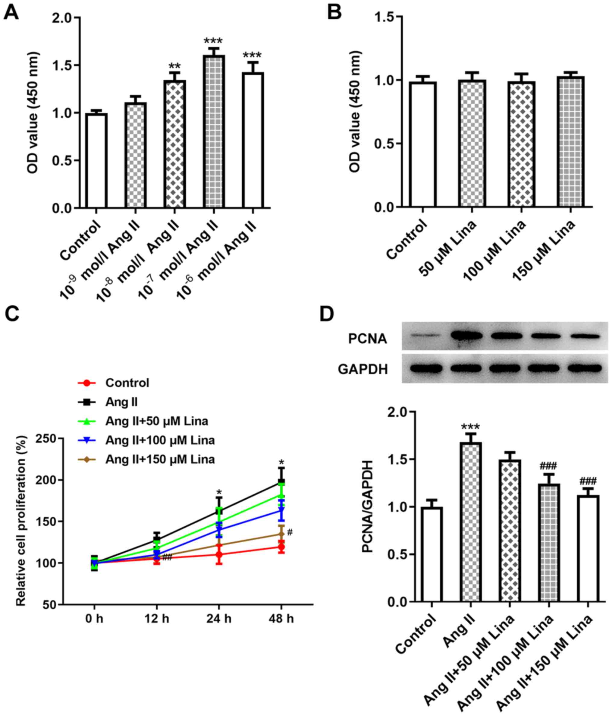
The viability of cells treated with different
concentrations of Ang II was detected using a CCK-8 assay. The
experimental concentration range of Ang II was
1×10⁻⁹−1×10⁻⁶ mol/l. The number of viable
cells increased in a concentration-dependent manner until the
concentration of Ang II reached 1×10⁻⁷ mol/l. When the
concentration continued to increase, the number of viable cells did
not increase further. This may be due to the excessive
concentration that caused damage to cells (Fig. 1A). Thus, Ang II at a concentration
of 1×10⁻⁷ mol/l was selected to treat A7r5 cells in
subsequent experiments. A similar method was employed to detect the
effect of linalool on cell viability. Linalool at a concentration
of 50–150 µmol/l had no effect on cell viability (Fig. 1B).
In addition, an MTT assay was used to detect the
proliferation of A7r5 cells treated with Ang II and linalool. The
number of cells treated with Ang II almost doubled at 48 h compared
with the control, while the proliferation of the cells treated with
linalool for the same time was inhibited compared with the Ang II
group. Compared with that of cells treated with Ang II alone, the
proliferation rate of groups treated with linalool additionally
showed a concentration-dependent decrease (Fig. 1C). PCNA plays a role in cell cycle
regulation and/or DNA replication, and is an objective indicator
for evaluating cell proliferation (19); therefore, its expression level in
each group of cells was determined via western blotting. The
results revealed that the expression of PCNA in groups treated with
linalool additionally decreased in comparison with that in the Ang
II-treated group (Fig. 1D).
Linalool inhibits the migration of Ang
II-induced VSMCs
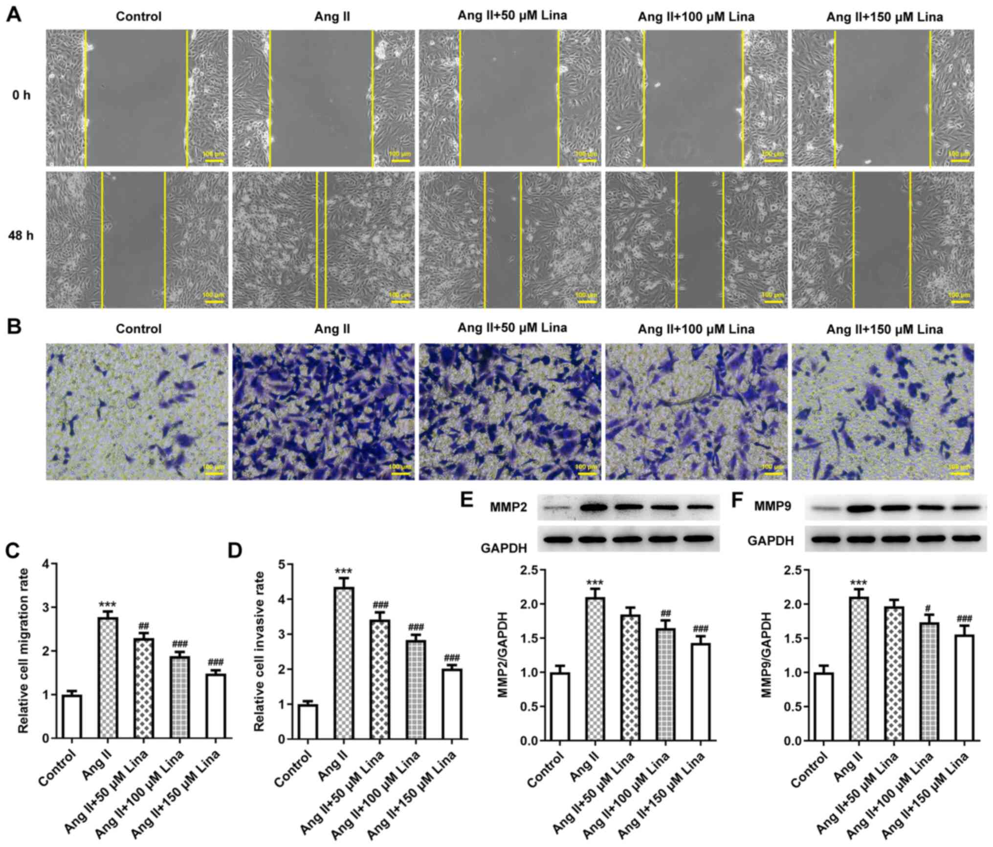
Wound-healing and Transwell assays were applied to
determinate cell migration. The wounds of cells treated with Ang II
alone were filled with numerous cells, while those of cells
simultaneously treated with linalool exhibited a lower number of
cells. Cells treated with linalool at a concentration of 150 µmol/l
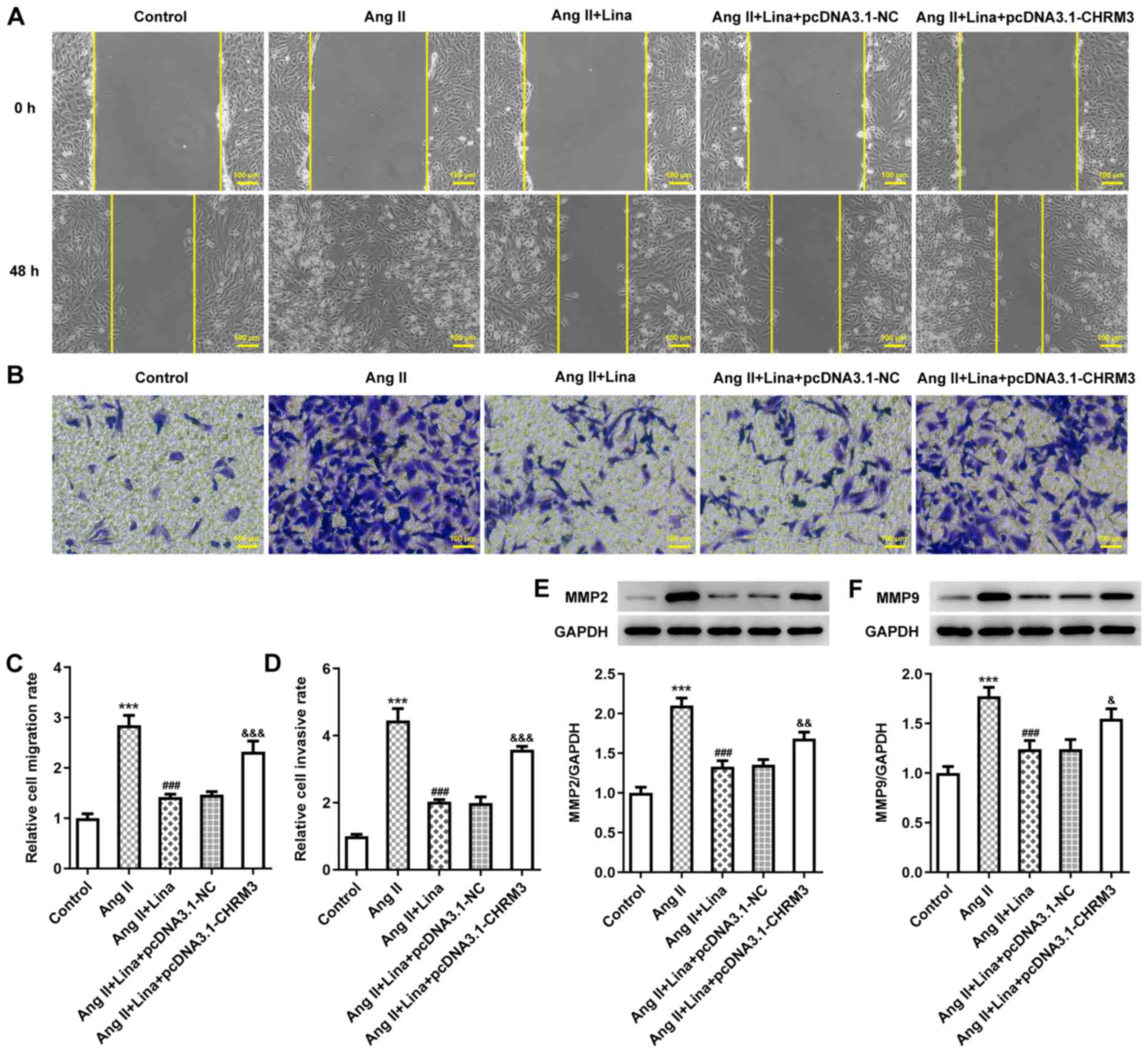
showed the least number of migrated cells (Fig. 2A and C).
Similarly, in the Transwell assay, the cells in the
Ang II group that migrated to the lower chamber were densely
packed, while in the group with the highest experimental
concentration of linalool, the number of migrated cells was the
least (Fig. 2B and D). The
expression levels of MMP2 and MMP9 were detected by western
blotting as they are indicators of migration (20), and they exhibited a decline in the
presence of linalool in a concentration-dependent manner compared
with the Ang II group (Fig. 2E and
F).
Linalool downregulates CHRM3
expression in Ang II-induced VSMCs
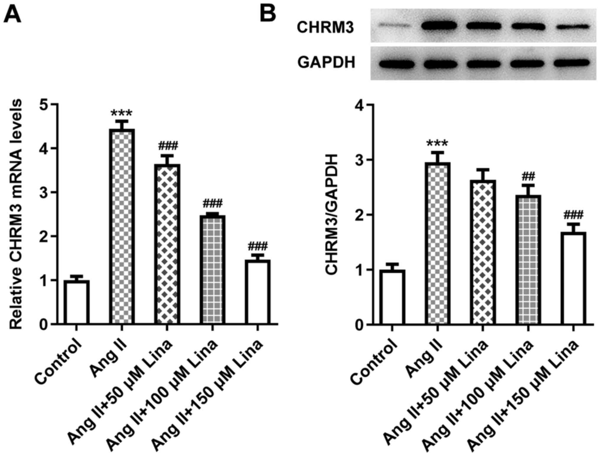
CHRM3 was the target of linalool predicted by the
SwissTargetPrediction website. RT-qPCR and western blotting were
used to detect CHRM3 expression in cells treated with Ang II and
different concentration of linalool. CHRM3 was lowly expressed in
normal A7r5 cells, and once the cells were stimulated with Ang II,
its expression significantly increased compared with the control
group, while the addition of linalool significantly decreased the
expression of CHRM3 in a concentration-dependent manner compared
with the Ang II group (Fig. 3A and
B).
Linalool inhibits the proliferation
and migration of Ang II-induced VSMCs by downregulating CHRM3
expression
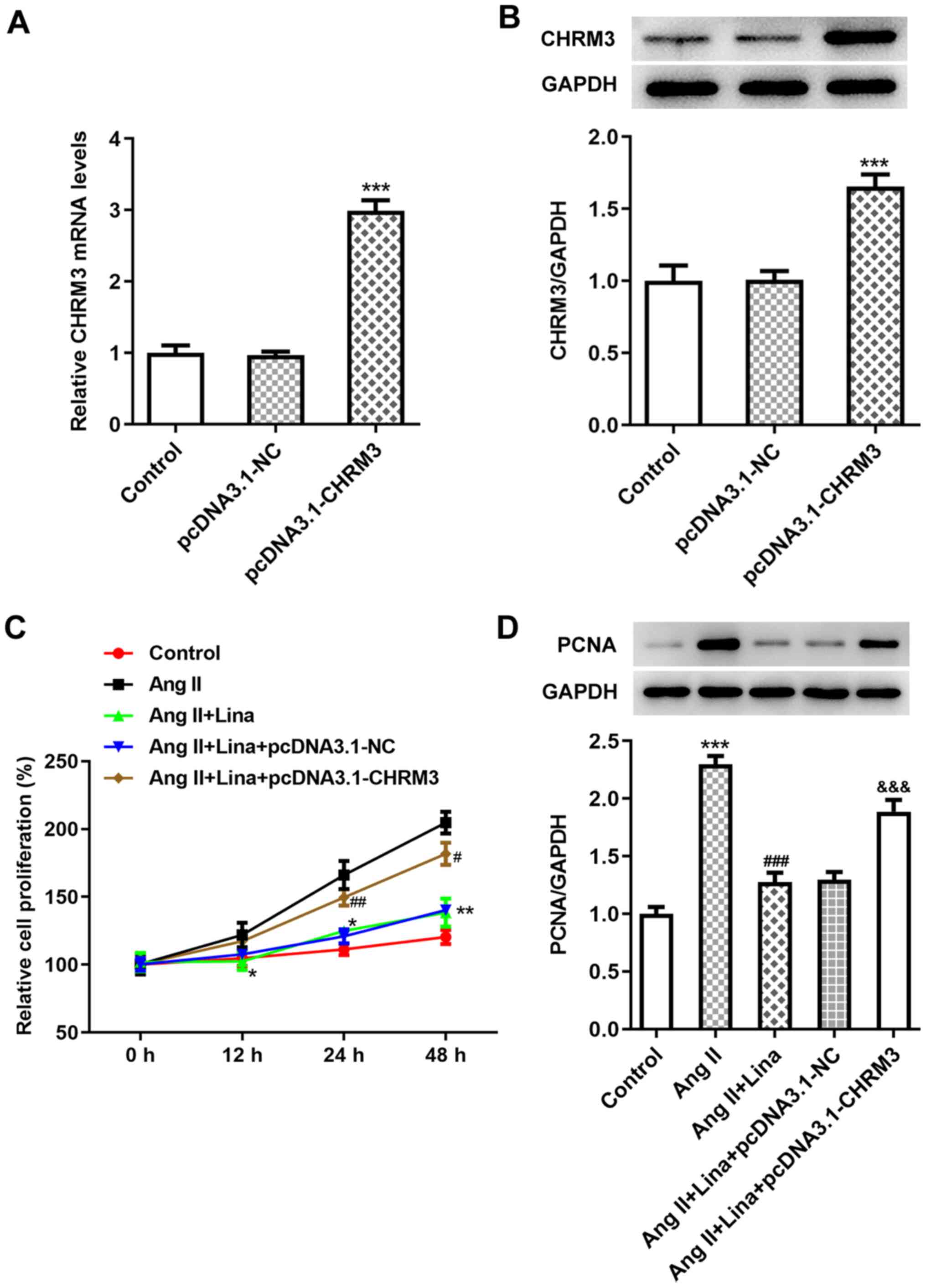
In order to study whether CHRM3 is involved in the
inhibitory mechanism of linalool, RT-qPCR and western blotting were
employed to detect the expression level of CHRM3 in transfected
cells. The results confirmed the efficiency of transfection
(Fig. 4A and B). Based on the
aforementioned experiments, linalool at a concentration of 150
µmol/l was used to treat transfected cells. The inhibitory effect
of linalool on the proliferation of non-transfected cells was
blocked. The cell proliferation of the Ang
II+linalool+pcDNA3.1-CHRM3 group was higher than that of the Ang
II+linalool+pcDNA3.1-NC group, indicating that CHRM3 overexpression
could affect the function of linalool (Fig. 4C). Furthermore, PCNA expression
was increased in the Ang II+linalool+pcDNA3.1-CHRM3 group compared
with the Ang II+linalool+pcDNA3.1-NC group (Fig. 4D). The migratory ability and
protein expression of the transfected cells were determined
(Fig. 5A-F). Similar results were
observed regarding the migratory ability of the cells, indicating
that linalool achieved its effect on cell proliferation and
migration by regulating CHRM3.
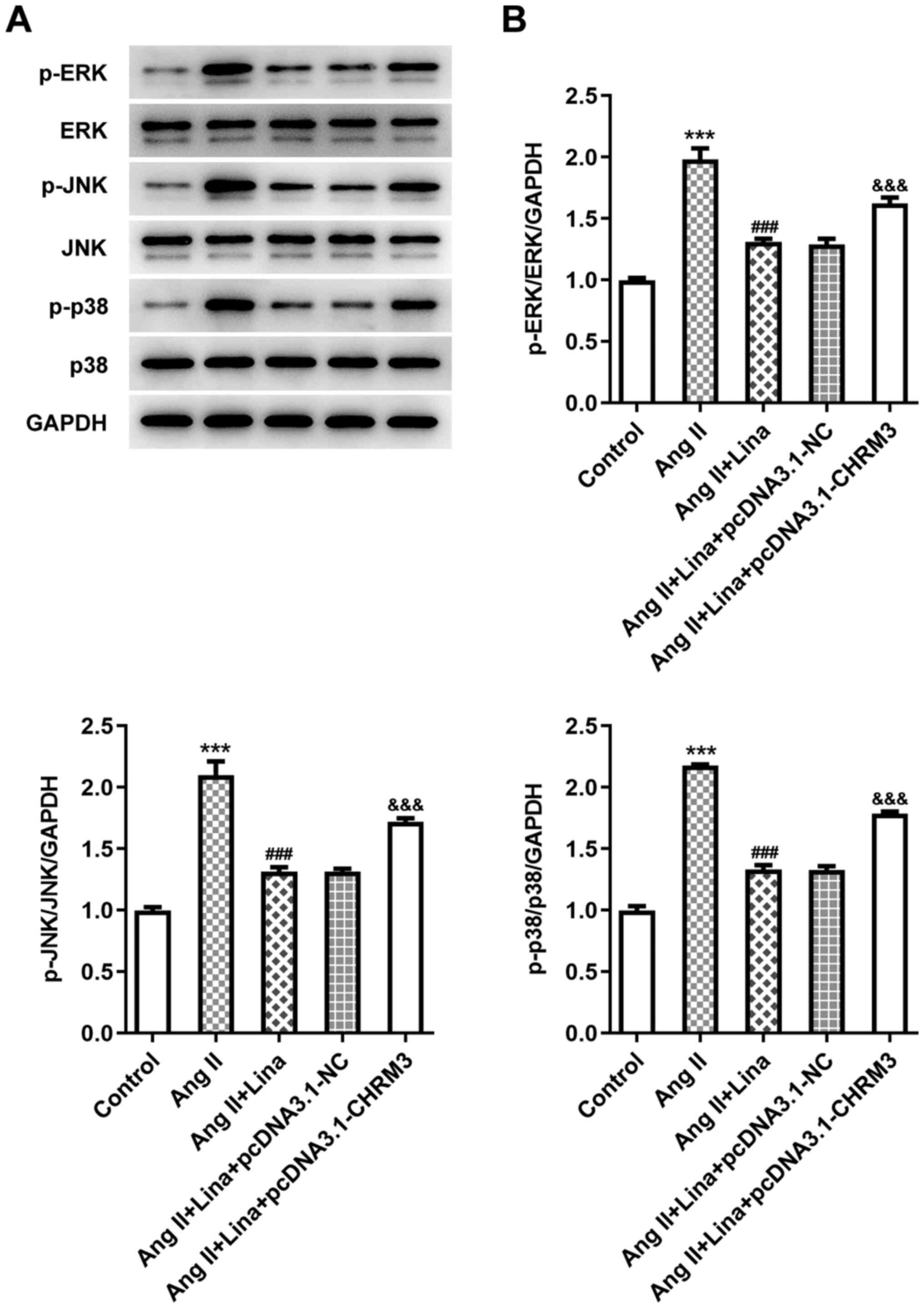
Linalool blocks the MAPK signaling
pathway by downregulating CHRM3 expression
To estimate whether linalool regulates cell
proliferation and migration via the MAPK signaling pathway, western
blotting was applied to detect the expression of MAPK-related
proteins in A7r5 and transfected cells. The expression levels of
p-ERK, p-JNK and p-p38 in Ang II-induced A7r5 cells were decreased
when cells were treated with Ang II+linalool, indicating that
linalool suppressed the activation of MAPK signaling. The
expression levels of these proteins in the Ang
II+linalool+pcDNA3.1-CHRM3 group were increased compared with the
Ang II+linalool+pcDNA3.1-NC group, with levels almost restored to
the expression levels in cells treated with only Ang II (Fig. 6A and B). These results suggested
that linalool hindered the MAPK signaling pathway by regulating
CHRM3.
Discussion
VSMC proliferation is considered to be a common
cause of vascular proliferative diseases, including hypertension
(21), atherosclerosis (22) and restenosis after balloon
angioplasty (23), which are the
most common cardiovascular diseases worldwide and the main causes
of mortality. Prolonged hypertension can cause VSMC proliferation
and fibrosis inside the arteries, leading to narrowing of the
vessel wall (24). Therefore,
developing a therapeutic target that can alleviate the
proliferation and migration of VSMCs is an important strategy,
which has medical relevance for the treatment of vascular diseases.
The present study applied Ang II to induce VSMCs to establish a
research model. After induction, the proliferation and migration of
cells were obviously abnormal, which allowed for the investigation
of the possible effects of linalool on VSMCs. The results showed
that the maximum experimental concentration of linalool (150 μM)
had no effect on normal VSMC viability, while linalool inhibited
the proliferation and migration of induced VSMCs.
The present study also explored the potential
mechanism of linalool. CHRM3, the target of linalool predicted by
the SwissTargetPrediction website, is a protein-coding gene. This
muscarinic cholinergic receptor belongs to a larger family of G
protein-coupled receptors. The functional diversity of these
receptors is determined by their binding to acetylcholine (25), in which CHRM3 regulates smooth
muscle contraction (26), and its
stimulation causes the secretion of glandular tissue. Diseases
associated with CHRM3 include Prune syndrome (27) and cholinergic urticaria (28). CHRM3 coding variants can increase
muscarinic cholinergic 3 receptor (M3R) signaling, which is
associated with higher blood pressure. Removal of M3R signals leads
to lower blood pressure and improves heart and kidney dysfunction
(29). In addition, CHRM3 is
upregulated in a large part of benign prostatic hyperplasia (BPH)
tissue, and the activation of CHRM3 also promotes the proliferation
of BPH cells (30).
Overexpression of CHRM3 or activation of CHRM3 by carbachol
promotes further cell proliferation, migration and castration
resistance (31). In the present
study, CHRM3 overexpression was found to promote the proliferation
and migration of VSMCs, and to inhibit the suppressive effect of
linalool on cell hyperplasia. This indicated that linalool exerts
its inhibitory role by downregulating CHRM3.
Furthermore, the expression level of proteins
related to the MAPK signaling pathway in VSMCs was found to
decrease with linalool treatment, indicating that linalool played a
role by blocking MAPK signaling. CHRM3 overexpression reversed the
decline, indicating that CHRM3 mediated this pathway. Linalool was
found to block the MAPK signaling pathway by downregulating CHRM3.
This association has also been verified in other studies where
CHRM3 mediates the MAPK signaling pathway. For example,
upregulation of microRNA-30e inhibits the MAPK signaling pathway
and its downstream genes by downregulating CHRM3 in prostate cancer
cells (32), suggesting that
CHRM3 can regulate the MAPK signaling pathway. Since the MAPK
signaling pathway is a universal pathway for cell proliferation,
differentiation and migration (33), after using Ang II to induce
abnormal cell proliferation, the present study explored the
underlying mechanism of linalool from the perspective of this
pathway. When the whole mechanism was reviewed, the Ang II receptor
caught our attention. We propose that linalool achieved inhibitory
effects by blocking the Ang II receptor. This can be regarded as a
novel viewpoint for research, which will be the direction of our
future research.
In conclusion, linalool was demonstrated to block
the MAPK signaling pathway by downregulating the expression of
CHRM3, thereby inhibiting the proliferation and migration of Ang
II-induced VSMCs. The present findings suggested a potential role
of linalool in inhibiting VSMC hyperplasia, although the specific
mechanisms involved remain to be further explored.
Acknowledgements
Not applicable.
Funding
No funding was received.
Availability of data and materials
The datasets used and/or analyzed during the current
study are available from the corresponding author on reasonable
request.
Authors' contributions
YL and XL designed the study and wrote the
manuscript. YZ, YX, YW and PX performed the experiments and
analyzed the data. PX examined the data and critically revised the
manuscript for important intellectual content. All authors confirm
the authenticity of all the raw data. All authors have read and
approved the final manuscript.
Ethics approval and consent to
participate
Not applicable.
Patient consent for publication
Not applicable.
Competing interests
The authors declare that they have no competing
interests.
References
|
1
|
Czuriga-Kovács KR, Czuriga D and Csiba L:
Influence of Hypertension, Alone and in Combination with Other
Vascular Risk Factors on Cognition. CNS Neurol Disord Drug Targets.
15:690–698. 2016. View Article : Google Scholar : PubMed/NCBI
|
|
2
|
Strandberg TE and Pitkala K: What is the
most important component of blood pressure: Systolic, diastolic or
pulse pressure? Curr Opin Nephrol Hypertens. 12:293–297. 2003.
View Article : Google Scholar : PubMed/NCBI
|
|
3
|
Kawabe H, Azegami T, Takeda A, Kanda T,
Saito I, Saruta T and Hirosi H: Features of and preventive measures
against hypertension in the young. Hypertens Res. 42:935–948. 2019.
View Article : Google Scholar : PubMed/NCBI
|
|
4
|
Devos P and Menard J: Bibliometric
analysis of research relating to hypertension reported over the
period 1997–2016. J Hypertens. 37:2116–2122. 2019. View Article : Google Scholar : PubMed/NCBI
|
|
5
|
Hurtubise J, McLellan K, Durr K, Onasanya
O, Nwabuko D and NdisAng JF: The Different Facets of Dyslipidemia
and Hypertension in Atherosclerosis. Curr Atheroscler Rep.
18:822016. View Article : Google Scholar : PubMed/NCBI
|
|
6
|
Chamarro R, Ponz A, Alonso MD, Gil R and
Laínez JM: [Arterial hypertension and intracerebral hemorrhage].
Neurologia. 18:731–739. 2003.PubMed/NCBI
|
|
7
|
Turin TC, Okamura T, Afzal AR, Rumana N,
Watanabe M, Higashiyama A, Nakao Y, Nakai M, Takegami M, Nishimura
K, et al: Hypertension and lifetime risk of stroke. J Hypertens.
34:116–122. 2016. View Article : Google Scholar : PubMed/NCBI
|
|
8
|
Arribas SM, Hinek A and González MC:
Elastic fibres and vascular structure in hypertension. Pharmacol
Ther. 111:771–791. 2006. View Article : Google Scholar : PubMed/NCBI
|
|
9
|
Shi L, Tian C, Sun L, Cao F and Meng Z:
The lncRNA TUG1/miR-145-5p/FGF10 regulates proliferation and
migration in VSMCs of hypertension. Biochem Biophys Res Commun.
501:688–695. 2018. View Article : Google Scholar : PubMed/NCBI
|
|
10
|
Owens GK, Kumar MS and Wamhoff BR:
Molecular regulation of vascular smooth muscle cell differentiation
in development and disease. Physiol Rev. 84:767–801. 2004.
View Article : Google Scholar : PubMed/NCBI
|
|
11
|
Herring BP, Hoggatt AM, Burlak C and
Offermanns S: Previously differentiated medial vascular smooth
muscle cells contribute to neointima formation following vascular
injury. Vasc Cell. 6:212014. View Article : Google Scholar : PubMed/NCBI
|
|
12
|
Montezano AC, Nguyen Dinh Cat A, Rios FJ
and Touyz RM: Angiotensin II and vascular injury. Curr Hypertens
Rep. 16:4312014. View Article : Google Scholar : PubMed/NCBI
|
|
13
|
Pereira I, Severino P, Santos AC, Silva AM
and Souto EB: Linalool bioactive properties and potential
applicability in drug delivery systems. Colloids Surf B
Biointerfaces. 171:566–578. 2018. View Article : Google Scholar : PubMed/NCBI
|
|
14
|
Anjos PJ, Lima AO, Cunha PS, De Sousa DP,
Onofre AS, Ribeiro TP, Medeiros IA, Antoniolli AR, Quintans-Júnior
LJ and Santosa MR: Cardiovascular effects induced by linalool in
normotensive and hypertensive rats. Z Naturforsch C J Biosci.
68:181–190. 2013. View Article : Google Scholar : PubMed/NCBI
|
|
15
|
Gong X, WAng B, Yan L, Lu X and Zhao X:
Linalool inhibits the growth of human T cell acute lymphoblastic
leukemia cells with involvement of the MAPK signaling pathway.
Oncol Lett. 20:1812020. View Article : Google Scholar : PubMed/NCBI
|
|
16
|
Xing X, Ma JH, Fu Y, Zhao H, Ye XX, Han Z,
Jia FJ and Li X: Essential oil extracted from erythrina
corallodendron L. leaves inhibits the proliferation, migration, and
invasion of breast cancer cells. Medicine (Baltimore).
98:e170092019. View Article : Google Scholar : PubMed/NCBI
|
|
17
|
Eguchi S, Kawai T, Scalia R and Rizzo V:
Understanding Angiotensin II Type 1 Receptor Signaling in Vascular
Pathophysiology. Hypertens. 71:804–810. 2018. View Article : Google Scholar : PubMed/NCBI
|
|
18
|
Livak KJ and Schmittgen TD: Analysis of
relative gene expression data using real-time quantitative PCR and
the 2(-Delta Delta C(T)) Method. Methods. 25:402–408. 2001.
View Article : Google Scholar : PubMed/NCBI
|
|
19
|
Strzalka W and Ziemienowicz A:
Proliferating cell nuclear antigen (PCNA): A key factor in DNA
replication and cell cycle regulation. Ann Bot (Lond).
107:1127–1140. 2011. View Article : Google Scholar : PubMed/NCBI
|
|
20
|
Urrutia G, Laurito S, Campoy E, Nasif D,
Branham MT and Roqué M: PAX6 Promoter Methylation Correlates with
MDA-MB-231 Cell Migration, and Expression of MMP2 and MMP9. Asian
Pac J Cancer Prev. 19:2859–2866. 2018.PubMed/NCBI
|
|
21
|
FAng G, Qi J, HuAng L and Zhao X: LncRNA
MRAK048635_P1 is critical for vascular smooth muscle cell function
and phenotypic switching in essential hypertension. Biosci Rep.
39:392019. View Article : Google Scholar
|
|
22
|
Basatemur GL, Jørgensen HF, Clarke MCH,
Bennett MR and Mallat Z: Vascular smooth muscle cells in
atherosclerosis. Nat Rev Cardiol. 16:727–744. 2019. View Article : Google Scholar : PubMed/NCBI
|
|
23
|
Jun MY, Karki R, Paudel KR, Sharma BR,
Adhikari D and Kim DW: Alkaloid rich fraction from Nelumbo nucifera
targets VSMC proliferation and migration to suppress restenosis in
balloon-injured rat carotid artery. Atherosclerosis. 248:179–189.
2016. View Article : Google Scholar : PubMed/NCBI
|
|
24
|
Liao H, Gong J, ZhAng W and Guo X:
Valsartan inhibits Angiotensin II-induced proliferation of vascular
smooth muscle cells via regulating the expression of mitofusin 2. J
Huazhong Univ Sci Technolog Med Sci. 32:31–35. 2012. View Article : Google Scholar : PubMed/NCBI
|
|
25
|
Borroto-Escuela DO, Agnati LF, Fuxe K and
Ciruela F: Muscarinic acetylcholine receptor-interacting proteins
(mAChRIPs): Targeting the receptorsome. Curr Drug Targets.
13:53–71. 2012. View Article : Google Scholar : PubMed/NCBI
|
|
26
|
Kruse AC, Hu J, Kobilka BK and Wess J:
Muscarinic acetylcholine receptor X-ray structures: Potential
implications for drug development. Curr Opin Pharmacol. 16:24–30.
2014. View Article : Google Scholar : PubMed/NCBI
|
|
27
|
Beaman GM, Galatà G, Teik KW, Urquhart JE,
Aishah A, O'Sullivan J, Bhaskar SS, Wood KA, Thomas HB, O'Keefe RT,
et al: A homozygous missense variant in CHRM3 associated with
familial urinary bladder disease. Clin Genet. 96:515–520. 2019.
View Article : Google Scholar : PubMed/NCBI
|
|
28
|
Tokura Y: New Etiology of Cholinergic
Urticaria. Curr Probl Dermatol. 51:94–100. 2016. View Article : Google Scholar : PubMed/NCBI
|
|
29
|
Deng AY, deBlois D, Laporte SA, Gelinas D,
Tardif JC, Thorin E, Shi Y, Raignault A and Menard A: Novel
Pathogenesis of Hypertension and Diastolic Dysfunction Caused by
M3R (Muscarinic Cholinergic 3 Receptor) Signaling. Hypertens.
72:755–764. 2018. View Article : Google Scholar : PubMed/NCBI
|
|
30
|
WAng N, Dong BJ, Quan Y, Chen Q, Chu M, Xu
J, Xue W, HuAng YR, YAng R and Gao WQ: Regulation of Prostate
Development and Benign Prostatic Hyperplasia by Autocrine
Cholinergic Signaling via Maintaining the Epithelial Progenitor
Cells in Proliferating Status. Stem Cell Reports. 6:668–678. 2016.
View Article : Google Scholar : PubMed/NCBI
|
|
31
|
WAng N, Yao M, Xu J, Quan Y, ZhAng K, YAng
R and Gao WQ: Autocrine Activation of CHRM3 Promotes Prostate
Cancer Growth and Castration Resistance via CaM/CaMKK-Mediated
Phosphorylation of Akt. Clin Cancer Res. 21:4676–4685. 2015.
View Article : Google Scholar : PubMed/NCBI
|
|
32
|
Zheng XM, ZhAng P, Liu MH, Chen P and
ZhAng WB: MicroRNA-30e inhibits adhesion, migration, invasion and
cell cycle progression of prostate cancer cells via inhibition of
the activation of the MAPK signaling pathway by downregulating
CHRM3. Int J Oncol. 54:443–454. 2019.PubMed/NCBI
|
|
33
|
Sun Y, Liu WZ, Liu T, Feng X, YAng N and
Zhou HF: Signaling pathway of MAPK/ERK in cell proliferation,
differentiation, migration, senescence and apoptosis. J Recept
Signal Transduct Res. 35:600–604. 2015. View Article : Google Scholar : PubMed/NCBI
|