Introduction
Glioblastoma (GBM) is the most frequent and fatal
primary brain tumor. The common therapy regimen consisting of
surgery, radiotherapy, and chemotherapy with Temozolomid (TMZ)
(1,2) lack long lasting effects. The
prognosis is dismal with a 5-year survival probability of 6.8%
(varying with age at diagnosis) (3,4)
which barely improved for decades (5), and a median overall survival (OS) of
about 15–18 months after diagnosis which still remains at this low
level despite several efforts (1–4).
Diverse therapeutic options are subject of actual or
recent clinical studies, among them targeted approaches consisting
of small-molecule kinase inhibitors (6–8) or
antibodies (7), immunotherapy
(9) or oncolytic viruses
(10). But on the whole, the
effects on OS remain rather low. Reasons for therapy failure
include development of resistances caused by genetic heterogeneity
or redundant signaling cascades, toxicity and other undesired
side-effects, and low abundant drug availability on the tumor site
due to inhibition by the blood-brain-barrier (6,7).
Nevertheless, some patients show a prolonged OS up
to several years.
To date, several different factors, clinical and
molecular, are discussed to cause or correlate with long-term
survival (LTS). The possibility of gross tumor resection, a high
Karnofsky performance status and a young age at diagnosis are
considered as clinical factors for prolonged survival (11), also the location and whether the
subventrical zone is affected or not might have an influence for
progression and survival (12).
Furthermore, for several patients, eligibility for a second surgery
after tumor recurrence, is an option associated with prolonged OS
(13).
On the molecular level, some expression signatures,
mutations, chromosomal aberrations and MGMT methylation
status are deemed to be factors influencing therapy response and
survival (14–16), but no common profile indicating
LTS is found for this highly heterogeneous tumor entity (17). In contrary, some biomarkers like
MGMT methylation are controversially discussed for outcome
prediction ability (18).
Although several studies describe potential
molecular features in connection with survival or prognosis
(14,15), studies analyzing microRNA (miR)
profiles are quite rare and mostly not concordant, describing
different sets of miRs to be differentially expressed and therefore
considered as potential biomarkers for survival (19–23).
MiRs are short non-coding RNAs of ~22 nt length
which derive from long primary transcripts which are matured in a
multistep process. They influence the transcription and translation
of targeted genes and are often deregulated in any kind of cancer
(24), also in glioblastoma
(25). Depending on the tissue,
kind of dysregulation and affected targets, they have oncogenic
and/or tumor suppressive character (26). They are considered as putative
targets for directed therapies consisting of miR-mimics for
upregulating repressed miRs, or anti-miRs or miR-sponges to
suppress overexpressed miRs (27–29).
The aim of this study is to compare miR expression
profiles of short-time and long-time survivors to determine,
whether there are survival-associated signatures. miRs showing
significant differential expression between the survival groups are
deemed as candidates for further analysis regarding potential
target genes and influence on tumor behavior. These findings could
contribute to more knowledge and deeper insights on molecular
features of this highly complex and heterogenous disease.
Identifying differential expressed miRs and their
potential target genes can give clues to factors affecting OS,
which, in turn, could be interesting candidates for developing
specific therapeutic approaches.
Materials and methods
Patient samples
Formalin-fixed paraffin-embedded (FFPE) samples of
GBM patients were retrieved from the archive of the Institute of
Pathology of the University Medicine Rostock. Tissue was fixed in
4% neutral buffered formalin (Grimm) and incubated overnight at
room temperature. Afterwards, it was embedded in molten paraffin
(Merck).
Inclusion criteria were diagnosed GBM WHO Grade IV,
sufficient availability of material and tumor content. For this
study, the number of patient samples for array-based microRNA
profile screening was limited, so from all potential available
samples, initially 24 were arbitrarily selected to obtain a
balanced distribution of sex and survival. One sample had to be
removed afterwards due to issues of data quality and completeness,
so a total of 23 patients were suitable for analysis.
Patient data (sex, age at diagnosis, OS, kind of
surgery, therapeutic treatments) were obtained from the department
of neurosurgery and patients were assigned to two groups: LTS and
short-term survivor (STS) using the median OS of the cohort as
discriminator. Molecular characteristics (EGFR
amplification, mutation status of IDH1 and IDH2) were
obtained by earlier projects and/or routine diagnostic
procedures.
Specimen collection was conducted in accordance with
the ethics guidelines for the use of human material, approved by
the Ethics Committee of the University of Rostock (Reference no. A
2009/34) and with informed written consent from all patients prior
to surgery.
MicroRNA extraction
For microRNA extraction from 10 µm FFPE sections,
the miRNeasy FFPE-Kit (Qiagen) was used following the
manufacturer's protocol. Concentration of extracted RNA was
determined with a Nanodrop spectrometer (Peqlab).
miR screening arrays
Analysis of microRNA expression was performed using
the Nanostring nCounter System with the Human v3 miRNA assay
(Nanostring). The analysis procedure was performed with 250 ng per
sample by a service provider lab (Transcriptome and Genome Analysis
Laboratory (TAL), Microarray and Deep-Sequencing Facility,
University Medicine Göttingen) and analyzed by the authors with the
nSolver 4.0 software (Nanostring) using standard settings for
background subtraction and housekeeping genes-based normalization.
Group-wise comparison (LTS vs. STS) was performed, delivering fold
change and p-values. Results were visualized as boxplots generated
by BoxPlotR (http://shiny.chemgrid.org/boxplotr/).
Determination of miR-targets
For determination of potential target genes of
miR-130b-3p, miR-146b-5p and miR-148a-3p, targeted literature
research was performed looking up PubMed (https://pubmed.ncbi.nlm.nih.gov) for corresponding
papers using search strings including the miR-name and
‘glioblastoma’. Furthermore, the online microRNA-target interaction
database miRTarBase (http://mirtarbase.cuhk.edu.cn) was consulted for
predicted potential targets of these three miRs.
Immunohistochemistry
Protein expression analysis for PTEN and TRAF6 was
performed by immunohistochemistry using 2 µm thick FFPE sections on
coated glass slides (Dako). For deparaffinization, rehydration and
antigen demasking, slides were incubated for 20 min at 97°C and pH
9 in EnVision FLEX Target Retrieval Solution, high pH (pH 9)
(Dako).
For slide processing, an automatic IHC system,
AutostainerLink48 (Dako) was used, according to the following
(routine) protocol. All steps were performed at room temperature.
Slides were rinsed with EnVision FLEX Wash Buffer (Dako). To reduce
unspecific background staining, slides were incubated for 5 min
with 100 µl of EnVision FLEX Peroxidase-Blocking Reagent,
readty-to-use (Dako), a phosphate buffer containing
H2O2, 15 mMol NaN3 and detergent,
and rinsed afterwards with wash buffer. For PTEN, a monoclonal
(clone 6H2.1) mouse-anti human PTEN antibody (Dako, cat. no. M3627)
was used, dilution 1:100 in EnVision FLEX Antibody Diluent (Dako).
For TRAF6, a monoclonal (clone EP592Y) rabbit-anti human TRAF6
antibody (Abcam; cat. no. ab40675), dilution 1:50, was used. Slides
were incubated with 100 µl of first antibodies for 20 min. and
rinsed afterwards with wash buffer. For TRAF6, a signal enhancement
step was inserted incubating slides for 15 min with 100 µl of
EnVision FLEX+ Rabbit (LINKER), ready-to-use (Dako, cat. no.
SM805). As secondary reagent, EnVision FLEX/HRP, ready-to-use
(Dako, cat. no. SM802), was used, containing dextran coupled with
peroxidase molecules and goat secondary antibodies against rabbit
and mouse immunoglobulins. Slides were incubated with 100 µl
thereof for 20 min and rinsed afterwards with wash buffer. As
substrate working solution 1 drop EnVision FLEX DAB+ Chromogen
(Dako, cat. no. DM827) was mixed with 1 ml EnVision FLEX Substrate
Buffer (Dako, cat. no. SM803). 200 µl thereof were applied to each
slide. Slides were incubated for 10 min and rinsed with wash buffer
afterwards. Finally, for counter staining, 100 µl of EnVision FLEX
Hematoxylin (Dako, cat. no. SM806) were applied and slides were
incubated for 5 min. Slides were rinsed with deionized water,
washed for 5 min with wash buffer and rinsed once more with water.
Slides were covered after dehydration with ethanol and xylol with
coverslips (epredia) and CV mount (Leica) as mounting medium using
the automated Coverslipper (Dako).
Stained slides were scanned using a Pannoramic Desk
DWII slide scanner (3DHistech) with Pannoramic Scanner 2.2.0
software (3DHistech). Visualization and analysis were performed
using CaseViewer 2.4 software (3DHistech). For presentation,
representative regions the magnification was set to 10×, and the
chosen sections were exported as JPEG.
Statistical analyses
Significance of differential miR expression was
determined using the nSolver Software 4.0 (Nanostring) applying a
paired, two-tailed Student's t-test. Statistical analyses
concerning OS and clinical data were performed using SPSS
Statistics version 28 (IBM) using the Kaplan-Meier method.
Significance was assumed for P≤0.05.
Results
Patients' characteristics
Samples of 23 GBM patients were finally included in
this study (Table I). Median age
at diagnosis was 59.96 years (47.36–80.82 years). Median OS was 380
days (30–2041 days). Patients with OS >median OS were classified
as LTS, the others as STS. 11 patients were male, 12 were female.
All but one were wild type for IDH1 R132 and 12 carried an
EGFR amplification. Sex and EGFR amplification status
were evenly distributed over both survival groups. 19 of 23
patients underwent gross-total resection and 4 had a subtotal
resection. 22 patients received radiotherapy, for one patient it is
unknown. 15 patients were treated with TMZ, 7 received no TMZ, for
one patient it is unknown. Of the patients without TMZ therapy,
five were grouped into the STS group and 2 into LTS. Regarding the
clinical data, only for the type of surgery, a significant
influence on OS could be observed (P=0.001). No significant
differences in OS were seen for age at diagnosis (P=0.566), sex
(P=0.927), EGFR status (P=0.437) and TMZ treatment (P=0.158)
(Fig. S1, Table II).
 | Table I.Characteristics of analyzed
patients. |
Table I.
Characteristics of analyzed
patients.
| Patient no. | Age at diagnosis
(y) | OS (d) | Sex | IDH1 status | EGFR status | Surgery | Rtx | TMZ | Survival
status |
|---|
| 1 | 67.36 | 528 | M | Wt | Amp | Gross-total
resection | Yes | Yes | LTS |
| 2 | 47.36 | 143 | F | Wt | Amp | Subtotal | Yes | Yes | STS |
| 3 | 47.72 | 427 | M | Wt | Amp | Gross-total
resection | Yes | Yes | LTS |
| 4 | 73.00 | 297 | F | Wt | Amp | Gross-total
resection | Yes | No | STS |
| 5 | 59.96 | 727 | M | Wt | Amp | Gross-total
resection | Yes | Yes | LTS |
| 6 | 69.63 | 380 | F | Wt | Amp | Gross-total
resection | Yes | Yes | STS |
| 7 | 50.05 | 2041 | M | Wt | Amp | Gross-total
resection | Yes | Yes | LTS |
| 8 | 64.47 | 148 | M | Wt | Amp | Gross-total
resection | Yes | Yes | STS |
| 9 | 76.46 | 883 | F | Wt | Amp | Gross-total
resection | Yes | No | LTS |
| 10 | 69.35 | 148 | F | Wt | Amp | Gross-total
resection | Yes | Yes | STS |
| 11 | 53.22 | 236 | M | Wt | Amp | Subtotal | N/A | N/A | STS |
| 12 | 57.18 | 687 | F | Wt | Norm | Gross-total
resection | Yes | Yes | LTS |
| 13 | 59.75 | 357 | F | Wt | Norm | Gross-total
resection | Yes | Yes | STS |
| 14 | 57.46 | 586 | F | Wt | Norm | Gross-total
resection | Yes | Yes | LTS |
| 15 | 49.17 | 253 | M | Wt | Norm | Gross-total
resection | Yes | Yes | STS |
| 16 | 77.23 | 777 | F | Wt | Norm | Gross-total
resection | Yes | Yes | LTS |
| 17 | 53.63 | 129 | M | Wt | Norm | Gross-total
resection | Yes | No | STS |
| 18 | 80.82 | 489 | F | Wt | Norm | Gross-total
resection | Yes | Yes | LTS |
| 19 | 49.62 | 120 | F | Wt | Norm | Gross-total
resection | Yes | No | STS |
| 20 | 66.29 | 55 | M | Wt | Norm | Subtotal | Yes | No | STS |
| 21 | 71.09 | 30 | M | R132H | Norm | Subtotal | Yes | No | STS |
| 22 | 70.50 | 397 | F | Wt | Amp | Gross-total
resection | Yes | No | LTS |
| 23 | 56.74 | 871 | M | Wt | Norm | Gross-total
resection | Yes | Yes | LTS |
| Table II.Overview of clinical parameters and
their influence on overall survival. |
Table II.
Overview of clinical parameters and
their influence on overall survival.
| Clinical
parameter | Variables, n
(%) | Influence on OS
(P-value) |
|---|
| Age at diagnosis,
years |
| 0.566 |
|
<60 | 11 (47.83%) |
|
|
>60 | 12 (52.17%) |
|
| Median age,
years | 59.96 |
|
| Age range,
years | 47.36-80.82 |
|
| Sex |
| 0.927 |
|
Female | 12 (52.17%) |
|
|
Male | 11 (47.83%) |
|
| EGFR status |
| 0.437 |
| EGRF
normal | 11 (52.17%) |
|
| EGFR
amplified | 12 (47.83%) |
|
| IDH1/2 status |
| ND |
|
Wild-type | 22 (95.65%) |
|
|
Mutated | 1 (4.35%) |
|
| Tmz |
| 0.158 |
|
Yes | 15 (65.2%) |
|
| No | 7 (30.4 %) |
|
|
Unknown: | 1 (4.35%) |
|
| Radiotherapy |
| ND |
|
Yes | 22 (95.65%) |
|
|
Unknown | 1 (4.35%) |
|
| Type of
surgery |
| 0.001 |
|
Gross-total | 19 (82.6%) |
|
|
Subtotal | 4 (17.4%) |
|
miR expression profiling
The miR-expression profiling by the Nanostring
nCounter software revealed on the whole just moderate changes in
miR expression comparing profiles of LTS and STS (Table SI) for most of the miRs analyzed.
But some miRs outperformed the moderate fold change values,
rendering them as potential candidates as significant targets.
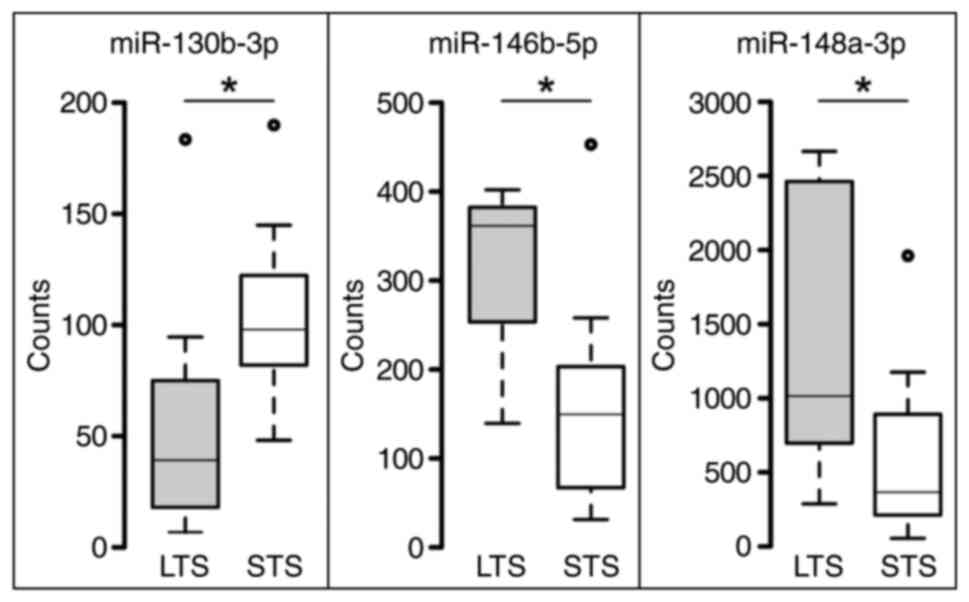
Finally, three miRs were choosen for further analysis: miR-130b-3p,
downregulated in LTS, miR-146b-5p and miR-148a-5p, both upregulated
in LTS (Table III, Fig. 1). All three of them fulfilled the
requirements for being further considered: a top fold change value
with high significance, and robust absolute expression values
(normalized counts) in both groups. MiRs showing high, significant
fold change values, but only very low counts, weren't considered
further.
| Table III.Top 10 significantly differentially
expressed miRs showing fold-change values of the LTS group (‘true’)
compared with the STS group (‘false’). |
Table III.
Top 10 significantly differentially
expressed miRs showing fold-change values of the LTS group (‘true’)
compared with the STS group (‘false’).
| Probe name | Accession no. | Fold-change LTS vs.
STS | P-value |
|---|
|
hsa-miR-146b-5p | MIMAT0002809 | 2.53 | 0.002 |
|
hsa-miR-130b-3p | MIMAT0000691 | −2.72 | 0.007 |
|
hsa-miR-148a-3p | MIMAT0000243 | 3.20 | 0.007 |
|
hsa-miR-302b-3p | MIMAT0000715 | 1.78 | 0.011 |
|
hsa-miR-3065-5p | MIMAT0015066 | 3.90 | 0.012 |
|
hsa-miR-301b-3p | MIMAT0004958 | −3.17 | 0.012 |
| hsa-miR-887-3p | MIMAT0004951 | 2.24 | 0.013 |
| hsa-miR-23a-3p | MIMAT0000078 | 1.94 | 0.017 |
| hsa-miR-21-5p | MIMAT0000076 | 1.79 | 0.019 |
| hsa-miR-142-3p | MIMAT0000434 | 2.02 | 0.019 |
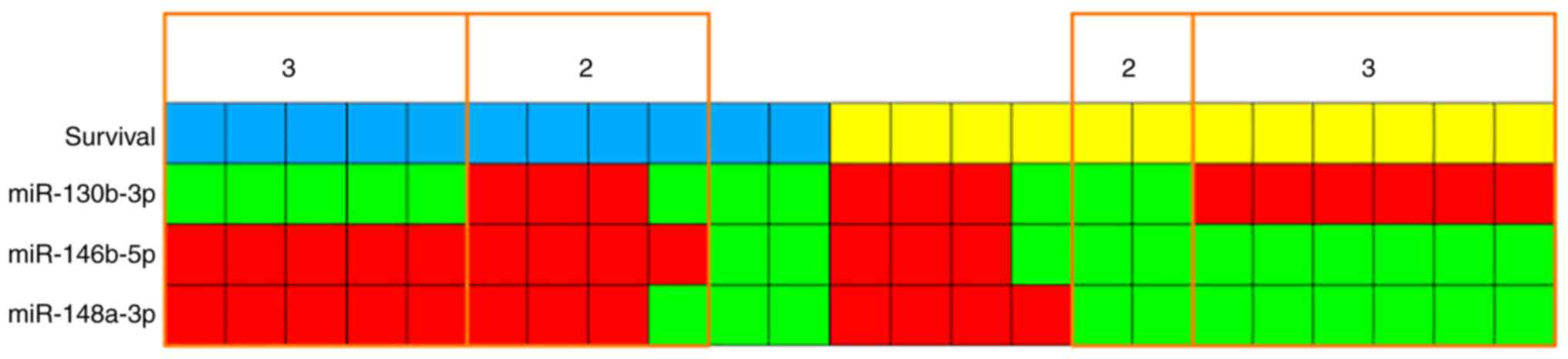
Although there are overlaps of expression values
between the two survival groups (Fig.
1), a kind of signature can be seen: low miR-130b-3p expression
and high expression of miR-146b-5p and miR-148a-3p for LTS; and
vice versa for STS. High and low expression states are separated by
the median expression values. Correlating survival and expression
status for each case shows that 11/23 cases (5 LTS and 6 STS)
completely correspond to this signature and another 6 cases (4 LTS
and 2 STS) in 2 of 3 miRs. Another 6 cases (2 LTS and 4 STS) show
just one miR but no case shows none (Fig. 2).
Determination of potential target
genes
Targeted PubMed search and specific investigation of
miRTarBase revealed several genes proposed to be potential targets
of miRs 130b-3p, 146b-5p and 148a-3p (Table IV). These two following were
chosen to start further analyses with: PTEN as target of
miR-130b-3p and important TSG in GBM, and TRAF6 as target of
miR-146b-5b, an invasion promoting factor. For miR-148a-3p the
situation is more complex, as it is described to function either as
oncogene or as TSG in GBM, so due to its ambivalent role, target
studies were delayed.
| Table IV.Literature and database predicted
target genes for miR-130b-3p, miR-146b-5p and miR 148a-3p. |
Table IV.
Literature and database predicted
target genes for miR-130b-3p, miR-146b-5p and miR 148a-3p.
| miR | Literature | miRTarBase |
|---|
| miR-130b-3p | PTEN (31); PPARγ (30); MST, SAV1 (32) | RUNX3, ZEB1, IGF1,
TP53INP1 |
| miR-146b-5p | TRAF6 (40,41); MMP16 (37); EGFR (34) | TRAF6, IRAK1,
MMP16, KIT, EGFR |
| miR-148a-3p | ITGA9 (49); DLGAP1 (46); MIG, BIM (52) | DNMT3B, DNMT1,
IKBKB, MMP7 |
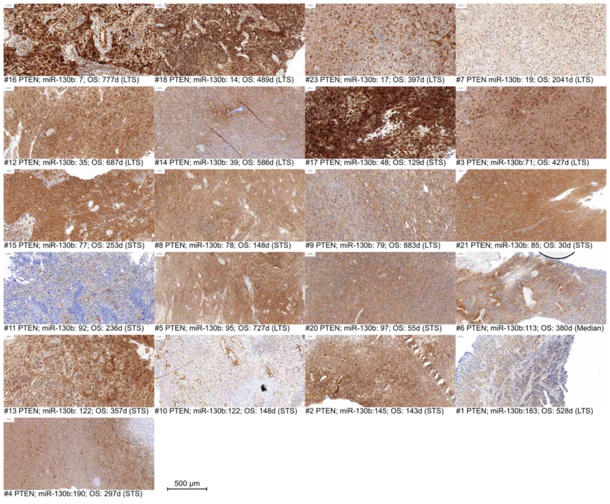
IHC analysis of PTEN and TRAF6
Immunohistochemistry of PTEN and TRAF6 was
successful in 22 of 23 cases. Although IHC is not considered as a
quantitative method, it is observable, that a partial loss or
decrease of PTEN expression occurs in some cases, mostly in samples
with a higher miR-130b expression (Fig. 3), especially samples #11, #10 and
#1.
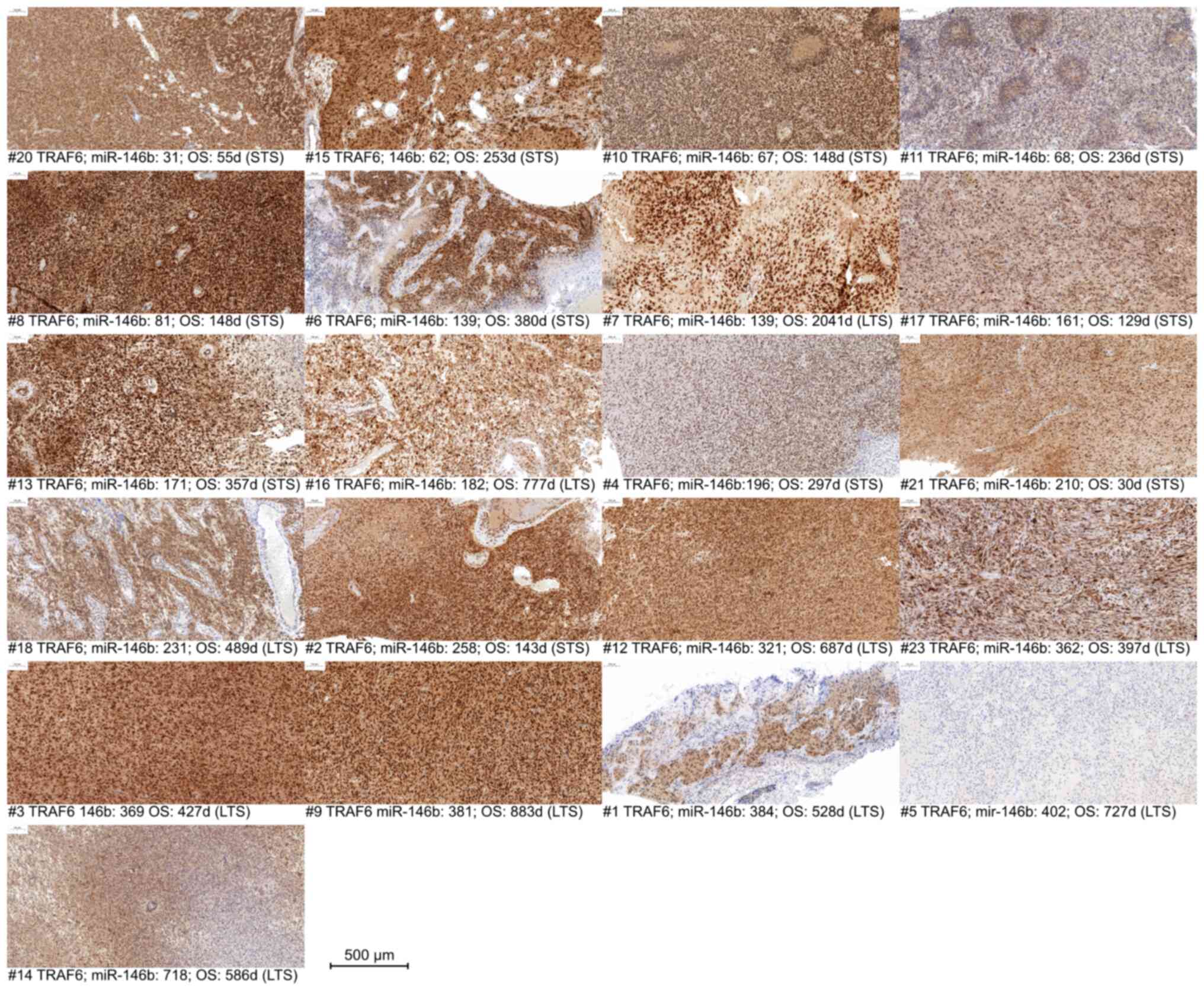
Similarly, for TRAF6, a reduction or partial loss in
expression is observable predominantly in samples with higher
miR-146b expression (Fig. 4),
especially samples #4, #1 and #5.
In summary, PTEN and TRAF6 could be considered as
putative targets of miR-130b and miR-146b, respectively, at least
in cases with higher miR expression levels.
Discussion
This study shows a significant differential miR
expression profile comparing LTS and STS, highlighting miR-130b-3p,
miR-146b-5p and miR-148a-3p as most significantly differentially
expressed miRs. As the definition of LTS is non-uniform throughout
the literature, in our study, the median OS of the cohort (380
days) was used as delimiter to split the cohort in two more or less
even groups. These data give the indication, that miR-130b-3p acts
as oncomir, while miR-146b-5p and miR-148a-3p have tumor suppressor
properties.
miR-130b is shown to be overexpressed in
glioblastoma in contrast to non-neoplastic brain tissue, as well as
in other tumor entities, and promotes cell proliferation by
inhibiting PPARγ, leading to decreased E-cadherin levels
(30). Furthermore, it was shown,
that suppression of miR-130b inhibits GBM cell proliferation and
invasion and induces apoptosis via the PTEN/AKT signaling pathway.
In this context, PTEN was shown to be a direct target of
miR-130b (31). Another study
shows more oncogenic properties of miR-130b, demonstrating that its
upregulation enhances a stem cell-like phenotype by inactivating
the Hippo signaling pathway with MST1/2 and SAV1 as
direct targets (32). Earlier,
miR-130b expression was associated with progression from lower to
high grade glioma (33). Together
with these, our data, showing a higher expression in STS samples,
underpin the oncogenic character of miR-130b.
miR-146b-5p is linked with tumor suppressive
properties. It is shown to target EGFR and hence, at least
in vitro, reduces invasion and migration of glioma cells
(34). In the cohort analyzed in
our study, immunohistochemistry showed, that the EGFR expression
was only dependent on the EGFR amplification status, but not
on the survival group (data not shown). But for in vitro
models it is shown that EGFR amplification is often lost
during cultivation (35), except
for special cell culture conditions (36). Another target deemed to be
regulated by miR-146b-5p is MMP16, whose inhibition leads to
decreased migration and invasion (37,38). MMP16 is a member of matrix
metalloproteases, important factors for migration and metastasis as
they contribute to proteolysis of the extracellular matrix,
enabling tumor cell migration (39). TRAF6 is another direct
target of miR-146b-5p whose repression inhibits proliferation and
progression, while promoting apoptosis. It is associated with a
better prognosis (40).
Furthermore, miR-146b-5p mediated TRAF6 repression is
considered to suppress TMZ resistance (41). TRAF6 is correlated with a
worse prognosis and oncogenic properties promoting invasion by
upregulating another matrix-metalloprotease, MMP9 (42). In context with our data these
studies underpin the correlation of miR-146-5p, TRAF6
expression and survival. Furthermore, miR-146b-5p inhibits GSCs and
radioresistance by targeting SMARCA5 (43) and the β-catenin pathway (44). Although most studies attest
miR-146b-5p tumor suppressive properties, some oncogenic potential
cannot be totally excluded, as its upregulation is also associated
with GBM recurrence (45). But
the data acquired in our own study are rather concordant with the
described tumor suppressive properties.
For miR-148a, its role in GBM seems to be
ambivalent, according to the current literature. Several studies
nominate different targets whose repression depict the oncogenic
character of miR-148a, where others describe tumor suppressive
properties. Repression of DLGAP1 leads to loss of cell
polarity, therefore promoting growth, migration, invasion and EMT
(46). Overexpression of miR-148a
deemed to be induced by NFκB and targeting QKI and
SKP1 activates TGFβ signaling and is associated with
progression and augmented tumor aggressiveness (47). In a TCGA based study, it is shown
that miR-148a targets FIH1, an HIF1 inhibitor and
therefore promotes HIF1a and NOTCH1 signaling, enhancing
vascularization, growth and survival (48). But also the opposite effect on
vascularization is described, as miR-148a has shown to have tumor
suppressive properties targeting ITGA9 (49), a cell adhesion factor involved in
NOTCH1 controlled vascularization, leading to decreased
angiogenesis (50). More
oncogenic properties are demonstrated with targets like
GADD45A, whose repression stimulates β-catenin and
MMP9, promoting invasion, migration and stemness (51). Also positively influencing EGFR
activity by targeting MIG2 and inhibiting apoptosis by
targeting BIM is described (52). Even exosomal delivery of miR-148a
seems to have a positive effect to proliferation and progression by
targeting CADM1, leading to STAT3 activation (53). On the other side, more tumor
suppressive properties are known. miR-148a targets ROCK-1, a
factor promoting migration in several tumors (54). Even therapeutic effects are
described, as in vivo experiments in mice show a prolonged
survival when miR-148a is co-delivered with miR-296-5p by
nanoparticles, targeting OCT4 and SOX2, reducing
stemness (29). Although studies
describing oncogenic properties of miR-148a in GBM predominate, our
own data show, that it is higher expressed in LTS, rendering it
more to the tumor suppressive side.
Considering the exemplarily selected targets PTEN
and TRAF6, the impression is not that clear. The expression of
these targets was assessed by IHC. This method is not considered as
quantitative and not sensitive enough to identify minor changes in
expression which is characteristic for miR-mediated regulation.
Also, there are much more factors influencing genes' translational
activity. But the data show, that at least at the upper ranges of
miR expression, a decrease in target gene expression is observable,
but there's no linearity.
To elucidate the downstream effects of differential
miR expression, identify the real target genes in these tumors, and
observe the effects on tumor cell behavior, functional approaches
with GBM in vitro models using transfection of miR mimics or
antagomiRs, followed by analyses on different levels are
needed.
This study is limited by a quite low number of
patients analyzed and a small selection of potential target genes.
To improve the value of robustness of the data, a consecutive study
based on the present data should include a higher number of
patients. Furthermore, next to functional studies, the selection of
potential target genes should be extended to elucidate more details
of the molecular mechanisms of tumor behavior underlying the
outcome. Also, the study is carried out on FFPE material, which
offers lesser quality and opportunities for deeper analysis. Fresh
material would serve as a source better quality nucleic acids and
proteins for deeper molecular analysis, and would even offer the
opportunity to be taken in culture to provide cell lines or
xenografts for in vitro or in vivo studies.
In summary, this study shows a significant
correlation of differential miR-expression and survival status. It
emphasizes the role of regulatory components/epigenetic factors, in
this case microRNAs, within the complex interactions determining
tumor behavior and outcome. These results hopefully help to clarify
the so far widely unconcordant approaches to determine outcome
predicting biomarkers. Although the regulatory impact to the few
selected, potential miR target genes is rather low, the connection
between miR expression and survival is significant. These data
provide a good basis for further, functional studies.
Supplementary Material
Supporting Data
Supporting Data
Acknowledgements
The authors would like to thank the technicians of
the Institute of Pathology (University Medicine Rostock, Rostock,
Germany), Mrs. Kerstin Westphal, Mrs. Heike Clasen and Mrs. Beate
Krause for performing immunohistochemistry, and Mrs. Susanne Höffer
for the sections.
Funding
Funding: No funding was received.
Availability of data and materials
The datasets used and/or analyzed during the current
study are available from the corresponding author on reasonable
request.
Authors' contributions
BS designed the study, acquired, analyzed and
interpreted data and wrote the manuscript. NL analyzed and
interpreted immunohistochemistry data. AZ analyzed and interpreted
immunohistochemistry data, contributed to the statistical analyses
and critically edited the manuscript. CH acquired the patient data
and substantially contributed to their analyses and interpretation,
and critically edited the manuscript. AE provided patient material,
substantially contributed to the concept of the study, and
critically contributed to the manuscript. BS and AZ confirm the
authenticity of all the raw data. All authors read and approved the
final manuscript.
Ethics approval and consent to
participate
The study was conducted in accordance with the
Declaration of Helsinki, and approved by the Ethics Committee of
the University of Rostock (approval no. A2009/34). Informed consent
was obtained from all subjects involved in the study.
Patient consent for publication
Not applicable
Competing interests
The authors declare that they have no competing
interests.
Authors' information
Dr Björn Schneider, ORCID-ID:
0000-0002-0282-7330.
References
|
1
|
Stupp R, Hegi ME, Mason WP, van den Bent
MJ, Taphoorn MJB, Janzer RC, Ludwin SK, Allgeier A, Fisher B,
Belanger K, et al: Effects of radiotherapy with concomitant and
adjuvant temozolomide versus radiotherapy alone on survival in
glioblastoma in a randomised phase III study: 5-year analysis of
the EORTC-NCIC trial. Lancet Oncol. 10:459–466. 2009. View Article : Google Scholar : PubMed/NCBI
|
|
2
|
Stupp R, Mason WP, van den Bent MJ, Weller
M, Fisher B, Taphoorn MJB, Belanger K, Brandes AA, Marosi C,
Bogdahn U, et al: Radiotherapy plus concomitant and adjuvant
temozolomide for glioblastoma. N Engl J Med. 352:987–996. 2005.
View Article : Google Scholar : PubMed/NCBI
|
|
3
|
Wen PY, Weller M, Lee EQ, Alexander BM,
Barnholtz-Sloan JS, Barthel FP, Batchelor TT, Bindra RS, Chang SM,
Chiocca EA, et al: Glioblastoma in adults: A society for
neuro-oncology (SNO) and european society of neuro-oncology (EANO)
consensus review on current management and future directions. Neuro
Oncol. 22:1073–1113. 2020. View Article : Google Scholar : PubMed/NCBI
|
|
4
|
Ostrom QT, Cioffi G, Gittleman H, Patil N,
Waite K, Kruchko C and Barnholtz-Sloan JS: CBTRUS statistical
report: Primary brain and other central nervous system tumors
diagnosed in the United States in 2012–2016. Neuro Oncol. 21 (Suppl
5):v1–v100. 2019. View Article : Google Scholar : PubMed/NCBI
|
|
5
|
Miller KD, Ostrom QT, Kruchko C, Patil N,
Tihan T, Cioffi G, Fuchs HE, Waite KA, Jemal A, Siegel RL and
Barnholtz-Sloan JS: Brain and other central nervous system tumor
statistics, 2021. CA Cancer J Clin. 71:381–406. 2021. View Article : Google Scholar : PubMed/NCBI
|
|
6
|
Heffron TP: Challenges of developing
small-molecule kinase inhibitors for brain tumors and the need for
emphasis on free drug levels. Neuro Oncol. 20:307–312. 2018.
View Article : Google Scholar : PubMed/NCBI
|
|
7
|
Le Rhun E, Preusser M, Roth P, Reardon DA,
van den Bent M, Wen P, Reifenberger G and Weller M: Molecular
targeted therapy of glioblastoma. Cancer Treat Rev. 80:1018962019.
View Article : Google Scholar : PubMed/NCBI
|
|
8
|
Perryman R, Renziehausen A, Shaye H,
Kostagianni AD, Tsiailanis AD, Thorne T, Chatziathanasiadou MV,
Sivolapenko GB, El Mubarak MA, Han GW, et al: Inhibition of the
angiotensin II type 2 receptor AT2R is a novel
therapeutic strategy for glioblastoma. Proc Natl Acad Sci USA.
119:e21162891192022. View Article : Google Scholar : PubMed/NCBI
|
|
9
|
Cao TQ, Wainwright DA, Lee-Chang C, Miska
J, Sonabend AM, Heimberger AB and Lukas RV: Next steps for
immunotherapy in glioblastoma. Cancers (Basel). 14:40232022.
View Article : Google Scholar : PubMed/NCBI
|
|
10
|
Todo T, Ito H, Ino Y, Ohtsu H, Ota Y,
Shibahara J and Tanaka M: Intratumoral oncolytic herpes virus G47∆
for residual or recurrent glioblastoma: A phase 2 trial. Nat Med.
28:1630–1639. 2022. View Article : Google Scholar : PubMed/NCBI
|
|
11
|
Anselmo P, Maranzano E, Selimi A,
Lupattelli M, Palumbo I, Bini V, Casale M, Trippa F, Bufi A,
Arcidiacono F and Aristei C: Clinical characterization of
glioblastoma patients living longer than 2 years: A retrospective
analysis of two Italian institutions. Asia Pac J Clin Oncol.
17:273–279. 2020. View Article : Google Scholar : PubMed/NCBI
|
|
12
|
Jiang H, Yu K, Li M, Cui Y, Ren X, Yang C,
Zhao X and Lin S: Classification of progression patterns in
glioblastoma: Analysis of predictive factors and clinical
implications. Front Oncol. 10:5906482020. View Article : Google Scholar : PubMed/NCBI
|
|
13
|
Pasqualetti F, Montemurro N, Desideri I,
Loi M, Giannini N, Gadducci G, Malfatti G, Cantarella M, Gonnelli
A, Montrone S, et al: Impact of recurrence pattern in patients
undergoing a second surgery for recurrent glioblastoma. Acta Neurol
Belg. 122:441–446. 2022. View Article : Google Scholar : PubMed/NCBI
|
|
14
|
Jovčevska I: Genetic secrets of long-term
glioblastoma survivors. Bosn J Basic Med Sci. 19:116–124.
2019.PubMed/NCBI
|
|
15
|
Gately L, McLachlan SA, Philip J, Rathi V
and Dowling A: Molecular profile of long-term survivors of
glioblastoma: A scoping review of the literature. J Clin Neurosci.
68:1–8. 2019. View Article : Google Scholar : PubMed/NCBI
|
|
16
|
Lei CG, Jia XY and Sun WJ: Establish
six-gene prognostic model for glioblastoma based on multi-omics
data of TCGA database. Yi Chuan. 43:665–679. 2021.PubMed/NCBI
|
|
17
|
Richardson TE, Kumar A, Xing C, Hatanpaa
KJ and Walker JM: Overcoming the odds: Toward a molecular profile
of long-term survival in glioblastoma. J Neuropathol Exp Neurol.
79:1031–1037. 2020. View Article : Google Scholar : PubMed/NCBI
|
|
18
|
Butler M, Pongor L, Su YT, Xi L, Raffeld
M, Quezado M, Trepel J, Aldape K, Pommier Y and Wu J: MGMT status
as a clinical biomarker in glioblastoma. Trends Cancer. 6:380–391.
2020. View Article : Google Scholar : PubMed/NCBI
|
|
19
|
Henriksen M, Johnsen KB, Andersen HH,
Pilgaard L and Duroux M: MicroRNA expression signatures determine
prognosis and survival in glioblastoma multiforme-a systematic
overview. Mol Neurobiol. 50:896–913. 2014. View Article : Google Scholar : PubMed/NCBI
|
|
20
|
Zhang W, Zhang J, Yan W, You G, Bao Z, Li
S, Kang C, Jiang C, You Y, Zhang Y, et al: Whole-genome microRNA
expression profiling identifies a 5-microRNA signature as a
prognostic biomarker in Chinese patients with primary glioblastoma
multiforme. Cancer. 119:814–824. 2013. View Article : Google Scholar : PubMed/NCBI
|
|
21
|
Srinivasan S, Patric IRP and Somasundaram
K: A ten-microRNA expression signature predicts survival in
glioblastoma. PLoS One. 6:e174382011. View Article : Google Scholar : PubMed/NCBI
|
|
22
|
Niyazi M, Zehentmayr F, Niemöller OM,
Eigenbrod S, Kretzschmar H, Schulze-Osthoff K, Tonn JC, Atkinson M,
Mörtl S and Belka C: MiRNA expression patterns predict survival in
glioblastoma. Radiat Oncol. 6:1532011. View Article : Google Scholar : PubMed/NCBI
|
|
23
|
Areeb Z, Stylli SS, Koldej R, Ritchie DS,
Siegal T, Morokoff AP, Kaye AH and Luwor RB: MicroRNA as potential
biomarkers in Glioblastoma. J Neurooncol. 125:237–248. 2015.
View Article : Google Scholar : PubMed/NCBI
|
|
24
|
Di Leva G, Garofalo M and Croce CM:
MicroRNAs in cancer. Annu Rev Pathol. 9:287–314. 2014. View Article : Google Scholar : PubMed/NCBI
|
|
25
|
Rolle K: miRNA Multiplayers in glioma.
From bench to bedside. Acta Biochim Pol. 62:353–365. 2015.
View Article : Google Scholar : PubMed/NCBI
|
|
26
|
Svoronos AA, Engelman DM and Slack FJ:
OncomiR or tumor suppressor? The duplicity of microRNAs in cancer.
Cancer Res. 76:3666–3670. 2016. View Article : Google Scholar : PubMed/NCBI
|
|
27
|
Chen L and Kang C: miRNA interventions
serve as ‘magic bullets’ in the reversal of glioblastoma hallmarks.
Oncotarget. 6:38628–38642. 2015. View Article : Google Scholar : PubMed/NCBI
|
|
28
|
Ananta JS, Paulmurugan R and Massoud TF:
Tailored nanoparticle codelivery of antimiR-21 and antimiR-10b
augments Glioblastoma Cell Kill by Temozolomide: Toward a
‘personalized’ anti-microRNA therapy. Mol Pharm. 13:3164–3175.
2016. View Article : Google Scholar : PubMed/NCBI
|
|
29
|
Lopez-Bertoni H, Kozielski KL, Rui Y, Lal
B, Vaughan H, Wilson DR, Mihelson N, Eberhart CG, Laterra J and
Green JJ: Bioreducible polymeric nanoparticles containing
multiplexed cancer stem cell regulating miRNAs inhibit Glioblastoma
growth and prolong survival. Nano Lett. 18:4086–4094. 2018.
View Article : Google Scholar : PubMed/NCBI
|
|
30
|
Gu JJ, Zhang JH, Chen HJ and Wang SS:
MicroRNA-130b promotes cell proliferation and invasion by
inhibiting peroxisome proliferator-activated receptor-γ in human
glioma cells. Int J Mol Med. 37:1587–1593. 2016. View Article : Google Scholar : PubMed/NCBI
|
|
31
|
Gu JJ, Fan KC, Zhang JH, Chen HJ and Wang
SS: Suppression of microRNA-130b inhibits glioma cell proliferation
and invasion, and induces apoptosis by PTEN/AKT signaling. Int J
Mol Med. 41:284–292. 2018.PubMed/NCBI
|
|
32
|
Zhu G, Wang Y, Mijiti M, Wang Z, Wu PF and
Jiafu D: Upregulation of miR-130b enhances stem cell-like phenotype
in glioblastoma by inactivating the Hippo signaling pathway.
Biochem Biophys Res Commun. 465:194–199. 2015. View Article : Google Scholar : PubMed/NCBI
|
|
33
|
Malzkorn B, Wolter M, Liesenberg F,
Grzendowski M, Stühler K, Meyer HE and Reifenberger G:
Identification and functional characterization of microRNAs
involved in the malignant progression of gliomas. Brain Pathol.
20:539–550. 2010. View Article : Google Scholar : PubMed/NCBI
|
|
34
|
Katakowski M, Zheng X, Jiang F, Rogers T,
Szalad A and Chopp M: MiR-146b-5p suppresses EGFR expression and
reduces in vitro migration and invasion of glioma. Cancer Invest.
28:1024–1030. 2010. View Article : Google Scholar : PubMed/NCBI
|
|
35
|
Pandita A, Aldape KD, Zadeh G, Guha A and
James CD: Contrasting in vivo and in vitro fates of glioblastoma
cell subpopulations with amplified EGFR. Genes Chromosomes Cancer.
39:29–36. 2004. View Article : Google Scholar : PubMed/NCBI
|
|
36
|
William D, Mokri P, Lamp N, Linnebacher M,
Classen CF, Erbersdobler A and Schneider B: Amplification of the
EGFR gene can be maintained and modulated by variation of EGF
concentrations in in vitro models of glioblastoma multiforme. PLoS
One. 12:e01852082017. View Article : Google Scholar : PubMed/NCBI
|
|
37
|
Li Y, Wang Y, Yu L, Sun C, Cheng D, Yu S,
Wang Q, Yan Y, Kang C, Jin S, et al: miR-146b-5p inhibits glioma
migration and invasion by targeting MMP16. Cancer Lett.
339:260–269. 2013. View Article : Google Scholar : PubMed/NCBI
|
|
38
|
Le Zhang, Wang J, Fu Z, Ai Y, Li Y and
Wang Y and Wang Y: Sevoflurane suppresses migration and invasion of
glioma cells by regulating miR-146b-5p and MMP16. Artif Cells
Nanomed Biotechnol. 47:3306–3314. 2019. View Article : Google Scholar : PubMed/NCBI
|
|
39
|
Stetler-Stevenson WG, Aznavoorian S and
Liotta LA: Tumor cell interactions with the extracellular matrix
during invasion and metastasis. Annu Rev Cell Biol. 9:541–573.
1993. View Article : Google Scholar : PubMed/NCBI
|
|
40
|
Liu J, Xu J, Li H, Sun C, Yu L, Li Y, Shi
C, Zhou X, Bian X, Ping Y, et al: miR-146b-5p functions as a tumor
suppressor by targeting TRAF6 and predicts the prognosis of human
gliomas. Oncotarget. 6:29129–29142. 2015. View Article : Google Scholar : PubMed/NCBI
|
|
41
|
Qian Z, Zhou S, Zhou Z, Yang X, Que S, Lan
J, Qiu Y and Lin Y: miR-146b-5p suppresses glioblastoma cell
resistance to temozolomide through targeting TRAF6. Oncol Rep.
38:2941–2950. 2017. View Article : Google Scholar : PubMed/NCBI
|
|
42
|
Sun J, Zhao B, Du K and Liu P: TRAF6
correlated to invasion and poor prognosis of glioblastoma via
elevating MMP9 expression. Neuroreport. 30:127–133. 2019.
View Article : Google Scholar : PubMed/NCBI
|
|
43
|
Wang H, Tan L, Dong X, Liu L, Jiang Q, Li
H, Shi J, Yang X, Dai X, Qian Z and Dong J: MiR-146b-5p suppresses
the malignancy of GSC/MSC fusion cells by targeting SMARCA5. Aging.
12:13647–13667. 2020. View Article : Google Scholar : PubMed/NCBI
|
|
44
|
Yang W, Yu H, Shen Y, Liu Y, Yang Z and
Sun T: MiR-146b-5p overexpression attenuates stemness and
radioresistance of glioma stem cells by targeting
HuR/lincRNA-p21/β-catenin pathway. Oncotarget. 7:41505–41526. 2016.
View Article : Google Scholar : PubMed/NCBI
|
|
45
|
Khwaja SS, Cai C, Badiyan SN, Wang X and
Huang J: The immune-related microRNA miR-146b is upregulated in
glioblastoma recurrence. Oncotarget. 9:29036–29046. 2018.
View Article : Google Scholar : PubMed/NCBI
|
|
46
|
Li Y, Li W, Zeng X, Tang X, Zhang S, Zhong
F, Peng X, Zhong Y, Rosol TJ, Deng X, et al: The role of
microRNA-148a and downstream DLGAP1 on the molecular regulation and
tumor progression on human glioblastoma. Oncogene. 38:7234–7248.
2019. View Article : Google Scholar : PubMed/NCBI
|
|
47
|
Wang H, Pan JQ, Luo L, Ning XJ, Ye ZP, Yu
Z and Li WS: NF-κB induces miR-148a to sustain TGF-β/Smad signaling
activation in glioblastoma. Mol Cancer. 14:22015. View Article : Google Scholar : PubMed/NCBI
|
|
48
|
Wong HA, Fatimy RE, Onodera C, Wei Z, Yi
M, Mohan A, Gowrisankaran S, Karmali P, Marcusson E, Wakimoto H, et
al: The cancer genome atlas analysis predicts microRNA for
targeting cancer growth and vascularization in Glioblastoma. Mol
Ther. 23:1234–1247. 2015. View Article : Google Scholar : PubMed/NCBI
|
|
49
|
Xu TJ, Qiu P, Zhang YB, Yu SY, Xu GM and
Yang W: MiR-148a inhibits the proliferation and migration of
glioblastoma by targeting ITGA9. Hum Cell. 32:548–556. 2019.
View Article : Google Scholar : PubMed/NCBI
|
|
50
|
Guichet P-O, Guelfi S, Teigell M, Hoppe L,
Bakalara N, Bauchet L, Duffau H, Lamszus K, Rothhut B and Hugnot
JP: Notch1 stimulation induces a vascularization switch with
pericyte-like cell differentiation of glioblastoma stem cells. Stem
Cells. 33:21–34. 2015. View Article : Google Scholar : PubMed/NCBI
|
|
51
|
Cui D, Sajan P, Shi J, Shen Y, Wang K,
Deng X, Zhou L, Hu P and Gao L: MiR-148a increases glioma cell
migration and invasion by downregulating GADD45A in human gliomas
with IDH1 R132H mutations. Oncotarget. 8:25345–25361. 2017.
View Article : Google Scholar : PubMed/NCBI
|
|
52
|
Kim J, Zhang Y, Skalski M, Hayes J, Kefas
B, Schiff D, Purow B, Parsons S, Lawler S and Abounader R:
microRNA-148a is a prognostic oncomiR that targets MIG6 and BIM to
regulate EGFR and apoptosis in glioblastoma. Cancer Res.
74:1541–1553. 2014. View Article : Google Scholar : PubMed/NCBI
|
|
53
|
Cai Q, Zhu A and Gong L: Exosomes of
glioma cells deliver miR-148a to promote proliferation and
metastasis of glioblastoma via targeting CADM1. Bull Cancer.
105:643–651. 2018. View Article : Google Scholar : PubMed/NCBI
|
|
54
|
Fang Z, Weng Y, Xiao F and Yu J: LncRNA
RP11-390F4.3 inhibits invasion and migration of glioblastoma cells
by downregulating ROCK1. Neuroreport. 32:888–893. 2021. View Article : Google Scholar : PubMed/NCBI
|