Introduction
Ankylosing spondylitis (AS) is a chronic,
progressive, painful, systemic, immune-mediated, highly inherited
multifactorial inflammatory arthritis that primarily affects the
spine and sacroiliac joints, while, in some cases, it also affects
the peripheral joints and extraarticular tissues (1). It is the major subtype of an
inter-related group of rheumatic diseases sharing clinical, genetic
and radiographic features, termed spondyloarthritides (SpA), which
includes psoriatic arthritis, arthritis associated with
inflammatory bowel disease, reactive arthritis, undifferentiated
spondyloarthritis and juvenile-onset spondyloarthritis (2). AS is the most common form of
inflammatory arthritis after rheumatoid arthritis (RA) in the
developed world. Patients with AS suffer from pain and are
characterized by stiffness and the loss of spinal mobility, which
are explained by spinal inflammation and/or structural damage, by
bone and joint erosion and, finally, ankylosis and fibrosis
(3). AS is a disease that affects
mainly young individuals, who generally develop the first symptoms
at an age <30 years, and males are more often affected than
females, with a ratio of roughly 2 to 1 (1). The prevalence of AS varies in
different countries. Notably, in a Northern European region
(Norway), the prevalence of AS is between 0.2 and 1.2%, and the
incidence of the disease ranges from 0.5 to 14 per 100 000
individuals per year (4). The
molecular pathogenesis of this complex disease involves genetic,
immunological, microbial and hormonal components (5). Of note, microbial infection plays a
pivotal role as a triggering factor of the host innate immunity and
AS development (1).
Endometriosis is an enigmatic, common, benign,
estrogen-dependent gynecological disease with an unknown etiology
and a poorly understood pathogenesis. It is characterized by the
presence of endometrial tissue external to the uterine cavity, most
commonly in the pelvic cavity, including the ovaries and the
uterosacral ligaments, and is associated with chronic pelvic pain,
dysmenorrhea, irregular menstrual bleeding, intestinal symptoms,
recurrent urinary tract infections, dyspareunia and infertility,
thus markedly affecting the quality of life of patients; however, a
low percentage of patients with this condition may be asymptomatic
(6,7). Endometriosis can appear as peritoneal
lesions, ovarian endometriotic cysts and deeply infiltrative
endometriosis (8). Endometriosis
affects 6–10% of women of childbearing age, and it has been
estimated that 176 million women worldwide are affected by
endometriosis, with significant costs for both affected women and
for society (9) (http://endometriosis.org). Genetic and epigenetic
factors, as well as environmental ones, including pollution agents
and toxins, contribute to the development of this disease (8,10).
The pathogenetic mechanisms leading to its development remain
unclear, although several theories have been suggested thus far
regarding the development of endometriosis. Of note, all cases
cannot be explained by one theory alone. Thus, apart from the most
accepted Sampson's retrograde menstruation hypothesis, other
processes related to angiogenesis, increased oxidative stress,
endothelial dysfunction and chronic inflammation have been also
enrolled in the development of this condition (11).
Women with endometriosis are at a high risk of
developing several other chronic diseases, including cancer
(12), endocrine disorders
(hypothyroidism) (12),
cardiovascular disorders (13,14)
and, mainly, a number of autoimmune diseases including RA, multiple
sclerosis, scleroderma, systemic lupus erythematosus, ulcerative
colitis, Crohn's disease, Sjögren's syndrome, coeliac disease and
autoimmune thyroid disorder (15,16).
Notably, recent findings have suggested that endometriosis can
increase the susceptibility for AS in these women (17).
The present review discusses the genetic factors
involved in the co-occurrence of AS and endometriosis in an attempt
to deepen the knowledge concerning the shared underlying
pathogenetic mechanisms and the relevant molecular and cellular
pathways. A more in-depth understanding of these associations may
allow physicians to develop novel therapeutic interventions for
women with endometriosis, thus using this information in clinical
practice.
Genetics of AS and endometriosis
The rapid development of technology, including
genome-wide association studies (GWAS) and next-generation
sequencing (NGS) approaches, has contributed to the identification
of hundreds genetic associations between common DNA sequence
variants and multifactorial diseases. Although single nucleotide
polymorphisms (SNPs) are the preferred genetic markers for
case-control association and GWA studies due to their abundance in
the human genome, the exact functional consequences of most of
these polymorphisms are still unclear. Therefore, various
techniques have been developed over the past few years in an
attempt to reveal the functional basis of genetic risks associated
with human diseases.
AS has a strong genetic predisposition and the
investigation of genetic factors associated with AS susceptibility
became more systematic following the discovery of the class I human
leukocyte antigen B27, HLA-B27 locus, which was suggested to
be a major contributor to AS hereditability of ~30% (18). In particular, various subtypes of
HLA-B27, such as B*2702, B*2703 and B*2710,
have been reported to increase the risk of developing AS, with
these causative variants modifying the biochemical structure of the
protein (19). Significant
progress has been made in the discovery of genetic associations
with AS by GWAS, as well as an Immunochip study over past decade
(20). These studies performed to
date have uncovered less than one-third of the overall genetic risk
in AS (21), for the heritability
of the disease, which is estimated as ~90% (22). Through these studies, >113 SNPs
affecting the risk of developing AS have been identified thus far,
including endoplasmic reticulum aminopeptidase (ERAP)-1 and
ERAP-2, that are involved in antigen presentation, genes
that are involved in the IL23/IL17 axis including interleukin
(IL)-23R, Runt-related transcription factor 3,
JAK2, IL-1R1, IL-2R, IL-6R, IL-12B, IL-27, caspase
recruitment domain family member 9 (CARD9), signal
transducer and activator of transcription (STAT)3 and
tyrosine kinase 2 (TYK2), genes modulating the activation
and differentiation of lymphocytes including zinc finger MIZ-type
containing 1 (ZMIZ1), IL-7, T-box transcription
factor 21 (TBX21) and IL-7R, and other genes such
asIL-1Ra, NOD2 (CARD15), cytochrome P450
(CYP)2D6 and TGF-1 (5,20,22–27).
Of note, genetic studies regarding the clinical manifestations of
AS, such as the extent of bony ankylosis or the presence of
anterior uveitis have been very fruitful, given that some genes
that were found to influence uveitis risk are not AS risk factors
(28). The findings have uncovered
novel pathways involved in the pathogenesis of the disease and have
led to the introduction of novel therapeutic treatments for AS;
however, much of the heritability of AS remains unknown and causal
variants for most loci have yet to be defined.
Endometriosis is a complex disease, in which both
genetic and environmental factors interact and contribute to risk,
thus leading to the disease phenotype. The familial association of
endometriosis suggests a genetic contribution to the disease. Thus,
the relative risk for women who have immediate relatives with
endometriosis was estimated at 2.3 in a study on Australian twins
and their families (29), while
the overall heritability has been estimated at ~50%, as shown from
monozygotic twin studies (30).
Gene association studies, GWAS and NGS techniques have played a
primary role in depicting the genetic contributions to
endometriosis development. The ‘candidate gene’ studies have
firstly assisted investigators in identifying genetic variants
associated with endometriosis over the past decades, taking into
account hundreds of SNPs (31,32),
while GWAS were adopted for endometriosis from 2007 onwards, aiming
at identifying common genetic variants of moderate effects
(31–35). Notably, the loci identified were
categorized according to the function of their gene products
involved in estrogen-induced cell growth, matrix remodeling, sex
hormone activity, glucose homeostasis, vascular function, cell
adhesion, migration, growth and differentiation, inflammation and
WNT/β-catenin signaling, hormone receptors and metabolism,
transcription regulation, immunity and oxidative stress (36). However, considering that a number
of studies have yielded controversial results (31,35),
due mainly to the enrolment of small populations (37), many of these loci were confirmed by
replication studies and further meta-analyses. As is typical for
GWAS results, the majority of the loci reside in intergenic regions
for which the functionality remains to be uncovered. Rahmioglu
et al (34) conducted a
meta-analysis that detected GWA significance for rs12700667 on
7p15.2, rs7521902 near WNT4, rs10859871 near VEZT,
rs1537377 near CDKN2B-AS1, rs7739264 near ID4 and
rs13394619 in GREB1. Notably, all these SNPs, apart from the
VEZT one, exhibited a stronger association with stage III/IV
of endometriosis, which are characterized by ovarian (cystic) or
deep infiltrating disease including extensive adhesions (34). In a further meta-analysis, Sapkota
et al (38) identified five
novel endometriosis risk loci exhibiting significance in GWA,
namely fibronectin 1 (FN1), Coiled-coil domain containing 17
(CCDC170), estrogen receptor 1 (ESR1), spectrin
repeat-containing nuclear envelope protein 1 (SYNE1) and
follicle-stimulating hormone beta subunit (FSHB), with all
of them being involved in sex steroid hormone pathways. In the
largest GWAS and replication meta-analysis of endometriosis that
has been performed to date, enrolling 60,674 cases and 701,926
controls of European and East Asian ancestry, 42 genome-wide
significant loci were identified, including 31 novel ones, such as
ADP ribosylation factor like GTPase 14 effector protein
(ARL14EP), Bcl-2-modifying factor (BMF), homeobox A10
(HOXA10), long intergenic non-protein coding RNA 629
(LINC00629), alpha 1–3-N-acetylgalactosaminyltransferase and
alpha 1–3-galactosyltransferase (ABO), bassoon presynaptic
cytomatrix protein (BSN), PDZ and LIM domain 5
(PDLIM5), potassium channel tetramerization domain
containing 9 (KCTD9), MLLT10 histone lysine
methyltransferase DOT1L cofactor (MLLT10), actin like 9
(ACTL9) and inhibitor of DNA binding 4 (ID4)
(39). The majority of SNPs
exhibited larger effect sizes for stage III/IV disease, and when
combined, they explained 5.61% of stage III/IV disease variance.
Notably, the authors have previously used, for the first time in
family studies, the whole exome sequencing technique, which allowed
for the identification of UDP glucuronosyltransferase family 2
member B28 (UGT2B28) and ubiquitin specific peptidase 17
like family member 2 (USP17L2) as novel,
endometriosis-associated genes, by analyzing a unique in the
literature three-generation family from Crete (Greece) with seven
members suffering from endometriosis (40).
Influence of autoimmunity, angiogenesis and
inflammation in AS and endometriosis
It is well-known that endometriosis is a
multifactorial condition involving, apart from hormonal and
genetic, pro-inflammatory, pro-angiogenic and immunologic
processes. All the multiple interconnected factors involved in
these processes may explain the complex and heterogeneous
manifestations of the disease. Although it is beyond the scope of
the present review to discuss the aforementioned processes in
detail, some aspects regarding their role in AS and endometriosis
are presented below.
There is increasing evidence to indicate that women
with endometriosis are more likely to have an additional autoimmune
disease, probably due to underlying shared pathogenic pathways.
Autoimmune diseases are characterized by the loss of
self-tolerance, causing immune-mediated tissue destruction and
multiorgan involvement. Considering that similar immunological
alterations occur in endometriosis, thus increasing the number of
macrophages and leading to various abnormalities in the function
and concentrations of B- and T-lymphocytes (41), it has been suggested that
endometriosis resembles an autoimmune disease. Endometriosis is
often associated with the presence of a large variety of antibodies
in the blood and peritoneal fluid of patients, including
antinuclear, antiphospholipid, antithyroid, anti-survivin,
anti-laminin-1, anti-carbonic anhydrase, anti-α-enolase and
anti-endometrial autoantibodies (42,43).
Of note, various theories have been put forth, suggesting that
changes in the function and regulation of the immune system may
prevent the ability to eliminate the endometrium of the pelvic
cavity, while several types of immune cells appear to play roles in
the destruction of cells in ectopic sites (41). Furthermore, it has been suggested
that defects in immune surveillance in women with endometriosis may
also lead to the development of autoimmune diseases (44). AS is an autoimmune disease and, as
a consequence, it develops through complex interactions between
genetic background and environmental factors, as described in
detail in the previous section of the present review.
Endometriosis, which is characterized by systemic
inflammation in the affected organs of the body, is caused by a
variety of inflammatory factors, including cytokines, macrophages
and prostaglandin. Of note, a strong support for a pivotal role of
inflammation and its associated pathways dealing with the
establishment of endometriosis derives from the detection and
confirmation of the genetic background of the condition through
GWAS and NGS techniques. It has been reported that women with
endometriosis appear to have an altered inflammatory profile upon
surgical lesion removal, thus suggesting that the ectopic lesions
may drive systemic inflammation in endometriosis (45). Moreover, endometriotic lesions have
been found to exhibit the production of estrogen themselves, thus
stimulating mast cells to support the inflammatory process
(46). In addition, the hormonal
alterations that have been observed in endometriosis are related to
the inflammatory imbalance that characterizes the disease,
considering that inflammation affects hormonal regulation (47). The association of endometriosis
with inflammation has also been strengthened by the initiation of
several inflammatory responses by a network of pro-inflammatory
proteins of the IL-1 cytokine family, while elevated levels of
various inflammatory factors have been detected in the peritoneal
fluid and peripheral blood of women with endometriosis (48). In particular, elevated levels of
pro-inflammatory cytokines, including IL-6, IL-8 and TNF-α have
been detected in women with endometriosis compared to those without
this condition; however, it remains unknown whether endometriosis
causes this altered peritoneal microenvironment or whether it can
be considered as a consequence of the disease (49). Notably, the stimulation of
peritoneal macrophages with LPS in patients with endometriosis has
led to elevated levels of IL-6, IL-10 and TNF-α (50). Of note, the anti-inflammatory
cytokine, IL-37, has been enrolled in the development of
endometriosis, given that it suppresses the proliferation,
migration and invasion of cells of the ectopic endometrium from
samples of ovarian endometrioma (51).
AS is a common inflammatory rheumatic disease that
causes, among other clinical manifestations, characteristic
inflammatory back pain and inflammation at other locations in the
axial skeleton including spine (1). Accumulating evidence has indicated
that inflammation plays a critical role in pathologic bone
formation in patients with AS (52). Considering that AS represents a
disease that is associated with chronic inflammation, it is
characterized by the key role of dendritic cells, natural killer
(NK) cells, macrophages and cells of adaptive immunity, with all
these cells producing a variety of cytokines that lead to the
development of the disease (39).
Thus, elevated levels of pro-inflammatory cytokines, including
IL-1B, IL-6, IL-8, IL-10, IL-17, IL-21 and IL-23 have been observed
in patients with AS compared with normal subjects, whereas
increased levels of interferon (IFN)-γ and TNF-α have been also
reported in some patients as a result of aggressive inflammatory
responses (5). Notably, the
IL-23/IL-17 axis that causes the production and secretion of
various inflammatory molecules in RA, also plays a prominent role
in AS (53).
Various studies have associated endometriosis with
angiogenesis thus far, demonstrating the highly angiogenic
properties of the human endometrium and its ability to attract
blood vessels from the surrounding tissue (54). Indeed, immune-related and
endometrial cells have been found to secrete high levels of various
cytokines, as well as growth factors that promote invasion to the
extracellular matrix, and the implantation and growth of the
ectopic endometrium by inducing angiogenesis (55). Vascular growth factors, with VEGF
representing one of the most potent angiogenic factors, are
involved in angiogenesis, that has been reported to be altered
under particular pathological conditions, including chronic
inflammation and endometriosis, thus indicating the angiogenic
potential of endometrial cells (56). Moreover, other factors reported to
be involved in the angiogenesis of endometriotic lesions include
TGF-α, TGF-β, basic fibroblast growth factor (bFGF) and
angiopoietin (57). Of note, the
eutopic endometrium in women with endometriosis is characterized by
a dysregulated angiogenic activity, due mainly to higher expression
levels of VEGF-A and angiopoietin-1 (58,59).
Furthermore, endometriotic lesions at various locations have
reported to exhibit lymphangiogenic properties, although the
different types of lesions exhibited no differences in the numbers
of vessels (60).
Angiogenesis, which is the process of the formation
of new capillaries from pre-existing vessels, has been implicated
in the pathogenesis of AS. It is regulated by pro-angiogenic
factors and inhibitors of angiogenesis (61). Thus, endothelial cell stimulating
angiogenesis factor, which is a specific mitogen for endothelial
cells, has been found in tissue or in the serum of patients with AS
(62). Furthermore, activating
transcription factor 6 has been reported to function as a key
regulator of angiogenesis by mediating FGF2 transcription in
chondrocytes, thus resulting in the pathogenesis of AS. FGF2 is a
well-investigated protein that is involved in both angiogenesis and
tissue regeneration (63). VEGF, a
pivotal factor of angiogenesis secreted by macrophages and other
cell types, has been reported to play a crucial role in the
pathogenesis of AS, given that its serum levels have been found to
be significantly higher in patients with AS than in controls
(64). Additionally, due to the
crucial role of angiogenesis in synovial membrane vascularity and
excessive bone formation, a contribution to the development of
synovitis and enthesitis that characterize AS may be suggested
(64).
Shared susceptibility loci between AS and
endometriosis
The association between endometriosis and an
increased risk of developing AS has posed an interesting question
regarding the putative role of a shared genetic background on the
co-occurrence of these diseases. Taking into consideration that
various genes involved in inflammation, angiogenesis, endothelial
dysfunction and immune deregulation (65) have been found to be associated with
endometriosis or AS, the present review summarizes the existing
knowledge dealing with the potential shared genetic background of
these diseases through an extensive search of the current
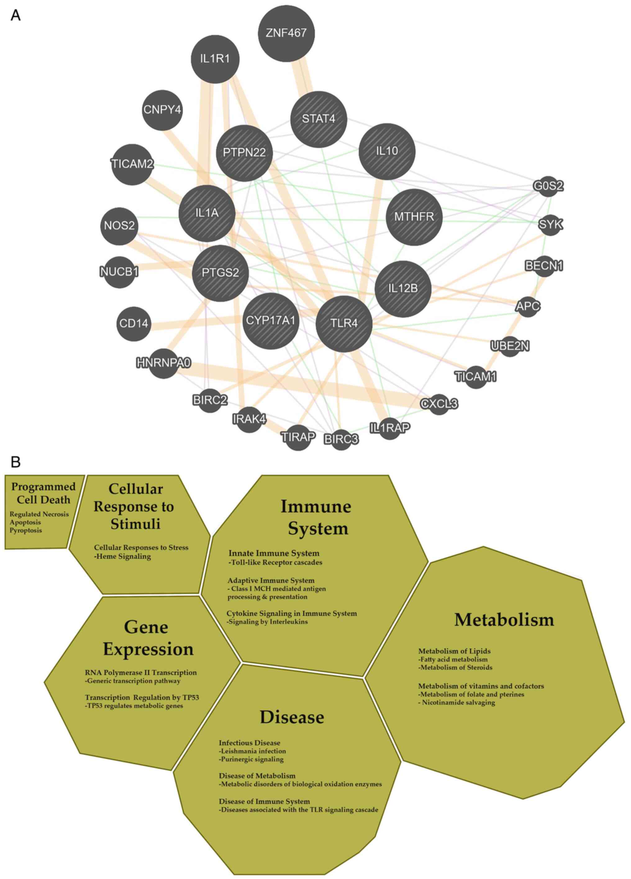
literature (Fig. 1).
The results of the literature research revealed that
IL-1A rs2856836 and rs3783550 (66,67),
IL-10 rs1800871 (68,69),
IL-12B rs17860508 (70,71),
protein tyrosine phosphatase non-receptor type 22 (PTPN22)
rs2476601 (16,72), cyclooxygenase (COX)-2 and
(PTGS)-2 rs20417 (73–75),
CYP17A1 rs743572 (76,77),
STAT4 rs7574865 (78,79),
Toll like receptor 4 (TLR4) rs4986791 (76,80)
and methylenetetrahydrofolate reductase (MTHFR) rs1801133
(81–83) SNPs are associated with both
diseases under investigation (Table
I). The common genetic targets and their corresponding genes
have been further analyzed towards estimating the candidate gene
regulatory network and the major categories of the biological
pathways using the GeneMANIA and the Reactome Knowledgebase
platforms, respectively (Fig. 1A and
B) (84,85).
 | Table I.An overview of the genetic
polymorphisms associated with the development of both endometriosis
and AS, as they have been confirmed by gene association studies
and/or genome-wide association studies. |
Table I.
An overview of the genetic
polymorphisms associated with the development of both endometriosis
and AS, as they have been confirmed by gene association studies
and/or genome-wide association studies.
| dbSNP ID | Endometriosis- and
AS-associated gene | Function | (Refs.) |
|---|
| rs2476601 | PTPN22 | Lymphoid-specific
phosphatase; downregulator of T-cell activation | (16,72) |
| rs7574865 | STAT4 | Transcription
factor involved in Th17 differentiation, monocyte activation and
IFN-γ production | (78,79) |
| rs2856836 | IL-1A | Pleiotropic
cytokine involved in inflammatory processes and hematopoiesis | (66,67) |
| rs3783550 |
|
|
|
| rs1800871 | IL-10 | Anti-inflammatory
cytokine; inhibitor of Th1 differentiation | (68,69) |
| rs17860508 | IL-12B | Cytokine acting on
T- and natural killer cells | (70,71) |
| rs1801133 | MTHFR | Key regulatory
enzyme in folate and homocysteine metabolism | (81–83) |
| rs20417 | COX-2
PTGS2 | Key enzyme required
for the conversion of arachidonic acid to prostaglandins | (73–75) |
| rs4986791 | TLR4 | Transmembrane
protein, member of the toll-like receptor family | (76,80) |
| rs743572 | CYP17A1 | Monooxygenase, a
member of the cytochrome P450 superfamily of enzymes | (76,77) |
Moreover, various microRNAs (miRs/miRNAs) have been
reported to be associated with both diseases, including
miR-20-a (22,86), miR-21 (22,87),
miR-196-b (22,88), miR-146-a (86,89,90),
miR-499a (86,90) and miR-214 (88,90).
Shared genetic polymorphisms and biological
mechanisms
Polymorphisms in inflammation and
autoimmune-related genes
The STAT proteins are members of a family of latent
cytosolic transcription factors, which are activated in response to
numerous cytokines, growth factors and hormones (91) (Fig.
1). STAT4, encoded by the STAT4 gene located at
2q32.2-q32.3, represents a key transcription factor that is
expressed in activated peripheral blood monocytes, dendritic cells
and macrophages in humans (92),
mediating IL-12 signaling that is critical for the development of
protective immunity in intracellular infection. Moreover, STAT4,
which is a member of the JAK/STAT pathway, appears to play a
central role in IFN signaling, which is involved in regulatory
networks related to the immunological processes that promote
chronic inflammation and tissue destruction (93). The STAT4 rs7574865 G/T
polymorphism, located within the third intron of the gene, has
previously been shown to be associated with an increased
susceptibility for AS and endometriosis (78,79).
The TT genotype of rs7574865 has been found to be significantly
more frequent in women with minimal or mild endometriosis than in
the controls (79); however, the
functional significance of this SNP as regards the pathogenesis of
endometriosis has not yet been determined. It has been hypothesized
that this SNP can affect the gene expression or mRNA splicing, thus
inducing strong Th1 and Th17 cytokine responses and IFN signaling
(79). Furthermore, the rs7574865
SNP has been shown to be significantly associated with an increased
AS susceptibility and radiographic severity, although its actual
functional consequence in AS remains to be identified (78). However, in a bioinformatics
analysis performed previously in the authors laboratory, the
polymorphic site was not found to disrupt any transcription factor
binding site, while preliminary experiments also conducted by the
authors demonstrated an impairment in STAT4 production, as well as
in STAT4 phosphorylation in the presence of the T allele (94). In particular, lower levels of the
produced protein and a disruption in the phosphorylation procedure
were observed, probably due to a defect in some stage of gene
expression or protein production when T1D were analyzed (94).
The lymphoid-specific phosphatase (LYP), which is
encoded by the PTPN22 gene, located on chromosome
1p13.3-13.1, is a potent downregulator of T-cell activation
(95) (Fig. 1). The PTPN22 rs2476601
(C1885T) SNP has been found to be associated with a number of
autoimmune diseases, including SLE (96), RA (97), T1D (98) and JIA (99), as well as with AS (72) and endometriosis (71). This SNP, which corresponds to the
functional A620W polymorphism, results in a variant that affects
the protein-protein interaction with tyrosine kinase Csk and codes
for a gain-of-function enzyme, which increases the inhibition of
T-cell receptor signaling. As a consequence, the authors have
previously demonstrated that it may have profound effects on the
function of the immune system (16) and, therefore, it may influence the
development of AS. Of note, subjects carrying the T-allele of
rs2476601 are considered to have increased numbers of auto-reactive
T-cells escaping negative selection, thus persisting in the
circulation, and thus being prone to autoimmunity (100). Various studies on patients with
endometriosis have revealed an aberrant function of cells involved
in the immune system (15,101), thus suggesting a critical role of
immunological factors in the development of the disease.
Accordingly, it has been suggested that alterations in
T-cell-mediated immunity may facilitate the implantation of
endometrial fragments or cells in ectopic regions (102). As recently reported by the
authors (16), patients with
endometriosis exhibit elevated levels of inflammatory cytokines,
decreased apoptosis and cell-mediated abnormalities, which are
indicative of autoimmune diseases.
COX is the rate-limiting enzyme for the synthesis of
prostaglandins and the names of its three isozymes are COX-1, COX-2
and COX-3 (103). COX-1 is COX-1
is constitutively expressed in the majority of cell types
regulating vascular homeostasis. COX-2 enzyme is encoded by the
COX-2 gene, located at chromosome 1q25.2-25.3 and consists
of 10 exons and nine introns (104). It is an inducible enzyme
exhibiting either a low or no expression in the majority of tissues
under normal physiological status (105); however, it can function upon
proper induction. Thus, it can be induced and produced in
inflammatory and injured sites, mediating the production of
prostaglandin that causes local inflammation and pain (106). As a consequence, this enzyme may
represent a key player in the inflammatory response (107). It has been previously reported
that the functionally important rs20417 SNP of COX-2 is
associated with AS (74) and
endometriosis (73). Previous
studies had demonstrated that the G allele of the rs20417 SNP
increases the expression of COX-2 when compared to C
(108), thus increasing the
formation of prostaglandins (75).
Notably, the excessive expression of COX-2 has been found to
be associated with the pathogenesis of endometriosis, since the
mRNA levels of COX-2 have been found to be 5-fold higher in
ectopic issue when compared to the eutopic endometrium, thus
leading to an abnormal production of prostaglandins (109). The −765G allele has been shown to
be associated with an increased risk of developing endometriosis of
stages II/III (75). Similarly, in
another study, the G allele of this SNP was shown to be associated
with an increased susceptibility of developing endometriosis of
stages III and IV, with the eutopic endometrial tissue of patients
presenting an elevated expression of COX-2 compared to the
controls (73). Of note, Liu et
al (74) found that allele G
of rs20417 was more frequent in patients AS, and was thus
associated with an increased risk of developing AS.
TLRs form a family of transmembrane protein
molecules, which are activated at the biochemical and structural
level by pathogen molecular patterns. TLRs activate the cellular
signaling pathways to induce immune-response genes, including
inflammatory cytokines (110).
TLRs have been extensively studied in endometriosis due to their
role in the regulation of the activation of immune and inflammatory
responses (111). TLR4 is an
important pathogen recognition receptor that mainly recognizes the
lipopolysaccharide (LPS) of Gram-negative bacteria, but also
structures from fungal and mycobacterial pathogens (112). TLR4 is essential for the innate
immune response and is composed of three domains. It is expressed
in macrophages, endometrial and endometriotic epithelial cells, and
alterations in its expression levels or function have been observed
thus far in the endometrium (111). The rs4986791 (T399I, C/T
transition) SNP of TLR4 has been investigated in a number of
studies focusing on inflammatory, infectious and autoimmune
diseases. For example, it has been found that the T allele of
rs4986791 is a risk factor for both endometriosis (80) and AS (76). The non-synonymous T399I variant is
located on the extracellular domain of TLR4, which is critical for
the LPS recognition and it has been reported that this polymorphism
results in a mild LPS-hyporesponsive phenotype (113). Although previous crystallography
analyses have demonstrated that the T399I change does not affect
the TLR4 structure (114), recent
approaches based on molecular dynamics have indicated that the
polymorphism may abrogate the stability of the hexamer complex,
thus leading to compromised TLR4 signaling (115). Of note, the genetic association
of rs4986791 SNP with AS could indicate the involvement of the
innate immune system in the progression of AS, considering that the
upregulation of TLR4 has been reported in this disease (116).
Polymorphisms in cytokine genes
The IL-1 gene cluster, a 360 kb region
containing nine genes, is located on the chromosomal 2q13 locus,
which represents a region harboring various inflammatory genes.
Notably, various modifications in the expression of the members of
this family have been shown to be associated with an increased
susceptibility for human diseases (117). The IL-1 cluster has also
emerged as a robust susceptibility locus for AS (66). IL-1A is a pro-inflammatory cytokine
that is involved in various immune responses, mainly produced by
activated macrophages, which augments the activation of T- and
B-lymphocytes and monocyte/macrophages, while it participates in
the induction of fibroblast proliferation (118). Both IL-1A rs2856836 (T/C)
and rs3783550 SNPs have been shown to be associated with
susceptibility to endometriosis and AS (38,66,67,119). rs2856836 is located at the
3′untranslated region (UTR) of the gene; however, limited data are
available regarding the effect of this SNP on the expression or
function of the IL-1A gene (119). However, increased expression
levels of IL-1A have been observed in the peritoneal fluid
of women with endometriosis, thus probably stimulating the activity
of the adhesion molecules and enhancing the implantation of
fragments of menstrual endometrium on peritoneal surfaces (120). The second IL-1A SNP,
rs3783550, which has been found to be associated with
endometriosis, has been found to overlap with binding sites of
three transcription factors, thus suggesting a possible regulatory
or functional role of this SNP (121). Taken together, genetic variants
of IL-1A may be involved in genetic susceptibility to
endometriosis and, in parallel, may induce inflammation in AS
through intracellular effects (122), thus being involved in the
initiation of disease.
IL-10 is a key immunomodulatory cytokine, produced
by Th2 cells and macrophages, with an ability to inhibit the
activation and function of macrophages, monocytes and T-cells
(123). The IL-10 gene
maps to chromosome 1q31-32 in humans, is composed of five exons and
four introns, and various genetic variants that are located in the
promoter region have been investigated thus far in relation to
human diseases. However, the functional promoter rs1800871
(−819C/T) SNP of the IL-10 gene has been found to be
associated with AS (69) and
endometriosis (68), considering
that it can influence both the mRNA, as well as the protein levels
of IL-10 (124). As
regards endometriosis, women with the TT genotype appear to have a
2-fold increased risk of developing endometriosis compared to those
carrying the C allele. Of note, the T allele has been shown to be
associated with lower IL-10 levels in comparison to the C allele in
women suffering from endometriosis; this finding may reflect a
mechanism that results in the upregulation of inflammation in the
peritoneal cavity (68). As
regards AS, the serum levels of IL-10 have been found to be
significantly higher in patients with AS than in healthy controls
(69). Again, the C allele of the
rs1800871 SNP has been found to be associated with an increased
risk of developing AS, with the CC genotype being associated with
higher IL-10 levels produced by peripheral blood mononuclear cells
in patients with AS as compared with the TT genotype (69).
IL-12 is involved in the differentiation of naive
T-cells into Th1 cells, regulates the activity of
antigen-presenting and NK cells (125) and induces IFN-γ production
(126). IL-12 is a heterodimeric
protein, consisting of two subunits, IL-12p35 and IL-12p40, encoded
by the IL-12A and IL-12B genes, respectively, which
are located on chromosome 5q31.1-33.1. Polymorphisms of the
IL-12B gene have been found to modulate the secretion, as
well as the activity of IL-12 (66,127). rs17860508 is a bi-allelic
promoter polymorphism of IL-12B, a potential functional
variant, and has been reported to be associated with the risk of
developing AS and endometriosis (70,71).
Notably, certain alleles may influence the release of specific
cytokines, thus affecting the regulation of the immune response.
The polymorphism under investigation, resulting from a 4-bp
micro-insertion (CTCT) combined with an AA/GC transition, has been
reported to be associated with alterations at the gene expression
levels, as well as mRNA stability in vitro (128). Of note, Zhao et al
(70) reported that cases carrying
the GC allele had a significantly increased risk of developing
ovarian endometriosis compared to individuals carrying the
CTCTAA/CTCTAA genotype, while the level of IL-12B mRNA
expression in the eutopic endometrial tissue of patients carrying
the GC/GC genotype was significantly higher than in those with the
CTCTAA/CTCTAA genotype. As regards the involvement of the
rs17860508 polymorphism in the development of AS, data have
indicated that it has an impact on the susceptibility for AS
through a modulation of the IL-12p40 levels, thus conferring a
variability in the immune response and contributing substantially
to the disease phenotype (71).
Polymorphisms in metabolism-related
genes
The CYP17A1 gene is located at chromosome
10q24.32 and codes for the CYP17A1 protein, which is one of the key
enzymes for glucocorticoid production and, most importantly, for
steroidogenic pathway producing estrogen (129). The T allele of CYP17A1
rs743572 SNP, which is located in the 5-UTR of the gene, 34 bp
upstream from the translation start site, has been found to be a
risk factor for AS and endometriosis (75,76).
Endometriosis is an estrogen-dependent disease and abnormal
expression levels of estrogen and consequent disruptions in
estrogen metabolism may be closely associated with the development
of endometriosis (130).
Mutations in the CYP17A1 gene have been shown to be
associated with gene expression, as well as with hormone levels;
Cong et al (76) reported
that the rs743572 TT genotype, as well as the T allele
significantly enhanced susceptibility to endometriosis. Notably,
cases carrying the TT genotype had the shortest menstrual cycle and
highest frequencies of intrauterine device use, although the SNP
under investigation had no significant influence on the severity
and specific characteristics of endometriosis (76).
The 5,10-methylenetetrahydrofolate reductase
(MTHFR) gene is located in chromosome 1p36.3, encoding a key
rate-limiting, regulatory enzyme in folate and homocysteine
metabolism, which catalyzes the conversion of
5,10-methylenetetrahydrofolate to 5-methyltetrahydrofolate
(131). The MTHFR
rs1801133 (Ala222Val) SNP, which is located in exon 4, has been
found to be significantly associated with both AS and endometriosis
(81–83). It has been previously suggested
that polymorphisms in the MTHFR gene may result in
alterations in gene expression levels and protein function, thus
leading to an abnormal metabolism of 5,10-methylenetetrahydrofolate
(82,83). Thus, the C677T missense mutation,
which has been characterized as a thermally unstable one, probably
influences the thermostability and activity of MHTFR protein, while
it has been also reported that the rs1801133 T allele results in
elevated plasma homocysteine levels, which, in combination with
endothelial injury and vascular inflammation, may result in the
development of cardiovascular disease, a well-known comorbidity of
endometriosis (13,132).
The role of miRNAs
The VEGFA-related miR-20-a, which has been
reported to be involved in the regulation of angiogenesis, has been
found to be expressed in decreased levels in the ovarian
endometrioma compared to the eutopic endometrium (133). Furthermore, it has been found
that the peritoneal fluid from women with endometriosis reduces the
expression of miR-20-a in primary stromal cell cultures from
patients (134). Moreover, Zhao
et al (70) revealed that
miR-20-a expression was upregulated in patients with AS.
It has previously reported in the literature that
miR-21 is associated with endometrial receptivity, which is
possibly associated with endometriosis (87), while it has also been found to be
upregulated in women with endometriosis (135). As regards AS, given than bone
loss is a feature of AS, this miRNA that has been implicated in the
activation of osteoclasts was investigated; a higher expression of
miR-21 was observed in patients with AS when compared with
the controls, with a parallel repression of its target programmed
cell death 4 (PDCD4) gene (136).
miR146-a has been shown to be significantly
associated with increased incidence rates of endometriosis, with
the G allele being a risk factor for the disease (137). In particular, miR-146a
expression has been found to be upregulated in ectopic vs. eutopic
endometrial tissues (138).
Furthermore, miR-146a expression has been found to be
upregulated in patients with AS, and to induce the expression of
TNF-α, IL-6 and IL-1B (139), while the frequency of the G
allele and GG genotype of the rs2910164 SNP of miR-146a was
shown to be significantly higher in patients with AS compared to
healthy controls (87,89). Previous research has demonstrated
the potential role of miR-196b in the pathogenesis of
endometriosis, by targeting proliferation, angiogenesis and
apoptosis (140). The
endometriosis-associated miR-214 has been found to regulate
major signaling pathways of critical genes participating in
inflammatory and cell proliferation pathways, including
angiogenesis, and may thus be implicated in the development of
endometriosis (70,141).
Conclusions and future perspectives
Based on the data presented in the present review,
a clear identification of a link between endometriosis and AS would
necessitate a radical shift in public health management. However,
although a relatively long list of genes identified to be involved
in the development either of endometriosis or AS is available, thus
far there appears little overlap between genetic factors affecting
susceptibility to both diseases. One remaining challenge in
immunogenetic research in endometriosis, as well as AS involves the
determination of the functional mechanisms underpinning the
identified genetic associations. Although GWAS have already
identified >100 SNPs associated with an increased susceptibility
for developing AS, there is currently a possible explanation for
only a small number of these genetic associations, thus minimizing
their potential for translation into therapeutic options. Thus,
apart from the efforts to analyze the biochemical pathways leading
to endometriosis, it remains an emerging public health issue of
reproductive-age women and its pathogenesis remains elusive. Of
note, endometriosis has been shown to be associated with various
autoimmune diseases, including AS, thus suggesting that this
condition may be considered a risk factor for AS, which requires
specific counseling and medical management. Considering that AS is
an autoimmune disease with a poorly-defined etiology (142), further elucidation of the
underlying pathophysiology may lead to the development of novel
therapeutics.
The treatment of AS has undergone substantial
changes over the past decades, thus leading to the establishment
and application of novel targeted therapies. The development of
efficient drugs targeting the IL-23/IL-17 axis represents a
successful paradigm of the value of genetics in the scientific
field of drug design (143).
Moreover, the targeting of TNF-α that is expressed in high levels
in patients with AS (144) is
another beneficial therapeutic option for a large percentage of
affected patients, while alternative promising therapeutic options
include JAK inhibitors that interrupt the JAK/STAT transduction of
IL-23/IL-17 and some other cytokine signals (145,146). Notably, it should be kept in mind
that the causal genes, which are regulated by the
disease-associated SNPs, are optimal targets for drug design,
considering the plausible importance of some well-analyzed
downstream molecular pathways (143). However, in spite of all this
progress being made in patient management, a significant proportion
of patients remain difficult to treat due to the heterogeneous
character of the disease at the clinical and/or the molecular
level. In this framework, polygenic risk scores involving the
testing of all known susceptibility variants may prove useful in
order to derive a personalized risk score for the disease or an
estimation for disease severity.
Immune and inflammatory dysfunctions have always
represented challenging therapeutic targets for endometriosis and
AS. Considering the lack of different studies focusing on the
co-occurrence of endometriosis with AS, further detailed studies
need to be conducted in an attempt to fully elucidate the
underlying pathophysiological mechanisms. Of note, it has been
suggested that clinicians should pay attention to the occurrence of
AS in patients with endometriosis and possible underlying
endometriosis in female patients with AS. As a consequence, a
suitable medication must be provided to these women.
In conclusion, the ultimate task may be the use of
all genetic information collected in order to better understand the
mechanisms leading to development of endometriosis and AS, in order
to design new mechanism-driven therapeutics, and to may in the
diagnosis and management of patients. The genetic profiling should
also play a role in the clinician's selection of therapy as part of
a precision medicine strategy towards the management of these
diseases.
Acknowledgements
Not applicable.
Funding
Funding: No funding was received.
Availability of data and materials
Not applicable.
Authors' contributions
MIZ, EE and GNG. designed the study and drafted the
manuscript. GNG, MIZ, DV, LP and DAS searched the literature for
related studies to be included in the review. GNG, EE, DV, LP and
MIZ analyzed and organized the data from the literature for
inclusion in the review. DAS, DV and LP critically revised the
manuscript. All authors have read and approved the final
manuscript. Data authentication is not applicable.
Ethics approval and consent to
participate
Not applicable.
Patient consent for publication
Not applicable.
Competing interests
DAS is the Managing Editor of the journal, but had
no personal involvement in the reviewing process, or any influence
in terms of adjudicating on the final decision, for this article.
All the other authors declare that they have no competing
interests.
References
|
1
|
Braun J and Sieper J: Ankylosing
spondylitis. Lancet. 369:1379–1390. 2007. View Article : Google Scholar : PubMed/NCBI
|
|
2
|
Rudwaleit M, van der Heijde D, Landewe R,
Listing J, Akkoc N, Brandt J, Braun J, Chou CT, Collantes-Estevez
E, Dougados M, et al: The development of Assessment of
SpondyloArthritis international Society classification criteria for
axial spondyloarthritis part II): Validation and final selection.
Ann Rheum Dis. 68:777–783. 2009. View Article : Google Scholar : PubMed/NCBI
|
|
3
|
Wanders A, Landewe R, Dougados M, Mielants
H, van der Linden S and van der Heijde D: Association between
radiographic damage of the spine and spinal mobility for individual
patients with ankylosing spondylitis: Can assessment of spinal
mobility be a proxy for radiographic evaluation? Ann Rheum Dis.
64:988–994. 2005. View Article : Google Scholar : PubMed/NCBI
|
|
4
|
Bakland G, Nossent HC and Gran JT:
Incidence and prevalence of ankylosing spondylitis in Northern
Norway. Arthritis Rheum. 53:850–855. 2005. View Article : Google Scholar : PubMed/NCBI
|
|
5
|
Zhu W, He X, Cheng K, Zhang L, Chen D,
Wang X, Qiu G, Cao X and Weng X: Ankylosing spondylitis: Etiology,
pathogenesis, and treatments. Bone Res. 7:222019. View Article : Google Scholar : PubMed/NCBI
|
|
6
|
Berkley KJ, Rapkin AJ and Papka RE: The
pains of endometriosis. Science. 308:1587–1589. 2005. View Article : Google Scholar : PubMed/NCBI
|
|
7
|
Maddern J, Grundy L, Castro J and Brierley
SM: Pain in endometriosis. Front Cell Neurosci. 14:5908232020.
View Article : Google Scholar : PubMed/NCBI
|
|
8
|
Symons LK, Miller JE, Kay VR, Marks RM,
Liblik K, Koti M and Tayade C: The immunopathophysiology of
endometriosis. Trends Mol Med. 24:748–762. 2018. View Article : Google Scholar : PubMed/NCBI
|
|
9
|
Vigano P, Parazzini F, Somigliana E and
Vercellini P: Endometriosis: Epidemiology and aetiological factors.
Best Pract Res Clin Obstet Gynaecol. 18:177–200. 2004. View Article : Google Scholar : PubMed/NCBI
|
|
10
|
Papageorgiou L, Andreou A, Zervou M,
Vlachakis D, Goulielmos GN and Eliopoulos E: A global population
genomic analysis shows novel insights into the genetic
characteristics of endometriosis. World Acad Sci J. 5:122023.
View Article : Google Scholar
|
|
11
|
Vercellini P, Frontino G, Pietropaolo G,
Gattei U, Daguati R and Crosignani PG: Deep endometriosis:
Definition, pathogenesis, and clinical management. J Am Assoc
Gynecol Laparosc. 11:153–161. 2004. View Article : Google Scholar : PubMed/NCBI
|
|
12
|
Kvaskoff M, Mu F, Terry KL, Harris HR,
Poole EM, Farland L and Missmer SA: Endometriosis: A high-risk
population for major chronic diseases? Hum Reprod Update.
21:500–516. 2015. View Article : Google Scholar : PubMed/NCBI
|
|
13
|
Rafi U, Ahmad S, Bokhari SS, Iqbal MA, Zia
A, Khan MA and Roohi N: Association of inflammatory
markers/cytokines with cardiovascular risk manifestation in
patients with endometriosis. Mediators Inflamm. 2021:34255602021.
View Article : Google Scholar : PubMed/NCBI
|
|
14
|
Vazgiourakis VM, Zervou MI, Papageorgiou
L, Chaniotis D, Spandidos DA, Vlachakis D, Eliopoulos E and
Goulielmos GN: Association of endometriosis with cardiovascular
disease: Genetic aspects (Review). Int J Mol Med. 51:292023.
View Article : Google Scholar : PubMed/NCBI
|
|
15
|
Shigesi N, Kvaskoff M, Kirtley S, Feng Q,
Fang H, Knight JC, Missmer SA, Rahmioglu N, Zondervan KT and Becker
CM: The association between endometriosis and autoimmune diseases:
A systematic review and meta-analysis. Hum Reprod Update.
25:486–503. 2019. View Article : Google Scholar : PubMed/NCBI
|
|
16
|
Zervou MI, Vlachakis D, Papageorgiou L,
Eliopoulos E and Goulielmos GN: Increased risk of rheumatoid
arthritis in patients with endometriosis: Genetic aspects.
Rheumatology (Oxford). 61:4252–4262. 2022. View Article : Google Scholar : PubMed/NCBI
|
|
17
|
Yin Z, Low HY, Chen BS, Huang KS, Zhang Y,
Wang YH, Ye Z and Wei JC: Risk of ankylosing spondylitis in
patients with endometriosis: A population-based retrospective
cohort study. Front Immunol. 13:8779422022. View Article : Google Scholar : PubMed/NCBI
|
|
18
|
Reveille JD: Genetics of
spondyloarthritis-beyond the MHC. Nat Rev Rheumatol. 8:296–304.
2012. View Article : Google Scholar : PubMed/NCBI
|
|
19
|
Bowness P: Hla-B27. Annu Rev Immunol.
33:29–48. 2015. View Article : Google Scholar : PubMed/NCBI
|
|
20
|
International Genetics of Ankylosing
Spondylitis Consortium (IGAS), . Cortes A, Hadler J, Pointon JP,
Robinson PC, Karaderi T, Leo P, Cremin K, Pryce K, Harris J, et al:
Identification of multiple risk variants for ankylosing spondylitis
through high-density genotyping of immune-related loci. Nat Genet.
45:730–738. 2013. View Article : Google Scholar : PubMed/NCBI
|
|
21
|
Cortes A, Pulit SL, Leo PJ, Pointon JJ,
Robinson PC, Weisman MH, Ward M, Gensler LS, Zhou X, Garchon HJ, et
al: Major histocompatibility complex associations of ankylosing
spondylitis are complex and involve further epistasis with ERAP1.
Nat Commun. 6:71462015. View Article : Google Scholar : PubMed/NCBI
|
|
22
|
Motta F, Carena MC, Selmi C and Vecellio
M: MicroRNAs in ankylosing spondylitis: Function, potential and
challenges. J Transl Autoimmun. 3:1000502020. View Article : Google Scholar : PubMed/NCBI
|
|
23
|
Jaakkola E, Crane AM, Laiho K, Herzberg I,
Sims AM, Bradbury L, Calin A, Brophy S, Kauppi M, Kaarela K, et al:
The effect of transforming growth factor beta1 gene polymorphisms
in ankylosing spondylitis. Rheumatology (Oxford). 43:32–38. 2004.
View Article : Google Scholar : PubMed/NCBI
|
|
24
|
Australo-Anglo-American Spondyloarthritis
Consortium (TASC), . Reveille JD, Sims AM, Danoy P, Evans DM, Leo
P, Pointon JJ, Jin R, Zhou X, Bradbury LA, et al: Genome-wide
association study of ankylosing spondylitis identifies non-MHC
susceptibility loci. Nat Genet. 42:123–127. 2010. View Article : Google Scholar : PubMed/NCBI
|
|
25
|
Robinson PC, Claushuis TA, Cortes A,
Martin TM, Evans DM, Leo P, Mukhopadhyay P, Bradbury LA, Cremin K,
Harris J, et al: Genetic dissection of acute anterior uveitis
reveals similarities and differences in associations observed with
ankylosing spondylitis. Arthritis Rheumatol. 67:140–151. 2015.
View Article : Google Scholar : PubMed/NCBI
|
|
26
|
Choi CB, Kim TH, Jun JB, Lee HS, Shim SC,
Lee B, Pope A, Uddin M, Rahman P and Inman RD: ARTS1 polymorphisms
are associated with ankylosing spondylitis in Koreans. Ann Rheum
Dis. 69:582–584. 2010. View Article : Google Scholar : PubMed/NCBI
|
|
27
|
Ellinghaus D, Jostins L, Spain SL, Cortes
A, Bethune J, Han B, Park YR, Raychaudhuri S, Pouget JG, Hübenthal
M, et al: Analysis of five chronic inflammatory diseases identifies
27 new associations and highlights disease-specific patterns at
shared loci. Nat Genet. 48:510–518. 2016. View Article : Google Scholar : PubMed/NCBI
|
|
28
|
Hanson A and Brown MA: Genetics and the
causes of ankylosing spondylitis. Rheum Dis Clin North Am.
43:401–414. 2017. View Article : Google Scholar : PubMed/NCBI
|
|
29
|
Treloar SA, O'Connor DT, O'Connor VM and
Martin NG: Genetic influences on endometriosis in an Australian
twin sample. simplesueT@qimr.edu.au. Fertil
Steril. 71:701–710. 1999. View Article : Google Scholar : PubMed/NCBI
|
|
30
|
Saha R, Pettersson HJ, Svedberg P,
Olovsson M, Bergqvist A, Marions L, Tornvall P and Kuja-Halkola R:
Heritability of endometriosis. Fertil Steril. 104:947–952. 2015.
View Article : Google Scholar : PubMed/NCBI
|
|
31
|
Falconer H, D'Hooghe T and Fried G:
Endometriosis and genetic polymorphisms. Obstet Gynecol Surv.
62:616–628. 2007. View Article : Google Scholar : PubMed/NCBI
|
|
32
|
Papageorgiou L, Zervou MI, Vlachakis D,
Matalliotakis M, Matalliotakis I, Spandidos DA, Goulielmos GN and
Eliopoulos E: Demetra Application: An integrated genotype analysis
web server for clinical genomics in endometriosis. Int J Mol Med.
47:1152021. View Article : Google Scholar : PubMed/NCBI
|
|
33
|
Nyholt DR, Low SK, Anderson CA, Painter
JN, Uno S, Morris AP, MacGregor S, Gordon SD, Henders AK, Martin
NG, et al: Genome-wide association meta-analysis identifies new
endometriosis risk loci. Nat Genet. 44:1355–1359. 2012. View Article : Google Scholar : PubMed/NCBI
|
|
34
|
Rahmioglu N, Nyholt DR, Morris AP, Missmer
SA, Montgomery GW and Zondervan KT: Genetic variants underlying
risk of endometriosis: Insights from meta-analysis of eight
genome-wide association and replication datasets. Hum Reprod
Update. 20:702–716. 2014. View Article : Google Scholar : PubMed/NCBI
|
|
35
|
Watanabe Y, Suzuki R, Kinoshita M and
Hirota M: Inguinal endometriosis with a disappearing mass
preoperatively: A case report. Int J Surg Case Rep. 91:1067812022.
View Article : Google Scholar : PubMed/NCBI
|
|
36
|
Kobayashi H, Imanaka S, Nakamura H and
Tsuji A: Understanding the role of epigenomic, genomic and genetic
alterations in the development of endometriosis (Review). Mol Med
Rep. 9:1483–1505. 2014. View Article : Google Scholar : PubMed/NCBI
|
|
37
|
Falconer H, Sundqvist J, Xu H, Vodolazkaia
A, Fassbender A, Kyama C, Bokor A and D'Hooghe TM: Analysis of
common variations in tumor-suppressor genes on chr1p36 among
Caucasian women with endometriosis. Gynecol Oncol. 127:398–402.
2012. View Article : Google Scholar : PubMed/NCBI
|
|
38
|
Sapkota Y, Steinthorsdottir V, Morris AP,
Fassbender A, Rahmioglu N, De Vivo I, Buring JE, Zhang F, Edwards
TL, Jones S, et al: Meta-analysis identifies five novel loci
associated with endometriosis highlighting key genes involved in
hormone metabolism. Nat Commun. 8:155392017. View Article : Google Scholar : PubMed/NCBI
|
|
39
|
Rahmioglu N, Mortlock S, Ghiasi M, Møller
PL, Stefansdottir L, Galarneau G, Turman C, Danning R, Law MH,
Sapkota Y and Christofidou P: The genetic basis of endometriosis
and comorbidity with other pain and inflammatory conditions. Nat
Genet. 55:423–436. 2023. View Article : Google Scholar : PubMed/NCBI
|
|
40
|
Albertsen HM, Matalliotaki C,
Matalliotakis M, Zervou MI, Matalliotakis I, Spandidos DA, Chettier
R, Ward K and Goulielmos GN: Whole exome sequencing identifies
hemizygous deletions in the UGT2B28 and USP17L2 genes in a
three-generation family with endometriosis. Mol Med Rep.
19:1716–1720. 2019.PubMed/NCBI
|
|
41
|
Matarese G, De Placido G, Nikas Y and
Alviggi C: Pathogenesis of endometriosis: Natural immunity
dysfunction or autoimmune disease? Trends Mol Med. 9:223–228. 2003.
View Article : Google Scholar : PubMed/NCBI
|
|
42
|
Wells M: Recent advances in endometriosis
with emphasis on pathogenesis, molecular pathology, and neoplastic
transformation. Int J Gynecol Pathol. 23:316–320. 2004. View Article : Google Scholar : PubMed/NCBI
|
|
43
|
Fernandez-Shaw S, Hicks BR, Yudkin PL,
Kennedy S, Barlow DH and Starkey PM: Anti-endometrial and
anti-endothelial auto-antibodies in women with endometriosis. Hum
Reprod. 8:310–315. 1993. View Article : Google Scholar : PubMed/NCBI
|
|
44
|
Sinaii N, Cleary SD, Ballweg ML, Nieman LK
and Stratton P: High rates of autoimmune and endocrine disorders,
fibromyalgia, chronic fatigue syndrome and atopic diseases among
women with endometriosis: A survey analysis. Hum Reprod.
17:2715–2724. 2002. View Article : Google Scholar : PubMed/NCBI
|
|
45
|
Monsanto SP, Edwards AK, Zhou J,
Nagarkatti P, Nagarkatti M, Young SL, Lessey BA and Tayade C:
Surgical removal of endometriotic lesions alters local and systemic
proinflammatory cytokines in endometriosis patients. Fertil Steril.
105:968–977.e5. 2016. View Article : Google Scholar : PubMed/NCBI
|
|
46
|
Graziottin A, Skaper SD and Fusco M: Mast
cells in chronic inflammation, pelvic pain and depression in women.
Gynecol Endocrinol. 30:472–477. 2014. View Article : Google Scholar : PubMed/NCBI
|
|
47
|
Grandi G, Mueller MD, Papadia A, Kocbek V,
Bersinger NA, Petraglia F, Cagnacci A and McKinnon B: Inflammation
influences steroid hormone receptors targeted by progestins in
endometrial stromal cells from women with endometriosis. J Reprod
Immunol. 117:30–38. 2016. View Article : Google Scholar : PubMed/NCBI
|
|
48
|
Bedaiwy MA and Falcone T: Peritoneal fluid
environment in endometriosis. Clinicopathological implications.
Minerva Ginecol. 55:333–345. 2003.PubMed/NCBI
|
|
49
|
Mahnke JL, Dawood MY and Huang JC:
Vascular endothelial growth factor and interleukin-6 in peritoneal
fluid of women with endometriosis. Fertil Steril. 73:166–170. 2000.
View Article : Google Scholar : PubMed/NCBI
|
|
50
|
Rana N, Braun DP, House R, Gebel H, Rotman
C and Dmowski WP: Basal and stimulated secretion of cytokines by
peritoneal macrophages in women with endometriosis. Fertil Steril.
65:925–930. 1996. View Article : Google Scholar : PubMed/NCBI
|
|
51
|
Jiang J, Yu K, Jiang Z and Xue M: IL-37
affects the occurrence and development of endometriosis by
regulating the biological behavior of endometrial stromal cells
through multiple signaling pathways. Biol Chem. 399:1325–1337.
2018. View Article : Google Scholar : PubMed/NCBI
|
|
52
|
Lories RJ and Haroon N: Evolving concepts
of new bone formation in axial spondyloarthritis: Insights from
animal models and human studies. Best Pract Res Clin Rheumatol.
31:877–886. 2017. View Article : Google Scholar : PubMed/NCBI
|
|
53
|
Jethwa H and Bowness P: The interleukin
(IL)-23/IL-17 axis in ankylosing spondylitis: New advances and
potentials for treatment. Clin Exp Immunol. 183:30–36. 2016.
View Article : Google Scholar : PubMed/NCBI
|
|
54
|
Maas JW, Le Noble FA, Dunselman GA, de
Goeij AF, Struyker Boudier HA and Evers JL: The chick embryo
chorioallantoic membrane as a model to investigate the angiogenic
properties of human endometrium. Gynecol Obstet Invest. 48:108–112.
1999. View Article : Google Scholar : PubMed/NCBI
|
|
55
|
Ulukus M and Arici A: Immunology of
endometriosis. Minerva Ginecol. 57:237–248. 2005.PubMed/NCBI
|
|
56
|
Griffioen AW and Molema G: Angiogenesis:
Potentials for pharmacologic intervention in the treatment of
cancer, cardiovascular diseases, and chronic inflammation.
Pharmacol Rev. 52:237–268. 2000.PubMed/NCBI
|
|
57
|
May K and Becker CM: Endometriosis and
angiogenesis. Minerva Ginecol. 60:245–254. 2008.PubMed/NCBI
|
|
58
|
McLaren J: Vascular endothelial growth
factor and endometriotic angiogenesis. Hum Reprod Update. 6:45–55.
2000. View Article : Google Scholar : PubMed/NCBI
|
|
59
|
Donnez J, Smoes P, Gillerot S,
Casanas-Roux F and Nisolle M: Vascular endothelial growth factor
(VEGF) in endometriosis. Hum Reprod. 13:1686–1690. 1998. View Article : Google Scholar : PubMed/NCBI
|
|
60
|
Malhotra N, Karmakar D, Tripathi V, Luthra
K and Kumar S: Correlation of angiogenic cytokines-leptin and IL-8
in stage, type and presentation of endometriosis. Gynecol
Endocrinol. 28:224–227. 2012. View Article : Google Scholar : PubMed/NCBI
|
|
61
|
Koch AE: Review: Angiogenesis:
Implications for rheumatoid arthritis. Arthritis Rheum. 41:951–962.
1998. View Article : Google Scholar : PubMed/NCBI
|
|
62
|
Brown MD, Hudlicka O, Makki RF and Weiss
JB: Low-molecular-mass endothelial cell-stimulating angiogenic
factor in relation to capillary growth induced in rat skeletal
muscle by low-frequency electrical stimulation. Int J Microcirc
Clin Exp. 15:111–116. 1995. View Article : Google Scholar : PubMed/NCBI
|
|
63
|
Ma Q, Reiter RJ and Chen Y: Role of
melatonin in controlling angiogenesis under physiological and
pathological conditions. Angiogenesis. 23:91–104. 2020. View Article : Google Scholar : PubMed/NCBI
|
|
64
|
Drouart M, Saas P, Billot M, Cedoz JP,
Tiberghien P, Wendling D and Toussirot E: High serum vascular
endothelial growth factor correlates with disease activity of
spondylarthropathies. Clin Exp Immunol. 132:158–162. 2003.
View Article : Google Scholar : PubMed/NCBI
|
|
65
|
Taylor HS, Kotlyar AM and Flores VA:
Endometriosis is a chronic systemic disease: Clinical challenges
and novel innovations. Lancet. 397:839–852. 2021. View Article : Google Scholar : PubMed/NCBI
|
|
66
|
Li L, Shi B, Zheng W, Xing W, Zhao Y, Li
F, Xin D, Jin T, Zhu Y and Yang X: Association of IL-1A and IL-1B
polymorphisms with ankylosing spondylitis among the Chinese Han
population: A case-control study. Oncotarget. 8:28278–28284. 2017.
View Article : Google Scholar : PubMed/NCBI
|
|
67
|
Badie A, Saliminejad K, Salahshourifar I
and Khorram Khorshid HR: Interleukin 1 alpha (IL1A) polymorphisms
and risk of endometriosis in Iranian population: A case-control
study. Gynecol Endocrinol. 36:135–138. 2020. View Article : Google Scholar : PubMed/NCBI
|
|
68
|
Juo SH, Wu R, Lin CS, Wu MT, Lee JN and
Tsai EM: A functional promoter polymorphism in interleukin-10 gene
influences susceptibility to endometriosis. Fertil Steril.
92:1228–1233. 2009. View Article : Google Scholar : PubMed/NCBI
|
|
69
|
Lv C, Wang Y, Wang J, Zhang H, Xu H and
Zhang D: Association of Interleukin-10 gene polymorphisms with
ankylosing spondylitis. Clin Invest Med. 34:E3702011. View Article : Google Scholar : PubMed/NCBI
|
|
70
|
Zhao W, Li Y, Zhao J and Kang S: A
functional promoter polymorphism in interleukin 12B gene is
associated with an increased risk of ovarian endometriosis. Gene.
666:27–31. 2018. View Article : Google Scholar : PubMed/NCBI
|
|
71
|
Ivanova M, Manolova I, Miteva L, Gancheva
R, Stoilov R and Stanilova S: Genetic variations in the IL-12B gene
in association with IL-23 and IL-12p40 serum levels in ankylosing
spondylitis. Rheumatol Int. 39:111–119. 2019. View Article : Google Scholar : PubMed/NCBI
|
|
72
|
Sode J, Bank S, Vogel U, Andersen PS,
Sørensen SB, Bojesen AB, Andersen MR, Brandslund I, Dessau RB,
Hoffmann HJ, et al: Genetically determined high activities of the
TNF-alpha, IL23/IL17, and NFkB pathways were associated with
increased risk of ankylosing spondylitis. BMC Med Genet.
19:1652018. View Article : Google Scholar : PubMed/NCBI
|
|
73
|
Kim HY, Cho S, Choi YS, Yang HI, Lee KE,
Seo SK and Lee BS: Cyclooxygenase-2 (COX-2) gene-765G/C
polymorphism and advanced-stage endometriosis in Korean women. Am J
Reprod Immunol. 68:238–243. 2012. View Article : Google Scholar : PubMed/NCBI
|
|
74
|
Liu H, Wang B and Zhang Q: Connection of
COX-2 variants to ankylosing spondylitis susceptibility. J Cell
Physiol. Feb 9–2021.(Epub ahead of print). doi:
10.1002/jcp.30302.
|
|
75
|
Cavalcanti V, Ponce TG, Mafra FA, André
GM, Christofolini DM, Barbosa CP and Bianco B: Evaluation of the
frequency of G-765C polymorphism in the promoter region of the
COX-2 gene and its correlation with the expression of this gene in
the endometrium of women with endometriosis. Arch Gynecol Obstetr.
293:109–115. 2016. View Article : Google Scholar : PubMed/NCBI
|
|
76
|
Schiotis R, Bartolome N, Sánchez A,
Szczypiorska M, Sanz J, Cuende E, Collantes Estevez E, Martínez A,
Tejedor D, Artieda M, et al: Both baseline clinical factors and
genetic polymorphisms influence the development of severe
functional status in ankylosing spondylitis. PLoS One.
7:e434282012. View Article : Google Scholar : PubMed/NCBI
|
|
77
|
Cong L, Fu Q and Gao T: CYP17A1 rs743572
polymorphism might contribute to endometriosis susceptibility:
Evidences from a case-control study. Medicine (Baltimore).
97:e114152018. View Article : Google Scholar : PubMed/NCBI
|
|
78
|
Liu Z, Zhang P and Dong J: Genetic
variants of STAT4 are associated with ankylosing spondylitis
susceptibility and severity in a Chinese Han population. Int J Clin
Exp Med. 7:5877–5881. 2014.PubMed/NCBI
|
|
79
|
Bianco B, Fernandes RFM, Trevisan CM,
Christofolini DM, Sanz-Lomana CM, de Bernabe JV and Barbosa CP:
Influence of STAT4 gene polymorphisms in the pathogenesis of
endometriosis. Ann Hum Genet. 83:249–255. 2019. View Article : Google Scholar : PubMed/NCBI
|
|
80
|
Marchionni E, Porpora MG, Megiorni F,
Piacenti I, Giovannetti A, Marchese C, Benedetti Panici P and
Pizzuti A: TLR4 T399I polymorphism and endometriosis in a cohort of
Italian Women. Diagnostics (Basel). 10:2552020. View Article : Google Scholar : PubMed/NCBI
|
|
81
|
Szczepanska M, Mostowska A, Wirstlein P,
Lianeri M, Marianowski P, Skrzypczak J and Jagodziński PP:
Polymorphic variants of folate and choline metabolism genes and the
risk of endometriosis-associated infertility. Eur J Obstet Gynecol
Reprod Biol. 157:67–72. 2011. View Article : Google Scholar : PubMed/NCBI
|
|
82
|
Li HH, Li XQ, Sai LT, Cui Y, Xu JH, Zhou
C, Zheng J, Li XF, Liu HX and Zhao YJ: Association of homocysteine
with ankylosing spondylitis: A systematic review and meta-analysis.
Adv Rheumatol. 61:172021. View Article : Google Scholar : PubMed/NCBI
|
|
83
|
Lu M, Peng K, Song L, Luo L, Liang P and
Liang Y: Association between genetic polymorphisms in
methylenetetrahydrofolate reductase and risk of autoimmune
diseases: A systematic review and meta-analysis. Dis Markers.
2022:45681452022. View Article : Google Scholar : PubMed/NCBI
|
|
84
|
Giacomini E, Minetto S, Li Piani L,
Pagliardini L, Somigliana E and Vigano P: Genetics and inflammation
in endometriosis: Improving knowledge for development of new
pharmacological strategies. Int J Mol Sci. 22:90332021. View Article : Google Scholar : PubMed/NCBI
|
|
85
|
Franz M, Rodriguez H, Lopes C, Zuberi K,
Montojo J, Bader GD and Morris Q: GeneMANIA update 2018. Nucleic
Acids Res. 46:W60–W64. 2018. View Article : Google Scholar : PubMed/NCBI
|
|
86
|
Zubrzycka A, Zubrzycki M, Perdas E and
Zubrzycka M: Genetic, epigenetic, and steroidogenic modulation
mechanisms in endometriosis. J Clin Med. 9:13092020. View Article : Google Scholar : PubMed/NCBI
|
|
87
|
Xu X, Li Z, Liu J, Yu S and Wei Z:
MicroRNA expression profiling in endometriosis-associated
infertility and its relationship with endometrial receptivity
evaluated by ultrasound. J Xray Sci Technol. 25:523–532.
2017.PubMed/NCBI
|
|
88
|
Chang CY, Lai MT, Chen Y, Yang CW, Chang
HW, Lu CC, Chen CM, Chan C, Chung C, Tseng CC, et al: Up-regulation
of ribosome biogenesis by MIR196A2 genetic variation promotes
endometriosis development and progression. Oncotarget.
7:76713–76725. 2016. View Article : Google Scholar : PubMed/NCBI
|
|
89
|
Xu HY, Wang ZY, Chen JF, Wang TY, Wang LL,
Tang LL, Lin XY, Zhang CW and Chen BC: Association between
ankylosing spondylitis and the miR-146a and miR-499 polymorphisms.
PLoS One. 10:e01220552015. View Article : Google Scholar : PubMed/NCBI
|
|
90
|
Li Z, Wong SH, Shen J, Chan MTV and Wu
WKK: The Role of MicroRNAS in ankylosing spondylitis. Medicine
(Baltimore). 95:e33252016. View Article : Google Scholar : PubMed/NCBI
|
|
91
|
Leonard WJ and O'Shea JJ: Jaks and STATs:
Biological implications. Annu Rev Immunol. 16:293–322. 1998.
View Article : Google Scholar : PubMed/NCBI
|
|
92
|
Frucht DM, Aringer M, Galon J, Danning C,
Brown M, Fan S, Centola M, Wu CY, Yamada N, El Gabalawy H and
O'Shea JJ: Stat4 is expressed in activated peripheral blood
monocytes, dendritic cells, and macrophages at sites of
Th1-mediated inflammation. J Immunol. 164:4659–4664. 2000.
View Article : Google Scholar : PubMed/NCBI
|
|
93
|
Wun CM, Piao Z, Hong KT, Choi JY, Hong CR,
Park JD, Park KD, Shin HY and Kang HJ: Effect of donor STAT4
polymorphism rs7574865 on clinical outcomes of pediatric acute
leukemia patients after hematopoietic stem cell transplant. Int
Immunopharmacol. 43:62–69. 2017. View Article : Google Scholar : PubMed/NCBI
|
|
94
|
Kofteridis D, Krasoudaki E, Kavousanaki M,
Zervou MI, Panierakis C, Boumpas DT and Goulielmos GN: STAT4 is not
associated with type 2 diabetes in the genetically homogeneous
population of Crete. Genet Test Mol Biomarkers. 13:281–284. 2009.
View Article : Google Scholar : PubMed/NCBI
|
|
95
|
Wu J, Katrekar A, Honigberg LA, Smith AM,
Conn MT, Tang J, Jeffery D, Mortara K, Sampang J, Williams SR, et
al: Identification of substrates of human protein-tyrosine
phosphatase PTPN22. J Biol Chem. 281:11002–11010. 2006. View Article : Google Scholar : PubMed/NCBI
|
|
96
|
Eliopoulos E, Zervou MI, Andreou A,
Dimopoulou K, Cosmidis N, Voloudakis G, Mysirlaki H, Vazgiourakis
V, Sidiropoulos P, Niewold TB, et al: Association of the PTPN22
R620W polymorphism with increased risk for SLE in the genetically
homogeneous population of Crete. Lupus. 20:501–506. 2011.
View Article : Google Scholar : PubMed/NCBI
|
|
97
|
Begovich AB, Carlton VE, Honigberg LA,
Schrodi SJ, Chokkalingam AP, Alexander HC, Ardlie KG, Huang Q,
Smith AM, Spoerke JM, et al: A missense single-nucleotide
polymorphism in a gene encoding a protein tyrosine phosphatase
(PTPN22) is associated with rheumatoid arthritis. Am J Hum Genet.
75:330–337. 2004. View
Article : Google Scholar : PubMed/NCBI
|
|
98
|
Bottini N, Musumeci L, Alonso A, Rahmouni
S, Nika K, Rostamkhani M, MacMurray J, Meloni GF, Lucarelli P,
Pellecchia M, et al: A functional variant of lymphoid tyrosine
phosphatase is associated with type I diabetes. Nat Genet.
36:337–338. 2004. View
Article : Google Scholar : PubMed/NCBI
|
|
99
|
Goulielmos GN, Chiaroni-Clarke RC,
Dimopoulou DG, Zervou MI, Trachana M, Pratsidou-Gertsi P,
Garyfallos A and Ellis JA: Association of juvenile idiopathic
arthritis with PTPN22 rs2476601 is specific to females in a Greek
population. Pediatr Rheumatol Online J. 14:252016. View Article : Google Scholar : PubMed/NCBI
|
|
100
|
Bottini N, Vang T, Cucca F and Mustelin T:
Role of PTPN22 in type 1 diabetes and other autoimmune diseases.
Semin Immunol. 18:207–213. 2006. View Article : Google Scholar : PubMed/NCBI
|
|
101
|
Zondervan KT, Becker CM, Koga K, Missmer
SA, Taylor RN and Vigano P: Endometriosis. Nat Rev Dis Primers.
4:92018. View Article : Google Scholar : PubMed/NCBI
|
|
102
|
Dmowski WP, Gebel HM and Braun DP: The
role of cell-mediated immunity in pathogenesis of endometriosis.
Acta Obstet Gynecol Scand Suppl. 159:7–14. 1994.PubMed/NCBI
|
|
103
|
Ozatik FY, Kaygisiz B and Erol K: The role
of cyclooxygenase enzymes in the effects of losartan and lisinopril
on the contractions of rat thoracic aorta. Eur J Med. 49:16–21.
2017.
|
|
104
|
Wang L, Jiang N, Lin QR, Qin CH, Hu YJ and
Yu B: Cyclooxygenase-2 (COX-2) polymorphism rs689466 may contribute
to the increased susceptibility to post-traumatic osteomyelitis in
Chinese population. Infect Dis (Lond). 49:817–823. 2017. View Article : Google Scholar : PubMed/NCBI
|
|
105
|
Eslami SM, Ghasemi M, Bahremand T, Momeny
M, Gholami M, Sharifzadeh M and Dehpour AR: Involvement of
nitrergic system in anticonvulsant effect of zolpidem in
lithium-pilocarpine induced status epilepticus: Evaluation of iNOS
and COX-2 genes expression. Eur J Pharmacol. 815:454–461. 2017.
View Article : Google Scholar : PubMed/NCBI
|
|
106
|
Tsai MH, Hsu LF, Lee CW, Chiang YC, Lee
MH, How JM, Wu CM, Huang CL and Lee IT: Resveratrol inhibits urban
particulate matter-induced COX-2/PGE2 release in human
fibroblast-like synoviocytes via the inhibition of activation of
NADPH oxidase/ROS/NF-κB. Int J Biochem Cell Biol. 88:113–123. 2017.
View Article : Google Scholar : PubMed/NCBI
|
|
107
|
Yin J, Xia W, Li Y, Guo C, Zhang Y, Huang
S, Jia Z and Zhang A: COX-2 mediates PM2.5-induced apoptosis and
inflammation in vascular endothelial cells. Am J Transl Res.
9:3967–3976. 2017.PubMed/NCBI
|
|
108
|
Oliveira-Filho J, Ornellas AC, Zhang CR,
Oliveira LM, Araújo-Santos T, Borges VM, Ventura LM, Reis FJ, Aras
R, Fernandes AM, et al: COX-2 rs20417 polymorphism is associated
with stroke and white matter disease. J Stroke Cerebrovasc Dis.
24:1817–1822. 2015. View Article : Google Scholar : PubMed/NCBI
|
|
109
|
Chishima F, Hayakawa S, Sugita K, Kinukawa
N, Aleemuzzaman S, Nemoto N, Yamamoto T and Honda M: Increased
expression of cyclooxygenase-2 in local lesions of endometriosis
patients. Am J Reprod Immunol. 48:50–56. 2002. View Article : Google Scholar : PubMed/NCBI
|
|
110
|
Medzhitov R and Janeway C Jr: Innate
immunity. N Engl J Med. 343:338–344. 2000. View Article : Google Scholar : PubMed/NCBI
|
|
111
|
Khan KN, Kitajima M, Fujishita A,
Nakashima M and Masuzaki H: Toll-like receptor system and
endometriosis. J Obstet Gynaecol Res. 39:1281–1292. 2013.
View Article : Google Scholar : PubMed/NCBI
|
|
112
|
Poltorak A, Smirnova I, He X, Liu MY, Van
Huffel C, McNally O, Birdwell D, Alejos E, Silva M, Du X, et al:
Genetic and physical mapping of the Lps locus: Identification of
the toll-4 receptor as a candidate gene in the critical region.
Blood Cells Mol Dis. 24:340–355. 1998. View Article : Google Scholar : PubMed/NCBI
|
|
113
|
Arbour NC, Lorenz E, Schutte BC, Zabner J,
Kline JN, Jones M, Frees K, Watt JL and Schwartz DA: TLR4 mutations
are associated with endotoxin hyporesponsiveness in humans. Nat
Genet. 25:187–191. 2000. View
Article : Google Scholar : PubMed/NCBI
|
|
114
|
Ohto U, Fukase K, Miyake K and Shimizu T:
Structural basis of species-specific endotoxin sensing by innate
immune receptor TLR4/MD-2. Proc Natl Acad Sci USA. 109:7421–7426.
2012. View Article : Google Scholar : PubMed/NCBI
|
|
115
|
Anwar MA and Choi S: Structure-activity
relationship in TLR4 mutations: Atomistic molecular dynamics
simulations and residue interaction network analysis. Sci Rep.
7:438072017. View Article : Google Scholar : PubMed/NCBI
|
|
116
|
Assassi S, Reveille JD, Arnett FC, Weisman
MH, Ward MM, Agarwal SK, Gourh P, Bhula J, Sharif R, Sampat K, et
al: Whole-blood gene expression profiling in ankylosing spondylitis
shows upregulation of toll-like receptor 4 and 5. J Rheumatol.
38:87–98. 2011. View Article : Google Scholar : PubMed/NCBI
|
|
117
|
Gajbhiye R, McKinnon B, Mortlock S,
Mueller M and Montgomery G: Genetic variation at chromosome 2q13
and its potential influence on endometriosis susceptibility through
effects on the IL-1 family. Reprod Sci. 25:1307–1317. 2018.
View Article : Google Scholar : PubMed/NCBI
|
|
118
|
Johnson K, Hashimoto S, Lotz M, Pritzker K
and Terkeltaub R: Interleukin-1 induces pro-mineralizing activity
of cartilage tissue transglutaminase and factor XIIIa. Am J Pathol.
159:149–163. 2001. View Article : Google Scholar : PubMed/NCBI
|
|
119
|
Sims AM, Timms AE, Bruges-Armas J,
Burgos-Vargas R, Chou CT, Doan T, Dowling A, Fialho RN, Gergely P,
Gladman DD, et al: Prospective meta-analysis of interleukin 1 gene
complex polymorphisms confirms associations with ankylosing
spondylitis. Ann Rheum Dis. 67:1305–1309. 2008. View Article : Google Scholar : PubMed/NCBI
|
|
120
|
Bulun SE: Endometriosis. N Engl J Med.
360:268–279. 2009. View Article : Google Scholar : PubMed/NCBI
|
|
121
|
Sapkota Y, Low SK, Attia J, Gordon SD,
Henders AK, Holliday EG, MacGregor S, Martin NG, McEvoy M, Morris
AP, et al: Association between endometriosis and the interleukin 1A
(IL1A) locus. Hum Reprod. 30:239–248. 2015. View Article : Google Scholar : PubMed/NCBI
|
|
122
|
Werman A, Werman-Venkert R, White R, Lee
JK, Werman B, Krelin Y, Voronov E, Dinarello CA and Apte RN: The
precursor form of IL-1alpha is an intracrine proinflammatory
activator of transcription. Proc Natl Acad Sci USA. 101:2434–2439.
2004. View Article : Google Scholar : PubMed/NCBI
|
|
123
|
Moore KW, de Waal Malefyt R, Coffman RL
and O'Garra A: Interleukin-10 and the interleukin-10 receptor. Ann
Rev Immunol. 19:683–765. 2001. View Article : Google Scholar : PubMed/NCBI
|
|
124
|
Edwards-Smith CJ, Jonsson JR, Purdie DM,
Bansal A, Shorthouse C and Powell EE: Interleukin-10 promoter
polymorphism predicts initial response of chronic hepatitis C to
interferon alfa. Hepatology. 30:526–530. 1999. View Article : Google Scholar : PubMed/NCBI
|
|
125
|
D'Andrea A, Rengaraju M, Valiante NM,
Chehimi J, Kubin M, Aste M, Chan SH, Kobayashi M, Young D, Nickbarg
E, et al: Production of natural killer cell stimulatory factor
(interleukin 12) by peripheral blood mononuclear cells. J Exp Med.
176:1387–1398. 1992. View Article : Google Scholar : PubMed/NCBI
|
|
126
|
Sepulcre J, Sanchez-Ibarrola A, Moreno C
and de Castro P: Association between peripheral IFN-gamma producing
CD8+ T-cells and disability score in relapsing-remitting multiple
sclerosis. Cytokine. 32:111–116. 2005. View Article : Google Scholar : PubMed/NCBI
|
|
127
|
Van Eden W, Van Der Zee R, Van Kooten P,
Berlo SE, Cobelens PM, Kavelaars A, Heijnen CJ, Prakken B, Roord S
and Albani S: Balancing the immune system: Th1 and Th2. Ann Rheum
Dis. 61 (Suppl 2):ii25–ii28. 2002. View Article : Google Scholar : PubMed/NCBI
|
|
128
|
Morahan G, Huang D, Wu M, Holt BJ, White
GP, Kendall GE, Sly PD and Holt PG: Association of IL12B promoter
polymorphism with severity of atopic and non-atopic asthma in
children. Lancet. 360:455–459. 2002. View Article : Google Scholar : PubMed/NCBI
|
|
129
|
Jahromi MS, Tehrani FR, Noroozzadeh M,
Zarkesh M, Ghasemi A and Zadeh-Vakili A: Elevated expression of
steroidogenesis pathway genes; CYP17, GATA6 and StAR in prenatally
androgenized rats. Gene. 593:167–171. 2016. View Article : Google Scholar : PubMed/NCBI
|
|
130
|
Tosti C, Biscione A, Morgante G, Bifulco
G, Luisi S and Petraglia F: Hormonal therapy for endometriosis:
From molecular research to bedside. Eur J Obstet Gynecol Reprod
Biol. 209:61–66. 2017. View Article : Google Scholar : PubMed/NCBI
|
|
131
|
Ueland PM and Rozen R: MTHFR polymorphisms
and disease. Landes Bioscience. Eurekah.com; Georgetown, Tex.:
2005
|
|
132
|
Luo Z, Lu Z, Muhammad I, Chen Y, Chen Q,
Zhang J and Song Y: Associations of the MTHFR rs1801133
polymorphism with coronary artery disease and lipid levels: A
systematic review and updated meta-analysis. Lipids Health Dis.
17:1912018. View Article : Google Scholar : PubMed/NCBI
|
|
133
|
Filigheddu N, Gregnanin I, Porporato PE,
Surico D, Perego B, Galli L, Patrignani C, Graziani A and Surico N:
Differential expression of microRNAs between eutopic and ectopic
endometrium in ovarian endometriosis. J Biomed Biotechnol.
2010:3695492010. View Article : Google Scholar : PubMed/NCBI
|
|
134
|
Braza-Boils A, Gilabert-Estelles J, Ramon
LA, Gilabert J, Marí-Alexandre J, Chirivella M, España F and
Estellés A: Peritoneal fluid reduces angiogenesis-related microRNA
expression in cell cultures of endometrial and endometriotic
tissues from women with endometriosis. PLoS One. 8:e623702013.
View Article : Google Scholar : PubMed/NCBI
|
|
135
|
Aghajanova L and Giudice LC: Molecular
evidence for differences in endometrium in severe versus mild
endometriosis. Reprod Sci. 18:229–251. 2011. View Article : Google Scholar : PubMed/NCBI
|
|
136
|
Huang CH, Wei JC, Chang WC, Chiou SY, Chou
CH, Lin YJ, Hung PH and Wong RH: Higher expression of whole blood
microRNA-21 in patients with ankylosing spondylitis associated with
programmed cell death 4 mRNA expression and collagen cross-linked
C-telopeptide concentration. J Rheumatol. 41:1104–1111. 2014.
View Article : Google Scholar : PubMed/NCBI
|
|
137
|
Farsimadan M, Ismail Haje M, Khudhur
Mawlood C, Arabipour I, Emamvirdizadeh A, Takamoli S, Masumi M and
Vaziri H: MicroRNA variants in endometriosis and its severity. Br J
Biomed Sci. 78:206–210. 2021. View Article : Google Scholar : PubMed/NCBI
|
|
138
|
Ohlsson Teague EM, Van der Hoek KH, Van
der Hoek MB, Perry N, Wagaarachchi P, Robertson SA, Print CG and
Hull LM: MicroRNA-regulated pathways associated with endometriosis.
Mol Endocrinol. 23:265–275. 2009. View Article : Google Scholar : PubMed/NCBI
|
|
139
|
Sun R, Wang X, Sun X, Zhao B, Zhang X,
Gong X, Wong SH, Chan MTV and Wu WKK: Emerging roles of long
non-coding RNAs in ankylosing spondylitis. Front Immunol.
13:7909242022. View Article : Google Scholar : PubMed/NCBI
|
|
140
|
Abe W, Nasu K, Nakada C, Kawano Y,
Moriyama M and Narahara H: miR-196b targets c-myc and Bcl-2
expression, inhibits proliferation and induces apoptosis in
endometriotic stromal cells. Hum Reprod. 28:750–761. 2013.
View Article : Google Scholar : PubMed/NCBI
|
|
141
|
Zhao L, Gu C, Ye M, Zhang Z, Han W, Fan W
and Meng Y: Identification of global transcriptome abnormalities
and potential biomarkers in eutopic endometria of women with
endometriosis: A preliminary study. Biomed Rep. 6:654–662. 2017.
View Article : Google Scholar : PubMed/NCBI
|
|
142
|
Wu J, Yan L and Chai K: Systemic
immune-inflammation index is associated with disease activity in
patients with ankylosing spondylitis. J Clin Lab Anal.
35:e239642021. View Article : Google Scholar : PubMed/NCBI
|
|
143
|
Nancy Z, Yan L, Hui S, Paul B and Liye C:
From the genetics of ankylosing spondylitis to new biology and drug
target discovery. Front Immunol. 12:6246322021. View Article : Google Scholar : PubMed/NCBI
|
|
144
|
Smeets TJ, Kraan MC, van Loon ME and Tak
PP: Tumor necrosis factor alpha blockade reduces the synovial cell
infiltrate early after initiation of treatment, but apparently not
by induction of apoptosis in synovial tissue. Arthritis Rheum.
48:2155–2162. 2003. View Article : Google Scholar : PubMed/NCBI
|
|
145
|
Gaffen SL, Jain R, Garg AV and Cua DJ: The
IL-23-IL-17 immune axis: From mechanisms to therapeutic testing.
Nat Rev Immunol. 14:585–600. 2014. View Article : Google Scholar : PubMed/NCBI
|
|
146
|
Sutton S, Clutterbuck A, Harris P, Gent T,
Freeman S, Foster N, Barrett-Jolley R and Mobasheri A: The
contribution of the synovium, synovial derived inflammatory
cytokines and neuropeptides to the pathogenesis of osteoarthritis.
Vet J. 179:10–24. 2009. View Article : Google Scholar : PubMed/NCBI
|